Dental ray
June 16, 2026
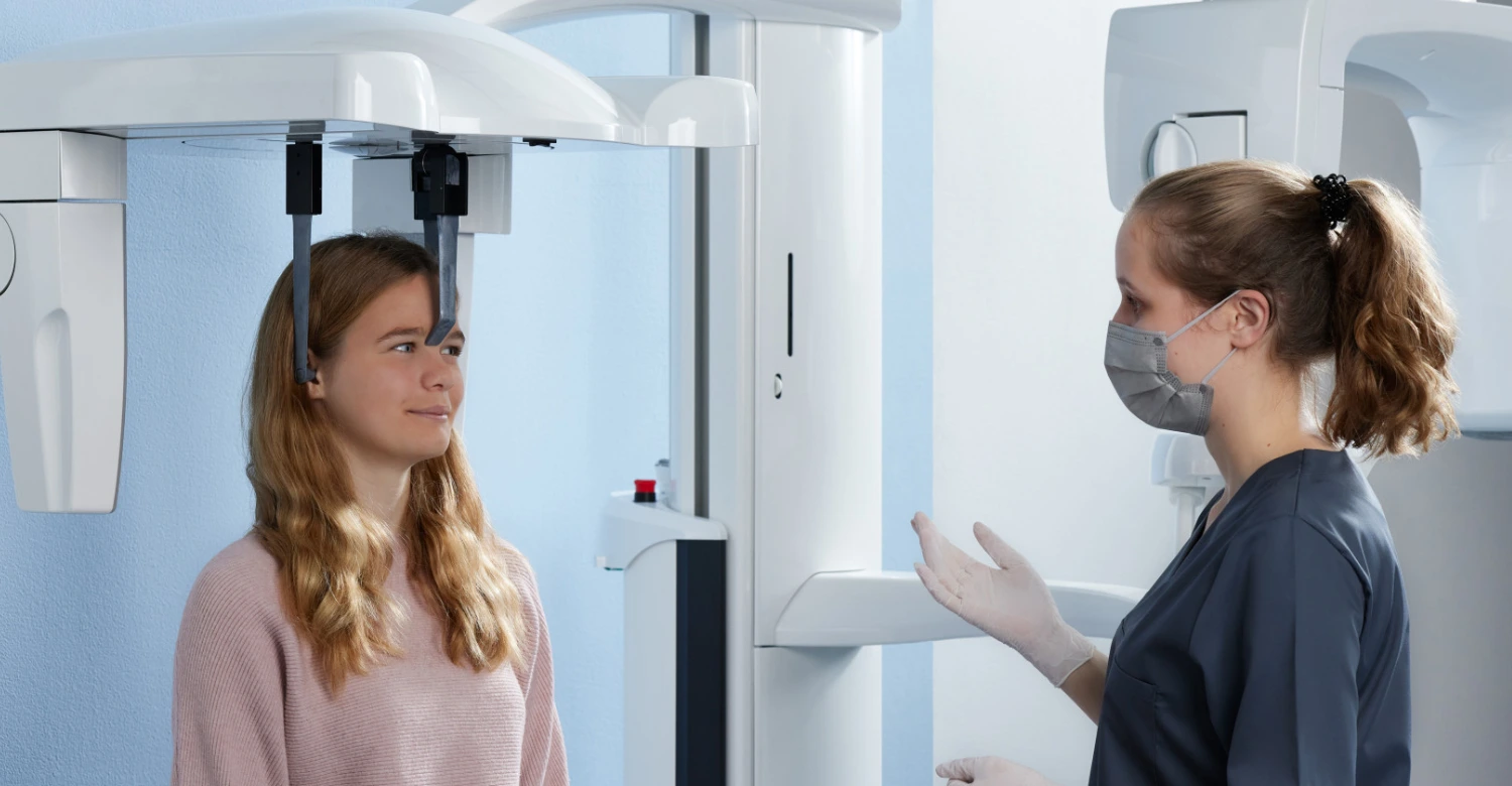
What is a cephalometric X ray machine?
.webp&w=3840&q=75)
A cephalometric X ray machine is a specialized imaging device used primarily in dentistry and orthodontics to capture detailed images of the teeth, jaw, and cranial structures. Unlike standard dental X-rays, it provides a lateral or frontal view of the skull, which helps orthodontists and oral surgeons plan treatments accurately. The technology has evolved significantly, offering both traditional and digital options. Clinics today prefer ceph x ray dental machines for their precision, speed, and ability to integrate with digital orthodontic software. By analyzing craniofacial structures, professionals can detect misalignments, plan braces, or prepare for maxillofacial surgeries efficiently. Using a cephalometric X ray machine enhances patient outcomes while minimizing the need for repeated imaging.
What is a Cephalometric X Ray Machine?
A cephalometric X ray machine is designed to capture the full skeletal structure of a patient’s head in a single image. It provides crucial data about jaw alignment, tooth positioning, and craniofacial proportions. Dentists and orthodontists rely on these images to create treatment plans, monitor progress, and ensure accurate surgical or orthodontic interventions. Unlike panoramic or intraoral X-rays, which focus on teeth or specific areas, a ceph x ray dental captures the entire skull in a standardized format, which is essential for reproducibility in follow-ups. Modern machines often feature digital sensors, offering high-resolution images instantly, reducing exposure time, and simplifying storage in patient records.
Overview of Ceph X Ray Technology
Ceph X-ray technology is a breakthrough in dental imaging, providing 2D or 3D visualizations of craniofacial structures. A cephalometric X ray machine employs precise positioning devices to ensure consistent head alignment, allowing for reproducible measurements. Digital sensors replace traditional film in most modern units, offering higher clarity and immediate access to images. Advanced software can analyze skeletal landmarks, helping orthodontists measure angles, distances, and growth patterns. This technology is particularly valuable for patients requiring braces, corrective jaw surgery, or long-term orthodontic monitoring. With features like adjustable exposure and image enhancement, ceph x-ray technology improves diagnostic accuracy while maintaining patient safety.
How It Differs From Standard Dental X Rays
A cephalometric X ray machine differs from standard dental X-rays in scope, purpose, and application. Traditional X-rays, like bitewings or periapical images, focus on individual teeth or small areas of the jaw. In contrast, cephalometric X-rays capture the entire craniofacial structure in a lateral or frontal view. This allows orthodontists to evaluate skeletal relationships, facial symmetry, and airway space. Unlike conventional X-rays, which are primarily used for cavities or periodontal issues, ceph x ray dental machines are essential for treatment planning, growth assessment, and orthodontic progress tracking. The ability to integrate digital images into treatment software adds efficiency and precision unmatched by standard X-ray methods.
Advantages in Orthodontics and Oral Surgery
Using a cephalometric X ray machine provides several advantages for orthodontics and oral surgery. It offers accurate skeletal mapping, facilitating precise brace placement, corrective jaw surgery planning, and craniofacial anomaly assessment. These images help professionals predict treatment outcomes, monitor growth, and adjust plans dynamically. Moreover, digital ceph X-rays reduce patient exposure to radiation and allow for easy storage, comparison, and sharing with multidisciplinary teams. Clinics can enhance patient education by visually demonstrating treatment needs and progress. Overall, this technology ensures safer, faster, and more effective interventions, making it an indispensable tool in modern dental and orthodontic practices.
Types of Cephalometric X Ray Machines
Cephalometric X ray machines come in various types, each suited for specific clinical needs. Traditional analog units capture images on film, while digital systems use sensors for instant imaging and enhanced resolution. Some machines are standalone cephalometers, while others are combined with panoramic units, known as pan ceph machines, allowing multiple imaging types from a single device. Choosing the right system depends on clinic size, patient volume, and budget. Digital units often integrate with imaging software, facilitating treatment simulations and precise measurements. By understanding the features of each type, dental professionals can select a machine that maximizes efficiency, patient safety, and diagnostic accuracy.
Traditional vs Digital Ceph X Ray Machines
Traditional cephalometric X-ray machines use film to record images, requiring chemical processing and longer examination times. They are reliable but less convenient for modern workflows. Digital machines, on the other hand, capture high-resolution images instantly and often integrate with treatment planning software. The cephalometric X-ray machine can be paired with advanced imaging programs to analyze skeletal and dental landmarks, reducing manual calculations. Digital units also reduce radiation exposure and allow images to be easily shared or stored in electronic medical records. Clinics that adopt digital ceph X-ray technology benefit from increased efficiency, improved patient experience, and more accurate treatment outcomes.
Pan Ceph Machine: Features and Benefits
A pan ceph machine combines panoramic and cephalometric imaging in a single unit, offering versatility for dental clinics. This type of cephalometric X ray machine allows practitioners to capture both full skull images and detailed panoramic views of the teeth and jaw without repositioning the patient. Advantages include reduced examination time, lower radiation exposure, and seamless integration with digital software for orthodontic and surgical planning. The ceph x ray dental functionality ensures that craniofacial assessments and growth monitoring are precise. Clinics using pan ceph machines can optimize workflow, improve patient comfort, and provide comprehensive diagnostic services from one device.
Choosing the Right Machine for Your Clinic
Selecting the right cephalometric X ray machine involves considering patient volume, imaging needs, and budget. Digital units with software integration offer advanced analysis, while traditional models remain effective for smaller practices. Clinics must evaluate radiation safety features, image clarity, maintenance requirements, and compatibility with other dental devices. A pan ceph machine may be preferred in multi-functional clinics to save space and streamline procedures. Consulting with manufacturers and comparing features ensures the chosen machine meets both clinical and operational requirements. Investing in the right equipment enhances diagnostic accuracy, patient safety, and treatment outcomes, making it a crucial decision for modern dental practices.
How Cephalometric X Rays Work
The operation of a cephalometric X-ray machine involves precise positioning, controlled exposure, and image capture of craniofacial structures. The patient is aligned using head supports, and the machine rotates or scans the skull in a standardized manner. Modern units use digital sensors to produce instant images, which can then be analyzed using specialized software. Ceph x ray dental machines allow orthodontists to measure skeletal angles, jaw relationships, and growth patterns accurately. The entire process is quick, often taking just a few minutes, and is designed to minimize radiation exposure. Understanding how cephalometric X-rays work helps clinics optimize workflow and ensure consistent imaging results.
The Science Behind Ceph X Rays
Cephalometric X-rays rely on controlled radiation to penetrate tissues and create a detailed skeletal image. The cephalometric X-ray machine uses a precise X-ray source and a detector placed at a fixed distance to ensure reproducible images. By capturing lateral, frontal, or posteroanterior views, orthodontists can measure craniofacial angles and assess growth patterns. The physics behind the technology allows for minimal radiation exposure while maintaining high image clarity. With advancements in digital sensors, the resulting images can be enhanced, stored, and analyzed in real time, providing accurate diagnostic data crucial for treatment planning, surgical procedures, and monitoring progress over time.
Step-by-Step Imaging Process
Operating a cephalometric X ray machine follows a structured procedure. First, the patient is positioned using chin rests and head supports to ensure alignment. The technician adjusts the machine’s height and angle for the desired view. Once ready, the exposure is triggered, and digital sensors capture the craniofacial image within seconds. Advanced software may then enhance the image, annotate anatomical landmarks, and allow precise measurements for orthodontic or surgical planning. The ceph x ray dental system ensures minimal radiation exposure while providing high-resolution, reproducible images. This step-by-step process guarantees reliable diagnostics and reduces the need for repeated scans.
Radiation Safety and Patient Protection
Patient safety is a top priority when using a cephalometric X ray machine. Digital units significantly reduce radiation exposure compared to traditional film-based systems. Clinics implement shielding techniques, such as lead aprons and thyroid collars, to protect sensitive tissues. Proper machine calibration and adherence to recommended exposure settings ensure safety without compromising image quality. The ceph x-ray dental technology is designed to comply with international radiation safety standards, making it suitable even for pediatric patients. Educating patients and staff about safety measures enhances trust, reduces anxiety, and ensures responsible use of X-ray imaging in dental and orthodontic practice.
Benefits of Using a Cephalometric X Ray Machine
A cephalometric X ray machine provides essential benefits for modern dental and orthodontic practices. By capturing precise craniofacial images, it allows clinicians to identify skeletal discrepancies, malocclusions, and growth irregularities with confidence. Treatment planning becomes more accurate, as orthodontists can analyze jaw relationships and tooth positioning. Clinics also benefit from streamlined workflows, especially when using digital systems that integrate with patient records. Ceph x ray dental technology reduces radiation exposure compared to repeated traditional X-rays, enhancing patient safety. Overall, investing in this machine improves diagnostic accuracy, optimizes treatment strategies, and contributes to better long-term oral health outcomes for patients of all ages.
Accurate Diagnosis for Orthodontic Treatments
Orthodontic success depends heavily on accurate diagnosis. With a cephalometric X ray machine, clinicians can visualize the full craniofacial structure, enabling precise analysis of jaw alignment, dental crowding, and skeletal asymmetries. This information guides decisions on braces, retainers, or surgical interventions. Digital systems enhance accuracy by allowing software-based measurements, eliminating manual errors. Ceph x ray dental images also provide a standardized format, making follow-ups and progress tracking reliable. By identifying subtle discrepancies early, orthodontists can plan treatments tailored to each patient, improving outcomes and reducing treatment time. Accurate diagnosis ensures efficient use of clinic resources and enhances patient satisfaction.
Enhanced Treatment Planning
Treatment planning is greatly enhanced with a cephalometric X ray machine. Detailed craniofacial images allow orthodontists and oral surgeons to simulate procedures, anticipate challenges, and determine the optimal approach. Digital ceph X-rays integrate with software for 3D modeling, angle measurement, and growth prediction. Ceph x ray dental imaging facilitates interdisciplinary collaboration, as images can be shared with surgeons, pediatric dentists, or speech therapists. Clinicians can visualize results before starting treatment, helping patients understand procedures and outcomes. This proactive approach minimizes errors, reduces the need for repeated imaging, and ensures more predictable results, benefiting both patients and clinic efficiency.
Time and Cost Efficiency for Clinics
A cephalometric X ray machine improves both time and cost efficiency for dental clinics. Digital imaging eliminates film development, reduces repeat scans, and allows instant access to patient images. Ceph x ray dental machines can be combined with panoramic imaging in pan ceph machines, saving time by performing multiple scans in a single session. Faster image acquisition increases patient throughput while maintaining diagnostic quality. Reduced radiation exposure minimizes safety concerns, which is especially important for pediatric patients. Clinics also benefit financially by reducing film, chemicals, and storage costs. Overall, investing in this technology streamlines operations, enhances patient experience, and supports sustainable, high-quality care.
Cephalometric X Ray Machine at Vitrin Clinic
At Vitrin Clinic, the cephalometric X-ray machine is an integral part of advanced diagnostic and treatment planning services. The clinic combines high-resolution imaging with patient-focused care, ensuring precise craniofacial measurements while maintaining comfort. With cutting-edge ceph x-ray dental technology, clinicians can capture both lateral and frontal images quickly and safely. This allows for accurate orthodontic assessments, surgical planning, and monitoring of pediatric growth. By integrating modern imaging equipment with experienced professionals, Vitrin Clinic provides reliable, efficient, and tailored dental care, earning trust among patients seeking high-quality orthodontic and oral surgery services.
Advanced Equipment for Precise Imaging
Vitrin Clinic uses state-of-the-art cephalometric X ray machines to ensure imaging precision. Digital sensors and advanced software allow orthodontists and surgeons to analyze skeletal structures in detail. Ceph x ray dental images can be enhanced for clarity, with measurements of angles, jaw relationships, and dental positions. The clinic’s equipment supports both traditional and panoramic imaging, including pan ceph machines, providing versatility in diagnostics. This ensures every patient receives accurate assessments and well-informed treatment plans. The combination of advanced equipment and expert interpretation allows Vitrin Clinic to deliver superior outcomes in orthodontics, oral surgery, and pediatric dentistry.
How Vitrin Clinic Ensures Patient Comfort
Patient comfort is a priority at Vitrin Clinic when using a cephalometric X ray machine. Ergonomic head supports and adjustable positioning minimize discomfort during imaging. The clinic’s staff provides clear instructions to help patients remain calm and still, ensuring high-quality images. Digital systems reduce exposure time, which further improves the patient experience. Ceph x ray dental technology allows instant image capture, reducing waiting time and eliminating the need for multiple exposures. Vitrin Clinic also uses shielding techniques to protect sensitive tissues, making the imaging process safe for adults and children alike. Comfort-focused practices enhance trust and encourage patients to adhere to recommended treatments.
Choosing the Right Ceph X Ray Machine
Selecting the right cephalometric X ray machine is crucial for clinics aiming to optimize diagnostics and patient care. Factors to consider include image quality, software integration, ease of use, radiation safety, and maintenance requirements. Digital units offer instant imaging and analysis, while pan ceph machines provide multi-functional capabilities. Budget constraints and patient volume also play a role in decision-making. Comparing top brands and models helps clinics find the balance between cost and technology. A well-chosen cephalometric X ray machine enhances accuracy, efficiency, and patient satisfaction, supporting both daily operations and long-term clinical success.
Factors to Consider for Your Clinic
When selecting a cephalometric X ray machine, clinics should evaluate several factors. Image resolution and consistency are critical for accurate diagnosis and treatment planning. Digital integration allows seamless storage and sharing of ceph x ray dental images. Radiation dose, patient comfort, and ergonomic design ensure safety and efficiency. Multi-function machines, such as pan ceph machines, save space and reduce workflow complexity. Maintenance, warranty, and technical support are also important considerations. By assessing these factors, clinics can invest in a machine that maximizes diagnostic capability, optimizes patient experience, and ensures long-term reliability and clinical success.
Comparing Top Brands and Models
Top brands in cephalometric X ray machines offer a variety of models, each with unique features. Some provide high-resolution digital imaging, software analysis, or integration with panoramic units. Clinics must compare factors like exposure time, image quality, ease of use, and cost. Ceph x ray dental devices with digital sensors are preferred for efficiency, while pan ceph machines offer versatility in imaging. Brand reputation, customer support, and service availability are also essential. Selecting the right model ensures precise diagnostics, enhances patient safety, and improves treatment outcomes. Careful comparison helps clinics make a technology investment that supports growth and clinical excellence.
Maintenance and Longevity Tips
Maintaining a cephalometric X ray machine is essential for consistent performance and patient safety. Regular calibration, cleaning, and software updates prevent errors and prolong device lifespan. Digital systems require proper care of sensors and storage devices, while ceph x ray dental units should be inspected for mechanical integrity. Pan ceph machines need additional maintenance due to their multi-functional design. Staff should be trained on proper operation, safety protocols, and troubleshooting. Scheduled professional servicing ensures compliance with regulatory standards. Effective maintenance reduces downtime, enhances image quality, and extends the life of the machine, making it a cost-effective investment for any dental clinic.
Common Applications of Ceph X Rays
A cephalometric X ray machine serves multiple clinical applications. In orthodontics, it is used to assess jaw alignment, tooth positioning, and growth patterns. Oral and maxillofacial surgeons use it for preoperative planning and evaluating facial symmetry. Pediatric dentists monitor craniofacial development and detect abnormalities early. Academic and research institutions utilize cephalometric imaging for anatomical studies and technological advancements. The versatility of ceph x ray dental technology ensures that clinicians across various specialties have a reliable, precise tool for diagnosis, treatment planning, and outcome evaluation, enhancing overall patient care and clinical efficiency.
Orthodontics
In orthodontics, a cephalometric X ray machine is indispensable for treatment planning. By analyzing skeletal structures, dental crowding, and jaw relationships, orthodontists can design braces, aligners, and surgical interventions with precision. Digital imaging allows software-assisted measurements, providing consistent and reproducible data. Ceph x ray dental units enable progress monitoring, ensuring timely adjustments and optimal outcomes. Lateral and frontal views captured by the machine help predict growth patterns, assess airway space, and guide patient-specific treatments. Accurate imaging reduces errors, shortens treatment duration, and improves patient satisfaction, making cephalometric X-rays a cornerstone of modern orthodontic practice.
Oral and Maxillofacial Surgery
Oral and maxillofacial surgeons rely on cephalometric X ray machines to plan complex procedures. Accurate imaging of the jaw, teeth, and craniofacial structures allows precise surgical planning, reducing risk and improving outcomes. Ceph x-ray dental technology provides detailed views of anatomical landmarks, helping surgeons assess bone structure, symmetry, and surgical access points. Panoramic and lateral images, often captured with pan ceph machines, streamline workflow by providing multiple perspectives in a single scan. By integrating advanced imaging with clinical expertise, surgeons can anticipate complications, customize approaches, and enhance patient safety during orthodontic and reconstructive procedures.
Pediatric Dentistry
Pediatric dentists benefit greatly from cephalometric X ray machines for monitoring growth and development. Early detection of skeletal or dental irregularities allows timely interventions, reducing the need for complex procedures later. Digital ceph x-ray dental units offer low-dose imaging, ensuring safety for children while providing clear and accurate views of craniofacial structures. The machine helps track jaw development, tooth eruption patterns, and airway conditions. Pediatric patients can undergo imaging with minimal discomfort, thanks to ergonomic design and quick capture times. Accurate, safe imaging supports preventive care, early orthodontic planning, and long-term oral health outcomes for young patients.
Research and Academic Uses
Cephalometric imaging is essential in dental research and academia. A cephalometric X ray machine provides precise craniofacial data for studying growth patterns, treatment outcomes, and anatomical variations. Digital ceph x ray dental systems allow researchers to analyze landmarks, measure angles, and compare large datasets efficiently. Academic institutions use these machines to train future dentists and orthodontists, offering hands-on experience in imaging, diagnosis, and treatment planning. Research findings contribute to technological advancements in orthodontics, surgical methods, and preventive care. By combining imaging precision with analytical tools, cephalometric X-rays support innovation, evidence-based practice, and high-quality education in dentistry.
Vitrin Clinic’s Expertise in Ceph X Ray Imaging
Vitrin Clinic has established itself as a leader in cephalometric imaging. Using the latest cephalometric X ray machines, the clinic ensures accurate diagnostics, efficient treatment planning, and high patient satisfaction. Ceph x ray dental technology is integrated with advanced software for precise analysis of craniofacial structures. Vitrin Clinic combines technology with expert interpretation, providing tailored care for orthodontics, oral surgery, and pediatric patients. By prioritizing both accuracy and patient comfort, the clinic offers reliable and effective imaging services that support successful treatment outcomes and reinforce its reputation as a trusted provider in advanced dental care.
Why Patients Trust Vitrin Clinic
Patients trust Vitrin Clinic because of its use of cutting-edge cephalometric X ray machines and commitment to personalized care. Digital imaging ensures high-resolution, reproducible images with minimal radiation exposure. Experienced clinicians interpret the data accurately, offering clear treatment plans and progress tracking. Ceph x ray dental technology allows quick imaging, reducing appointment times and enhancing comfort. The clinic’s focus on safety, precision, and patient education builds confidence. Combining modern equipment with professional expertise, Vitrin Clinic provides reliable diagnostics and effective interventions, making it a preferred choice for orthodontic, surgical, and pediatric dental care.
Success Stories and Case Studies
Vitrin Clinic’s success stories highlight the impact of cephalometric X ray machines on treatment outcomes. Patients undergoing orthodontic and surgical procedures benefit from accurate diagnostics, resulting in improved alignment, symmetry, and facial aesthetics. Ceph x ray dental imaging allows clinicians to track progress, adjust treatment plans, and predict results with precision. Case studies demonstrate reduced treatment time, fewer complications, and higher patient satisfaction. By leveraging advanced imaging technology alongside clinical expertise, Vitrin Clinic consistently delivers superior outcomes, reinforcing the importance of precise, patient-focused cephalometric imaging in modern dental practice.
Integrating Technology With Personalized Care
Vitrin Clinic integrates cephalometric X ray machines with a patient-centered approach. Digital imaging supports customized treatment planning, while clinicians provide clear explanations and guidance. Ceph x ray dental images help patients visualize treatment goals, understand procedures, and track progress. Personalized care ensures comfort, safety, and collaboration, enhancing the overall experience. By combining technology with empathy, the clinic ensures optimal outcomes for orthodontic, surgical, and pediatric patients. This integration of advanced cephalometric imaging with tailored care exemplifies modern dentistry, where precision and patient well-being go hand in hand.
FAQs

Dr. Rifat Alsaman has more than 5 years of clinical experience in dentistry and currently serves as the Head of the Medical Team at Vitrin Clinic. He is dedicated to providing exceptional patient care, overseeing treatment planning, and ensuring the highest clinical standards across the team. His expertise, attention to detail, and commitment to continuous professional development have helped countless patients achieve healthier, more confident smiles.

%20(1).webp&w=3840&q=75)
.webp&w=3840&q=75)


