Gingival Esthetics
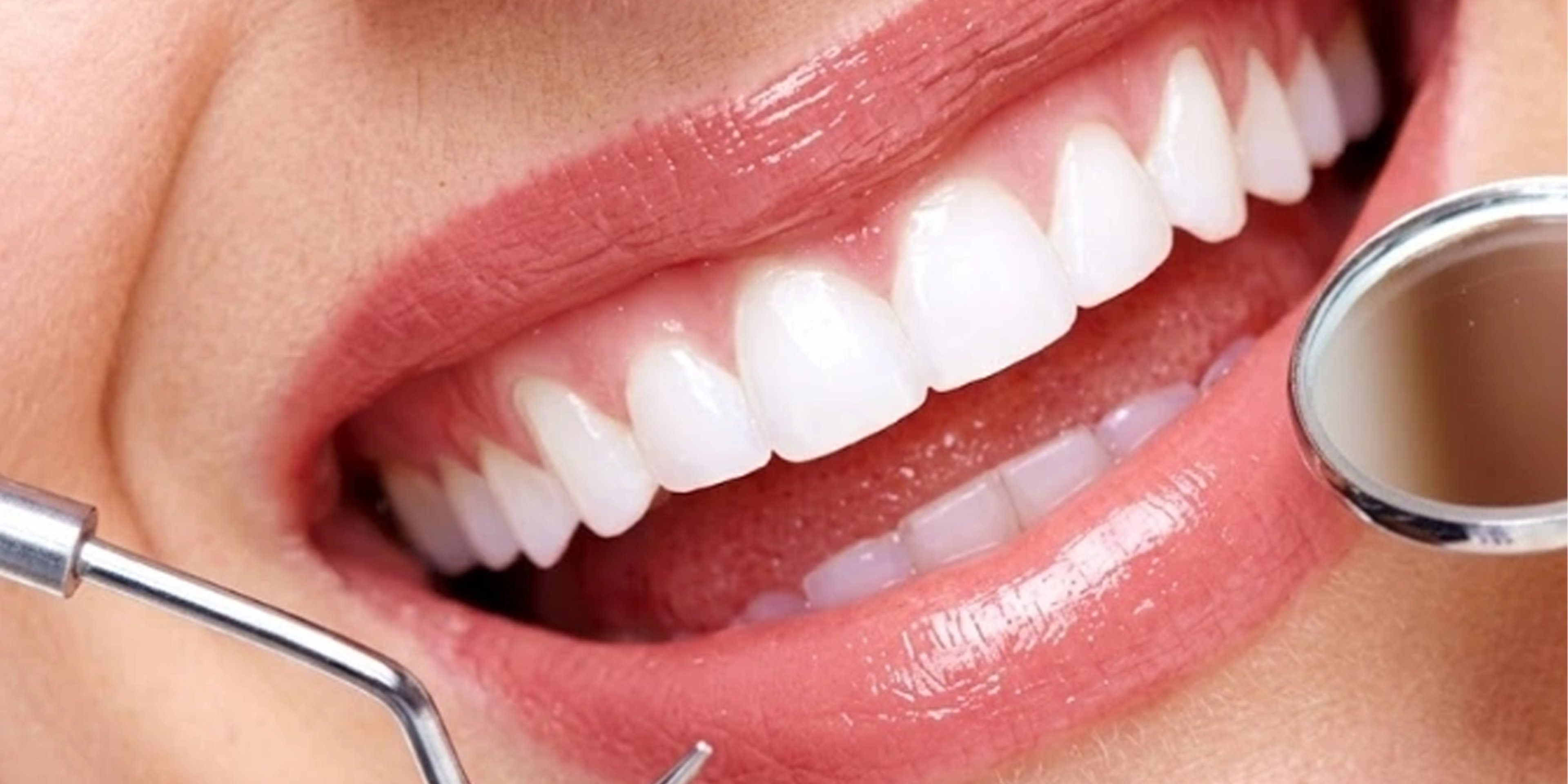
A gummy smile affects your gingival esthetic line the visible boundary between teeth and gums which should follow the natural curve of your upper lip. Identifying the root cause is the essential first step to choosing the right treatment and maximising results.
Treatment Process
A clear step-by-step overview of how the treatment is planned and performed, from the initial consultation to the final results, ensuring comfort, safety, and predictable outcomes.
01
Clinical Gum Line Measurement
We document gum exposure precisely in millimeters and evaluate gum health, symmetry, and the underlying bone structure. This baseline measurement guides every treatment decision that follows.
02
Orthodontic & Bite Analysis
We study tooth alignment and the way your teeth meet to understand how any shifting in tooth position might alter gum display determining whether orthodontic intervention forms part of your plan.
03
Orthognathic (Jaw) Evaluation
Where skeletal factors are suspected, we use panoramic X-rays, 3D scans, and cephalometric analysis to evaluate jaw position and plan any necessary surgical adjustments with precision.
04
Smile Line & Lip Mobility Study
Your smile is dynamic not static. We analyse how much gum shows during your natural expressions and lip movement, not just at rest, ensuring the treatment addresses what actually appears when you smile.
05
Digital Smile Design Preview
Using DSD technology, we create a visual simulation of your refined esthetic gingival proportions and tooth-to-gum relationship so you see and approve your expected outcome before any treatment begins.
What Is Gingival Esthetics?
Gingival esthetics clinically referred to as periodontal plastic surgery encompasses the surgical and non-surgical procedures used to optimise the shape, position, symmetry, and proportion of the gum tissue as it frames the teeth.
A harmonious smile depends as much on the gingival architecture as on the teeth themselves: uneven gum levels, excessive gum display, receded gum margins, or disproportionate tooth-to-gum ratios all affect the visual balance of the smile in ways that no restorative work alone can correct.
The American Academy of Periodontology defines gingival excess gum tissue covering the teeth as a mucogingival malformation.
Peer-reviewed literature consistently confirms that excessive gingival display carries significant psychosocial impact: patients report esthetic shame, reduced self-esteem, and avoidance of smiling in social and professional contexts.
Correcting the gingival architecture through evidence-based surgical techniques produces measurable and durable improvements in both smile esthetics and patient-reported quality of life.
Gingival esthetic treatment is not an isolated cosmetic procedure.
Within a comprehensive smile plan, it is typically performed before any restorative work because crown lengths, veneer designs, and restoration shade selections are all made against a stable, correctly positioned gum line.
Restorations placed before gingival surgery are placed in the wrong position relative to the final gum margin, requiring correction or replacement once the soft tissue has healed. The sequence is fixed: gingival esthetics first, restorations after.
Gingival Esthetic Procedures at Vitrin Clinic
Vitrin Clinic offers the full spectrum of evidence-based gingival esthetic procedures, selected on the basis of the clinical diagnosis and the patient's specific anatomical presentation.
The following are the primary procedures used each with a distinct indication that determines when it is the appropriate clinical choice.
Esthetic Crown Lengthening
Esthetic crown lengthening (ECL) is the surgical repositioning of the gingival margin to expose the correct anatomical length of the clinical crown.
Where bone levels need to be repositioned to maintain the biologic width the supracrestal tissue attachment osseous surgery is performed in addition to soft tissue repositioning. This is the distinction between a gingivectomy (soft tissue only) and a crown lengthening procedure (soft tissue plus bone).
A systematic review published in PMC/NIH searching PubMed, Cochrane CENTRAL, Scopus, and Web of Science confirmed that crown lengthening surgery achieves an average increase in clinical crown length of 1.4 to 3.3 mm at 6-month follow-up, with gingival margin rebound occurring primarily within the first three postoperative months.
This rebound is why restorations should not be placed until at least three months post-surgery a timing rule that is non-negotiable at Vitrin Clinic.
The open-flap technique with full tissue elevation and direct visualisation of the bone crest is indicated for cases requiring osseous resection.
The flapless technique is appropriate for cases where soft tissue repositioning alone is sufficient. A 2025 meta-analysis of RCTs confirmed that both techniques achieve comparable clinical outcomes, with technique selection based on case requirements rather than operator preference.
Indication APE, short crowns, pre-restorative
Anaesthesia Local
Crown Gain 1.4–3.3 mm average
Healing 6–8 weeks soft tissue, 3 months stable
Restoration Wait Minimum 3 months post-surgery
Technique Open-flap or flapless case dependent
Gingivectomy
Gingivectomy is the excision of excess gingival tissue without bone surgery indicated where the bone level is at a normal position relative to the CEJ and where the correction required is limited to the soft tissue margin.
It is the simpler of the two gingival surgical procedures and carries a shorter recovery period, but it is only appropriate where bone surgery is not needed to establish the correct biologic width.
Gingivectomy can be performed using a scalpel, electrosurgery, or laser each with advantages in specific clinical situations.
Laser gingivectomy offers excellent haemostasis, reduced post-operative discomfort, and precise tissue management, but requires bone assessment before selection to confirm that bone surgery is not required.
Selecting gingivectomy in a case that requires osseous surgery produces an inadequate result with gingival rebound toward the preoperative position.
Indication Soft tissue excess, normal bone level
Anaesthesia Local
Technique Scalpel, laser, or electrosurgery
Recovery Shorter than crown lengthening
Bone Surgery Not included — tissue only
Limitation Contraindicated if bone surgery needed
Soft Tissue Grafting
Where gum recession has exposed root surfaces causing sensitivity, esthetic concerns, or increased caries risk soft tissue grafting restores the missing gingival tissue.
The connective tissue graft, harvested from the palate, is the most evidence-supported grafting technique for root coverage.
It adds both keratinized tissue volume and root coverage in a single procedure.
The free gingival graft adds keratinized tissue where volume is the primary concern. Evidence-based outcomes for connective tissue grafting consistently demonstrate root coverage of 80 to 100% at sites with Miller Class I and II recession.
Indication Gum recession, root exposure
Graft Type Connective tissue or free gingival
Root Coverage 80–100% (Miller I–II)
Donor Site Palate or acellular dermal matrix
Gingival Esthetic Procedure Selection by Cause
The following comparison clarifies which procedure is clinically appropriate for each cause of excessive gingival display based on published evidence and clinical guidelines.
The wrong procedure applied to the wrong diagnosis produces a predictably inadequate or unstable result.
Cause of Gummy Smile | Crown Lengthening (+ Bone) | Gingivectomy (Soft Tissue Only) | ||
|---|---|---|---|---|
Altered Passive Eruption (APE) | Primary indication bone surgery required in most APE cases | Only for Type IA APE where bone is at correct level | ||
Hypermobile Upper Lip | Not indicated gum and bone levels are normal | Not indicated | ||
Vertical Maxillary Excess | Adjunctive only insufficient as sole treatment for VME | Insufficient does not address skeletal dimension | ||
Gingival Enlargement | Indicated if bone involvement present | Primary once cause is managed (medication, disease) | ||
Pre-Restorative (Short Crowns) | Required to expose sound tooth structure and establish ferrule | Only where bone surgery is confirmed unnecessary | ||
Result Permanence | Permanent stable after 3 months tissue maturation | Permanent when correctly indicated |
Compiled from: PMC12637009 — Open-Flap vs. Flapless ECL Meta-Analysis of RCTs, 2025 · Kuwait University, College of Dentistry — Clinical Considerations for Crown Lengthening, PMC11614317, 2024 · PubMed PMID 27535216 — Pre-Restorative Crown Lengthening Surgery Outcomes: Systematic Review · PubMed PMID 37730094 — Contemporary Techniques for Excessive Gingival Display: Altered Passive Eruption and Lip Hypermobility.
The Vitrin Clinic Standard
Bone sounding performed before any procedure is selected
The bone level relative to your CEJ is measured before any gingival procedure is recommended.
This single step determines whether you need gingivectomy or crown lengthening and getting it wrong produces a predictably inadequate result.
Cause identified before treatment selected
Your gummy smile has a specific cause and the treatment at Vitrin Clinic is selected to match that cause.
The same procedure is not applied to every presentation regardless of what is driving it.
No restorations within three months of surgery
Veneers and crowns are not placed until at least three months after gingival surgery and tissue stability is confirmed at reassessment.
The three-month wait is not negotiable placing restorations before the gum has settled means placing them in the wrong position.
Gum surgery planned against a digital smile design
The target gum line is designed digitally before your surgery begins mapping each tooth's gingival zenith against your facial landmarks.
The surgery follows the design, not the operator's intraoperative judgement alone.
Gingival Esthetics Cost: Vitrin Clinic vs. UK vs. USA
The cost of gingival esthetic treatment varies depending on the procedure required, the extent of correction, and the country of treatment. The following table provides a transparent comparison of average costs across the United Kingdom, United States, and Vitrin Clinic in Istanbul from non-surgical Botox correction to full surgical crown lengthening allowing patients to evaluate their options with accurate financial context before their consultation.
Procedure | 🇬🇧 UK | 🇺🇸 USA | 🇹🇷 Turkey |
|---|---|---|---|
Esthetic crown lengthening Soft tissue + bone, per arch | $1,500 – $3,500 | $1,000 – $4,000 | From $300 |
Gingivectomy Soft tissue only, laser or scalpel | $380 – $1,270 | $200 – $1,500 | From $100 |
Soft tissue graft Connective tissue / recession, per site | $760 – $1,525 | $600 – $1,200 | From $200 |
Botox for hypermobile lip Non-surgical gummy smile correction | $255 – $635 | $200 – $600 | From $80 |
Consultation & scans | Charged separately | Charged separately | Free & included |
Estimated saving | — | — | Up to 70% less |
* Prices are averages for reference only and may vary based on the procedure selected, number of teeth or sites treated, case complexity, and individual clinical requirements. A personalised quote is provided following your free consultation.
Clinical Evidence & References
Kuwait University, College of Dentistry, Kuwait
Clinical Considerations for Crown Lengthening: A Comprehensive Review
Cureus · November 2024Qali M, Alsaegh H, Alsaraf S · DOI: 10.7759/cureus.72934 · PMC11614317
Key finding: Crown lengthening is indicated for exposing sound tooth structure for restorative purposes, increasing clinical crown height, creating ferrule, and relocating subgingival margins. Correct diagnosis through meticulous examination including biologic width assessment and bone sounding is the prerequisite for selecting the correct technique and achieving predictable outcomes.
Journal of Clinical Periodontology Systematic Review
Pre-Restorative Crown Lengthening Surgery Outcomes: A Systematic Review
Journal of Clinical Periodontology · December 2016Pilalas I, Tsalikis L, Tatakis DN · DOI: 10.1111/jcpe.12617 · PubMed PMID: 27535216
Key finding: Crown lengthening surgery results in increased crown length of 1.4 to 3.3 mm at 6-month follow-up. Gingival margin rebound occurs primarily within the first three postoperative months confirming that restorative treatment should not be initiated until tissue stability is confirmed at a minimum of three months post-surgery.
ScienceDirect Clinical Guidance Review
Gummy Smiles: Etiologies, Diagnoses, and Formulating a Clinically Effective Treatment Protocol
Journal of Oral and Maxillofacial Surgery · 2024DOI: 10.1016/j.joms.2024.01.003 · Comprehensive aetiological and procedural selection framework
Key finding: A structured diagnostic protocol identifying whether the gummy smile is caused by APE, lip hypermobility, VME, gingival enlargement, or dentoalveolar extrusion is essential before procedure selection.
Each aetiology has a specific, evidence-matched treatment approach. Applying a single procedure without aetiological diagnosis leads to predictably inadequate or unstable outcomes.
Frequently Asked Questions
Answers to common questions about the treatment, including suitability, procedure details, recovery, and long-term care — helping you feel informed and confident before moving forward.