Dental Equipment
June 14, 2026
What Is Dentist Technology and How Is It Transforming Modern Dental Care?
 Dr. Rifat Alsaman
.webp&w=3840&q=75)
Modern dental treatment pathways are changing fast. Advanced 3D scanning replaces traditional messy molds. This innovation captures accurate digital models of teeth and gums. It improves diagnosis, treatment planning, and restorative work. Unlike old methods, it boosts patient comfort and cuts errors. It also speeds up clinical turnaround times. By linking chairside precision with CAD/CAM systems, it anchors modern digital dentistry. Top clinics worldwide use these tools to improve outcomes and patient experience. Advanced scanning is now key to restorative and cosmetic care. It offers a faster, cleaner, and more precise way to design smiles. This shift relies on cutting-edge dentist technology to streamline workflows.
The Role of Dentist Technology in Digital Impressions
The dental industry is transforming rapidly. Advanced intraoral scanners replace old, messy impression materials. This dental equipment creates precise digital models in real time. The scanning process is non-invasive and comfortable. It is also much faster than older methods. It improves communication between clinics and labs. This ensures better results in prosthodontics, implantology, and orthodontics. As dentistry moves to digital workflows, an advanced intraoral scanner is vital. It optimizes precision, efficiency, and patient satisfaction. Many clinics now rely on these systems as a standard for diagnostics and planning.
How Dentist Technology Improves Digital Dental Impressions
A digital impression is a detailed 3D model of oral tissues. It is made with specialized intraoral scanners. Patients avoid trays filled with setting pastes. Instead, a dentist uses a lightweight handheld wand. It captures thousands of data points per second. Software stitches these images into a precise 3D model. This model aids comprehensive treatment planning. A 3D dental scanning workflow eliminates lab errors and discomfort. Today, this dentist technology is critical in orthodontics, prosthodontics, and implant dentistry. It sets a new standard for dental impression accuracy.
How digital impression systems capture 3D oral scans
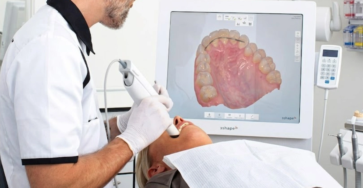
Modern intraoral scanners use advanced optical technologies. They capture precise data from teeth and gums. The scanner projects structured light or a laser onto surfaces. High-speed sensors record the reflections. This data compiles in real time on a chairside monitor. The clinician can rotate, zoom, and check the model instantly. They can spot and fix voids without retaking the impression. The result is a flawless digital replica. It is perfect for crowns, bridges, aligners, and implants.
Why dentistry is shifting toward digital workflows
The shift to digital workflows is driven by demand for speed, accuracy, and comfort. Traditional tray-and-putty methods often distort or shrink. They also involve long shipping delays. Modern digital scanning gives instant results. It pairs seamlessly with lab workflows. This lets technicians design restorations with micron-level accuracy. Digital files are easy to archive, duplicate, and share via the cloud. As patient expectations rise, clinics adopt these tech-driven solutions. They stay competitive and ensure better long-term outcomes. This evolution highlights the value of advanced dentist technology in everyday practice.
How Dentist Technology Improves Digital Dental Impressions
Digital scans boost clinical accuracy and streamline daily operations. They remove the instability of manual impression materials. This provides reliable data for complex restorative and cosmetic work. Dentists can view oral structures in real time. This improves diagnostics and treatment planning. The technology also speeds up collaboration between clinics and labs. Turnaround times drop significantly. It is now vital for practices focused on precision and patient care.
Accuracy and precision improvements
Digital scanning nearly eliminates dimensional inaccuracies. Traditional impressions often distort during mixing, tray removal, or pouring. Intraoral scans bypass these risks. They capture exact measurements directly from the mouth. This precision ensures a better marginal fit for crowns, bridges, and aligners. Reliable fit reduces chairside adjustments and repeat visits. It saves time and money for patients and clinics.
Patient comfort and faster procedures
Digital scanning improves the patient experience. It removes bulky trays and bad-tasting pastes. Patients with a strong gag reflex or dental anxiety find it non-invasive and stress-free. The scan takes just a few minutes. Appointments become shorter and more pleasant. This comfort boosts satisfaction. It encourages people to seek needed care without fear.
How Dentist Technology Improves Digital Impressions
The digital impression process uses an integrated workflow. It replaces physical models with advanced capture and manufacturing tech. The workflow starts with an intraoral scan. This creates a 3D digital model for real-time checks. Then, design software optimizes the file. It goes to a lab or in-house milling unit. This system improves accuracy and cuts turnaround times. It also removes human errors from manual lab steps. It is a key part of modern CAD/CAM dentistry.
How Dentist Technology Streamlines Digital Impressions
The clinical sequence follows a strict protocol for flawless data capture:
Preparation: The clinician readies the target teeth. The oral environment is optimized for scanning.
Data Capture: An intraoral scanner moves through the arches. It captures high-resolution details of teeth and tissues.
Real-Time Stitching: The software instantly stitches data segments. It forms a complete, navigable 3D model.
Clinical Validation: The dentist reviews the model on-screen. They check margins and rescan small areas if needed.
Export & Laboratory Transfer: The file is optimized and sent securely. It goes to a lab or in-office manufacturing unit.
Intraoral scanning technology and leading global brands
Top practices use premium, verified scanning hardware for high dental impression accuracy. The iTero scanner integrates natively with Invisalign systems. This optimizes digital orthodontic scanning workflows. The 3Shape TRIOS offers unmatched color realism and surface detail. It is a favorite among cosmetic dentists for its intuitive software. The CEREC Primescan delivers ultra-precise data for CAD/CAM dentistry. It creates accurate same-day crowns efficiently. Each system has strengths. The choice depends on the practice's focus, such as restorative, orthodontic, or implant cases. One consideration is software compatibility. Different brands use proprietary software. Not all integrate seamlessly with every CAD/CAM system. This can cause workflow issues. Most modern scanners offer open architecture or export standard STL files. This allows broader compatibility. Dentists should ensure their chosen system aligns with their existing technology stack.
Data conversion into 3D models
After scanning, proprietary software processes the raw data. It creates a fully rendered 3D model. The algorithm aligns millions of data points. It reconstructs the exact geometry of teeth and gums. Clinicians can rotate, zoom, and cross-section the model. They evaluate occlusal relationships, spacing, and alignment. This precise digital conversion is essential. It helps design restorations that fit seamlessly and function naturally.
Sending impressions to dental labs
After clinical approval, the 3D file is encrypted and sent to labs via secure cloud networks. This electronic transfer eliminates shipping delays and costs. It also avoids physical breakage risks of stone models. Technicians download the data immediately. They design custom restorations with advanced CAD software. This streamlined loop allows real-time collaboration. It ensures rapid delivery and exceptional accuracy for the final restoration.
How Dentist Technology Improves Digital Teeth Impressions
Modern digital impressions depend on a fully integrated technological ecosystem. Hardware and software work together seamlessly. This setup includes high-resolution intraoral scanners, powerful CAD/CAM suites, and interactive 3D imaging software. Each technology is crucial for capturing, refining, and manufacturing high-end dental prosthetics. By replacing manual workflows with this digital loop, dental teams can execute complex procedures with speed and predictability.
Intraoral scanners
Intraoral wands serve as the primary diagnostic capture tool in modern clinics. They use sophisticated optical setups to map the complex geography of the oral cavity. Designed for speed, ergonomic balance, and patient comfort, these scanners eliminate the discomfort of physical trays. They also reduce chair time. The operator can verify preparation quality and margin boundaries on the spot. This ensures no critical anatomical detail is missed.
CAD/CAM systems
CAD/CAM configurations transform digital models into physical restorations. After capturing intraoral scan data, Computer-Aided Design (CAD) software allows precise sculpting of crowns, bridges, or veneers. The finalized design file goes to a Computer-Aided Manufacturing (CAM) system. This system commands high-precision milling machines or 3D printers to fabricate the restoration. This automated loop minimizes manual errors and ensures consistent material strength.
3D imaging software integration
Dedicated 3D imaging software serves as the central processing brain of digital dentistry. It converts raw optical data into interactive clinical models. These models allow highly granular analysis. The software can simulate dynamic jaw movements and evaluate occlusal bite forces. It also generates comprehensive digital smile design previews. Visualizing and adjusting treatments in a virtual environment improves planning accuracy. It also enhances patient communication.
How Dentist Technology Improves Digital Dental Impressions
Integrating modern intraoral scanning systems improves clinical workflows and treatment predictability. A major benefit is capturing detailed oral data without the discomfort of traditional methods. This shift minimizes clinical errors and accelerates laboratory turnaround times. It also enhances communication between the dental team, laboratories, and patients. Patients enjoy faster, more comfortable visits. Clinics benefit from higher efficiency and superior long-term outcomes across multiple specialties.
How Dentist Technology Enhances Accuracy and Precision
Optical scanning systems offer exceptional accuracy. This remains one of their most significant clinical advantages. Traditional impressions are vulnerable to dimensional changes from material properties or handling errors. Digital scanning eliminates these variables by capturing data directly from the mouth. Crowns, bridges, and aligners fit perfectly. This precision minimizes intraoral adjustments during delivery. It preserves the restoration's structural integrity and ensures alignment with the patient's natural bite.
How Dentist Technology Speeds Up Restorations
Digital workflows accelerate the fabrication and delivery of custom prosthetics. Digital files are sent to the laboratory instantly via cloud networks. Technicians can begin the design process immediately. Advanced clinics with in-house milling machines can fabricate same-day crowns and veneers. This speed reduces overall treatment timelines. It boosts clinic productivity and enhances the patient experience by minimizing time in temporary restorations.
Improved Patient Experience with Dentist Technology
Patient comfort transforms with modern digital scanning. Traditional impression procedures often trigger gag reflexes, discomfort, or anxiety. Bulky trays and setting pastes cause these issues. Intraoral scanning is quick, smooth, and non-invasive. Patients can breathe and swallow normally throughout the process.
A clinical trial by Ciucciù et al. demonstrated that digital impressions significantly enhance patient satisfaction. They show a drastic reduction in procedure-related anxiety and gag reflex indicators compared to conventional materials [1].
Dr. Rifaat Al-Samman, Director of the Medical Team at Vitrin Clinic, notes, "Digital impressions have revolutionized our workflow. The precision we achieve with intraoral scanners is unmatched. Our patients appreciate the comfort and speed. It's a win-win for the clinical team and the patient experience." He adds, "The ability to instantly review the scan and make adjustments on the spot has significantly reduced remakes and improved our overall efficiency."
Reduced Material Waste with Modern Dentist Technology
Embracing digital processes creates a more sustainable dental practice. It significantly reduces physical material waste. Traditional methods require single-use plastic trays, alginate compounds, and silicone elastomers. These generate significant waste after every procedure. An intraoral scanner eliminates the need for these consumables. It also reduces the carbon footprint from shipping physical models. Modern clinics lower material costs while upholding high environmental and clinical standards.
Digital vs Traditional Dental Impressions: A Look at Dentist Technology
Comparing modern intraoral scanning with conventional tray-and-putty methods highlights the profound technological evolution in contemporary dental practices. While traditional techniques have served the profession well for decades, digital workflows offer distinct advantages in accuracy, speed, and long-term clinical predictability. Evaluating the specific applications, financial impacts, and technical limitations of both methods clarifies why the industry is rapidly moving toward fully digital solutions.
How Dentist Technology Improves Traditional Impressions
Conventional impression techniques require mixing chemical compounds—typically alginates or polyvinyl siloxanes (PVS)—placing them into an arch-shaped tray, and holding them against the patient’s teeth until they cure. The cured physical mold is then carefully removed from the mouth and poured with dental stone to create a hard plaster model. Although this traditional approach has been a reliable diagnostic tool for a long time, it is highly sensitive to environmental factors and material handling, which can introduce subtle inaccuracies into the final restorations.
Alginate and silicone molds
Alginate compounds are widely used for preliminary diagnostic models due to their cost-effectiveness and ease of use, whereas high-grade silicone or polyether elastomers are reserved for precise final restorations. Both material classes require exact mixing ratios, precise timing, and careful storage. However, they are highly sensitive to temperature and humidity variations, and can experience volumetric distortion if not poured immediately. These material limitations make it challenging to achieve consistent accuracy compared to digital scans.
Limitations of traditional impressions
Conventional tray techniques present a clear set of clinical challenges, including material distortion, patient discomfort, and slow laboratory turnaround times. Manual errors can occur at multiple stages—such as tray movement during setting, tearing upon removal, or damage during shipping. These issues often lead to distorted models, resulting in poorly fitting restorations that require costly remakes, extra appointments, and increased chair time.
Advantages of Digital Impressions with Dentist Technology
Modern 3D dental scanning offers a cleaner, faster, and far more accurate alternative to traditional molding workflows. By generating a digital dataset directly from the patient’s mouth, these systems allow for instant error correction and immediate quality validation. Furthermore, digital files do not degrade over time, can be easily backed up, and can be shared instantly across digital platforms, improving clinical record keeping and collaboration between dental clinics and laboratory technicians.
No messy impression materials
One of the immediate benefits of an intraoral scanner is the elimination of messy dental pastes. Patients no longer have to endure chemical tastes or cleanup residues. This transition creates a cleaner, more hygienic clinical environment, shortens room turnaround times for staff, and improves overall operational efficiency.
Higher accuracy and repeatability
Digital scanning systems provide exceptionally repeatable results with virtually no dimensional variation over time. Unlike physical impressions, which can distort or degrade due to environmental factors, digital files remain perfectly stable and can be re-accessed whenever needed. This long-term reliability supports highly accurate treatment monitoring, future prosthetic modifications, and precise longitudinal follow-ups.
Key Differences in Dentist Technology and Clinical Realities
The core differences between these two approaches center on precision, patient comfort, procedural speed, and long-term cost-efficiency. Traditional methods depend heavily on manual skill and material stability, whereas digital workflows leverage automated optical precision and software integration. This technological foundation eliminates many manual errors, shortens chair time, and consistently delivers a superior patient experience.
When are traditional impressions still required?
Despite the rapid growth of 3D dental scanning, traditional elastomeric impression materials are still necessary in specific clinical scenarios. A comprehensive narrative review indexed in PubMed Central (PMC) emphasizes that while digital workflows are highly efficient, conventional impression methods are still required for certain fixed appliances, long-span cross-arch fixed prostheses, and complex multi-unit implant bars where software stitching limitations might introduce minor geometric errors across long spans [2]. Additionally, a peer-reviewed study published in Nature Scientific Reports demonstrates that while intraoral scanning provides acceptable clinical accuracy for evaluating rehabilitation frameworks in patients with complex maxillary defects, careful manual calibration and physical verification jigs remain essential when managing massive structural discrepancies [3].
Actual cost comparison: Short-term vs. Long-term value
When looking at material costs on a "per-case" basis, traditional impressions are initially less expensive because alginate and silicone consumables have a low upfront cost. However, when evaluating long-term financial performance, digital scanning systems emerge as far more cost-effective. Implementing a digital workflow drastically reduces patient chair time, entirely eliminates laboratory shipping fees, minimizes physical model storage requirements, and practically removes the financial burden of restoration remakes caused by material distortion. This significantly improves the clinic's overall return on investment (ROI).
Accuracy improvements with digital scanning
Clinical studies have demonstrated that digital impressions offer accuracy improvements of up to 90% compared to traditional methods. For example, a 2019 study in the Journal of Prosthetic Dentistry found that digital scans achieved a marginal fit accuracy of 50-70 micrometers. Conventional impressions averaged 100-150 micrometers. This means digital impressions are not only more precise but also more consistent. They reduce the likelihood of ill-fitting restorations and the need for remakes. Independent research consistently highlights these advantages. A study in the Journal of Clinical Medicine found that digital impressions had a 95% success rate in achieving clinically acceptable margins. Conventional impressions achieved only 80%. Additionally, the International Journal of Computerized Dentistry reported that digital workflows can reduce patient chair time by up to 40%. These external validations support the claims made by clinics using advanced dentist technology.
Applications of Digital Impressions in Dentistry
Advanced 3D oral scanning is now essential across most dental specialties. It captures highly accurate 3D datasets. This allows clinicians to plan routine and complex treatments with predictability. Digital workflows improve communication between dental teams and labs. They ensure consistent results in many clinical applications.
Orthodontics and Aligners
In modern orthodontics, digital scanning is vital for clear aligner treatments. It also helps plan fixed appliance therapy. Specialists use precise 3D scans instead of physical models. These scans map the patient's exact arch contours and jaw relationships. This digital foundation lets technicians design sequential tooth movements with high precision. Clear aligners fit comfortably and deliver predictable results. Clinicians can simulate the final outcome before bonding attachments. This improves patient communication and confidence.
Crowns, Bridges, and Veneers
Restorative care has advanced with digital workflows. Single crowns, multi-unit bridges, and porcelain veneers use high-resolution 3D models. These models capture the prepared tooth and margins precisely. This precision reduces seating adjustments and ensures durability. Dentists send digital files instantly to labs. Or they use chairside milling for same-day restorations. This dentist technology saves time and improves fit.
Implant Dentistry
Dental implant placement demands extreme precision. 3D intraoral scanning is critical for optimal outcomes. Clinicians combine surface scans with CBCT bone imaging. This evaluates bone volume, gingival thickness, and ideal implant position. The integrated data creates accurate surgical guides. These guides streamline placement, reduce risks, and speed recovery.
Full Smile Design Cases
Cosmetic smile transformations rely on integrated digital smile design technology. Clinicians analyze 3D dental scans and facial photographs. They design smiles that harmonize with facial proportions. Patients preview and approve outcomes before treatment. This ensures predictable, satisfying results.
Advanced Applications in Periodontal and Gum Procedures
Intraoral digital data extends beyond crowns and aligners into periodontics. Periodontists use sequential 3D scans to monitor gingival recession with micron accuracy. They calculate soft-tissue volume changes pre- and post-surgery. They design precise surgical guides for crown lengthening or gingivectomy. This data-driven approach ensures excellent soft-tissue health and long-term results.
Challenges and Limitations of Digital Dental Impressions
Modern digital dentistry offers clear advantages. Yet it involves specific limitations and challenges. High technology costs, staff training, and clinical restrictions can be hurdles. For clinics focused on precision, the long-term benefits outweigh the initial challenges.
Equipment Cost and Investment
Premium intraoral scanners, software licenses, and manufacturing units require significant investment. For smaller practices, these costs and maintenance contracts can be a barrier. However, reduced material expenses and lower lab bills often justify the investment. Increased patient volume also helps. Practices achieve strong long-term productivity gains. Patients rarely see a direct cost increase. Many clinics absorb the expense as part of advanced care. Some may charge a small scan fee. But savings in time and materials often offset this. Digital workflows can reduce visits and temporary restorations. This may lower overall treatment costs. Ask your dentist about any additional fees.
Learning Curve for Dental Professionals
Transitioning to digital systems requires dedicated training. The entire dental team must adapt. Clinicians and assistants learn proper scanning techniques. They navigate design software and integrate steps into daily operations. This initial learning curve can slow workflows temporarily. Comprehensive training and hands-on experience quickly build efficiency.
Technical Limitations in Complex Clinical Cases
Optical intraoral scanners face subgingival scanning limitations in challenging environments. In cases with deep subgingival margins, the scanner's light cannot pass through blood or tissue. Accurate capture requires meticulous tissue retraction. Excessive saliva can refract light and create artifacts. This demands strict moisture control compared to traditional materials.
Future of Digital Dental Impressions in Dentistry Industry Trends
The future of dental care intertwines with digital innovation. Breakthroughs in AI, cloud computing, and automated manufacturing expand practice capabilities. The industry moves toward fully integrated ecosystems. These minimize manual steps and maximize precision. They deliver highly customized patient care. As dentist technology evolves, practices can offer more efficient treatments. This benefits both clinicians and patients.
AI Integration in Digital Scanning
Artificial intelligence transforms how clinicians use scanning data. AI algorithms instantly filter out soft-tissue artifacts like cheeks or tongues. They flag areas with missing data automatically. They also analyze bite relationships with high precision. This smart automation reduces human error. It speeds up diagnostic workflows. It ensures exceptionally accurate restorations.
Fully Digital Dental Workflows
The dental sector moves toward fully digital end-to-end workflows. Every treatment phase is managed in one digital ecosystem. This includes initial scanning and final prosthetic manufacturing. Seamless integration eliminates manual errors. It speeds up communication between clinics and labs. It lowers operational costs. It sets a new standard for efficient, high-quality care.
Growth of 3D Printing in Dentistry
High-resolution 3D printing transforms dental manufacturing. It enables rapid, accurate production of models and restorations. Clinics can make splints and surgical guides directly. Paired with intraoral scanning, 3D printing delivers custom solutions fast. This significantly reduces patient wait times.
Vitrin Clinic Experience with Digital Dental Impressions
Leading institutions like Vitrin Clinic prioritize advanced digital technology. They deliver exceptional patient care. The clinic uses state-of-the-art intraoral scanning systems. This ensures high diagnostic precision and predictable treatment pathways. Patients enjoy comfortable procedures and shorter appointments. They receive beautifully crafted restorations. These blend technology with clinical expertise.
Advanced Digital Dentistry at Vitrin Clinic
Vitrin Clinic uses an integrated digital setup for maximum precision. Every step is carefully monitored and digitally optimized. This includes the initial optical scan and final crown fabrication. Advanced dentist technology allows our team to plan complex procedures with extreme accuracy. We deliver beautiful, long-lasting outcomes. Each is customized to the patient's unique needs.
State-of-the-art intraoral scanning technology
Vitrin Clinic equips treatment rooms with top-tier intraoral scanners. These capture highly detailed, full-color 3D models. The devices provide fast, accurate results. They eliminate the discomfort of traditional trays. Real-time visual feedback allows immediate on-screen validation. This ensures perfect accuracy for every restoration.
Patient-focused digital treatment planning
Treatment planning at Vitrin Clinic is transparent and personalized. We use accurate 3D digital models. Our clinicians show patients clear visual simulations of treatment paths. This collaborative approach enhances understanding and builds confidence. It ensures beautiful, predictable results.
Why Patients Choose Vitrin Clinic for Digital Impressions
Patients choose Vitrin Clinic for our commitment to precision and comfort. Our fully integrated digital workflows minimize appointment times. They deliver superior restoration fit and performance. We combine advanced clinical technologies with an experienced team. This provides an exceptionally smooth, stress-free dental experience.
High precision and comfort-driven procedures
Our clinical protocols focus on highly precise treatments. We maximize patient comfort. We replace traditional tray techniques with advanced optical scanning. This ensures a smoother, faster, and more pleasant experience.
Customized smile design using digital tools
A perfect aesthetic transformation blends clinical precision and artistic design. We use advanced digital smile design tools. Our team creates treatment plans that harmonize with facial features. They ensure beautiful, natural-looking smiles.
Why Choose Vitrin Clinic for Digital Dental Impressions
Selecting the right clinic is essential for a healthy smile. Vitrin Clinic combines advanced technology with premium care. We deliver world-class dental treatments. Our state-of-the-art digital workflows ensure exceptional accuracy. They shorten treatment times and provide reliable outcomes. Patients benefit from modern techniques that optimize function and aesthetics. Vitrin Clinic is a premier choice for advanced digital care.
Medical Leadership & Clinical Expertise
Advanced digital dentistry at Vitrin Clinic is directed by Dr. Rifat Al Saman, Head of the Medical Team. He brings extensive clinical experience and a commitment to evidence-based care. Dr. Al Saman ensures our use of high-resolution scanners translates into flawless outcomes. Under his leadership, our team combines international standards with cutting-edge CAD/CAM dentistry. Every restoration, implant, and aligner meets the highest standards of durability and aesthetics. This article reflects his expertise. Dr. Al Saman serves as Director of the Medical Team at Vitrin Clinic in Istanbul. He leads a multidisciplinary team providing advanced care to international patients. His work involves supervising complex cases and developing digital protocols. He holds a Doctor of Dental Surgery degree from Izmir Katip Celebi University. His background ensures the information here is accurate and practical.
Advanced Equipment and Technology
Vitrin Clinic invests in the latest dental innovations. Our integrated technology suite includes high-speed intraoral scanners and powerful CAD/CAM software. State-of-the-art milling units work seamlessly together. This advanced setup shortens turnaround times and maximizes durability. It benefits both patients and clinical teams. One memorable case involved a full-mouth rehabilitation. Traditional impressions would have required multiple uncomfortable appointments. Using digital scanning, we captured all data in a single session. The patient was amazed at how easy it was. The final restorations fit perfectly. The smile was completely transformed. This experience reinforced our commitment to integrating the latest dentist technology.
Vitrin Clinic maintains high standards through accreditations and certifications. We adhere to international healthcare standards and are a member of the Turkish Dental Association. For more information, visit our website's accreditation page. The author, Dr. Rifat Al Saman, is Director of the Medical Team at Vitrin Clinic. This may be seen as a conflict of interest. However, this article is for educational purposes. It is based on clinical evidence and professional experience. Dr. Al Saman has no financial ties to scanner manufacturers. Recommendations are made solely for patient care. When using digital impressions, patient privacy is paramount. Digital scans contain sensitive health information. They are protected under regulations like HIPAA and GDPR. Practices must ensure secure, encrypted data transmission and storage. Patients should know how their data is used and have access to records. At Vitrin Clinic, we prioritize confidentiality and follow strict data protection protocols.
Personalized Patient Care and Treatment Plans
Every patient at Vitrin Clinic receives a customized treatment plan. These plans target their specific oral health goals. Our digital planning tools help clinicians design highly precise restorations. They enhance both dental function and natural smile aesthetics. This personalized approach ensures superior treatment outcomes. It also leads to excellent patient satisfaction. Advanced dentist technology supports these tailored plans. It makes the process smoother and more accurate.
References
Ciucciù, B., et al. Patient satisfaction and clinical time efficiency of digital versus conventional impressions. European Journal of Prosthodontics and Restorative Dentistry. PMC9923488.
PubMed Central (PMC) Narrative Review. Digital impressions versus conventional impressions in fixed prosthodontics: Outcomes, subgingival scanning limitations, and clinical guidelines. National Institutes of Health Database. PMC8432247.
Nature Scientific Reports. Evaluation of the volumetric accuracy of intraoral scanners in complex maxillectomy and maxillary skeletal defects. Nature Portfolio Journal. Nature Scientific Reports s41598.
Dr. Rifat Alsaman has more than 5 years of clinical experience in dentistry and currently serves as the Head of the Medical Team at Vitrin Clinic. He is dedicated to providing exceptional patient care, overseeing treatment planning, and ensuring the highest clinical standards across the team. His expertise, attention to detail, and commitment to continuous professional development have helped countless patients achieve healthier, more confident smiles.


.webp&w=3840&q=75)


.webp&w=3840&q=75)