Dental Equipment
June 8, 2026
CT Scan for Teeth: See What Traditional X-Rays Miss
.webp&w=3840&q=75)
A CT scan for teeth provides a detailed 3D view of the teeth, jawbone, nerves, and surrounding structures that traditional dental X-rays cannot fully capture. It helps dentists identify hidden infections, impacted teeth, bone loss, and complex root anatomy with greater accuracy. Unlike standard X-rays, CT scans reveal depth and spatial relationships between oral structures. This advanced imaging is especially useful for dental implant planning and oral surgery procedures. Dentists can evaluate bone density and locate important nerves before treatment begins. CT scans also assist in diagnosing cysts, tumors, and temporomandibular joint (TMJ) conditions. By providing comprehensive diagnostic information, CT scans improve treatment planning and patient outcomes.
What Is a CT Scan for Teeth?
Understanding advanced dental imaging involves recognizing 3D technology capturing detailed views of dental and jaw structures. Knowledge about cone beam computed tomography reveals that specialized equipment rotates around head creating three-dimensional reconstruction. Professional use of dental ct enables visualization of bone, teeth, nerves, and soft tissues from multiple angles. This advanced imaging represents significant improvement over traditional flat x-rays providing comprehensive diagnostic information. Modern dental imaging technology revolutionizes treatment planning through precise three-dimensional anatomical visualization capabilities.
Definition of Dental CT Imaging
Dental ct represents specialized cone beam computed tomography capturing three-dimensional images of oral and maxillofacial structures. Understanding definition helps patients recognize that dental ct differs from medical CT scans through focused imaging. Knowledge reveals that dental ct uses lower radiation doses while providing high-resolution images of teeth. Professional dental ct technology specifically designed for oral structures optimizing image quality while minimizing exposure. Specialized dental imaging provides superior detail for treatment planning compared to traditional diagnostic methods.
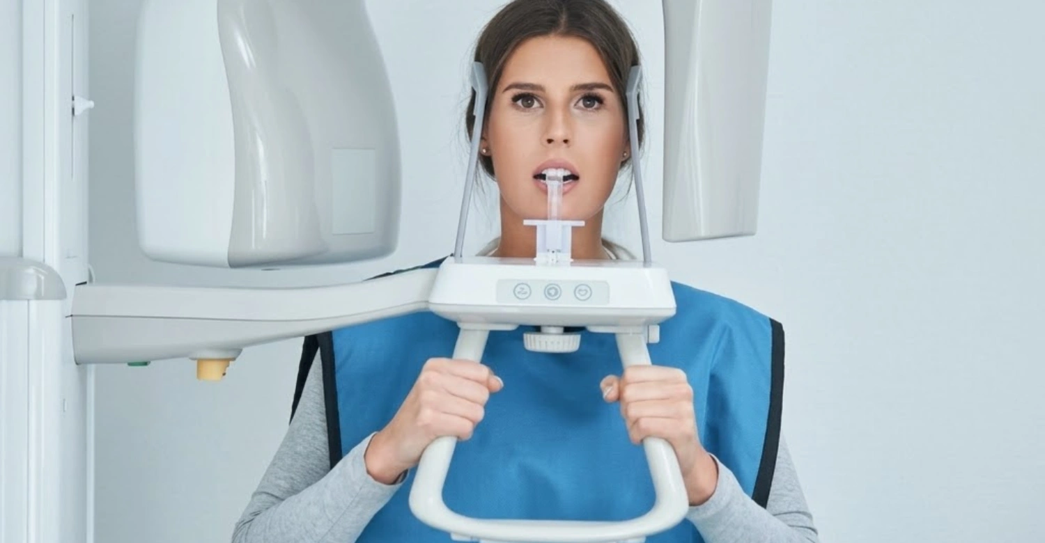
How a CT Scan for Teeth Works
CT scan for teeth works through rotating x-ray beam capturing multiple images reconstructed into three-dimensional model. Understanding mechanism helps patients recognize that scanning creates comprehensive views from single imaging session. Knowledge about technology reveals that computer software processes images creating detailed 3D representation accurately. Professional interpretation of images enables precise measurements and treatment planning with optimal accuracy levels. Advanced technology completes scanning quickly producing detailed images within seconds of x-ray exposure.
Difference Between Traditional X-Rays and CT Scan for Teeth
Traditional x-rays create flat two-dimensional images while ct scan for teeth produces three-dimensional detailed reconstructions. Understanding differences helps patients recognize when advanced imaging provides superior diagnostic value over standard methods. Knowledge reveals that three-dimensional visualization shows relationships between structures invisible on flat x-rays clearly. Professional assessment determines when detailed scanning necessary based on complexity and diagnostic requirements. Advanced imaging recommended for complex cases where standard x-rays provide insufficient information for treatment.
2D X-ray vs 3D Imaging
2D x-ray shows flat overlapping structures while 3D imaging from cbct scan for teeth reveals separate distinct layers. Understanding dimensional differences helps patients appreciate diagnostic advantages of cbct scan for teeth technology. Knowledge reveals that cbct scan for teeth eliminates structure overlap providing clear individual tissue visualization. Professional use of cbct scan for teeth enables precise measurements impossible with traditional two-dimensional imaging. Three-dimensional imaging revolutionizes treatment planning through accurate spatial relationship visualization of anatomical structures.
When a CT Scan Is More Accurate
Advanced imaging proves more accurate for implant planning, impacted teeth evaluation, and complex anatomical assessments. Understanding accuracy advantages helps patients recognize when three-dimensional scanning recommended over standard imaging methods. Knowledge reveals that detailed visualization shows bone quality, nerve locations, and sinus relationships precisely. Professional reliance on advanced imaging prevents complications through detailed pre-treatment anatomical assessment and planning. Complex cases require three-dimensional scanning ensuring safe accurate treatment addressing all anatomical considerations.
Why You May Need a CT Scan for Teeth
Patients may need ct scan for teeth for implant planning, wisdom teeth evaluation, infection detection, and orthodontic assessment. Understanding indications helps patients recognize when advanced imaging provides essential diagnostic information for treatment. Knowledge reveals that detailed visualization is recommended when traditional methods provide insufficient detail for planning procedures. Professional judgment determines necessity based on procedure complexity and specific diagnostic requirements for cases. Advanced imaging ensures comprehensive evaluation when standard methods cannot adequately assess anatomical structures.
Dental Implant Planning
Dental implant planning requires ct scan for teeth measuring bone dimensions, density, and vital structure locations. Understanding implant needs helps patients recognize why advanced imaging is essential for successful implant placement. Knowledge reveals that three-dimensional visualization shows exact bone availability determining implant size and position. Professional implant planning uses detailed imaging preventing nerve damage and ensuring adequate bone support. Accurate pre-surgical assessment dramatically improves implant success rates and long-term treatment outcomes.
Wisdom Teeth Evaluation
Wisdom teeth evaluation uses ct of jaw showing tooth position, root shape, and nerve proximity accurately. Understanding wisdom tooth assessment helps patients recognize why ct of jaw necessary for safe extraction planning. Knowledge about ct of jaw reveals that imaging shows relationship between roots and inferior alveolar nerve. Professional evaluation with ct of jaw prevents nerve damage during extraction through detailed anatomical mapping. Complex wisdom tooth cases require ct of jaw ensuring safe removal without complications from anatomical variations.
Detecting Hidden Tooth Infections
Detecting hidden infections requires advanced imaging revealing abscesses, bone destruction, and infection extent invisible otherwise. Understanding infection detection helps patients recognize that three-dimensional visualization shows problems missed on standard x-rays. Knowledge reveals that detailed imaging displays infection spread determining comprehensive treatment approach for resolution. Professional use of advanced imaging identifies infection sources enabling appropriate intervention preventing serious complications. Advanced imaging detects early bone changes indicating infection before visible on traditional two-dimensional radiographs.
Orthodontic Treatment Planning
Orthodontic treatment planning benefits from three-dimensional imaging showing tooth positions, root angulations, and jaw relationships. Understanding orthodontic applications helps patients recognize when advanced imaging improves treatment precision and outcomes. Knowledge reveals that detailed visualization enables virtual treatment simulation predicting tooth movement accurately beforehand. Professional orthodontic planning uses advanced imaging for complex cases requiring detailed three-dimensional assessment comprehensively. Advanced imaging improves orthodontic outcomes through comprehensive visualization of dental and skeletal relationships clearly.
Jawbone Assessment and Bone Loss Detection
Jawbone assessment uses ct of jaw measuring bone density, dimensions, and detecting periodontal bone loss accurately. Understanding bone evaluation helps patients recognize that ct of jaw provides precise measurements for planning. Knowledge about ct of jaw reveals that imaging quantifies bone loss determining treatment needs objectively. Professional assessment with ct of jaw guides bone grafting decisions through accurate remaining bone measurement. Detailed bone evaluation through ct of jaw essential for implants, extractions, and periodontal treatments.
Types of CT Scan for Teeth
Types include dental ct for general imaging, cbct xray for diagnosis, ct of jaw for complex cases, and ct scan of mouth for comprehensive evaluation. Understanding types helps patients recognize different imaging protocols serving specific diagnostic purposes and clinical needs. Knowledge reveals that cbct scan for teeth represents most common dental imaging providing optimal detail. Professional selection of imaging type depends on diagnostic requirements and anatomical area needing evaluation. Various imaging modalities available optimizing image quality while minimizing radiation exposure appropriately.
Dental CT (3D Dental Imaging)
Dental ct provides comprehensive three-dimensional imaging of teeth, bone, sinuses, and surrounding structures for diagnosis. Understanding dental ct helps patients recognize broad applications for this advanced imaging technology. Knowledge reveals that dental ct captures entire maxillofacial region enabling complete anatomical assessment thoroughly. Professional use of dental ct ranges from implant planning to pathology detection requiring visualization. Comprehensive dental ct imaging provides detailed information guiding treatment decisions across multiple dental specialties.
CBCT Xray for Dental Diagnosis
CBCT xray represents cone beam computed tomography specifically designed for dental and maxillofacial imaging applications. Understanding cbct xray helps patients recognize that this technology optimized for oral structures specifically. Knowledge about cbct xray reveals that lower radiation doses achieved compared to medical CT scans. Professional reliance on cbct xray provides high-resolution images essential for accurate dental diagnosis and planning. Specialized cbct xray technology revolutionized dental imaging through excellent detail with minimal radiation exposure.
CT of Jaw for Complex Cases
CT of jaw focuses on mandible and maxilla providing detailed bone structure visualization for complex treatments. Understanding ct of jaw applications helps patients recognize when focused imaging necessary for specific areas. Knowledge reveals that ct of jaw shows bone quality, pathology, and anatomical variations affecting treatment planning. Professional use of ct of jaw essential for trauma assessment, tumor evaluation, and reconstruction planning. Detailed ct of jaw imaging guides surgical procedures requiring precise anatomical knowledge for success.
CT Scan of Mouth for Full Oral Evaluation
CT scan of mouth captures comprehensive view of entire oral cavity including teeth, bone, and soft tissues. Understanding ct scan of mouth helps patients recognize complete evaluation capabilities of this imaging modality. Knowledge about ct scan of mouth reveals that full arch visualization possible from single scan session. Professional use of ct scan of mouth enables comprehensive assessment for full mouth reconstruction and complex cases. Complete ct scan of mouth imaging provides all necessary information for extensive treatment planning efficiently.
CBCT Scan for Teeth vs Traditional CT
CBCT scan for teeth uses cone beam technology with lower radiation while traditional CT uses fan beam. Understanding differences helps patients recognize that cbct scan for teeth specifically optimized for dental applications. Knowledge reveals that cbct scan for teeth provides superior resolution for hard tissues with minimal exposure. Professional preference for cbct scan for teeth in dental settings due to excellent image quality and safety. Specialized cbct scan for teeth technology offers advantages over medical CT for oral structure imaging.
How a CT Scan for Teeth Is Performed
Performance involves positioning patient, rotating scanner around head, and computer reconstruction creating three-dimensional images quickly. Understanding procedure helps patients recognize that ct scan for teeth quick, painless, and non-invasive imaging process. Knowledge reveals that scanning completed within seconds with minimal patient cooperation required for success. Professional operation of equipment ensures optimal image quality for accurate diagnosis and treatment planning. Simple straightforward procedure makes advanced imaging accessible and comfortable for most patients.
Step-by-Step Procedure
Procedure includes positioning, stabilization, scanner rotation, image capture, and computer processing creating final three-dimensional reconstruction. Understanding steps helps patients know what to expect during imaging appointment at dental facility. Knowledge reveals that entire scanning process typically completed within 10-20 seconds of actual imaging. Professional guidance ensures proper positioning enabling clear images without artifacts affecting diagnostic quality significantly. Quick efficient procedure minimizes patient time while providing comprehensive diagnostic information for treatment.
Preparation Before the Scan
Preparation includes removing metal objects, jewelry, and sometimes dental appliances that create imaging artifacts significantly. Understanding preparation helps patients arrive ready for imaging ensuring optimal image quality results. Knowledge reveals that metal creates scatter affecting image clarity requiring removal beforehand for quality. Professional instruction about preparation ensures successful imaging without need for retakes due to artifacts. Simple preparation steps enable clear images providing accurate diagnostic information for treatment planning.
During the CT Scan
During scan, patient stands or sits still while machine rotates around head capturing images quickly. Understanding scan process helps patients remain motionless ensuring clear images without blur or movement artifacts. Knowledge reveals that movement during scanning creates artifacts reducing diagnostic image quality for interpretation. Professional instruction to remain still ensures successful imaging completed in single quick rotation. Brief scanning time makes procedure comfortable for most patients including children and anxious individuals.
After the Scan
After scan, patient free to resume normal activities immediately as procedure non-invasive requiring no recovery. Understanding post-scan protocol helps patients recognize no restrictions following imaging appointment at facility. Knowledge reveals that images processed quickly enabling same-day treatment planning in many cases. Professional review of images determines diagnosis and treatment recommendations promptly for patient care. No post-procedure care required allowing immediate return to daily activities without restrictions.
Benefits of CT Scan for Teeth
Benefits include accurate diagnosis, three-dimensional visualization, faster treatment planning, and improved safety through detailed anatomical assessment. Understanding benefits helps patients appreciate value of ct scan for teeth for complex dental treatments. Knowledge reveals that advanced imaging improves outcomes through precise pre-treatment planning and comprehensive evaluation. Professional use prevents complications through comprehensive anatomical visualization before procedures and interventions. Advanced imaging provides significant advantages over traditional methods improving treatment precision and patient safety.
Accurate Diagnosis
Accurate diagnosis achieved through detailed imaging revealing information invisible on traditional two-dimensional x-rays clearly. Understanding diagnostic accuracy helps patients recognize that advanced imaging detects problems earlier enabling intervention. Knowledge reveals that three-dimensional visualization shows relationships between structures preventing misdiagnosis from overlapping anatomy. Professional diagnostic confidence increases with detailed imaging enabling appropriate treatment recommendations and planning. Superior imaging quality ensures problems identified accurately before they progress to advanced stages.
3D Imaging Advantages
3D imaging advantages include viewing structures from multiple angles, precise measurements, and virtual treatment simulation capabilities. Understanding dimensional benefits helps patients appreciate why advanced imaging superior to flat traditional radiographs. Knowledge reveals that three-dimensional visualization enables comprehensive assessment impossible with traditional two-dimensional methods. Professional treatment planning improved dramatically through three-dimensional reconstruction and measurement capabilities for precision. Advanced visualization enables better patient communication through clear demonstration of anatomical conditions requiring treatment.
Faster Treatment Planning
Faster treatment planning results from comprehensive information provided from single imaging session reducing appointments. Understanding efficiency helps patients recognize that advanced imaging streamlines diagnostic process significantly reducing delays. Knowledge reveals that detailed visualization eliminates need for multiple traditional x-rays from different angles. Professional efficiency improved through advanced imaging enabling same-day treatment planning in many cases. Comprehensive imaging reduces delays allowing prompt treatment initiation when time-sensitive conditions present requiring intervention.
Improved Patient Safety
Improved safety achieved through detailed visualization showing vital structures like nerves and sinuses preventing damage. Understanding safety benefits helps patients recognize that advanced imaging reduces complication risks significantly. Knowledge reveals that three-dimensional visualization enables avoidance of anatomical structures during surgery through mapping. Professional confidence in safety increases with detailed anatomical visualization before procedures and interventions. Advanced pre-treatment assessment prevents injuries through comprehensive understanding of individual anatomical variations and relationships.
Is CT Scan for Teeth Safe?
Yes, dental imaging is safe using significantly lower radiation than medical CT scans while providing diagnostic benefits. Understanding safety helps patients recognize that cbct scan for teeth uses minimal radiation doses effectively. Knowledge reveals that benefits typically outweigh minimal radiation risks substantially for diagnostic purposes. Professional judgment determines when imaging benefits justify radiation exposure for appropriate diagnostic purposes. Modern technology minimizes exposure while maintaining image quality ensuring safety standards maintained consistently.
Radiation Exposure Explained
Radiation exposure from dental imaging comparable to few days of natural background radiation received daily. Understanding exposure levels helps patients recognize that advanced imaging uses minimal radiation appropriately. Knowledge reveals that cbct delivers 10-100 times less radiation than medical CT scans. Professional protocols minimize exposure through collimation and optimized scanning parameters carefully controlled. Low radiation doses make imaging safe for most patients when clinically indicated.
Safety of CBCT Xray
Safety of cbct xray ensured through low radiation doses, focused imaging fields, and appropriate use guidelines. Understanding cbct xray safety helps patients feel confident about recommended advanced imaging procedures. Knowledge reveals that cbct xray technology specifically designed minimizing exposure while maintaining diagnostic quality. Professional adherence to safety protocols ensures cbct xray used appropriately when benefits outweigh risks. Modern cbct xray equipment incorporates dose reduction features protecting patients during necessary imaging.
Who Should Avoid Dental CT Scans
Pregnant women should avoid advanced dental imaging unless absolutely necessary as radiation affects developing babies. Understanding contraindications helps patients inform dentists about pregnancy before imaging procedures scheduled. Knowledge reveals that alternative imaging used during pregnancy unless advanced scanning essential for treatment. Professional caution exercised for pregnant patients postponing imaging when possible until after delivery. Children receive imaging only when absolutely necessary using lowest possible radiation doses available.
When Dentists Recommend Dental CT
Dentists recommend dental ct for implant planning, root canal complications, impacted teeth, and TMJ disorders requiring imaging. Understanding indications helps patients recognize when dental ct provides essential diagnostic information for treatment. Knowledge reveals that dental ct recommended when traditional x-rays provide insufficient detail for planning. Professional judgment determines dental ct necessity based on procedure complexity and diagnostic requirements carefully. Advanced imaging ensures comprehensive evaluation when standard methods cannot adequately assess anatomical structures.
Dental Implant Planning
Dental implant planning requires detailed imaging measuring bone dimensions accurately before surgical placement procedures. Understanding implant imaging helps patients recognize why advanced visualization essential for successful outcomes. Knowledge reveals that three-dimensional imaging shows bone quality determining implant size and position. Professional implant planning relies on detailed visualization preventing complications through comprehensive assessment. Accurate measurements ensure proper implant selection and placement success rates improve significantly.
Root Canal Complications
Root canal complications require advanced imaging identifying extra canals, calcifications, or infections missed previously. Understanding endodontic applications helps patients recognize when detailed visualization necessary for treatment success. Knowledge reveals that three-dimensional imaging shows canal anatomy guiding treatment accurately for resolution. Professional use of advanced imaging improves root canal success through comprehensive canal visualization. Complex cases benefit from detailed imaging revealing anatomical variations affecting treatment outcomes.
Impacted Teeth Assessment
Impacted teeth assessment uses detailed imaging showing exact tooth position and surrounding structure relationships. Understanding impaction imaging helps patients recognize why advanced visualization necessary for safe extraction. Knowledge reveals that three-dimensional imaging displays root proximity to nerves preventing damage during surgery. Professional surgical planning relies on detailed imaging for complex impaction cases requiring precision. Detailed imaging ensures safe extraction through comprehensive pre-surgical anatomical assessment and mapping.
Jaw Pain and TMJ Disorders
Jaw pain and TMJ disorders benefit from ct of jaw evaluating joint structures and bone changes. Understanding TMJ imaging helps patients recognize when ct of jaw provides diagnostic information for treatment. Knowledge reveals that ct of jaw shows joint anatomy, arthritis, and structural abnormalities causing pain. Professional TMJ assessment uses ct of jaw determining appropriate treatment approaches for disorders. Detailed joint evaluation through ct of jaw guides treatment decisions for complex jaw problems.
Expert Tips Before Getting a CT Scan for Teeth
Preparation includes wearing comfortable clothing without metal, informing dentist about medical conditions, and following instructions carefully. Understanding preparation helps patients ensure successful imaging producing optimal diagnostic results for treatment. Knowledge reveals that proper preparation prevents need for repeated scanning due to artifacts affecting quality. Professional guidance about preparation ensures efficient imaging appointments without delays or quality issues. Simple preparation steps enable high-quality images providing accurate information for treatment planning.
What to Wear
Wear comfortable clothing without metal zippers, buttons, or accessories that create imaging artifacts significantly. Understanding clothing requirements helps patients dress appropriately for imaging appointments at dental facilities. Knowledge reveals that metal creates scatter on images reducing clarity and diagnostic quality. Professional recommendation to avoid metal ensures clear images without artifacts requiring scan repetition. Appropriate clothing choices enable successful imaging completed in single session without quality issues.
Things to Inform Your Dentist About
Inform dentist about pregnancy, metal implants, pacemakers, and recent radiation exposure before imaging procedures. Understanding disclosure importance helps patients provide relevant medical information ensuring safety during imaging. Knowledge reveals that certain conditions require protocol modifications or alternative imaging methods instead. Professional assessment of medical history determines imaging appropriateness and necessary precautions for safety. Complete disclosure enables safe appropriate imaging tailored to individual patient needs and conditions.
How to Prepare for Accurate Results
Prepare by removing jewelry, hairpins, glasses, and dental appliances before imaging procedures begin. Understanding preparation helps patients ensure artifact-free images producing diagnostic quality results for interpretation. Knowledge reveals that removable items create shadows on images affecting interpretation and accuracy. Professional instruction about removal ensures clear images enabling accurate diagnosis and treatment planning. Thorough preparation prevents repeat scans due to poor image quality from artifacts.
Cost of CT Scan for Teeth
Cost varies from $250-600 depending on facility, imaging extent, and geographic location affecting pricing. Understanding costs helps patients plan financially for advanced imaging when recommended for treatment. Knowledge reveals that investment worthwhile considering improved treatment outcomes and safety benefits significantly. Professional discussion about costs enables informed decisions about necessity and timing of procedures. Comprehensive imaging provides value through preventing complications and improving treatment precision significantly.
Factors Affecting Price
Factors include imaging volume, geographic location, facility type, and radiologist interpretation fees affecting total cost. Understanding price variations helps patients recognize why costs differ between providers and facilities. Knowledge reveals that comprehensive ct scan of mouth costs more than focused limited imaging. Professional facilities with advanced equipment may charge higher fees for services provided. Multiple factors contribute to final cost requiring discussion with facility about specific pricing.
Insurance Coverage
Insurance coverage varies with many plans covering medically necessary imaging for specific clinical indications. Understanding coverage helps patients verify benefits before procedures reducing unexpected expenses significantly. Knowledge reveals that diagnostic imaging often covered while routine screening may not receive benefits. Professional documentation of medical necessity improves insurance approval chances significantly for procedures. Verify coverage details before scheduling avoiding surprise bills after imaging procedures completed.
Is Dental CT Worth the Cost?
Dental ct worth cost considering improved diagnosis accuracy, treatment success, and complication prevention benefits. Understanding value helps patients recognize that investment prevents expensive treatment failures long-term. Knowledge reveals that advanced imaging saves money through proper treatment planning preventing complications. Professional recommendation based on clinical benefit outweighing cost considerations for patient care. Advanced imaging provides value through superior outcomes justifying expense for complex dental treatments.
CT Scan for Teeth at Vitrin Clinic
Vitrin Clinic provides comprehensive imaging services using advanced cbct scan for teeth technology for patients. Understanding Vitrin Clinic capabilities helps patients access quality imaging when advanced visualization is needed. Knowledge about Vitrin Clinic reveals commitment to accurate diagnosis through state-of-the-art equipment. Professional excellence at Vitrin Clinic ensures optimal image quality for treatment planning purposes. Patient-centered approach at Vitrin Clinic prioritizes comfort during imaging procedures.
Advanced CBCT Scan for Teeth Technology
Advanced cbct scan for teeth technology at Vitrin Clinic provides high-resolution images with minimal radiation exposure. Understanding technology benefits helps patients recognize quality imaging available at Vitrin Clinic facilities. Knowledge reveals that Vitrin Clinic invests in latest equipment ensuring diagnostic accuracy. Professional operation of technology ensures optimal images for diagnosis and planning purposes. State-of-the-art equipment at Vitrin Clinic provides superior quality and safety.
Experienced Dental Specialists
Experienced dental specialists at Vitrin Clinic interpret images providing accurate diagnoses for patients. Understanding expertise helps patients recognize quality interpretation available at Vitrin Clinic for imaging. Knowledge reveals that Vitrin Clinic employs professionals trained in image analysis and interpretation. Professional expertise ensures accurate interpretation guiding appropriate treatment recommendations for optimal care. Specialized training at Vitrin Clinic provides superior diagnostic accuracy from images.
Comfortable and Quick Scanning Process
Comfortable quick scanning process at Vitrin Clinic ensures positive experience for patients. Understanding procedure helps patients feel confident about appointments at Vitrin Clinic facilities. Knowledge reveals that Vitrin Clinic prioritizes patient comfort during imaging procedures. Professional staff at Vitrin Clinic ensures efficient scanning completed quickly minimizing appointment time. Patient-focused approach makes imaging comfortable and stress-free at Vitrin Clinic facilities.
Why Choose Vitrin Clinic for Dental CT
Choose Vitrin Clinic for advanced technology, experienced specialists, comprehensive care, and patient-centered approach to imaging. Understanding advantages helps patients select quality provider for advanced imaging when needed. Knowledge reveals that Vitrin Clinic offers complete services with expert interpretation available. Professional excellence at Vitrin Clinic ensures accurate imaging providing optimal treatment outcomes. Comprehensive care at Vitrin Clinic makes imaging accessible, comfortable, and accurately interpreted.
FAQs

Dr. Rifat Alsaman has more than 5 years of clinical experience in dentistry and currently serves as the Head of the Medical Team at Vitrin Clinic. He is dedicated to providing exceptional patient care, overseeing treatment planning, and ensuring the highest clinical standards across the team. His expertise, attention to detail, and commitment to continuous professional development have helped countless patients achieve healthier, more confident smiles.
.webp&w=3840&q=75)


.webp&w=3840&q=75)

