
The Enamel Dentin Junction is the specialized boundary connecting tooth enamel with the underlying dentin. This interface creates a strong transition between two tissues with different structures and mechanical properties. Enamel provides a hard, mineral-rich outer covering that protects the tooth from daily forces. Dentin lies beneath enamel and provides resilience because it contains more organic material and water. The interface helps these tissues work together during biting, chewing, and other functional stresses. Its microscopic architecture also influences how cracks move through the tooth. Understanding this boundary helps explain important aspects of tooth anatomy, tooth strength, and dental restoration. Research using advanced imaging has shown that its complex structure contributes to the durability of natural teeth. At Vitrin Clinic, understanding tooth structure supports conservative treatment planning and appropriate cosmetic or restorative procedures.
What Is the Enamel Dentin Junction?
It represents the natural transition between enamel and dentin within a tooth. It is not simply a flat dividing line between two tissues. Instead, the interface has a complex three-dimensional architecture that helps connect enamel and dentin mechanically. Enamel contains a very high mineral content, making it extremely hard but relatively brittle. Dentin contains more collagen and fluid, giving it greater flexibility and toughness. Their different properties create mechanical challenges during normal chewing forces. The interface helps distribute these forces between the two tissues. It also contributes to the tooth's ability to resist crack development and progression. Scientists study this region using microscopy, imaging techniques, and mechanical testing. These investigations provide insights into natural tooth durability and guide improvements in restorative dentistry.
Enamel Dentin Junction Definition
The Enamel Dentin Junction is the anatomical interface where tooth enamel meets underlying dentin. It forms during tooth development as specialized cells produce these mineralized tissues. Its structure differs from both enamel and dentin because it represents their biological and mechanical connection. Microscopic studies describe this interface as an irregular, three-dimensional boundary rather than a perfectly smooth surface. This architecture increases interaction between the adjacent tissues. Enamel is highly mineralized and resistant to surface wear. Dentin is less mineralized and contains a collagen-rich organic framework. The interface therefore connects materials with substantially different physical characteristics. This transition is important because abrupt changes between hard and resilient tissues could increase mechanical stress. The specialized structure helps create a functional connection within the natural tooth. This definition is essential for understanding tooth microstructure and dental biomechanics. Pediatric dentistry: Teeth Abnormalities In Children
Why Is the EDJ Important?
This interface is important because it helps enamel and dentin function together during repeated mechanical loading. Enamel can withstand substantial compressive forces but has limited resistance to certain forms of tensile stress. Dentin provides a more resilient foundation beneath the hard enamel layer. The interface helps transfer mechanical forces between these tissues. Its complex geometry can influence how stresses develop inside the tooth. It also affects how cracks initiate and travel through dental tissues. Researchers consider this region important when studying natural tooth fracture resistance. Understanding its behavior can also inform restorative material design. Modern dental materials attempt to reproduce some characteristics of natural tissue transitions. This knowledge has relevance for crowns, veneers, fillings, and other restorations. Preserving healthy tooth structure remains important because natural tissue relationships are difficult to reproduce completely.
Clinical Note
The interface is clinically relevant because treatment can alter the relationship between enamel and dentin. Tooth preparation may remove enamel and expose underlying dentin during restorative procedures. Exposed dentin can respond differently because it contains microscopic tubules and has different physical properties. Conservative dentistry therefore aims to preserve healthy enamel whenever treatment allows. The amount of remaining tooth structure can influence restoration stability and biological considerations. Tooth wear, erosion, fractures, and extensive preparation can also change the natural tissue interface. Clinicians evaluate these factors before selecting restorative approaches. At Vitrin Clinic, tooth structure assessment can help guide personalized treatment planning. Digital imaging and clinical examination may provide information about remaining enamel and dentin. Protecting natural tissues can support predictable restorative outcomes and long-term tooth function. Neurological connections: Oral Microbiome And Migraine
Where Is the Enamel Dentin Junction Located?
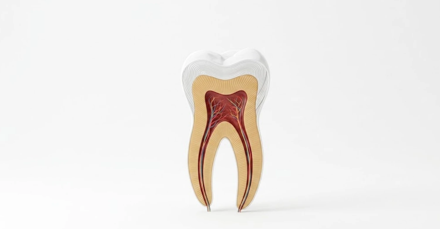
The Enamel Dentin Junction is located inside the crown of a tooth, directly beneath the enamel layer. Enamel covers the external crown surface and forms the tooth's primary protective barrier. Dentin occupies the deeper portion beneath enamel and continues toward the root. The interface follows the internal contour of the enamel throughout the crown. Its shape therefore differs according to the tooth's anatomy and location. Near cusps and ridges, its three-dimensional configuration reflects the complex external shape of enamel. Around pits and developmental structures, the interface can also show considerable morphological variation. This location makes the boundary important for understanding the relationship between external tooth form and internal structure. It should not be confused with the cementoenamel junction, which occurs externally near the cervical region. Knowing these locations helps clinicians and students interpret tooth anatomy accurately.
Location of the EDJ in a Tooth
This boundary lies immediately beneath the enamel of the anatomical crown. It generally follows the contour created by the enamel's inner surface. In a healthy tooth, it remains enclosed within the mineralized crown rather than being directly visible clinically. The interface extends across the crown and surrounds areas containing dentin beneath enamel. Its exact morphology varies between teeth and between different regions of the same tooth. Cusps may have complex internal contours because their external anatomy experiences substantial functional forces. The interface also contributes to the structural relationship between enamel thickness and underlying dentin. Understanding its location is particularly useful when interpreting microscopic sections and advanced dental imaging. It also helps explain why enamel removal can expose dentin during tooth preparation. Clinicians consider this relationship when planning conservative restorative and cosmetic procedures.
Relationship Between the Enamel Layer and Dentin Layer
This structure creates the transition between the enamel layer and dentin layer. Enamel forms the highly mineralized outer covering of the crown. Dentin forms the supporting tissue beneath enamel and extends through much of the tooth. These tissues differ considerably in mineral composition, organic content, hardness, and elasticity. Enamel is harder but generally less tolerant of tensile deformation. Dentin is softer but provides greater resilience and energy absorption. Their interface allows these contrasting materials to function as a coordinated structure. This relationship is particularly important during chewing because teeth experience repeated loading cycles. The supporting dentin can help reduce the consequences of stresses affecting enamel. Meanwhile, enamel protects dentin from direct environmental and mechanical exposure. Preserving both tissues helps maintain the natural biomechanical behavior of the tooth.
EDJ and Tooth Anatomy
This transition is an important feature of tooth anatomy because it separates two major mineralized tissues. Understanding its position helps explain the internal organization of the dental crown. Enamel forms the external protective shell, while dentin provides most of the tooth's internal bulk. The interface follows the changing contours of the crown rather than forming a simple geometric plane. Its morphology can therefore reflect developmental patterns and functional adaptation. Tooth anatomy studies often examine this boundary alongside enamel thickness, dentin distribution, and pulp location. Researchers can also use the interface to investigate developmental changes and evolutionary differences. Three-dimensional imaging has expanded understanding of its complex internal shape. These findings demonstrate that tooth anatomy involves interconnected structures rather than isolated tissue layers. Accurate anatomical knowledge supports diagnosis, restorative planning, and conservative dental treatment. Systemic link: Can Tooth Infection Cause Gallbladder Problems
What Does the Enamel Dentin Junction Look Like?
The Enamel Dentin Junction appears as a distinct microscopic boundary between two mineralized dental tissues. It is not completely smooth when examined using high-resolution microscopy. Instead, researchers describe a complex, undulating, and three-dimensional morphology. This architecture varies across different regions of the tooth. The interface can contain microscopic features that increase the mechanical connection between enamel and dentin. Its appearance also depends on the imaging technique, specimen preparation, and scale of observation. Under microscopy, enamel shows highly organized mineral structures, while dentin displays tubules within its mineralized matrix. The boundary separates these different microstructures while maintaining their physical connection. Advanced imaging has provided increasingly detailed views of this transition. These observations help researchers understand why natural teeth can tolerate repeated mechanical loading without immediately separating into distinct tissue layers.
Enamel Dentin Interface Under a Microscope
Under a microscope, this boundary can appear irregular rather than perfectly straight. Its three-dimensional architecture reflects the developmental interaction between enamel-forming and dentin-forming tissues. Different microscopy methods reveal different structural details at this boundary. Light microscopy may show the interface as a distinct transition between enamel and dentin. Electron microscopy can reveal finer mineral and structural characteristics. Advanced three-dimensional techniques can demonstrate its complex spatial organization. Enamel contains densely packed mineral structures arranged into organized patterns. Dentin contains tubules extending through a collagen-containing mineralized matrix. The interface connects these contrasting microstructures. Researchers study this region to understand crack resistance, mineralization, and tissue development. Microscopic observations have helped establish that the boundary has functional characteristics beyond simply separating enamel from dentin.
EDJ Tooth Microstructure
It forms an important component of the tooth's microstructure. Its architecture connects highly mineralized enamel with the more resilient dentin beneath it. At microscopic scales, enamel contains organized hydroxyapatite-rich structures. Dentin contains mineralized collagen and numerous microscopic dentinal tubules. The interface accommodates these different structural organizations. Researchers have found that its morphology can vary according to tooth region and developmental history. Three-dimensional studies are particularly valuable because traditional two-dimensional sections cannot capture the complete architecture. Microstructural analysis can also help explain mechanical behavior under chewing forces. The interface contributes to the overall organization of the tooth rather than functioning as an isolated boundary. Understanding these details helps researchers investigate natural fracture resistance and develop improved biomimetic restorative materials. This field continues to connect dental anatomy with materials science and biomechanics. Nutritional aid: Omega 3 For Periodontitis
How Enamel and Dentin Connect
This interface connects enamel and dentin through a complex biological and structural transition. The interface develops during tooth formation as specialized cells produce and mineralize neighboring tissues. Its irregular morphology creates a mechanically integrated boundary between enamel and dentin. This connection is important because the two tissues have different hardness and deformation characteristics. Enamel provides surface protection, while dentin contributes underlying toughness and support. During chewing, forces pass through these interconnected tissues rather than acting on enamel alone. The interface helps manage differences between their mechanical behaviors. Its structure may also influence how microscopic cracks develop and change direction. Natural teeth therefore use tissue organization as part of their fracture-resistance strategy. Researchers study these characteristics to inspire restorative materials that better reproduce natural tooth biomechanics.
What Is the Function of the Enamel Dentin Junction?
The Enamel Dentin Junction functions as a structural transition that allows enamel and dentin to work together. Enamel provides exceptional hardness and resistance to surface wear. Dentin provides a more compliant and resilient foundation beneath that hard tissue. The interface helps transfer stresses between these materials during normal tooth function. Its three-dimensional morphology can also influence crack behavior within the crown. This is important because enamel can fracture when subjected to unfavorable stress conditions. A supportive dentin foundation can reduce some consequences of enamel brittleness. The interface therefore contributes to the overall mechanical performance of the natural tooth. Its function is also relevant to restorative dentistry because replacement materials must interact with remaining tooth tissues. Preserving natural tissue relationships can help maintain the biological and mechanical characteristics of the tooth.
How the EDJ Connects Enamel and Dentin
The interface creates a mechanically integrated transition between enamel and dentin. These tissues differ substantially in hardness, elasticity, mineralization, and organic composition. A simple flat boundary would provide limited adaptation between such contrasting materials. Instead, the natural interface has a complex three-dimensional structure. This morphology increases the interaction between adjacent tissue regions. Forces generated during chewing can therefore be transferred through a broader structural transition. Dentin can deform slightly under loading, while enamel remains comparatively rigid. The interface accommodates these differences while maintaining tissue continuity. Researchers describe this relationship as an important component of natural tooth biomechanics. Understanding this connection also helps explain why preserving natural enamel and dentin can be valuable during dental treatment. The interface is therefore both an anatomical boundary and a functional structural connection.
How the EDJ Supports Tooth Strength
This boundary contributes to tooth strength by connecting a hard outer tissue with a tougher internal tissue. Enamel resists abrasion and compressive loading but can be susceptible to crack formation. Dentin provides greater capacity for deformation and energy absorption. Their combination produces a tooth structure that balances hardness with resilience. The interface helps transfer loads between these materials during functional activity. Its irregular morphology may also reduce the effects of abrupt mechanical transitions. Studies of natural teeth show that tissue architecture strongly influences fracture behavior. This is why tooth strength cannot be understood by measuring enamel hardness alone. The surrounding dentin and interface also contribute to overall performance. These principles are relevant when dentists determine how much natural tissue should remain during restorative treatment.
How the EDJ Helps Resist Cracks
This structure can influence how cracks move through the tooth. Enamel is highly mineralized and comparatively brittle, making crack development mechanically important. Dentin is tougher and can absorb greater deformation before failure. When a crack approaches the interface, changes in material properties can affect its direction and progression. The irregular architecture may also create a more complex pathway for crack movement. Researchers study these mechanisms to understand why natural teeth can resist catastrophic fracture. Crack behavior depends on many factors, including tooth geometry, loading direction, defects, and tissue condition. Therefore, the interface should not be considered the only factor controlling fracture resistance. Nevertheless, its structure forms an important part of the tooth's natural defense against crack propagation. These findings also inspire biomimetic approaches in restorative material development.
Dr. Rifat Alsaman's Clinical Opinion
This transition matters clinically because natural tooth tissues work as an integrated structure. Dr. Rifat Alsaman emphasizes the value of preserving healthy tooth structure whenever clinically appropriate. Removing unnecessary enamel can alter the relationship between the external surface and underlying dentin. Excessive preparation may also increase the amount of dentin exposed during restorative treatment. Conservative planning therefore begins with understanding the existing anatomy. Each tooth should be assessed according to its condition, function, and restorative requirements. Cosmetic treatment should focus on achieving the desired result while respecting biological structures. At Vitrin Clinic, this principle supports individualized treatment planning for restorative and cosmetic procedures. The goal is not simply to change appearance but to maintain predictable function. Careful assessment can help balance aesthetics, strength, tissue preservation, and long-term oral health. Comprehensive Breakdown Of Tooth Anatomy
Why Is the Enamel Dentin Junction Important for Tooth Strength?
The Enamel Dentin Junction is important because tooth strength depends on the interaction between enamel and dentin. Enamel provides exceptional hardness but has limited capacity for deformation. Dentin provides flexibility and toughness beneath the enamel shell. Their combination creates a natural structure capable of handling repeated chewing forces. The interface helps manage mechanical differences between these tissues. It can influence stress distribution and crack behavior within the crown. Tooth fracture is therefore affected by more than enamel thickness or hardness alone. Researchers use mechanical testing and microscopy to investigate these interactions. Findings from dental biomechanics show that hierarchical organization contributes substantially to natural tooth performance. This knowledge has influenced research into restorative materials designed to imitate natural tissue behavior. Maintaining healthy tooth structure remains important because artificial materials may not perfectly reproduce this complex architecture.
EDJ and Stress Distribution
It contributes to how mechanical stresses are transferred between enamel and dentin. Enamel is rigid, while dentin has greater capacity for elastic deformation. During biting, these differences create complex stress patterns within the tooth. The interface provides a transition between these contrasting mechanical environments. Its three-dimensional morphology can influence the concentration and direction of local stresses. Tooth geometry also affects stress distribution, particularly around cusps, grooves, and contact areas. Repeated loading can create microscopic fatigue even when no visible fracture exists. The combination of tissue properties and interface architecture helps the tooth tolerate these repeated forces. Researchers use finite-element models and mechanical testing to investigate these processes. Such studies contribute to better understanding of natural tooth durability and restorative material performance.
EDJ and Crack Propagation
This interface can affect the path followed by cracks traveling through enamel. Cracks develop when local stresses exceed the tissue's resistance to fracture. When a crack reaches the interface, the difference between enamel and dentin properties becomes mechanically important. The crack may change direction, slow down, or continue into deeper tissue depending on multiple conditions. Interface morphology can contribute to this behavior by creating a complex transition zone. Dentin's greater toughness can also influence how fracture energy is dissipated. However, crack resistance depends on tooth shape, loading conditions, defects, and tissue quality. Researchers therefore study the entire tooth structure rather than the interface alone. These findings help explain why natural teeth often show sophisticated resistance to catastrophic fracture.
Difference in the Mechanical Properties of Enamel and Dentin
The interface connects tissues with markedly different mechanical properties. Enamel is the hardest tissue in the human body and contains a very high proportion of minerals. Dentin contains substantially more organic material and water than enamel. Consequently, dentin is less hard but generally more resilient and tougher. Enamel primarily provides resistance against surface wear and deformation. Dentin supports enamel and can absorb mechanical energy through controlled deformation. This contrast creates a functional combination of hardness and toughness. The interface helps connect these different behaviors within one tooth. Researchers measure properties such as hardness, elastic modulus, fracture toughness, and fatigue resistance. Values vary according to testing methods, tooth location, hydration, and specimen preparation. These differences explain why preserving the natural enamel-dentin relationship remains important in restorative dentistry. Inner tooth anatomy: Dental Pulp
What Happens When the Enamel Dentin Junction Is Exposed?
The Enamel Dentin Junction normally remains protected beneath the enamel layer inside the tooth crown. It does not usually become directly exposed during normal oral function. However, extensive tooth wear, erosion, fractures, or aggressive preparation can remove enough enamel to expose dentin. Once dentin becomes exposed, sensitivity can occur because dentinal tubules can communicate with the pulp. Patients may notice short, sharp discomfort from cold, heat, sweetness, or touch. The severity depends on the amount of tissue loss and the individual tooth. Dentists evaluate the cause before recommending treatment. Management may involve reducing damaging habits, improving preventive care, or restoring lost structure. At Vitrin Clinic, evaluation can help determine whether exposed dentin requires monitoring or restorative intervention. Early assessment may help prevent progressive tissue loss.
Can the EDJ Become Exposed?
This boundary itself is normally protected by enamel and remains inside the tooth crown. It can become closer to the external surface when significant enamel loss occurs. However, clinicians generally describe the resulting condition as dentin exposure rather than direct clinical exposure of the microscopic interface. Enamel can be lost through erosion, abrasion, attrition, fractures, developmental defects, or dental preparation. As enamel becomes thinner, underlying dentin may eventually become visible or exposed. This can change the tooth's appearance and increase sensitivity. The degree of exposure depends on the location and severity of tissue loss. Professional evaluation is important because similar symptoms can have different causes. Preserving remaining enamel and dentin is generally preferable whenever clinically possible. Treatment should address both the underlying cause and the resulting structural problem.
Causes of Enamel and Dentin Exposure
This structure can become clinically relevant when enamel loss exposes the underlying dentin. Common contributors include dental erosion, abrasive brushing, tooth grinding, and traumatic fractures. Acid exposure from dietary sources can gradually soften enamel and accelerate tissue loss. Mechanical abrasion can remove weakened tooth surfaces through repeated friction. Bruxism can create substantial functional wear over time. Dental procedures may also expose dentin when enamel is intentionally or unintentionally removed. Developmental defects can produce areas with reduced enamel thickness or altered mineralization. Gum recession can expose root dentin, although this area does not contain enamel over its entire surface. Diagnosis requires identifying the specific mechanism responsible for tissue loss. Addressing the cause can help prevent continued damage and reduce sensitivity.
Symptoms of Exposed Dentin
This transition becomes clinically significant when enamel loss exposes underlying dentin. Exposed dentin may produce sensitivity because fluid movement can occur within dentinal tubules. Typical symptoms include brief, sharp discomfort triggered by cold drinks or foods. Sweet, acidic, hot, or tactile stimuli can also trigger sensitivity. Pain that lingers after removing the stimulus may suggest a different pulpal condition. The location and intensity of symptoms vary among individuals. Some patients have exposed dentin without noticeable sensitivity. Others experience significant discomfort that affects eating or drinking habits. A dental examination can determine whether sensitivity results from exposed dentin or another condition. Treatment depends on the underlying cause and may include preventive measures or restorative approaches. Persistent or spontaneous pain should receive professional evaluation.
Tips for Patients
It is protected when patients maintain the enamel and dentin surrounding it. Use a soft-bristled toothbrush and avoid aggressive horizontal brushing. Fluoride toothpaste can support enamel resistance against acid-related mineral loss. Limit frequent exposure to acidic foods and beverages throughout the day. Avoid using excessive pressure when brushing sensitive areas. Patients who grind their teeth may benefit from professional assessment and appropriate protective strategies. Do not ignore persistent sensitivity because it can have multiple causes. Regular dental examinations can identify enamel loss before significant structural damage develops. If cosmetic treatment is planned, discuss conservative preparation options with your dentist. At Vitrin Clinic, individualized assessments can help match treatment choices with existing tooth structure. Protecting natural tissues is an important part of maintaining long-term dental health. Aesthetic care: Does Coffee Stain Veneers Or Crowns
Enamel Dentin Junction vs. Cementoenamel Junction
The Enamel Dentin Junction and cementoenamel junction are different anatomical structures within a tooth. The first lies between enamel and dentin inside the crown. The cementoenamel junction occurs externally where enamel meets cementum near the tooth's cervical region. These structures therefore have different locations, developmental characteristics, and clinical relevance. The internal interface supports the relationship between enamel and dentin. The cervical junction marks the transition between enamel covering the crown and cementum covering the root. Confusing these terms can lead to inaccurate descriptions of tooth anatomy. Dental students and patients may encounter both terms during discussions about tooth structure. Understanding their differences also helps explain sensitivity, gum recession, and restorative procedures. At Vitrin Clinic, accurate anatomical assessment supports appropriate treatment planning and conservative care.
What Is the Difference Between EDJ and CEJ?
This interface separates enamel from dentin within the tooth. The cementoenamel junction separates enamel from cementum around the cervical tooth region. The two structures therefore involve different dental tissues and occupy different anatomical positions. The internal interface remains beneath the enamel of the crown. The cementoenamel junction is closer to the gumline and can sometimes be clinically visible. Root surfaces below this junction are generally covered by cementum rather than enamel. Their developmental origins also differ because different mineralized tissues form during tooth development. Clinically, the internal interface relates mainly to tooth microstructure and mechanical behavior. The cervical junction has greater relevance to periodontal anatomy and root exposure. Distinguishing these structures improves communication between dentists, researchers, students, and patients.
EDJ vs. CEJ Location
The interface is located inside the anatomical crown, directly beneath enamel. The cementoenamel junction is located externally near the cervical area of the tooth. The internal interface follows the inner contour of enamel across the crown. The cervical junction follows the external boundary between crown and root covering tissues. These locations can be visualized differently depending on imaging and anatomical sectioning techniques. The internal boundary is generally not visible during a routine clinical examination. The cervical boundary may become visible when gingival recession exposes the root surface. This distinction is important when interpreting dental diagrams and clinical terminology. Understanding location also helps explain why different tissue exposures produce different symptoms. Accurate anatomical identification supports better diagnosis and treatment planning.
Why These Terms Should Not Be Confused
This boundary should not be confused with the cementoenamel junction because they describe different boundaries. One connects enamel and dentin, while the other connects enamel and cementum. Their anatomical positions are also significantly different. Confusing them can create misunderstandings when discussing tooth structure or clinical treatment. The internal interface is associated with enamel support, dentin, and tooth biomechanics. The cervical junction is associated with the transition from crown tissues toward the root. Gum recession can expose areas around the cervical junction. Enamel loss can instead expose dentin beneath the crown surface. These conditions require different clinical evaluations and management strategies. Clear terminology is particularly important when dentists explain treatment findings to patients. Vitrin Clinic uses detailed tooth assessment to distinguish structural and periodontal concerns accurately. Structural guide to Parts Of A Tooth
Enamel Dentin Junction vs. Dentin Enamel Junction
This boundary and the term dentin enamel junction generally describe the same anatomical boundary. Both expressions refer to the interface where enamel meets underlying dentin. Dental literature may use different word orders depending on the author or scientific context. The abbreviation EDJ is commonly used for this interface. Another related expression is enamel-dentin interface. These terms should therefore be interpreted according to their surrounding context. The structure itself remains the same anatomical transition between enamel and dentin. Understanding terminology is especially useful when searching scientific publications or dental research databases. Different naming conventions can otherwise make relevant studies difficult to identify. For SEO and patient education, using common variations can also improve discoverability. However, clinical explanations should prioritize clear and anatomically accurate terminology.
Are Enamel Dentin Junction and Dentin Enamel Junction the Same?
This term and dentin enamel junction generally refer to the same anatomical structure. The difference is primarily the order in which enamel and dentin are named. Scientific publications may use either expression when describing their interface. Researchers may also use the broader term enamel-dentin interface. These terms describe the boundary connecting the outer enamel with underlying dentin. The structure is important because enamel and dentin have different mechanical characteristics. Their connection allows these tissues to function together within the tooth. Using multiple terms can help researchers locate relevant scientific literature. Patients may encounter different terminology depending on the source they read. Dentists can clarify these terms by explaining that they identify the same tissue boundary.
What Does Enamel Dentin Interface Mean?
It is also described as an enamel-dentin interface. The word interface emphasizes the region where two different tissues meet and interact. Enamel and dentin have different mineral content, structure, hardness, and mechanical behavior. Their interface creates a transition between these contrasting materials. Researchers examine this region when studying tooth development, fracture resistance, and tissue biomechanics. The term can also appear in studies of dental materials and restorative techniques. Understanding the terminology makes scientific information easier to interpret. It also helps connect patient-friendly explanations with technical dental literature. Although several names exist, they commonly refer to the same anatomical relationship. This interface is an important feature of natural tooth microstructure. Erosion risks: Is Diet Soda Bad For Teeth
How Does the Enamel Dentin Junction Develop?
The Enamel Dentin Junction develops during tooth formation through coordinated interactions between enamel-forming and dentin-forming cells. Dentin formation begins before mature enamel formation in the developing crown. Odontoblasts produce the initial dentin matrix before becoming embedded along the pulp boundary. Ameloblasts subsequently produce enamel over the developing dentin surface. These developmental processes create a highly organized relationship between the two tissues. The interface therefore reflects biological signaling as well as mineralized tissue formation. Its final morphology varies according to tooth type and anatomical region. Developmental studies help explain why the mature boundary has complex three-dimensional characteristics. Understanding formation also provides insights into enamel defects, dentin abnormalities, and tooth development disorders. Researchers continue studying these processes to improve regenerative dental approaches.
How Enamel Develops
This interface forms in association with the development of enamel during tooth formation. Specialized ameloblast cells produce the enamel matrix after appropriate developmental signaling. The matrix gradually becomes highly mineralized through complex maturation processes. Enamel formation occurs in organized stages involving secretion, mineral deposition, and maturation. During maturation, mineral content increases while organic components and water decrease. Mature enamel becomes one of the hardest biological tissues in the human body. Its organization reflects the coordinated activity of developing ameloblasts. Enamel formation also depends on interactions with neighboring developing dentin. Disturbances during development can affect enamel thickness, mineralization, or structural quality. Understanding these processes helps explain why developmental enamel defects can influence tooth function and appearance.
How Dentin Develops
The interface develops alongside dentin formation during crown development. Dentin is produced by specialized cells called odontoblasts. These cells originate from the dental papilla during tooth development. Odontoblasts first produce an organic matrix that later becomes mineralized. Dentin formation begins before enamel formation reaches its mature stages. This developmental sequence creates the foundation over which enamel is subsequently deposited. Dentin retains a significant organic component, particularly collagen, within its mineralized structure. It also contains microscopic dentinal tubules extending through the tissue. These features contribute to dentin's distinctive mechanical and biological properties. Dentin continues to form throughout life through primary, secondary, and sometimes tertiary processes. This ongoing activity distinguishes dentin from mature enamel, which has very limited biological repair capacity.
Formation of the EDJ
This boundary forms through coordinated developmental events involving odontoblasts and ameloblasts. Dentin formation begins first along the future interface. Odontoblasts deposit and mineralize dentin matrix during crown development. Ameloblasts then produce enamel over the developing dentin surface. Their coordinated activities establish the boundary between the two mineralized tissues. Developmental signaling between epithelial and mesenchymal tissues influences this process. The resulting interface is highly organized and reflects the shape of the developing tooth. Its morphology is not simply created by mechanical contact between mature tissues. Instead, it develops as part of the biological construction of the tooth. Research into this process contributes to regenerative dentistry and biomimetic material development. Understanding natural formation may eventually help researchers reproduce aspects of this complex tissue relationship.
Clinical Note
This structure reflects developmental events that influence the final structure of every tooth. Disturbances during tooth formation can alter enamel thickness, mineralization, or dentin organization. Such developmental changes may affect sensitivity, appearance, fracture resistance, or restorative requirements. Clinicians therefore consider tooth history when evaluating unusual structural findings. Enamel developmental defects can sometimes create areas with increased vulnerability to wear or decay. Dentin developmental abnormalities can similarly affect internal tooth structure. Modern imaging can provide additional information when conventional examination is insufficient. Early diagnosis allows dentists to monitor structural changes and select appropriate preventive strategies. At Vitrin Clinic, understanding developmental anatomy supports individualized assessment before cosmetic or restorative treatment. Preserving unaffected tooth structure remains an important clinical priority. Outer layer facts: Tooth Enamel
What Is the Clinical Importance of the Enamel Dentin Junction?
The Enamel Dentin Junction has clinical importance because restorative procedures interact with enamel and dentin differently. Enamel generally provides a favorable substrate for many adhesive procedures when sufficient healthy structure remains. Dentin has greater moisture and contains microscopic tubules, making adhesion more technically complex. Tooth preparation can change the natural distribution of these tissues. Excessive removal may expose more dentin and reduce remaining enamel support. The interface also matters when evaluating cracks, tooth wear, and structural defects. Understanding tissue relationships helps dentists select suitable restorative materials and preparation designs. Clinical decisions should consider function, remaining tooth structure, aesthetics, and biological factors. Conservative approaches can help preserve the natural architecture of the tooth. At Vitrin Clinic, comprehensive assessment supports treatment planning based on individual tooth structure.
EDJ in Restorative Dentistry
This transition is relevant to restorative dentistry because restorations must interact with both enamel and dentin. Adhesive bonding can behave differently depending on which tissue forms the prepared surface. Enamel generally provides a more predictable bonding substrate than dentin. Dentin contains tubules and more organic material, creating additional clinical considerations. Restorative preparation should therefore preserve healthy enamel whenever possible. Material selection also depends on location, loading, remaining structure, and aesthetic requirements. Modern adhesive systems have improved dentin bonding considerably compared with earlier techniques. However, clinical outcomes still depend on moisture control, preparation, material handling, and operator technique. Understanding natural tissue architecture helps clinicians make more informed restorative decisions. Conservative dentistry aims to restore function while minimizing unnecessary removal of healthy tissue.
EDJ and Tooth Wear
It becomes clinically relevant when tooth wear progressively removes enamel. Tooth wear can result from erosion, attrition, abrasion, or combinations of these processes. Enamel loss may eventually expose underlying dentin and alter the tooth's appearance. Dentin is softer than enamel and may wear more rapidly once exposed. This can accelerate changes in tooth shape under continued mechanical or chemical stress. Patients may also develop sensitivity when dentinal tubules become exposed. Diagnosis should identify whether chemical, mechanical, functional, or behavioral factors contribute to the wear. Treatment focuses on controlling the cause and protecting remaining tooth structure. Depending on severity, dentists may recommend preventive monitoring or restorative rehabilitation. Early intervention can help preserve tooth structure before extensive loss occurs.
EDJ and Dental Fractures
This interface is involved in the structural environment where dental fractures develop. Enamel and dentin respond differently when teeth experience excessive or repeated forces. Cracks may begin within enamel and progress toward deeper structures. The transition into dentin can alter crack direction and fracture behavior. Large restorations, tooth wear, trauma, and heavy functional loading can increase fracture risk. Cracks may also develop around existing restorations or anatomical stress concentrations. Symptoms can include pain during biting or sensitivity to temperature. However, some cracks remain difficult to detect clinically. Dentists may combine visual examination, bite testing, transillumination, and imaging when appropriate. Early evaluation can help determine whether a tooth requires monitoring, restoration, or more extensive treatment.
What We Notice Clinically
The interface is rarely assessed directly during routine clinical examination. Instead, dentists evaluate the enamel and dentin surrounding the interface. Changes in tooth color, sensitivity, wear patterns, or fractures can indicate structural compromise. Areas with substantial enamel loss may reveal underlying dentin. Cracks can also provide clues about stress affecting the tooth. Clinical evaluation considers symptoms alongside tooth anatomy and functional loading. A visible defect does not always indicate severe internal damage. Conversely, a small surface crack can sometimes extend deeper than expected. Advanced imaging may be useful in selected cases when conventional examination cannot provide sufficient information. At Vitrin Clinic, clinicians can combine examination findings with treatment goals before recommending restorative or cosmetic procedures.
Dr. Rifat Alsaman's Opinion
This boundary should be considered when planning treatments that modify natural tooth structure. Dr. Rifat Alsaman emphasizes evaluating the amount and quality of remaining enamel and dentin. This assessment can influence preparation depth, restorative material selection, and treatment predictability. Cosmetic goals should be balanced with structural preservation whenever possible. Patients may benefit from understanding how much natural tooth structure a proposed procedure requires. Conservative preparation can be particularly valuable when healthy enamel remains available. The appropriate approach depends on tooth condition, alignment, function, and aesthetic objectives. At Vitrin Clinic, treatment planning focuses on individualized clinical assessment rather than applying one approach universally. Long-term tooth health remains an important consideration alongside immediate cosmetic improvement. Protective tissue: Cementum
Can the Enamel Dentin Junction Repair Itself?
The Enamel Dentin Junction does not regenerate as a complete anatomical structure after significant structural damage. Mature enamel has no living cells capable of rebuilding lost enamel tissue. Dentin has greater biological activity because odontoblasts can continue producing certain forms of dentin. Secondary dentin forms naturally throughout life at a slow rate. Tertiary dentin may develop in response to specific irritation or injury. These processes can provide some protection for the pulp but do not recreate lost enamel. Mineral deposition and remineralization can strengthen early enamel lesions before substantial tissue loss occurs. However, remineralization is different from true regeneration of destroyed tissue. Clinical treatment therefore focuses on preventing progression and restoring lost structure when necessary. Protecting remaining natural tissues remains fundamental to long-term oral health.
Can Enamel Regenerate?
This structure cannot regenerate by producing new mature enamel after significant enamel destruction. Mature enamel contains no living ameloblast cells because these cells disappear after tooth development. Early enamel mineral loss can sometimes be reversed through remineralization. Fluoride, saliva, and calcium-phosphate availability contribute to this natural repair process. Remineralization restores minerals within existing enamel rather than creating an entirely new enamel layer. Once substantial enamel tissue is physically lost, the body cannot naturally replace its original structure. Dental materials can restore missing areas when clinically necessary. Preventive care is therefore essential for maintaining enamel throughout life. Limiting acidic exposure and controlling plaque can help reduce mineral loss. Professional evaluation is recommended when enamel appears thin, damaged, or unusually sensitive.
Can Dentin Repair Itself?
This transition lies next to dentin, which has greater biological activity than mature enamel. Dentin can respond to irritation through natural defensive processes. Odontoblasts can produce secondary dentin throughout life under normal conditions. Stronger irritation may stimulate formation of tertiary dentin through different cellular mechanisms. These responses can increase the distance between the irritant and the pulp. However, dentin does not regenerate lost tissue in exactly the same way as developing dentin. Existing dentinal tubules and structural organization are not completely restored after major destruction. The extent of response depends on injury severity, location, and remaining pulp health. Dentists therefore aim to protect dentin and preserve pulp vitality whenever possible. Preventing unnecessary tissue removal supports these natural protective mechanisms.
Secondary and Tertiary Dentin
It borders dentin that can continue changing throughout a person's lifetime. Secondary dentin develops slowly after root formation and continues under normal physiological conditions. This process gradually reduces the internal dimensions of the pulp chamber. Tertiary dentin forms in response to specific irritation, such as caries, trauma, or restorative procedures. Its formation represents a defensive response rather than complete tissue regeneration. The characteristics of tertiary dentin can vary depending on the cells responsible for its formation. These changes may help protect the pulp from advancing irritation. However, severe injury can exceed the tooth's natural repair capacity. Understanding these processes helps dentists evaluate pulp protection during restorative treatment. Conservative dentistry aims to preserve healthy dentin and maintain favorable biological conditions. Anatomy focus: Tooth Root Attachment
How Can Dentists Protect the Enamel Dentin Junction?
The Enamel Dentin Junction is protected primarily by preserving the enamel and dentin surrounding it. Dentists can minimize unnecessary tooth preparation during restorative and cosmetic procedures. Accurate diagnosis helps determine whether treatment is genuinely necessary. When restoration is required, preparation should follow the principles appropriate for the tooth and selected material. Managing tooth wear can also prevent progressive loss of enamel and dentin. Fluoride-based preventive strategies can support enamel mineral stability. Treating bruxism and controlling dietary acid exposure may further reduce structural damage. Regular examinations can identify early changes before substantial tissue loss occurs. Patients should also receive personalized advice based on their individual risk factors. At Vitrin Clinic, treatment planning can prioritize natural tissue preservation while addressing functional and aesthetic needs.
Preserving Natural Tooth Structure
This interface benefits indirectly when healthy enamel and dentin are preserved. Natural tooth tissues provide a complex combination of hardness, toughness, and biological function. Removing unnecessary structure can change the mechanical environment within the remaining tooth. Conservative dentistry therefore seeks to remove only tissue that requires treatment. Modern adhesive techniques can support less invasive restorative approaches in appropriate cases. Digital planning may also help clinicians evaluate tooth anatomy before certain procedures. The ideal preparation depends on the clinical problem and restoration being considered. Preserving enamel can be particularly valuable because mature enamel cannot biologically regenerate. Patients should ask about conservative alternatives when multiple treatment approaches are available. Long-term planning should consider both immediate aesthetics and future restorative needs.
Protecting Enamel and Dentin
The interface remains protected when patients reduce factors that damage enamel and dentin. Effective oral hygiene helps control bacterial plaque and reduces the risk of dental caries. Fluoride exposure can strengthen enamel against acid-mediated mineral loss. Limiting frequent acidic drinks can reduce repeated periods of enamel softening. Avoiding excessive brushing pressure can also reduce mechanical abrasion. Patients with grinding habits should seek professional evaluation because repeated forces can accelerate tooth wear. Balanced nutrition supports general oral health and normal saliva function. Regular dental examinations allow early detection of structural changes. Sensitivity should not automatically be assumed to result from exposed dentin. A dentist can identify the cause and recommend appropriate management.
Preventing Tooth Wear and Damage
This boundary remains better protected when excessive tooth wear is prevented. Tooth wear may involve chemical erosion, mechanical abrasion, attrition, or combinations of these processes. Acidic foods and beverages can contribute to erosive mineral loss when consumed frequently. Grinding and clenching can increase mechanical loading on enamel and dentin. Aggressive brushing can contribute to abrasion, particularly when enamel has already been softened. Prevention should therefore address both chemical and mechanical causes. Dentists may recommend behavioral changes, protective appliances, fluoride products, or restorative treatment. The appropriate strategy depends on the severity and cause of tissue loss. Early management can help preserve tooth structure and reduce future sensitivity. Patients should seek evaluation when teeth become visibly shorter, thinner, rough, or increasingly sensitive.
Tips for Patients
This structure is protected when daily habits support healthy enamel and dentin. Brush gently twice daily using a soft-bristled toothbrush. Use fluoride toothpaste according to professional recommendations. Avoid frequent sipping of acidic beverages throughout the day. Do not brush immediately after significant acidic exposure. Give saliva time to restore the oral environment before brushing. Avoid using teeth to open packages or bite hard non-food objects. Discuss nighttime grinding if you notice morning jaw fatigue or unusual tooth wear. Attend regular dental examinations for early detection of structural changes. Report persistent sensitivity, fractures, or changes in tooth shape. At Vitrin Clinic, personalized preventive guidance can complement restorative and cosmetic treatment planning. Tooth layers: Dentin
Enamel Dentin Junction and Cosmetic Dentistry
The Enamel Dentin Junction is relevant to cosmetic dentistry because aesthetic procedures can modify enamel and sometimes expose dentin. Veneers, crowns, and other restorations require careful evaluation of available tooth structure. The amount of enamel remaining can influence preparation design and bonding considerations. Conservative cosmetic dentistry aims to achieve aesthetic improvement while minimizing unnecessary removal. Treatment planning should account for tooth position, occlusion, enamel thickness, and existing restorations. Digital smile planning can help evaluate proposed changes before treatment begins. Patients should understand whether a procedure requires enamel preparation or deeper reduction. Different restorative materials also have different preparation requirements. At Vitrin Clinic, cosmetic treatment planning can integrate aesthetic goals with structural preservation. Protecting healthy tooth tissues remains an important principle throughout cosmetic treatment.
Why the EDJ Matters for Veneers
This transition matters when veneers are planned because veneer preparation can affect enamel thickness. Many veneer approaches rely substantially on bonding to enamel. Preserving enamel can support favorable adhesion and maintain natural tooth structure. However, veneer suitability depends on tooth position, color, function, occlusion, and existing restorations. Excessive preparation can expose more dentin and change the restorative substrate. Patients with significant wear or previous restorations may require different approaches. A dentist should evaluate the tooth before recommending veneers based solely on aesthetic goals. Digital planning can help determine the amount of correction needed. Conservative preparation is generally preferred when it can achieve the required result safely. Vitrin Clinic evaluates individual tooth structure before developing cosmetic treatment plans.
EDJ Considerations for Dental Crowns
It is relevant to crowns because crown preparation can involve both enamel and dentin. A crown requires sufficient reduction to create space for restorative material. However, unnecessary removal can weaken the remaining tooth structure. The preparation design depends on the crown material, tooth position, functional demands, and clinical condition. Dentin exposure is common during many crown preparations because adequate restorative space may require deeper reduction. Adhesive and conventional cementation approaches also have different considerations. Dentists evaluate margins, remaining walls, ferrule, occlusion, and pulp status before treatment. The goal is to provide adequate restoration thickness while preserving sound tooth structure. At Vitrin Clinic, crown planning can consider both structural requirements and aesthetic expectations.
Preserving Enamel During Cosmetic Treatment
This interface remains protected when cosmetic treatment follows a conservative preparation strategy. Not every cosmetic correction requires extensive removal of natural enamel. Treatment options should be selected according to the patient's anatomy and desired outcome. Digital planning can help determine whether minimal preparation is sufficient. Existing tooth position may influence how much enamel must be modified. Occlusal forces should also be considered before placing aesthetic restorations. Patients should understand the difference between additive and subtractive approaches. Additive techniques may preserve more natural tissue in selected cases. However, not every patient is suitable for minimal-preparation treatment. Professional examination remains essential before choosing a cosmetic procedure.
Dr. Rifat Alsaman's Clinical Opinion
The interface deserves consideration whenever cosmetic dentistry changes the natural tooth surface. Dr. Rifat Alsaman recommends evaluating available enamel before selecting a restorative approach. Preserving healthy enamel can support both structural integrity and predictable adhesive treatment when appropriate. Cosmetic dentistry should not be based solely on achieving a particular shape or color. Tooth function, occlusion, existing restorations, and long-term maintenance also matter. Some patients may benefit from conservative veneers, while others require more comprehensive rehabilitation. The correct approach depends on individual anatomy and clinical findings. At Vitrin Clinic, treatment planning can balance aesthetics with preservation of natural tooth tissues. Patients should receive clear explanations about preparation requirements before consenting to treatment. Structural support: What Is Alveolar Bone And Why Do Teeth Need It
Enamel Dentin Junction at Vitrin Clinic
The Enamel Dentin Junction is considered as part of the broader tooth structure during treatment assessment at Vitrin Clinic. Dentists evaluate enamel, dentin, tooth shape, existing restorations, and functional factors. The objective is to understand how much healthy structure remains before recommending treatment. Clinical examination may be complemented by appropriate imaging when necessary. Cosmetic planning also considers facial aesthetics, tooth proportions, and the patient's desired outcome. Restorative planning focuses on function, durability, and preservation of sound tissues. Different patients may require different treatment approaches even when their aesthetic concerns appear similar. A personalized assessment helps identify suitable options. Vitrin Clinic combines clinical evaluation with individualized treatment planning for cosmetic and restorative dentistry. The focus remains on achieving an appropriate result while respecting natural tooth structure.
How Vitrin Clinic Evaluates Tooth Structure
This boundary forms part of the anatomical framework considered during comprehensive tooth evaluation. Clinicians assess enamel thickness, visible dentin, tooth wear, fractures, restorations, and structural defects. They also consider occlusion because biting forces influence restorative requirements. Appropriate imaging may provide additional information about internal anatomy when indicated. Cosmetic patients are evaluated according to both aesthetic and biological considerations. Restorative patients require assessment of remaining tooth walls and supporting structures. These findings help determine whether conservative or more comprehensive treatment is appropriate. Treatment recommendations should follow clinical evidence rather than aesthetic preference alone. At Vitrin Clinic, individualized evaluation helps create treatment plans suited to each patient's dental condition. This approach supports informed decision-making and long-term oral health.
Personalized Treatment Planning
This structure is relevant to personalized treatment because every tooth has different anatomy and structural demands. Treatment planning considers enamel availability, dentin exposure, tooth position, occlusion, and existing restorations. Patient expectations also influence the final restorative or cosmetic strategy. A treatment that works well for one tooth may not suit another. Digital records and clinical photographs can support communication during cosmetic planning. Dentists may compare conservative and more comprehensive options before selecting a treatment pathway. Patients should understand preparation requirements, maintenance needs, and expected limitations. Personalized planning can reduce unnecessary procedures and help align treatment with long-term goals. At Vitrin Clinic, the treatment approach is tailored to individual clinical findings and aesthetic priorities.
Preserving Natural Enamel and Dentin
This transition remains part of the natural architecture that dentists aim to protect. Preserving enamel and dentin can help maintain the tooth's original mechanical and biological characteristics. Conservative treatment removes less healthy structure when clinically appropriate. Restorations should provide adequate function without unnecessary reduction of sound tissues. Preventive care can also delay the need for invasive restorative procedures. Patients can contribute by maintaining oral hygiene and addressing harmful habits early. Dentists can support preservation through accurate diagnosis and appropriate treatment selection. At Vitrin Clinic, natural tooth structure is considered during both cosmetic and restorative planning. Long-term maintenance is included when discussing treatment choices and expected outcomes.
Why Choose Vitrin Clinic for Restorative and Cosmetic Dentistry?
It represents one example of why detailed tooth anatomy matters in restorative and cosmetic dentistry. Vitrin Clinic focuses on evaluating individual tooth structure before recommending treatment. Clinical assessment considers enamel, dentin, occlusion, aesthetics, and existing dental work. This approach helps distinguish cosmetic concerns from structural problems requiring restorative care. Treatment options can then be discussed according to the patient's specific needs. Conservative principles are considered whenever they are clinically appropriate. Patients can also receive guidance about maintenance and long-term oral health. Modern dentistry requires more than changing tooth appearance. Successful treatment should consider function, biology, aesthetics, and durability together. Vitrin Clinic integrates these factors when developing personalized dental treatment plans.
Comprehensive Tooth Structure Assessment
This interface is part of the detailed anatomy considered during comprehensive dental assessment. Dentists examine tooth surfaces, restorations, wear patterns, fractures, and visible structural changes. They also evaluate occlusion because biting forces influence restorative requirements. Imaging may be recommended when additional information is clinically necessary. For cosmetic cases, tooth proportions and surrounding structures are also assessed. This information helps determine whether veneers, crowns, bonding, whitening, or other options may be appropriate. No single cosmetic procedure is suitable for every patient. Comprehensive assessment can reduce the risk of choosing treatment based only on appearance. Vitrin Clinic uses individualized evaluation to connect aesthetic goals with appropriate clinical planning.
Conservative and Personalized Treatment
The interface highlights the importance of preserving natural tissue during dental treatment. Conservative dentistry aims to remove only the structure that requires modification. The appropriate degree of conservation depends on the tooth condition and selected restoration. Personalized planning allows dentists to consider anatomy rather than relying on a standardized preparation. This can be especially relevant for cosmetic treatments where patients may request significant aesthetic changes. Digital planning can help visualize potential outcomes and guide treatment discussions. Patients should understand the advantages and limitations of each proposed approach. Vitrin Clinic emphasizes personalized treatment planning based on clinical findings and patient goals. The objective is to balance aesthetics, function, preservation, and long-term maintenance.
Focus on Long-Term Oral Health
This boundary is part of a natural tooth structure that patients ideally preserve throughout life. Long-term oral health requires more than achieving an attractive immediate result. Restorations need appropriate maintenance and periodic professional evaluation. Preventive care can help protect surrounding enamel, dentin, gums, and supporting tissues. Treatment plans should therefore consider future dental needs as well as current concerns. Excessive intervention can create additional restorative requirements over time. Conservative strategies may help preserve options for future treatment when clinically appropriate. At Vitrin Clinic, long-term oral health is considered alongside aesthetic and functional objectives. Patients receive treatment recommendations based on their individual dental condition and goals. Infectious risks: Meningococcal Disease And Oral Health
Key Takeaways About the Enamel Dentin Junction
The Enamel Dentin Junction is the specialized interface connecting enamel and dentin within the tooth crown. It has a complex three-dimensional structure rather than a simple flat boundary. Enamel provides hardness and surface protection, while dentin provides resilience and support. Their interface helps these contrasting tissues function together during chewing. It can also influence stress distribution and crack propagation within the tooth. The interface develops during tooth formation through coordinated enamel and dentin development. It cannot regenerate as a complete structure after significant tissue destruction. Protecting natural enamel and dentin is therefore important for long-term tooth health. Conservative restorative and cosmetic treatment can help preserve healthy tissue when clinically appropriate. Vitrin Clinic considers individual tooth structure when planning restorative and cosmetic procedures. Understanding this anatomy helps patients make better-informed decisions about dental care.
Reference
FAQs

Dr. Rifat Alsaman has more than 5 years of clinical experience in dentistry and currently serves as the Head of the Medical Team at Vitrin Clinic. He is dedicated to providing exceptional patient care, overseeing treatment planning, and ensuring the highest clinical standards across the team. His expertise, attention to detail, and commitment to continuous professional development have helped countless patients achieve healthier, more confident smiles.

.webp&w=3840&q=75)


