General Dentistry
September 22, 2026
What Are Dental Impressions and How Are They Used in Dentistry?
 Dr. Rifat Alsaman
Dental Impressions are records of the teeth and oral tissues used to reproduce the shape of the mouth. Dentists use them for crowns, bridges, dentures, orthodontic appliances, implants, and diagnostic models. Traditional impressions use trays and materials such as alginate or polyvinyl siloxane. Digital workflows use intraoral scanners to create three-dimensional virtual records. The appropriate method depends on the treatment, anatomy, accuracy requirements, and patient factors. Modern dentistry increasingly combines digital scanning with established conventional techniques. Research shows that digital workflows can improve patient comfort and reduce procedure time in many restorative cases.
What Are Dental Impressions?
A dental impression records the shape and position of oral structures. It can reproduce teeth, gums, supporting tissues, implant components, and surrounding anatomy. Traditional methods create a physical negative reproduction. Digital methods create a three-dimensional computer file instead. Dentists and laboratories use these records to design restorations and appliances. Accuracy depends on material selection, tray design, tissue management, scanning technique, and clinical conditions.
What is a dental impression?
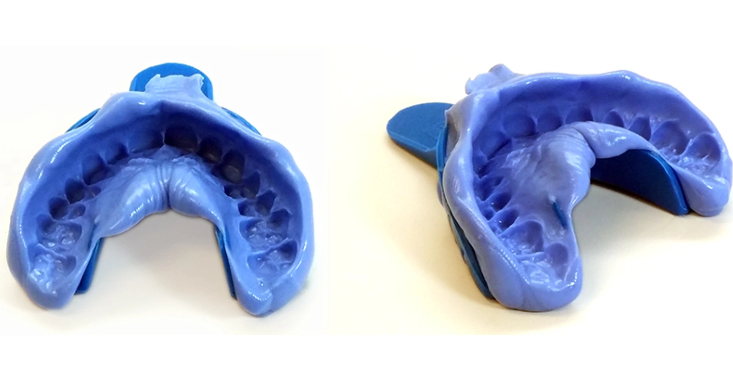
A dental impression is a reproduction of oral structures used for diagnosis, planning, fabrication, or documentation. Conventional techniques place impression material inside a tray. The tray is positioned over selected teeth and tissues until the material sets. It is then removed carefully and inspected. The resulting negative form can be poured with dental stone to create a model. Digital techniques follow a different process. An intraoral scanner captures optical data and produces a virtual three-dimensional representation. Both approaches can support restorative and prosthetic treatment when performed correctly.
What does a dental impression capture?
An impression can capture teeth, gingival contours, edentulous areas, implant components, and other relevant oral structures. The exact anatomy recorded depends on the treatment objective. Crown impressions must capture preparation margins and neighboring teeth accurately. Denture impressions may need to record supporting tissues and functional borders. Implant impressions must reproduce implant positions precisely. Orthodontic records can capture tooth positions and arch relationships. Digital scanning records similar information as virtual surface data. The dentist determines which structures need to be captured before selecting the technique.
Why do dentists take dental impressions?
Dentists take impressions when accurate anatomical records are required for treatment. These records help laboratories fabricate crowns, bridges, dentures, retainers, night guards, and other appliances. Impressions can also support diagnosis and treatment planning. Implant procedures may require records of implant positions and surrounding structures. Orthodontic workflows can use digital models to assess tooth movement and appliance design. The selected technique depends on clinical requirements. A physical impression is not always necessary because digital scanning can provide a suitable alternative for many treatments.
How dental impressions are used to create dental restorations and appliances
Impression records transfer clinical information from the dental chair to the laboratory or digital design workflow. A technician can use the record to create a crown, bridge, denture, appliance, or other restoration. Physical impressions may be poured into stone models. Digital records can enter CAD/CAM systems for computer-assisted design and manufacturing. The quality of the original record influences subsequent stages. Errors may affect fit, contacts, contours, or appliance performance. Dentists therefore inspect impressions or digital scans before sending them for fabrication.
Dental impressions versus dental molds
The terms are sometimes used interchangeably, but they describe different stages. An impression records the mouth's shape as a negative reproduction. A mold can refer more broadly to a form used to reproduce an object. In dentistry, an impression is the more precise clinical term. The impression may later produce a positive dental model. Digital scanning changes this workflow because no physical negative mold is required. Instead, the scanner creates virtual surface information.
Dental impressions versus dental scans
Traditional impressions use physical materials and trays. Digital scans use an intraoral scanner to capture optical information. Both methods aim to provide accurate anatomical records. Digital workflows can reduce material handling and may improve patient comfort. Evidence also suggests that intraoral scanning can reduce working time in many restorative situations. However, digital scanning is not automatically appropriate for every clinical situation. Full-arch, mobile-tissue, and completely edentulous cases may present additional challenges.
Find out how nail biting can affect the jaw
Why Are Dental Impressions Needed?
Impressions provide detailed records needed for many dental treatments. They can support restorative, prosthetic, orthodontic, implant, and diagnostic workflows. The required level of detail depends on the procedure. A crown preparation requires accurate margins. A denture requires information about supporting tissues. Implant treatment requires accurate implant positioning. Orthodontic treatment may require precise arch and tooth records. Digital scanning can provide these records without conventional impression material in suitable cases.
Dental impressions for crowns and bridges
Crowns and bridges require accurate records of prepared teeth and surrounding structures. The impression must reproduce preparation margins, neighboring teeth, and occlusal relationships. This information allows the laboratory to design a restoration that fits the prepared site. Digital scanning can capture preparation geometry and transfer it electronically. Conventional materials remain widely used when clinically appropriate. Moisture control and tissue management can influence the quality of either workflow. The dentist evaluates the record before fabrication begins.
Dental impressions for dentures
Dentures require records of the oral tissues supporting the prosthesis. Preliminary impressions can help create diagnostic models and custom trays. Final impressions aim to capture relevant tissues and borders more precisely. Functional considerations are particularly important because dentures interact with mobile oral tissues. An accurate record can contribute to retention, stability, and comfort. Complete dentures may present greater challenges for digital scanning because mobile soft tissues can be difficult to capture consistently.
Dental impressions for orthodontic treatment
Orthodontic records help document tooth positions and arch relationships. Traditional impressions can produce physical study models. Digital scans can create virtual models for treatment planning and appliance fabrication. Scanning can also simplify digital storage and communication. Research in orthodontics reports satisfactory accuracy and improved patient comfort with digital scanning, although results vary between scanners and clinical situations. The orthodontic team selects the workflow according to treatment requirements.
Dental impressions for dental implants
Implant impressions must accurately reproduce implant positions and relationships. Conventional workflows may use impression copings and impression material. Digital workflows can use scan bodies attached to implants. The scanner records the geometry of these components and surrounding tissues. The resulting information can support CAD/CAM restoration design. Implant position, scan strategy, tissue conditions, and scanner performance can influence accuracy. Recent reviews report comparable prosthetic outcomes between many digital and conventional workflows, with digital methods often reducing procedure time.
Dental impressions for night guards and mouth guards
Night guards and mouth guards need to fit the patient's teeth and oral anatomy. An impression or digital scan can provide the information required for fabrication. The appliance design depends on its purpose and the patient's dental condition. A professionally obtained record can reduce problems caused by inaccurate fit. Patients should not assume that an online impression kit provides the same clinical assessment. The dentist must also evaluate tooth position, restorations, occlusion, and oral health.
Dental impressions for diagnostic models and treatment planning
Diagnostic models can help dentists evaluate tooth position, arch form, spacing, occlusion, and restorative needs. Traditional models provide a physical reference. Digital models provide electronic records that can be measured and shared easily. Digital files can also integrate with other planning technologies. The usefulness of either method depends on record quality. A dentist may choose conventional, digital, or combined records based on the planned treatment.
Learn more about nail biting and teeth
How Are Dental Impressions Taken?
Traditional impression procedures involve several controlled steps. The dentist first examines the mouth and determines which structures must be recorded. A suitable tray and material are selected. The material is mixed and loaded into the tray. The tray is positioned carefully and held still while the material sets. After removal, the record is inspected for defects. Digital scanning follows a different workflow. The scanner captures surfaces progressively and produces a virtual model.
What happens before a dental impression?
Before taking an impression, the dentist examines the patient's teeth and oral tissues. The treatment objective determines the required area and level of detail. The dentist may assess tooth preparation, gingival condition, moisture control, implant components, or existing appliances. Any significant mobility or oral pain should be identified beforehand. The dentist then selects an appropriate tray and impression technique. Good preparation reduces the risk of repeating the procedure.
How a dental impression tray is selected
Tray selection depends on arch size, anatomy, treatment area, and impression technique. The tray should provide sufficient material thickness around the structures being recorded. It must also remain stable during setting. Stock trays come in different shapes and sizes. Custom trays can provide more controlled material thickness and border extension. Implant procedures may require specialized tray designs. The dentist chooses the tray according to clinical requirements rather than simply selecting the largest available option.
How impression material is placed in the tray
The impression material is prepared according to manufacturer instructions. Correct mixing or dispensing is important because setting characteristics can affect working time. The material is placed evenly inside the selected tray. Some techniques use different viscosities together. A heavy-body material can provide tray support while a light-body material records fine details. The dentist must work within the material's available working time. Incorrect handling can create voids, distortions, or incomplete anatomy.
How the tray is positioned in the mouth
The tray is inserted carefully to avoid unnecessary movement. It must cover the intended anatomy without excessive pressure. The dentist maintains appropriate positioning while the material sets. Movement during setting can distort the impression. Tissue management may also be necessary around prepared teeth. The patient should remain relaxed and follow the dentist's instructions. Once the material has set adequately, the dentist removes the tray with controlled movement.
How long does a dental impression take?
The clinical time varies according to the impression type, material, treatment area, and patient. Traditional impressions require preparation, tray loading, insertion, setting, removal, and inspection. Digital scanning involves scanner preparation and intraoral scanning. Systematic reviews generally find digital scanning faster than conventional impression procedures in many restorative situations. However, scanning time varies with the arch size, scanner, operator, and clinical conditions.
What happens after the impression is removed?
After removal, the dentist checks the impression for accuracy. Important anatomy should be completely captured without major voids, tears, bubbles, or distortions. If critical areas are missing, the impression may need to be repeated. An acceptable physical record is disinfected and prepared for laboratory transfer. Digital records are checked on the computer before transmission. The dentist may also compare the record with the clinical situation before approving fabrication.
How the impression is sent to a dental laboratory
Physical impressions are packaged and transferred according to appropriate infection-control procedures. Digital records can be transmitted electronically through compatible laboratory systems. Digital communication can reduce physical transportation and simplify information sharing. The laboratory receives the required clinical information and design instructions. The dentist and technician may communicate about preparation margins, shade, material, implant components, or appliance requirements.
How the dental model or restoration is produced
A conventional impression can be poured with dental stone to create a positive model. The laboratory then uses this model for fabrication or further design steps. Digital impressions can enter CAD software directly. The technician can design restorations or appliances using the digital model. Manufacturing may involve milling, printing, or other laboratory processes. The chosen workflow depends on the restoration and laboratory capabilities.
Explore common brushing and flossing mistakes
What Materials Are Used for Dental Impressions?
Dental impression materials have different physical and handling characteristics. Alginate is widely used for preliminary records and selected appliances. Polyether provides detailed reproduction and useful dimensional stability. PVS and VPS materials are widely used for precise restorative impressions. Polysulfide is another elastomeric material with particular handling characteristics. Putty, heavy-body, and light-body materials can be combined in specific techniques. Material selection depends on accuracy requirements, moisture conditions, working time, and clinical purpose.
What is dental impression material?
Dental impression material is a substance designed to reproduce oral structures. It must flow sufficiently during placement and then set into a stable form. Different materials offer different levels of elasticity, detail reproduction, working time, and dimensional stability. The dentist selects the material based on the procedure. Some materials are more suitable for preliminary models. Others are designed for highly detailed restorative or implant records. Manufacturer instructions should be followed carefully.
Alginate dental impression material
Alginate is an elastic hydrocolloid commonly used for preliminary impressions. It is relatively easy to mix and generally economical. Dentists may use it for diagnostic models, orthodontic records, study casts, and selected appliances. Alginate can lose dimensional accuracy if it is stored improperly or delayed before pouring. It therefore requires appropriate handling and timely laboratory processing. It may not provide the same precision required for some fixed restorative procedures.
Polyether dental impression material
Polyether is an elastomeric impression material known for detailed reproduction and dimensional stability. It can perform well in situations requiring precise records. Its handling properties differ from those of PVS materials. Polyether may also be less forgiving when removal from certain undercut areas is required. The dentist considers the patient's anatomy and treatment requirements before selecting it. Proper tray selection and technique remain essential.
PVS and VPS dental impression materials
Polyvinyl siloxane and vinyl polysiloxane describe closely related silicone-based impression materials. They are widely used for crowns, bridges, implants, and other detailed restorative procedures. These materials can provide high detail reproduction and dimensional stability. Different viscosities allow dentists to combine heavy-body and light-body materials. Moisture control remains important despite improvements in material formulations. The dentist selects the appropriate viscosity and technique according to the procedure.
Polysulfide impression material
Polysulfide is an elastomeric impression material with a long history in prosthodontics. It provides good flexibility and tear resistance. Its relatively long working and setting characteristics can be useful in selected situations. However, modern practices may prefer other materials depending on the treatment. Material selection should consider accuracy, patient tolerance, handling, and laboratory requirements. Polysulfide remains part of the broader family of conventional impression materials.
Impression putty and heavy-body materials
Putty and heavy-body materials provide bulk and support within impression techniques. They can be combined with lighter materials to record fine anatomical details. Putty can be used in specific two-step or putty-wash techniques. Heavy-body materials can provide stability while light-body material flows around detailed areas. Correct proportioning and positioning are important. The technique must match the material manufacturer's instructions.
Light-body and wash impression materials
Light-body materials have lower viscosity than heavy-body materials. They can flow around prepared teeth and capture fine surface details. In wash techniques, light-body material works with a supporting putty or heavy-body layer. The material must remain within its working time. Moisture, blood, saliva, and tissue movement can affect detailed reproduction. Careful isolation and tissue management help produce a reliable record.
How dentists choose an impression material
Dentists consider the treatment, required accuracy, oral anatomy, moisture conditions, working time, removal requirements, and laboratory workflow. A material suitable for an orthodontic study model may not be ideal for an implant restoration. Patient comfort also matters. Digital scanning can provide another option for many treatments. The final decision should be based on clinical requirements rather than marketing claims about one material.
Discover important information about aortic dissection and dental treatment
Alginate vs PVS vs Polyether Dental Impressions
Alginate, PVS, and polyether differ in composition and clinical applications. Alginate is commonly used for preliminary records. PVS is frequently selected for detailed restorative and prosthetic work. Polyether can provide accurate detail and dimensional stability in selected situations. The dentist must consider moisture, working time, flexibility, tissue undercuts, and laboratory requirements. No material is universally appropriate for every clinical situation.
Alginate dental impressions
Alginate is commonly selected when a preliminary or diagnostic record is required. It can be useful for study models, orthodontic records, and some removable appliances. Its handling is relatively straightforward. However, dimensional changes can occur if the impression is stored improperly. For highly precise fixed restorations, dentists may select an elastomeric material or digital workflow instead.
PVS dental impressions
PVS materials are commonly used when detailed and stable impressions are required. They are available in different viscosities and setting times. Their dimensional stability supports many laboratory workflows. They can be useful for crowns, bridges, implant restorations, and other prosthetic procedures. Good moisture control and correct handling remain important. Digital scanning can also replace conventional PVS techniques in many suitable cases.
Polyether dental impressions
Polyether can provide detailed impressions with good dimensional stability. Its material properties can make it useful for selected restorative procedures. However, removal characteristics and patient factors must be considered. The dentist determines whether polyether is suitable based on anatomy and clinical objectives. Modern digital workflows can provide an alternative in many cases.
How accuracy, moisture tolerance, working time, and clinical purpose differ
Each material has different physical properties. Alginate is commonly used for preliminary records but requires careful storage. PVS offers detailed reproduction and dimensional stability. Polyether also provides detailed records but has distinct handling and removal characteristics. Moisture control remains relevant for conventional materials. Digital scanning avoids impression material but introduces scanner-specific factors. Clinical requirements should guide the final selection.
Comparison of traditional dental impression materials
Material | Common use | Main consideration |
Alginate | Diagnostic and preliminary records | Requires timely processing |
PVS/VPS | Crowns, bridges, implants | Detailed reproduction |
Polyether | Detailed restorative records | Handling and removal |
Polysulfide | Selected prosthodontic applications | Longer working characteristics |
Putty | Support in wash techniques | Technique-sensitive |
Light-body | Fine detail recording | Requires adequate moisture control |
What Are Dental Impression Trays?
A dental impression tray holds impression material against the teeth and oral tissues. Trays may be stock or custom-made. Their design affects material thickness, stability, coverage, and accuracy. Some procedures require specialized trays. Implant impressions may use open-tray or closed-tray approaches. Triple-tray systems can record opposing teeth and occlusal relationships in selected restorative procedures. Correct tray selection is essential for obtaining a complete record.
What is a dental impression tray?
An impression tray is a device designed to carry impression material into the mouth. It provides support while the material sets. Trays can be made from plastic, metal, or other suitable materials. Their shape should correspond to the patient's dental arch and treatment area. The dentist checks coverage and stability before taking the impression. Poor tray selection can contribute to inadequate material thickness or incomplete anatomy.
Stock trays versus custom impression trays
Stock trays are manufactured in standard sizes and shapes. They are convenient and commonly used for routine impressions. Custom trays are fabricated specifically for a patient's anatomy or treatment requirements. They can provide more controlled material thickness and may improve certain impression techniques. Custom trays can be particularly useful in complete denture workflows. The dentist chooses between them according to clinical objectives.
Disposable dental impression trays
Disposable trays are designed for single-patient use. They can simplify infection-control procedures and reduce the need for tray sterilization. They are available in different sizes and designs. Their rigidity and fit should be appropriate for the impression material. A disposable tray still requires careful selection. Single-use status does not automatically guarantee an accurate impression.
Triple-tray impressions
Triple-tray systems can record a prepared tooth, opposing teeth, and bite relationship in one procedure. They are commonly associated with selected crown and indirect restoration workflows. The technique requires stable occlusion and adequate clinical conditions. It may not be suitable for every patient or restoration. The dentist evaluates whether the system provides enough information for the planned laboratory workflow.
Open-tray implant impressions
Open-tray implant impressions use impression copings that extend through openings in the tray. After the material sets, the coping screws are loosened before removing the impression. This technique can provide direct transfer of implant positions. It may be selected when multiple implants or complex positioning requires a particular approach. Proper coping stabilization and material support remain essential.
How the tray affects impression accuracy
Tray stability influences impression accuracy. Excessive movement can distort the material during setting. Insufficient material space can also reduce detail reproduction. A tray that does not cover the required anatomy can create missing information. Custom trays can help control material thickness. The dentist therefore evaluates tray size, rigidity, extension, and stability before starting the impression.
Learn more about medical diagnostic technology
What Are Digital Dental Impressions?
Digital impressions use intraoral scanners to capture three-dimensional information about oral surfaces. The scanner records images or optical measurements and combines them into a virtual model. Digital records can support crowns, bridges, implants, orthodontics, aligners, and other workflows. Studies generally report faster procedures and better patient comfort with digital scanning in many clinical settings. However, accuracy depends on the scanner, operator, scanning area, tissue conditions, and clinical situation.
How a dental impression scanner works
An intraoral scanner uses optical technology to capture surface information. The scanner is moved across the teeth and tissues according to a specific scanning strategy. Software combines multiple images or measurements into a three-dimensional model. The clinician reviews the model for missing areas or scanning artifacts. Additional scanning can be performed when necessary. The final file can then be transferred to compatible design or laboratory software.
What is an intraoral scanner?
An intraoral scanner is a handheld digital device used to record oral structures. It produces a virtual three-dimensional model rather than a physical impression. Different scanners use different optical technologies and software systems. Scanner performance varies by device, operator, anatomy, moisture, and scanning area. Digital scanning is now used widely in restorative and orthodontic workflows. It can also support selected implant procedures.
How digital dental impressions are created
The scanner captures a sequence of images while moving around the dental arch. Software aligns the captured information to build a continuous virtual model. The operator monitors the scan and adds data to incomplete areas. The final model is checked before being exported. Digital files can be stored electronically and sent directly to a laboratory. This can simplify communication and reduce physical handling.
Digital dental impressions versus traditional impressions
Digital impressions eliminate the need for conventional tray material. They can reduce unpleasant taste, nausea, and gagging for some patients. Evidence generally shows a patient preference for digital scanning in many studies. Traditional impressions remain useful for certain clinical situations. Completely edentulous arches and mobile soft tissues can challenge digital scanning. The appropriate method therefore depends on clinical circumstances.
Advantages and limitations of digital impressions
Digital scanning can improve patient comfort, simplify laboratory communication, and reduce material handling. Digital files can also be stored and transferred easily. However, scanning requires appropriate equipment and trained operators. Saliva, blood, reflective surfaces, deep margins, and difficult anatomy can complicate scanning. Long-span and edentulous cases can also present accuracy challenges. A digital workflow should therefore be selected according to the treatment.
When a digital scan may be appropriate
Digital scanning may be appropriate for crowns, bridges, orthodontic records, aligners, selected implant restorations, and many restorative procedures. It can be particularly useful when patient comfort is important. It also supports digital laboratory workflows. The dentist should confirm that the scanner and workflow are suitable for the planned treatment. Digital technology should complement clinical judgment rather than replace it.
When conventional impressions may still be used
Conventional impressions can remain appropriate when anatomy, tissue mobility, treatment complexity, or laboratory requirements make them preferable. Completely edentulous arches are one important example. Evidence indicates that intraoral scanning may have limitations around mobile tissues in these cases. Conventional techniques may therefore remain valuable. Dentists can also combine conventional and digital records when clinically appropriate.
Explore cosmetic dentistry in Turkey
Are Digital Dental Impressions Better Than Traditional Impressions?
Digital and conventional impressions can both produce clinically useful records. Research does not support treating one method as universally superior. Digital scanning often provides advantages in comfort and procedure time. Conventional techniques can remain useful for specific anatomy and treatment situations. The best workflow depends on the restoration, arch, tissue condition, equipment, and clinician experience.
Accuracy and detail
Accuracy depends on the specific clinical situation and technique. Digital scanners can provide highly detailed records for many restorative applications. Conventional materials can also reproduce fine anatomy when used correctly. Recent reviews report comparable clinical acceptability for many conventional and digital workflows. However, accuracy may decrease in challenging full-arch or edentulous situations. The dentist should evaluate the required level of accuracy before selecting a method.
Patient comfort and gag reflex
Digital scanning generally avoids the bulky impression tray and setting material used conventionally. Multiple systematic reviews report improved comfort and patient preference for digital scanning. A 2025 pediatric meta-analysis included seven studies involving 337 children. Digital scanning was faster and associated with lower reported gagging and discomfort. Individual experiences still vary.
Treatment workflow and laboratory communication
Digital records can be transferred electronically to laboratories. This can reduce physical transportation and simplify communication. Digital models can enter CAD/CAM systems directly. Clinicians can also retain digital records for future comparison or treatment planning. Conventional impressions require physical handling, disinfection, packaging, and laboratory processing. Both workflows can support successful restorations when the records are accurate.
Scanning difficult areas and full-arch cases
Scanning becomes more challenging when tissues are mobile, surfaces are difficult to isolate, or the arch is completely edentulous. Moisture and reflective surfaces can also interfere with optical capture. Research has identified limitations in digital scanning of mobile peripheral tissues. Long-span scans can also accumulate errors depending on the scanner and scanning strategy. Clinical judgment remains essential.
Why the appropriate technique depends on the clinical situation
Treatment requirements should determine the impression method. A simple crown may be suitable for digital scanning. A complete denture may require a conventional approach. Implant cases may support either method depending on implant number and configuration. Patient tolerance can also influence the workflow. Dentists should select the technique that provides adequate information for the intended treatment.
Clinical Note: Digital does not automatically mean better for every case
Digital dentistry provides important advantages, but technology does not replace clinical assessment. A scanner can only reproduce information that it successfully captures. Difficult anatomy can compromise digital records. Conventional materials can remain highly useful when selected and handled correctly. Evidence supports digital scanning for many applications while recognizing important limitations. The clinical objective should therefore guide technology selection.
Find out more about opioids and dental pain
Are Dental Impressions Painful?
Most impression procedures are not painful. Patients may experience pressure, fullness, or temporary discomfort when a conventional tray is inserted. Some people experience gagging because the tray or material stimulates sensitive areas. Digital scanning can reduce these sensations for many patients. Systematic reviews consistently report better patient comfort with digital methods in many studied situations. Pain is not normally expected during routine impression taking.
What does taking a dental impression feel like?
A conventional impression can feel unusual because the tray occupies space around the teeth. The material may feel bulky while it sets. Patients usually need to remain still for a short period. Digital scanning typically involves a smaller handheld scanner moving around the mouth. The sensation can be easier for some patients. Individual experiences vary based on anatomy, gag reflex, anxiety, and previous dental experiences.
Why some patients experience pressure or discomfort
Pressure can occur when the tray is seated against the teeth and surrounding tissues. The impression material may also create a feeling of fullness. A properly selected tray should not require excessive force. If a patient experiences sharp pain, the dentist should be informed immediately. Discomfort can also result from inflamed tissues or an existing dental problem rather than the impression procedure itself.
Dental impressions and the gag reflex
The gag reflex can make conventional impressions difficult for some patients. Tray size, posterior extension, material volume, anxiety, and individual sensitivity can contribute. Digital scanning may reduce gagging because it avoids a large impression tray. Research supports better patient-reported comfort with digital scanning in many studies. Patients should tell the dental team about a strong gag reflex before treatment.
What can make an impression more difficult?
A strong gag reflex, limited mouth opening, sensitive tissues, anxiety, saliva, or unusual anatomy can complicate impression taking. Loose teeth may also require additional assessment. Extensive restorations or deep preparation margins can create technical challenges. The dentist may modify the technique or choose another method. Digital scanning can provide an alternative when conventional impressions are poorly tolerated.
How dentists can improve patient comfort
Dentists can select an appropriate tray, minimize unnecessary material, and explain each step before starting. Patient positioning and controlled breathing can also help. Some clinicians use distraction or behavioral techniques for anxious patients. Digital scanning can provide another option when conventional materials trigger gagging. Communication is important. Patients should signal discomfort rather than trying to tolerate severe symptoms silently.
When discomfort during an impression may need further evaluation
Significant pain is not expected from routine impression taking. Persistent discomfort, bleeding, or tooth mobility should be reported. A loose tooth may require evaluation before an impression is attempted. Existing gum inflammation can also increase sensitivity. The dentist should determine whether symptoms arise from the impression procedure or an underlying oral condition.
Discover the connection between heartburn and jaw pain
Can Dental Impressions Pull Teeth Out?
A properly performed impression should not normally pull out a healthy tooth. The material is designed to record oral structures rather than extract them. However, a severely mobile tooth can present a different situation. The dentist should assess mobility before placing a tray. Careful technique and appropriate tray removal can reduce unnecessary forces. Patients with significant tooth mobility should inform the dental team beforehand.
Why dental impressions normally do not pull teeth out
Impression materials set around teeth but do not normally bond strongly enough to remove stable teeth. The dentist uses controlled movements to separate the tray from the oral structures. Elastomeric materials have sufficient flexibility for routine removal. Proper tray selection also reduces unnecessary retention. A stable tooth should remain in place during routine impression removal.
Why a patient may feel pressure during impression removal
Removal can create pressure because the impression material engages the contours of the teeth. This sensation is usually brief. Dentists use controlled movements to break the seal and remove the tray. Patients may feel temporary pressure around individual teeth. Sharp pain or unexpected movement should be reported immediately. The dentist can then evaluate the situation.
What happens if a tooth is loose?
A loose tooth may require assessment before conventional impression taking. The degree and cause of mobility matter. Periodontal disease, trauma, infection, or other conditions can contribute to mobility. The dentist may modify the technique or choose digital scanning. Treatment of the underlying condition may be necessary before fabrication of an appliance.
What dentists assess before taking an impression
Dentists assess tooth stability, gum health, existing restorations, oral tissues, and treatment requirements. They also consider the tray and material. If a tooth appears significantly mobile, the clinician may investigate the cause. The impression technique should not create unnecessary risk. Digital scanning can sometimes avoid the mechanical forces associated with conventional tray removal.
When a loose tooth requires dental evaluation before an impression
Significant mobility, pain, bleeding, swelling, or recent trauma should be discussed before an impression. These symptoms may indicate an underlying dental condition. The dentist can determine whether the planned impression should proceed. Treating the underlying problem may be more important than obtaining an immediate impression.
Learn about myotonic dystrophy and oral health
What Makes a Good or Bad Dental Impression?
A good impression captures the required anatomy clearly and without significant distortion. It should include relevant teeth, soft tissues, margins, and supporting structures. A poor impression may contain bubbles, tears, voids, drag marks, incomplete anatomy, or distortion. Digital scans can have holes, stitching errors, artifacts, or incomplete surfaces. The dentist decides whether the record is clinically acceptable before fabrication.
What does a good dental impression look like?
A good conventional impression reproduces the required anatomy with clear surface detail. Preparation margins should be visible when relevant. There should be adequate material around the recorded structures. The tray should remain stable and free from major distortion. Digital scans should show complete, continuous anatomy without missing areas. The dentist checks the record before sending it for laboratory processing.
What can cause an inaccurate impression?
Inaccuracy can result from movement, poor tray selection, inadequate material mixing, incorrect setting time, moisture contamination, insufficient material, or improper removal. Digital scans can be affected by saliva, blood, reflective surfaces, scanning strategy, and operator technique. Long scanning paths may also accumulate errors. Accurate results require both appropriate technology and proper clinical technique.
Bubbles, voids, tears, and missing anatomy
Bubbles can obscure important anatomical details. Voids may result from air entrapment or incomplete material placement. Tears can occur during removal, especially around undercuts. Missing anatomy means the laboratory lacks essential information. Digital records can show similar problems as holes or incomplete scans. The dentist should determine whether the defect affects the treatment area. If it does, a new record may be necessary.
Tray positioning and impression material problems
Poor tray positioning can cause uneven material thickness or missing anatomy. Incorrect material proportions can change setting characteristics. Excessive movement can distort the record. Inadequate tissue management can obscure preparation margins. The dentist should follow the material manufacturer's instructions and use a suitable tray. Digital scanning has different technical requirements but still depends on correct scanning technique.
How dentists determine whether an impression needs to be retaken
The dentist examines the record against the treatment requirements. A minor defect away from the relevant anatomy may not require repetition. A missing crown margin or implant position can make the record unacceptable. Digital scans can often be corrected by rescanning a missing area. Conventional impressions may require a complete repeat. The final decision belongs to the treating clinician.
Clinical Note: Why impression accuracy matters for the final restoration
The laboratory relies on the clinical record to design the restoration or appliance. Inaccurate information can affect fit, contacts, occlusion, contours, and adaptation. Digital and conventional workflows can both produce accurate results when properly performed. Systematic reviews report comparable clinical outcomes for many restorative applications. Accuracy therefore depends on technique and clinical circumstances.
Find out how to get feeling back in your mouth after the dentist
Dental Impressions for Crowns
Crowns require accurate records of prepared teeth and surrounding structures. The impression or scan should capture the preparation margin, adjacent teeth, and occlusal relationships. Traditional PVS techniques remain widely used. Digital scanning can also capture preparations and transfer data electronically. The dentist evaluates gingival conditions and margin visibility before recording the preparation. The laboratory then uses the information to design the crown.
Why a dental crown impression is needed
A crown must fit the prepared tooth precisely. The laboratory needs information about the preparation shape, margin, neighboring teeth, and bite. An accurate impression or digital scan provides these details. The record also helps establish appropriate proximal contacts and occlusal relationships. Without sufficient information, the laboratory may be unable to fabricate the restoration accurately.
How a crown impression captures the prepared tooth
The impression material flows around the prepared tooth and records its surfaces. Light-body material can capture detailed margins while heavier material supports the overall record. Retraction may be necessary to expose the preparation margin. Moisture control is important. Digital scanning captures the preparation optically and can provide immediate visual feedback. The dentist checks the record before laboratory fabrication.
PVS and other materials used for crown impressions
PVS materials are frequently used for detailed crown impressions. Different viscosities can be combined to support accurate margin reproduction. Polyether can also be used in selected restorative situations. Alginate is generally more appropriate for preliminary records than definitive crown impressions. Digital scanning provides another workflow when clinically suitable. The material or scanning method should match the required precision.
Digital impressions for dental crowns
Digital scanning can capture crown preparations without conventional impression material. The clinician can inspect the virtual model immediately. Missing areas can often be rescanned before the patient leaves. Digital files can then be sent electronically to a laboratory. Research comparing digital and conventional crown workflows has found similar clinical outcomes in many situations, while digital techniques often improve comfort.
What happens after the crown impression is taken
The dentist checks the record and confirms that the preparation and surrounding anatomy are adequately captured. A physical impression is processed and sent to the laboratory. A digital record can be transmitted electronically. The laboratory uses the information to design and fabricate the crown. The restoration is later evaluated clinically for fit, contacts, occlusion, and appearance.
Explore how tongue posture affects teeth
Dental Impressions for Dentures
Dentures require detailed records of teeth, gums, supporting tissues, and functional anatomy. Conventional techniques remain important because mobile tissues can be difficult to reproduce digitally. Preliminary impressions can help create diagnostic models and custom trays. Final impressions are designed to capture the supporting anatomy more accurately. Denture impressions can influence retention, stability, comfort, and overall prosthetic function.
Why dentures require accurate impressions
A denture must interact closely with the tissues supporting it. An inaccurate record can contribute to poor adaptation, instability, or discomfort. Complete dentures can be particularly challenging because tissues move during function. The impression must capture the relevant anatomy while considering functional borders. The dentist chooses the technique according to the patient's anatomy and prosthetic requirements.
Preliminary versus final denture impressions
A preliminary impression generally provides an initial record for diagnosis and custom tray fabrication. A final impression is intended to capture the anatomy more precisely for denture fabrication. The two stages serve different purposes. The final technique may include border molding and controlled tissue recording. Digital approaches are developing, but conventional methods remain important for many completely edentulous cases.
How impressions capture the gums and supporting tissues
Denture impressions record the shape of the supporting mucosa and relevant anatomical structures. Border extension can influence denture stability. Functional movements may therefore be considered during impression procedures. The dentist evaluates the tissues and selects an appropriate technique. Digital scanners may have difficulty capturing mobile tissues consistently in completely edentulous arches.
Why denture impressions can affect fit and comfort
The impression provides the laboratory with information about the tissues supporting the denture. Inadequate extension or distorted tissue records can affect fit. An accurate record supports better adaptation to the intended anatomy. However, denture comfort also depends on jaw relationships, tooth arrangement, occlusion, base design, and clinical adjustment. An impression is one important stage rather than the entire fitting process.
Discover more about craniofacial disorders and dental health
Dental Impressions for Dental Implants
Implant impressions record implant positions for the fabrication of implant-supported restorations. Conventional techniques can use impression copings. Digital techniques can use scan bodies. Accuracy is particularly important because implant restorations connect to fixed implant positions. Implant number, distribution, angulation, soft tissue conditions, scanner characteristics, and technique can affect results. Current evidence supports both conventional and digital workflows for many implant-supported restorations.
Why implant impressions require precise positioning
Implant restorations must connect accurately with implants. Small recording errors can affect the fit of the final prosthesis. Multiple implants may increase the complexity of transferring three-dimensional positions. The dentist therefore uses specialized components and controlled techniques. Digital workflows can simplify data transfer but still require accurate scan-body positioning and scanning.
Implant impression copings
Impression copings are components used to transfer implant positions into a conventional impression. They attach to the implants and become incorporated into the impression. The laboratory uses them to reproduce implant positions on a working model. Proper seating and stabilization are essential. The dentist verifies the components before recording the impression.
Open-tray versus closed-tray implant impressions
Open-tray techniques allow impression coping screws to be accessed through tray openings. The screws can be loosened before impression removal. Closed-tray techniques retain the copings differently and may be used for selected implant situations. The choice depends on implant position, accessibility, number of implants, and clinical requirements. The dentist determines which technique is appropriate.
Digital implant impressions and scan bodies
Digital implant workflows use scan bodies to identify implant position and orientation. The scanner captures the scan body and surrounding anatomy. Software then uses this information during restoration design. The scan body must be fully seated and correctly identified. Scanning strategy also affects the quality of the digital record. The laboratory receives the digital file along with the necessary implant information.
Factors that influence implant impression accuracy
Accuracy can be influenced by implant distribution, scan-body seating, tissue conditions, scanner performance, operator technique, and arch length. Long-span cases can be more demanding than single implants. Conventional impressions have their own technique-sensitive factors. Recent systematic reviews report similar prosthetic outcomes across many digital and conventional workflows. Clinical selection remains essential.
Clinical Note: Why implant impressions require careful clinical and laboratory coordination
Implant restoration fabrication involves multiple stages and professionals. The clinical record must communicate implant positions accurately. The laboratory also needs the correct implant system and component information. Digital communication can streamline this process. Conventional workflows can also be effective when properly performed. Close coordination reduces avoidable errors.
Learn how to get rid of numbness after the dentist
Note : All images used are for editorial and illustrative purposes only and may not originate from the original news provider or associated company.
What We Notice Clinically
Patients often have questions about impressions because the procedure looks different from routine dental treatment. Some patients expect an impression to change their teeth. Others worry that impression material can damage teeth. Understanding the purpose of the record can reduce anxiety. The clinical technique should always match the planned treatment. Modern digital workflows provide another option for many patients.
Common Patient Concerns About Dental Impressions
Common concerns include gagging, discomfort, loose teeth, unpleasant taste, and the fear of swallowing material. These concerns are understandable. Digital scanning can reduce several of these issues for suitable cases. Systematic reviews show that patients often prefer intraoral scanning. Patients should tell the dental team about previous difficult experiences.
Why patients sometimes confuse impressions with treatment itself
An impression is a diagnostic or fabrication record. It is not the restorative treatment itself. The dentist may take an impression before creating a crown, denture, appliance, or other restoration. The impression transfers anatomical information to the laboratory. Understanding this distinction helps patients understand why multiple appointments may be needed.
Common misconceptions about impression materials and discomfort
Impression materials are not designed to damage stable teeth. Temporary pressure and gagging can occur. Severe pain is not expected. Patients may also believe that digital scanning is always more accurate. Evidence shows that accuracy depends on the clinical situation and scanning conditions. The chosen technique should therefore be individualized.
Dr. Rifat Alsaman's Clinical Perspective on Dental Impressions
Dr. Rifat Alsaman, Head of the Medical Team at Vitrin Clinic and cosmetic dentist, emphasizes that impression selection should follow the treatment objective. The required record differs between a crown, implant restoration, denture, and orthodontic appliance. Digital scanning can improve comfort and workflow for suitable cases. However, conventional techniques remain clinically relevant when anatomy or treatment requirements demand them. The important goal is an accurate and usable record.
Do Dental Impressions Have Side Effects?
Most patients experience no lasting effects from routine impression procedures. Temporary gagging, pressure, unpleasant taste, or mild tissue irritation can occur. Serious complications are uncommon. Patients with loose teeth, significant gum inflammation, oral pain, or allergies should inform the dental team beforehand. Digital scanning can provide an alternative when conventional impressions are poorly tolerated.
Common temporary effects after an impression
Temporary effects can include gagging, nausea, pressure, altered taste, or mild soft-tissue irritation. These effects usually resolve after the procedure. The experience depends on the material, tray, technique, and individual sensitivity. Digital scanning may reduce several sensory problems because it avoids impression material. Evidence generally favors digital methods for patient comfort.
Gagging, nausea, or temporary discomfort
Gagging can occur when a conventional tray stimulates sensitive areas near the back of the mouth. Anxiety can increase the perceived difficulty of the procedure. Controlled breathing and appropriate positioning may help. Digital scanning can reduce gagging for many patients. A 2025 meta-analysis in children found lower reported gag reflex with digital scanning.
Rare problems related to impression procedures
Rare problems can involve significant tissue irritation, allergic reactions, or complications involving severely mobile teeth. These situations are uncommon. Dentists should obtain relevant medical and dental history before the procedure. Patients should report unusual symptoms immediately. The clinician can determine whether further assessment is necessary.
What patients should tell their dentist before an impression
Patients should mention strong gagging, previous difficult impressions, loose teeth, oral pain, bleeding, swelling, allergies, or sensitivity. They should also mention relevant medical conditions and medications when appropriate. This information helps the dental team select a suitable technique. Patients should not hide discomfort during the procedure.
When unexpected symptoms should be discussed with a dentist
Persistent pain, bleeding, swelling, significant tooth mobility, or unusual tissue changes should be discussed with a dentist. These symptoms may reflect an underlying dental problem rather than the impression itself. Prompt evaluation can clarify the cause. Patients should seek urgent care if severe swelling affects breathing or swallowing.
Explore the link between atherosclerosis and periodontal disease
How Can You Get Relief From Discomfort During Dental Impressions?
Patient preparation can make impression procedures easier. Tell the dental team about gagging before starting. Slow breathing and appropriate positioning may help. The dentist can explain the procedure and provide short pauses when needed. Digital scanning may offer an alternative for suitable cases. Evidence suggests that patients generally experience better comfort with intraoral scanning than conventional impressions.
Practical ways to reduce gagging
Patients can focus on slow breathing through the nose when possible. Sitting in an appropriate position can also help. Eating immediately before the appointment may not be comfortable for patients prone to nausea. The dental team can use smaller trays when clinically appropriate. Digital scanning can also reduce the need for conventional tray materials.
Breathing and positioning during the procedure
Controlled breathing can help patients remain relaxed. The dentist may position the patient appropriately to reduce unnecessary stimulation. Avoiding sudden head or tongue movement can also help. Patients should follow instructions from the dental team. If discomfort becomes excessive, they should communicate immediately.
Communicating discomfort with the dental team
Patients should agree on a simple signal for stopping before the impression begins. They should tell the dentist if the tray causes pain or excessive gagging. Communication allows the clinician to modify the technique. Digital scanning may also be considered when appropriate. Patient comfort is an important part of modern dental care.
When digital scanning may provide an alternative
Digital scanning can be useful when conventional impressions cause significant gagging or discomfort. Research generally reports improved comfort with intraoral scanning. However, digital scanning is not suitable for every treatment. Completely edentulous arches and difficult mobile tissues may still require conventional approaches.
Medical Tip: Tell your dentist about a strong gag reflex before the procedure
A strong gag reflex is important clinical information. Tell your dentist before the appointment begins. The clinician can consider tray size, patient positioning, scanning, or other suitable techniques. Early communication can make the procedure more predictable and comfortable.
Dental Impression Kits: Are DIY Impressions Safe?
Home impression kits are marketed for products such as mouth guards and cosmetic appliances. However, they do not replace a professional dental examination. An inaccurate impression can result in a poorly fitting appliance. Dental professionals also assess oral health before recommending treatment. Patients should understand the difference between obtaining an impression and receiving dental care.
What is a dental impression kit?
A dental impression kit usually contains impression material, trays, and instructions. The user places the material into a tray and attempts to record the teeth. Such kits may be marketed for custom mouth guards or cosmetic products. The quality depends heavily on correct tray selection and technique. They do not provide a clinical diagnosis.
How professional dental impressions differ from DIY kits
Professional impressions are taken after clinical examination and treatment assessment. The dentist selects the appropriate tray and material. The clinician also checks the record for accuracy. Digital scanning provides another professional alternative. DIY kits rely on the user's ability to capture the required anatomy. They may not identify underlying oral disease or tooth mobility.
DIY dental impression kits for mouth guards and grillz
Some products can be fabricated from home impressions. However, patients should consider fit and oral health before using them. Poorly fitting appliances can cause discomfort or interfere with oral function. Cosmetic appliances should not replace professional dental assessment. A dentist can determine whether the requested appliance is appropriate.
Risks of an inaccurate home impression
An inaccurate impression can produce an appliance that does not fit properly. This may affect comfort, retention, speech, or function. The impression may also miss important anatomical areas. More importantly, a DIY process does not identify untreated cavities, gum disease, infection, or tooth mobility. Professional assessment remains important.
Why DIY impressions should not replace a dental examination
An impression records anatomy but does not diagnose disease. A dental examination evaluates teeth, gums, occlusion, restorations, and other oral structures. Patients seeking an appliance should therefore receive appropriate professional assessment. Digital scanning can also provide a more controlled alternative.
When a professionally taken impression is necessary
Professional records are particularly important for crowns, bridges, dentures, implant restorations, orthodontic appliances, and complex mouth guards. Treatment-specific impressions require controlled technique and accurate anatomical capture. The dentist can determine whether conventional or digital methods are suitable.
How Much Do Dental Impressions Cost?
The price depends heavily on the treatment rather than the impression alone. Some clinics include the impression or digital scan within the restoration fee. Others may list it separately. Cost can depend on the material, laboratory, treatment complexity, and geographic location. Digital scanning may also be bundled into a broader treatment plan. Patients should request an itemized quotation.
What determines the cost of dental impressions?
Cost can depend on the impression material, tray type, laboratory workflow, treatment, and number of arches. A simple diagnostic impression may cost less than an implant impression. Digital scanning can also be included within crown, veneer, orthodontic, or implant fees. The quoted price should therefore be interpreted within the complete treatment plan.
Are dental impressions included in the cost of treatment?
Many clinics bundle impressions into the cost of a restoration or appliance. Others may charge diagnostic records separately. Patients should ask what the quoted treatment includes. This is particularly important when comparing clinics internationally. A procedure-only price may not represent the total treatment cost.
Average dental impression costs in the United States
There is no standardized national price for a standalone impression. Fees vary by treatment and dental office. Diagnostic impressions may be bundled into examination or appliance fees. Crown, bridge, denture, orthodontic, and implant impressions can have different pricing structures. Patients should ask whether the impression is included in the final restoration fee.
Average dental impression costs in the United Kingdom
UK pricing also varies according to treatment type and provider. Many clinics include impressions within the fee for crowns, dentures, orthodontics, or other appliances. Private practices may itemize digital scans or laboratory records differently. Patients should request a complete treatment quotation instead of comparing impression fees alone.
Average dental impression costs in Turkey
In Turkey, impression and scanning fees vary between clinics and treatment packages. International dental patients may receive bundled treatment estimates that include diagnostics, laboratory work, and restoration. At Vitrin Clinic, the treatment plan should be based on the patient's individual dental requirements. Patients should confirm which diagnostic and laboratory services are included.
Traditional versus digital impression costs
Digital scanning requires specialized equipment and software. Conventional impressions require trays, materials, disinfection, and laboratory processing. The final price depends on how the clinic structures its treatment fees. Digital scanning may not appear as a separate charge when included in a restorative package. The most useful comparison is therefore the complete treatment cost.
Cost estimates vary by treatment, clinic, laboratory, material, and whether the impression is bundled into a restoration or appliance
A standalone impression price can be misleading. A crown quotation may include scanning, laboratory design, fabrication, and fitting. An implant quotation may include scan bodies, imaging, laboratory components, and restoration. Patients should request an itemized estimate. This provides a clearer basis for comparing treatment plans.
Dental Impressions Cost Comparison: US, UK, and Turkey
International pricing should be compared carefully because clinics package services differently. There is no universal national fee for an impression. Treatment type, laboratory involvement, technology, and restoration design influence the final price. Digital scanning may be included within the treatment rather than charged separately. Patients should compare complete treatment packages rather than isolated impression fees.
United States: Average Dental Impression Cost
The United States has wide regional and provider-level price variation. A conventional impression may be included within the fee for a crown, denture, or appliance. Implant-related records can involve additional laboratory components. Insurance may also affect the patient's final expense. An itemized estimate is the most reliable way to understand the actual cost.
United Kingdom: Average Dental Impression Cost
UK fees differ between NHS and private treatment settings. Private restorative and prosthetic fees can include impressions as part of the overall procedure. Digital scanning may also be bundled into treatment. Patients should confirm whether laboratory and restoration costs are included.
Turkey: Average Dental Impression Cost
Turkish clinics commonly structure pricing around complete treatment plans. Digital scanning may be incorporated into restorative or cosmetic packages. The final price depends on the planned procedure and laboratory workflow. International patients should confirm what diagnostics, scanning, laboratory work, restoration, and follow-up include.
What may be included in the quoted price
A treatment quotation may include consultation, imaging, scanning, conventional impressions, laboratory design, restoration, fitting, and follow-up. Not every clinic includes all components. Patients should ask for a written breakdown. This prevents confusion when comparing international treatment prices.
Consultation, scanning or impression, laboratory work, restoration, and follow-up
These components can represent separate stages of one treatment. A low impression fee does not necessarily mean lower overall treatment cost. The complete package should be evaluated. Patients should also ask whether additional appointments or remakes carry separate charges.
What Happens After a Dental Impression?
After an impression is taken, the dentist checks whether it contains the required anatomy. A conventional impression may be disinfected and sent to the laboratory. The laboratory can pour a model and begin fabrication. Digital scans can be transmitted electronically. The laboratory then uses the record to design the restoration or appliance. If essential information is missing, a new record may be needed.
How dentists evaluate the impression
Dentists inspect the record for completeness, distortion, bubbles, tears, voids, and missing anatomy. They pay particular attention to treatment-critical areas. Crown margins, implant positions, and denture borders require appropriate recording. Digital scans are checked for holes or incomplete areas. The dentist determines whether the record is acceptable.
How the dental laboratory uses the impression
The laboratory uses the record to understand the patient's anatomy. Physical impressions can be poured into stone models. Digital files can enter CAD software. Technicians use this information to design restorations or appliances. Additional clinical instructions may accompany the record. Communication between dentist and laboratory remains important.
How a dental model is produced
A physical impression can be filled with dental stone. After setting, the stone creates a positive reproduction of the recorded anatomy. The model can then be trimmed and prepared for laboratory work. Digital workflows produce virtual models instead. These can be manipulated, measured, and integrated into CAD/CAM systems.
How the impression contributes to the final restoration or appliance
The record transfers information about tooth position, shape, margins, occlusion, and supporting anatomy. The laboratory uses this information during design. The quality of the record can influence the accuracy of the final product. However, clinical fitting and adjustment remain necessary. An impression is one part of a broader treatment workflow.
When a new impression may be required
A new record may be required when important anatomy is missing or distorted. A crown margin may not be visible. An implant position may be inadequately recorded. A denture border may also require modification. Digital scans can often be corrected by rescanning. Conventional impressions usually require a new impression when critical areas are defective.
Tips for Patients
Patients can improve their experience by communicating clearly before the procedure. Tell your dentist about gagging, previous difficult impressions, loose teeth, or oral pain. Follow pre-appointment instructions. Remain still while the material sets or the scanner is used. Ask which technique will be used and why. Understanding the process can reduce anxiety.
Tell your dentist about gagging or previous difficulties
Previous difficulty with impressions is important information. The dentist can consider alternative techniques or digital scanning. Strong gagging may affect conventional impression tolerance. Early communication allows the team to prepare appropriately.
Follow pre-appointment instructions
Follow instructions regarding food, medication, oral hygiene, or other preparation. Most routine impressions require little special preparation. However, specific treatments may have additional requirements. Ask the clinic if you are unsure.
Avoid moving your tongue or jaw during the impression
Movement can interfere with conventional impression setting and digital scanning. Follow the dentist's instructions during the recording process. Relax your jaw unless instructed otherwise. The dental team will guide you throughout the procedure.
Communicate if you need a short break
Patients can usually signal when they need a pause. Communicating early is better than suddenly moving during the procedure. The dentist can remove the tray or pause scanning when clinically appropriate. Comfort and safety should remain priorities.
Ask which material or scanning method will be used
Different treatments require different recording methods. Ask why the dentist selected a particular material or scanner. Understanding the reason can make the procedure easier. It also helps patients understand how their treatment will proceed.
Ask whether the impression is included in your treatment cost
A scan or impression may be included in the restoration fee. Some clinics may list diagnostic records separately. Ask for an itemized estimate before treatment begins. This is especially useful for international patients comparing treatment packages.
Follow instructions after the impression is taken
Most patients have no special restrictions after an impression. Follow any instructions provided by the dental team. If a temporary restoration or appliance is involved, additional guidance may apply. Report unusual pain, swelling, or mobility.
Medical Tips for a More Comfortable Dental Impression
Good communication can improve the impression experience. Patients should disclose relevant sensitivities and oral symptoms. Digital scanning may be considered when conventional impressions cause significant gagging. However, the dentist must determine whether scanning is clinically appropriate. Patients should not attempt to diagnose impression-related problems independently.
Discuss allergies and sensitivities before the procedure
Tell the dental team about known allergies or previous reactions. This allows the clinician to select appropriate materials and precautions. Patients should also mention strong sensory sensitivity or gagging. Relevant information should be provided before the procedure starts.
Inform the dentist about loose teeth or oral pain
Loose teeth and pain may indicate an underlying condition. The dentist should evaluate these findings before taking an impression. A digital scan may reduce mechanical pressure in some situations. However, the underlying cause still requires appropriate dental assessment.
Ask about digital scanning if gagging is a concern
Digital scanning can be a useful alternative for suitable cases. Evidence shows improved patient comfort with digital methods in many studies. However, not every case can be scanned reliably. The dentist should determine whether digital recording is appropriate.
Do not attempt to diagnose an impression-related problem yourself
Unusual symptoms may have several causes. Pain, swelling, bleeding, or mobility can indicate dental disease rather than an impression reaction. A dentist can evaluate the symptoms clinically. Avoid relying on online information for diagnosis.
Follow your dentist's instructions for the planned treatment
The impression is part of a larger treatment plan. Patients should follow instructions about preparation, restoration delivery, appliance use, and follow-up. This supports a smoother treatment process. Questions should be directed to the treating dental team.
At Vitrin Clinic: Dental Impressions and Digital Treatment Planning
At Vitrin Clinic, dental impressions can form part of a broader digital treatment workflow. The process begins with a clinical examination and treatment assessment. Digital diagnostics can help evaluate teeth, restorations, occlusion, and relevant oral structures. A 3D intraoral scanner can create a digital record when clinically appropriate. The resulting information can support restorative, cosmetic, orthodontic, or implant treatment planning. Conventional impressions may still be considered when they provide advantages for a specific clinical situation. The objective is to select the recording method that matches the planned treatment. Digital information can then be coordinated with laboratory workflows for restoration design and fabrication.
Comprehensive dental examination before treatment
A clinical examination helps determine what information is needed. The dentist evaluates teeth, gums, restorations, occlusion, and treatment objectives. This assessment guides the choice between conventional and digital recording. A scan should not replace the clinical examination. It provides additional information for treatment planning.
Digital diagnostics and treatment assessment
Digital diagnostics can support treatment planning by providing detailed records of dental structures. Vitrin Clinic can combine relevant digital information with clinical findings. Depending on the treatment, diagnostic imaging may also be indicated. The dentist determines which records are necessary.
3D intraoral scanning and digital dental impressions
Vitrin Clinic uses 3D intraoral scanning as part of its digital workflow when clinically appropriate. The scanner records dental surfaces and creates a virtual model. This can support restorative and cosmetic planning. Digital scanning can also improve communication with dental laboratories. Research supports its use across many restorative applications, while recognizing limitations in challenging cases.
Selecting the appropriate impression or scanning workflow
The appropriate workflow depends on the treatment. Crowns, veneers, bridges, implants, orthodontic appliances, and dentures have different recording requirements. Vitrin Clinic's clinical team evaluates these requirements before selecting the workflow. Conventional impressions remain available when they are clinically preferable.
Coordinating digital information with treatment planning
Digital records can be integrated into treatment planning and laboratory communication. This can help clinicians and technicians work from the same information. Digital workflows can also simplify record storage and transfer. The treatment plan remains based on clinical assessment.
Dr. Rifat Alsaman's Clinical Perspective on Digital Dental Impressions
Dr. Rifat Alsaman, Head of the Medical Team at Vitrin Clinic and cosmetic dentist, considers digital scanning valuable when it supports the specific treatment objective. Patient comfort, anatomy, restoration type, and laboratory requirements all influence workflow selection. Digital technology can streamline restorative planning, but clinical judgment remains central.
How Vitrin Clinic Supports Your Treatment Journey
Vitrin Clinic coordinates dental assessment, diagnostics, treatment planning, restorative care, and follow-up for international patients. The workflow can incorporate digital scanning and other diagnostic technologies when clinically indicated. Treatment recommendations are based on individual dental findings rather than a single standard approach. Laboratory coordination supports the fabrication stage. International patients can also receive assistance with treatment scheduling and care coordination.
Initial consultation and assessment
The process begins with a consultation and dental assessment. The clinical team reviews the patient's concerns and treatment goals. Appropriate diagnostic records are then selected. This may include digital scans, X-rays, or other imaging when indicated.
Personalized digital treatment planning
Digital records can support individualized treatment planning. They allow clinicians to evaluate dental structures and communicate information with laboratories. The final plan depends on the patient's clinical condition and treatment objectives. Digital planning can be particularly useful for restorative and cosmetic workflows.
Coordination between clinical and laboratory workflows
Vitrin Clinic coordinates relevant clinical information with laboratory processes. Digital files can be transmitted electronically when compatible with the laboratory workflow. This can reduce physical handling and simplify communication. Conventional records can also be used when appropriate.
Monitoring treatment progress
Treatment progress can be reviewed through clinical examinations and appropriate digital records. Digital models can provide useful references in selected cases. Monitoring depends on the treatment type and clinical requirements. The dentist determines which follow-up records are necessary.
Follow-up and aftercare
Follow-up allows the dental team to evaluate the completed treatment. Restorations and appliances may require clinical checks and adjustments. International patients receive guidance regarding follow-up and aftercare. Any persistent discomfort should be reported promptly.
Choosing the appropriate restorative or implant treatment when clinically indicated
An impression or scan is only one part of treatment planning. Vitrin Clinic evaluates whether crowns, veneers, bridges, implants, or other restorative options are appropriate. The recommendation depends on clinical findings. Digital records help support the planning process but do not determine treatment independently.
When Should You Ask Your Dentist About Dental Impressions?
Patients may encounter impressions during several stages of dental treatment. Crowns, bridges, dentures, orthodontic appliances, implants, and mouth guards may require physical or digital records. Patients should ask which method is planned and why. They can also ask whether the procedure is included in the treatment fee. Questions are especially useful when comparing conventional and digital workflows.
Before a crown or bridge
Ask whether the restoration will use a conventional impression or digital scan. Patients can also ask how the preparation will be recorded. Understanding the process can reduce anxiety. The dentist can explain the selected workflow.
Before dentures or orthodontic appliances
Denture and orthodontic workflows may require detailed records. Ask whether preliminary and final impressions are necessary. Digital scanning may be appropriate for some orthodontic applications. Completely edentulous cases can require conventional techniques because of mobile tissues.
During implant treatment
Implant patients should ask how implant positions will be recorded. The dentist may use conventional copings or digital scan bodies. The choice depends on implant configuration and treatment requirements. Laboratory compatibility also matters.
When replacing an inaccurate or damaged appliance
A new impression or scan may be required when an appliance needs replacement. The dentist can determine whether the existing record remains usable. A new record may be preferable if tooth positions or oral structures have changed.
When discomfort or a loose tooth affects impression taking
Significant discomfort or tooth mobility should be discussed before the procedure. The dentist can assess the underlying cause. Digital scanning may sometimes reduce mechanical pressure. The appropriate method depends on the clinical findings.
Warning Signs That Need Professional Dental Evaluation
Impressions themselves rarely cause significant complications. However, symptoms identified before or after an impression can indicate an underlying dental condition. Significant tooth mobility, persistent pain, swollen gums, infection, or difficulty opening the mouth should be evaluated. These findings may require treatment before an appliance or restoration is fabricated.
Significant tooth mobility
Significant mobility can result from periodontal disease, trauma, infection, or other conditions. The dentist should assess the tooth before taking a conventional impression. Digital scanning may sometimes reduce mechanical forces. However, the cause of mobility still requires diagnosis.
Persistent tooth pain or sensitivity
Persistent pain or sensitivity should not be attributed automatically to the impression. It may indicate decay, pulp disease, a cracked tooth, or another condition. Dental assessment can identify the cause. Treatment planning may need to change before fabrication proceeds.
Bleeding or swollen gums
Bleeding or swelling can make impression taking more difficult. Inflamed tissues may obscure preparation margins or affect record quality. The underlying gum condition should be assessed. Appropriate periodontal management may be necessary before definitive restorative records.
Oral infection or swelling
Oral infection can affect treatment planning and patient comfort. Significant swelling may require prompt evaluation. Definitive restorative procedures may need to wait until the infection is appropriately managed. Patients should not ignore facial or oral swelling.
Difficulty opening the mouth
Limited mouth opening can make conventional tray placement difficult. It can also affect digital scanning access. The dentist can assess the cause and choose an appropriate technique. Persistent or sudden limitations may require further evaluation.
When urgent dental assessment may be appropriate
Facial swelling, difficulty breathing, difficulty swallowing, rapidly worsening pain, or systemic symptoms require urgent medical or dental assessment. These symptoms may indicate a spreading dental infection. They should not be managed solely with home remedies. Emergency services may be appropriate when breathing or swallowing is affected.
Conclusion
Dental impressions are important records used across restorative, prosthetic, orthodontic, and implant dentistry. Traditional techniques remain clinically valuable, while digital scanning has expanded modern treatment workflows. The appropriate method depends on anatomy, treatment type, accuracy requirements, patient comfort, and clinical expertise. Research generally supports digital scanning as a comfortable and efficient option for many procedures. However, conventional techniques remain useful in challenging situations, including some completely edentulous cases. Patients should discuss the planned technique, expected comfort, and associated costs with their dentist.
What patients should know about dental impressions
An impression records oral anatomy for diagnosis or treatment fabrication. It does not constitute treatment itself. Traditional impressions use trays and materials. Digital methods use intraoral scanners. Both approaches can support accurate dental care when properly selected and performed.
Traditional and digital impressions have different clinical applications
Neither technique is appropriate for every situation. Digital scanning can improve comfort and workflow. Conventional techniques remain valuable for specific anatomical and prosthetic conditions. The clinical objective should guide selection.
Impression material and tray selection depend on the treatment
Different materials have different properties. Alginate, PVS, polyether, and other materials serve different purposes. Tray design also influences accuracy. The dentist selects the combination based on the treatment requirements.
Accurate impressions can contribute to the fit of restorations and appliances
Accurate records provide essential information to dental laboratories. They can contribute to appropriate restoration and appliance fabrication. Clinical fitting remains necessary after fabrication.
Digital scanning may improve comfort for some patients
Systematic reviews consistently report patient preference and improved comfort with digital scanning in many settings. Digital methods can reduce gagging and unpleasant taste for some patients. However, individual experiences vary.
When to discuss concerns with your dentist
Discuss gagging, allergies, loose teeth, pain, swelling, or previous difficult impressions before the procedure. Your dentist can determine the most appropriate recording method. Early communication can improve comfort and treatment planning.
Reference
FAQs
Dr. Rifat Alsaman has more than 5 years of clinical experience in dentistry and currently serves as the Head of the Medical Team at Vitrin Clinic. He is dedicated to providing exceptional patient care, overseeing treatment planning, and ensuring the highest clinical standards across the team. His expertise, attention to detail, and commitment to continuous professional development have helped countless patients achieve healthier, more confident smiles.


.webp&w=3840&q=75)


