General Dentistry
September 27, 2026
Tooth Autotransplantation in Young Patients: Who Can Benefit From It?
 Dr. Rifat Alsaman
Tooth Autotransplantation in Young Patients offers a biological way to replace a missing or badly damaged permanent tooth. The procedure moves a natural tooth from one position to another in the same mouth. Unlike an implant, the moved tooth keeps its periodontal ligament and living connection to nearby tissues. This matters greatly while the jaw and face are still growing. Donor-tooth development, recipient-site health, available space and periodontal condition all shape suitability. A 2025 systematic review reported 94.2% survival among children and adolescents receiving immature donor teeth. The evidence base remains limited, so these figures guide decisions rather than predict individual results. At Vitrin Clinic, assessment combines clinical examination with digital diagnostics when clinically appropriate. The final decision always follows evaluation by the treating dental team.
What Is Tooth Autotransplantation in Young Patients?
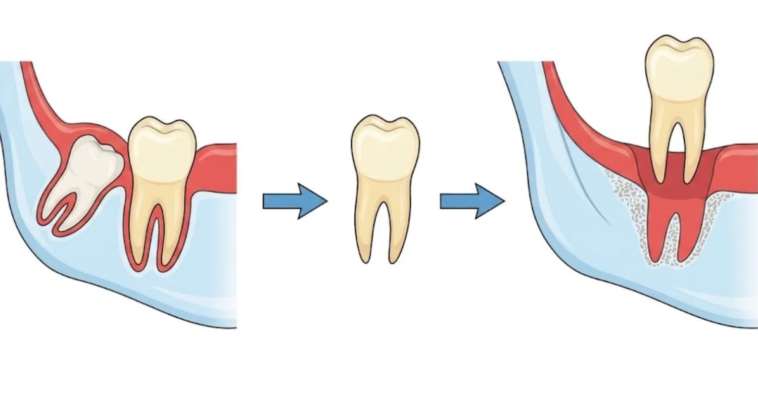
Tooth Autotransplantation in Young Patients moves a suitable natural tooth to a new site in the same mouth. The moved tooth is called the donor tooth. Its new position is called the recipient site. Success depends on protecting the periodontal tissues during removal and placement. Developing teeth often have an advantage because their roots are still forming. An open root tip can support further root growth and, in selected cases, pulp revascularization. Still, an incomplete root does not guarantee success. A 2025 review of 404 teeth in patients under 18 found 85.4% success and 94.2% survival. These are group figures, not personal predictions.
How Does Tooth Autotransplantation Work?
The dental team first studies the donor tooth, the recipient area and root development. Nearby structures and available space are also assessed. Imaging shows tooth position and root shape before surgery. During the procedure, the donor tooth is removed gently to protect periodontal ligament cells. The recipient site is shaped to hold the tooth without excess pressure. The tooth is then placed according to the planned position and bite. Short-term stabilization may follow, depending on the clinical situation. Developing teeth can keep forming their roots after transplantation in suitable cases. Pediatric studies mostly used donor teeth with about one-half to three-quarters root formation. Regular follow-up then tracks healing and development over time.
What Is the Difference Between Tooth Autotransplantation and Dental Implants?
Tooth Autotransplantation in Young Patients uses a natural tooth taken from elsewhere in the same mouth. A dental implant is an artificial fixture placed into the jawbone after tooth loss. The transplanted tooth can keep its periodontal ligament and natural biological connection. An implant fuses directly with bone through osseointegration and has no ligament. This difference matters greatly during growth. Natural teeth can move with the developing jaw, while implants stay fixed in place. For that reason, implants are usually delayed in growing patients. The final choice depends on growth, anatomy, tooth development and orthodontic needs.
Which Teeth Are Commonly Used for Autotransplantation?
Donor teeth must be removable without harming the patient's overall dental function. Premolars are common donors, especially when orthodontic treatment already calls for their removal. Third molars may replace a lost molar in suitable patients. Canines and other permanent teeth are occasionally used when their anatomy is favorable. Selection depends on root shape, root development, tooth size, access and periodontal health. A 2025 meta-analysis found favorable outcomes across premolars, molars, third molars and canines. Large differences between studies limit direct comparisons between donor groups. Careful imaging therefore guides each individual choice.
Developing Premolars as Donor Teeth
Developing premolars are often useful donors when their size and stage match the recipient site. Their roots may still be forming during adolescence. This can support continued root growth after transplantation in selected cases. Orthodontic plans sometimes free up a premolar for this purpose. The team confirms that removing it will not create a new functional problem. Not every developing premolar qualifies, because individual anatomy decides.
Other Potential Permanent Teeth
Third molars may replace a lost first or second molar when safe removal is possible. Canines can suit selected cases with favorable orthodontic and surgical conditions. Mature teeth can also be moved, though their pulp usually needs different management. Outcomes have been favorable across different root-development groups, with wide variation between studies. Donor choice considers root shape, ligament condition, recipient anatomy, bite and future orthodontic needs. Vitrin Clinic can review these factors using clinical and digital records when appropriate.
Learn more about Tooth Autotransplantation in Young Patients
Why Is Tooth Autotransplantation Considered for Young Patients?
Tooth Autotransplantation in Young Patients is considered because a natural tooth can grow and adapt with the jaw. It preserves periodontal tissues and avoids placing an implant during active growth. It may suit traumatic loss, congenital absence or severe damage to a permanent tooth. A systematic review of developing teeth reported 97.9% overall survival. Survival was about 95.9% at five years and 96.9% at ten years. These results describe study groups, not guaranteed individual outcomes. Donor choice, recipient-site health, surgical technique and follow-up remain decisive.
Tooth Autotransplantation for Missing Permanent Teeth
Tooth Autotransplantation in Young Patients can replace a missing permanent tooth with the patient's own natural tooth. The cause of absence shapes the plan. Congenital absence, trauma, developmental problems and severe damage each create different needs. The recipient site needs enough space and suitable bone. Orthodontic treatment may be needed before or after surgery. The transplanted tooth may then take part in ongoing dental development.
Tooth Autotransplantation After Tooth Loss
After a permanent tooth is lost, early assessment can protect future options. Dental trauma is a common cause in young people. The dentist checks the socket, bone, gums and neighboring teeth. Timing matters because the site changes after tooth loss. A donor tooth is chosen only if its stage and shape fit the space. Gentle handling and ligament preservation support favorable results.
Congenitally Missing Teeth and Tooth Replacement
Some permanent teeth never develop at all. This can cause spacing, bite and orthodontic problems during growth. Options include orthodontic space closure, space maintenance, prosthetic replacement or transplantation. The best path depends on the missing tooth, available donors, bite and facial growth. A transplanted tooth can fill the gap without an implant during growth. The donor must, however, be expendable within the overall plan.
Tooth Loss Caused by Trauma, Infection, or Severe Dental Damage
Trauma, infection or extensive decay can leave a permanent tooth beyond predictable repair. The dentist first checks whether the original tooth can be saved. If not, transplantation becomes one possible replacement. The recipient site must be free of active infection that could disturb healing. The donor tooth must be healthy and suitable for transfer. Thorough examination and imaging therefore come before any decision.
Understand the details of Folate Deficiency and Oral Health
Can a Tooth Be Transplanted in a Teenager?
Yes, in suitable cases. Tooth Autotransplantation in Young Patients is often planned during adolescence, when many permanent roots are still forming. Age alone, however, does not decide suitability. The dentist evaluates root development, donor anatomy, bone, space, gum health and bite. A 2025 review of 404 teeth in patients under 18 reported 94.2% survival. These findings support treatment for carefully selected teenagers. Orthodontic needs should also be reviewed before surgery.
Is Tooth Autotransplantation Suitable for Children?
It can be, but careful evaluation of dental development comes first. The donor tooth must reach a suitable developmental stage. The recipient site needs enough space and healthy supporting tissue. Younger children still have much growth ahead, so long-term planning is essential. The team considers eruption patterns, nearby developing teeth and future orthodontics. Research focuses on tooth development rather than a child's exact age. Treatment is considered only after thorough clinical and radiographic assessment.
Tooth Autotransplantation in Adolescents
Tooth Autotransplantation in Young Patients is often timed for adolescence, when several donor teeth are still developing. Teenagers may face trauma, missing teeth or crowding that frees up a donor tooth. The team confirms that moving the donor will not create another problem. The recipient site must accept the tooth without forcing it. Long-term checks then track root growth, pulp health, gums and bite.
Why Skeletal and Dental Growth Matters
Natural teeth keep erupting and adapting as the jaw grows. Implants cannot do this because they fuse directly with bone. This difference strongly shapes treatment in growing patients. It is a key reason Tooth Autotransplantation in Young Patients is considered before growth ends. The team considers expected growth, eruption of nearby teeth, bite and orthodontic timing. Planning looks at both today's needs and tomorrow's development.
Developing Jawbone and the Transplantation Site
The recipient site must offer enough bone and gum tissue for stable healing. In young patients, that bone keeps changing after surgery. The dentist measures its width, height and shape beforehand. Space between neighboring teeth must fit the donor without harming them. Orthodontics can create or hold space when needed. Imaging may reveal limits a clinical exam cannot detect.
Root Development and Donor-Tooth Maturity
Immature roots often carry more healing potential after transplantation. An open root tip may allow continued root growth and possible pulp revascularization. X-rays show how far the root has formed. More mature teeth may need planned root canal care. Curved or unusual roots can make gentle removal and placement harder. Pediatric studies commonly used teeth with one-half to three-quarters root formation. These ranges guide decisions but are not fixed rules for every patient.
What Is the Best Age for Tooth Autotransplantation?
There is no single ideal birthday for this treatment. Dental development tells clinicians more than age does. Root formation, skeletal growth, recipient anatomy, bite and orthodontic needs all matter. Studies often report treatment during late childhood and adolescence. Early professional review after trauma or a missing tooth helps keep options open.
Explore the facts on Tirzepatide Dental Side Effects
Which Young Patients May Be Candidates for Tooth Autotransplantation?
Tooth Autotransplantation in Young Patients suits cases where several biological and anatomical conditions line up. A suitable donor must exist without harming overall function. The recipient site needs enough bone, gum tissue and space. Root development and donor shape affect surgical feasibility. Healthy gums support healing after surgery. Growth stage and bite influence the tooth's long-term position. Candidates often include young people with a missing, lost or unrestorable permanent tooth. A clinical examination confirms whether treatment is appropriate.
Adequate Donor Tooth Availability
The donor must be healthy, accessible and suitably developed. Its removal must fit the wider treatment plan. A useful tooth should not be sacrificed simply to fill another gap. Orthodontic plans sometimes make a premolar available. Imaging shows root shape, developmental stage and nearby structures. A good donor lowers surgical trauma and supports healing.
Suitable Recipient Site and Bone Support
The new site must give the tooth enough bone and soft tissue. Infection or active inflammation should be treated first. The space should roughly match the donor tooth's size. Heavy reshaping of the site adds surgical trauma. Radiographs show bone contours and neighboring roots. Growth-related changes in the jaw are also considered. A healthy site does not guarantee success, but a poor one complicates healing.
Sufficient Space for the Transplanted Tooth
The donor tooth must fit without being forced into place. Crown and root sizes are compared with the available gap. Neighboring roots must stay protected. Orthodontics may open or hold the space before surgery. Space needs can change as nearby teeth erupt. Digital scans and radiographs help measure the gap when appropriate.
Healthy Periodontal and Oral Tissues
Living ligament cells on the donor root drive attachment after transplantation. Gentle surgery helps protect them. The recipient gums should be healthy and free of inflammation. Existing gum problems should be treated first. Good home hygiene after surgery limits plaque and inflammation. Parents can help younger patients follow cleaning instructions.
Favorable Root Development
Developing roots may continue maturing after transplantation. An open apex can also allow pulp revascularization in selected teeth. Imaging confirms the exact developmental stage. Root length, shape, curvature and canal anatomy affect surgery and aftercare. Many pediatric studies used teeth with partial root formation. Root stage is important but is never assessed alone.
When Autotransplantation May Not Be Appropriate
Treatment may be unsuitable without a good donor tooth. Poor bone, too little space or unfavorable root shape can also rule it out. Severe gum disease or active infection needs attention first. A donor with an important functional role may be worth keeping in place. Sometimes orthodontic or restorative options suit the long-term bite better. The decision weighs risks, benefits and alternatives, not age alone.
Check out the connection between X-linked Hypophosphatemia and Dental Damage
How Is Tooth Autotransplantation Diagnosed and Planned?
Tooth Autotransplantation in Young Patients is planned through close study of both donor tooth and recipient site. The process starts with dental history and a clinical examination. Radiographs show root development, tooth position and bone anatomy. CBCT may be added when three-dimensional detail is clinically needed. Intraoral scans help measure tooth size and available space. Orthodontic review may follow if the space needs changing. Evidence links careful case selection with favorable outcomes. At Vitrin Clinic, digital tools support individualized planning when clinically appropriate.
Clinical Examination Before Tooth Autotransplantation
The exam begins at the missing or damaged tooth area. The dentist checks neighboring teeth, gums, bite, space and signs of infection. The possible donor is checked for decay, gum health, mobility and function. Past trauma, orthodontics and restorations are reviewed. Eruption patterns and developing teeth are assessed in younger patients. Findings are then combined with imaging.
Dental X-Rays and 3D Imaging
Standard X-rays show root development, tooth position and nearby structures. CBCT adds three-dimensional detail when plain images cannot answer a clinical question. Its use should be justified because it involves radiation exposure. It helps assess complex roots, bone dimensions and nearby roots. Vitrin Clinic uses CBCT and digital X-rays when clinically appropriate. Imaging supports, but never replaces, clinical examination.
Evaluating Donor Tooth Root Development
Radiographs show whether the donor root is immature, partly formed or complete. An open apex may allow further development after transfer. The dentist also checks root length, curvature, width and canal shape. These features affect removal, placement, splinting and possible root canal care. Root stage is only one part of donor selection. The full clinical picture decides.
Assessing Bone, Space, Occlusion, and Adjacent Teeth
The recipient site must support the tooth while protecting its neighbors. Available space is compared with donor size and the planned final position. The bite is checked to avoid heavy early contact. Adjacent roots must stay clear during placement. Future eruption and jaw growth are also considered. Digital scans measure crowns and gaps, while radiographs show roots and bone.
Digital Treatment Planning for Growing Patients
Digital planning brings clinical, radiographic and three-dimensional information together. Scans show tooth size, available space and bite relationships. CBCT adds anatomical detail when clinically indicated. Together they help judge donor and recipient compatibility. They also help coordinate surgery with orthodontic treatment. Plans must allow for future eruption and skeletal growth. Surgical findings can still differ from imaging, so clinical judgment remains essential.
Why Precise Planning Matters Before Surgery
Donor and recipient anatomy vary widely between patients. The donor must come out with minimal ligament damage. The site must hold the tooth without excess pressure or bone removal. Planning reveals root curves, nearby structures and space limits in advance. It also flags likely orthodontic or root canal needs. Evidence consistently links careful selection and technique with better outcomes.
Discover how Peri-Implant Bone Loss is Detected
What Happens During Tooth Autotransplantation?
Tooth Autotransplantation in Young Patients follows several carefully coordinated surgical steps. The team first confirms the donor's position and prepares the recipient site. The donor tooth is then removed as gently as possible. The site is adjusted to receive it without unnecessary bone removal. The tooth is placed at the planned depth, angle and bite. Stabilization may limit movement during early healing. Throughout, the priority is protecting the periodontal ligament. Details vary with each patient's anatomy and clinical circumstances.
Preparing the Recipient Site
The dentist shapes the site to receive the donor tooth. The goal is enough room while keeping healthy supporting bones. Too much preparation removes needed support. Too little prevents proper seating or creates pressure around the root. Any infection is addressed beforehand. Final shaping depends on the donor's root shape, size and stage.
Removing the Donor Tooth
The donor is removed with minimal force to protect its ligament. Root shape, tooth position and surrounding bone affect difficulty. Developing roots need especially careful handling. Time and handling outside the mouth are kept to a minimum. The dentist checks the tooth before placing it.
Placing the Tooth Into the Recipient Site
The tooth is set at the planned angle and depth. Excess pressure is avoided to protect the ligament and blood supply. Contacts with neighboring teeth are checked. The bite is adjusted to avoid heavy early loading. Minor changes may be needed during surgery. Clinical judgment guides each adjustment.
Stabilizing and Monitoring the Transplanted Tooth
A splint or suture may hold the tooth during early healing. Its type and duration depend on mobility and surgical findings. Overly rigid fixation is not always ideal, since slight movement can aid ligament healing. Follow-up visits check mobility, gums, bite and comfort. Radiographs track root growth and detect early problems. Patients and parents should follow every postoperative instruction.
Protecting the Periodontal Ligament During the Procedure
Ligament cells on the root drive healing and reattachment. Rough handling can damage them and raise complication risk. The root surface should not be scraped, dried or handled unnecessarily. The recipient site is prepared with equal care. This living ligament separates transplantation biologically from implant placement. Still, root stage, site health, splinting and follow-up matter too.
How Successful Is Tooth Transplantation in Young Patients?
Tooth Autotransplantation in Young Patients shows favorable survival when cases are well chosen and managed. Results vary because studies differ in patients, donor teeth, follow-up and definitions. A 2025 review of 404 teeth in under-18s found 85.4% success and 94.2% survival. Another review of developing teeth reported 97.9% overall survival. Five-year survival in that review was about 95.9%. These figures describe study populations, not individual patients. Outcomes depend on donor development, ligament care, site health, technique and follow-up. Long-term monitoring therefore remains essential.
Tooth Autotransplantation Success Rate
Survival usually means the tooth remains present and working. Success adds stricter criteria, such as healthy gums and no progressive resorption. That explains why success figures run lower than survival figures. For Tooth Autotransplantation in Young Patients, both measures are commonly reported. The pediatric review reported 85.4% success against 94.2% survival. Follow-up length and case selection also shape these numbers. Personal risk still needs an individual assessment.
What Factors Influence Long-Term Success?
Several factors shape long-term outcomes. Root stage affects biological healing potential. Donor choice affects surgical difficulty and ligament healing. The recipient site must provide bone, gum tissue and space. Gentle handling protects ligament cells on the root. Splinting, hygiene and regular follow-up support early healing. Bite, orthodontic treatment and pulp status matter over time.
Root Development
Immature roots may keep growing after transfer. An open apex may allow pulp revascularization in selected cases. A review of patients under 18 found donors commonly had one-half to three-quarters root formation. Root stage alone, however, does not guarantee success. Site conditions, technique, gum health and follow-up matter equally.
Donor Tooth Selection
The donor must be healthy, accessible and sized for the site. Root shape affects removal and aftercare. Premolars are frequent choices, while third molars suit some molar gaps. A 2025 review found favorable results across several donor types, with wide variation. Selection follows individual anatomy, not a universal preference.
Recipient-Site Condition
Healthy bone and gum tissue must surround the planned position. The space must also suit the patient's bite. Active infection or inflammation needs treatment first. Nearby roots and future growth are also considered. Poor site conditions make predictable healing harder.
Surgical Technique and Tooth Handling
Gentle removal protects ligament cells on the root. The tooth spends minimal time outside the mouth. The site is prepared without excess bone removal. Placement avoids unnecessary pressure on the root. Stabilization suits the clinical situation. Even careful technique cannot remove every risk.
Follow-Up and Oral Hygiene
Regular visits check mobility, gums, bite and radiographic changes. Good hygiene limits plaque and inflammation around healing tissue. Dietary limits may apply early on. Developing teeth may need longer monitoring. Visits can reveal resorption, ankylosis or pulp problems early. Parents can help younger patients keep appointments.
Find out the link in Peripheral Artery Disease and Periodontitis
Why Success Rates Differ Between Studies
Some studies include only immature teeth, while others include mature ones. Definitions of success and survival also differ. Longer studies catch late complications that shorter ones miss. Surgical, splinting and root canal protocols vary between centers. Recent reviews noted substantial differences between pediatric studies. Comparing raw percentages can therefore be misleading.
How Long Can an Autotransplanted Tooth Last?
After Tooth Autotransplantation in Young Patients, a well-healed tooth can last many years. One review of developing teeth reported 95.9% survival at five years. Ten-year survival reached 96.9% in the same analysis. No individual lifespan is guaranteed, however. Resorption, ankylosis, pulp problems or gum disease can shorten it. Regular checkups and good hygiene support longevity.
What Is Tooth Autotransplantation Healing and Recovery Like?
Tooth Autotransplantation in Young Patients requires a healing period while tissues adapt to the new site. Early recovery may bring mild soreness, swelling or slight tooth mobility. These symptoms should ease gradually. Patients receive guidance on eating, cleaning, activity and splinting. Follow-up visits check ligament healing, mobility, bite and surrounding tissues. Developing teeth need extra monitoring of root growth. Radiographs help track development and detect complications. Recovery varies with surgical complexity and individual healing.
What to Expect Immediately After Surgery
The tooth and nearby gums may feel tender. Mild swelling is common in the first days. Biting may feel different while tissues heal. Some mobility can occur, depending on the splinting method. Patients should avoid touching the area with fingers or tongue. Medication should be taken exactly as prescribed. Parents should watch younger patients closely. Rising pain, swelling, discharge or fever needs prompt review.
How Long Does the Transplanted Tooth Take to Heal?
Healing happens in stages rather than on a fixed day. Gums settle over the first weeks. Ligament attachment develops over a longer period. Bone remodeling can continue for months. Developing roots may keep forming during this time. Follow-up timing depends on root stage and surgical findings. Healing is an ongoing process, not a single event.
When Can Patients Return to Normal Eating and Activities?
Soft foods are usually advised early on. Hard, sticky or chewy foods may be avoided temporarily. Contact sports may need a pause or a mouthguard. The dentist decides when normal function can resume. Patients should not test the tooth by biting hard foods. Any sudden change in mobility, pain or bite should be reported.
Follow-Up Appointments and Dental Monitoring
Symptoms alone cannot show how well the tooth is healing. Visits check mobility, gums, attachment, bite and inflammation. Radiographs track root growth and detect resorption. Root canal assessment may become necessary if the pulp fails. Schedules vary by case and clinical protocol. Missed visits can delay detection of complications. Long-term monitoring matters because the tooth may serve for decades.
Monitoring Root Development and Tooth Vitality
Developing teeth may keep forming roots after transfer. Pulp revascularization can occur but is never guaranteed. Vitality tests and radiographs help assess pulp health. Mature donors have less revascularization potential and may need root canal care. Persistent symptoms or radiographic changes need further evaluation. Monitoring separates normal development from complications.
Why Long-Term Follow-Up Matters in Growing Patients
Dental and jaw growth continue long after surgery. The transplanted tooth may erupt and adapt further. Nearby teeth may shift as the patient grows. Orthodontics can also change the final relationships. Periodic checks catch resorption, ankylosis, pulp or gum problems early. Tooth Autotransplantation in Young Patients therefore needs monitoring well into adulthood. Ten-year survival data show it can be a lasting solution.
Read about Alveolar Ridge Preservation After Tooth Extraction
What Are the Risks and Complications of Tooth Autotransplantation?
Tooth Autotransplantation in Young Patients can involve complications affecting the pulp, gums, root or bone. Possible problems include root resorption, ankylosis, pulp necrosis, infection and excess mobility. Risk depends on donor maturity, surgical handling, site health and follow-up. Immature donors often heal well but are not risk-free. Patients should know both expected symptoms and warning signs. Early detection allows timely management. The dentist should explain individual risks before surgery.
Tooth Autotransplantation Risks and Complications
Root resorption can gradually wear away root structure. Ankylosis fuses the root directly to the surrounding bone. Pulp complications may need root canal treatment. Infection may develop around the surgical area. Excess mobility can signal delayed ligament healing. Risk profiles differ between immature and mature donors. Careful selection and gentle surgery reduce, but cannot remove, these risks.
Root Resorption
Resorption is loss of root structure after transplantation. Inflammatory resorption often links to root damage or pulp infection. Replacement resorption occurs when bone gradually replaces root tissue. Early stages may cause no symptoms at all. Radiographs are therefore key for detection. Immature roots lower, but do not remove, this risk. Treatment depends on type, severity and cause.
Ankylosis
Ankylosis joins the root directly to bone, bypassing the ligament. In growing patients, the tooth may stop following its neighbors vertically. It can then appear to sink below the bite line. Exams may show changed position, a distinct tapping sound or reduced mobility. Radiographs can support the diagnosis. Gentle handling aims to prevent it.
Pulp Necrosis and Endodontic Complications
The pulp may lose vitality after transplantation, especially in mature teeth. Immature teeth may revascularize, though not always. Mature teeth often need planned root canal treatment. Symptoms alone do not reveal pulp status. Clinical tests and radiographs guide decisions. Untreated pulp infection can trigger inflammatory resorption.
Infection or Inflammation
Signs include rising pain, swelling, redness, discharge or tenderness. Good hygiene supports a cleaner healing environment. Antibiotics should be used only under professional guidance. Not every postoperative symptom means bacterial infection. Infection may also coexist with pulp or gum problems. Clinical and radiographic exams find the source. Worsening symptoms need prompt review.
Mobility or Problems With Periodontal Healing
Some early mobility is normal while ligament tissue heals. It does not automatically mean failure. Increasing or lasting mobility, however, needs evaluation. Heavy biting early on can disturb healing. Patients should follow diet and activity limits. Follow-up shows whether mobility reflects normal recovery or a problem.
What Warning Signs Should Patients Watch For?
Watch for symptoms that worsen instead of improving. Rising pain, persistent swelling, discharge and fever need review. Sudden bite changes may mean the tooth has moved. A new tooth can feel odd at first, which is often normal. Young children may struggle to describe symptoms. Parents should therefore observe recovery closely.
Persistent Pain or Increasing Swelling
Mild soreness is expected, but worsening pain is not. It may signal inflammation, infection or heavy bite contact. Severe pain should never be assumed normal. Self-treatment should not replace professional review. The dentist examines the site, tooth, gums and bite. Imaging may follow if needed.
Discharge, Fever, or Signs of Infection
Discharge or fever can indicate infection. Swelling with rising pain also needs attention. The source may be the gums, surgical site or pulp. Antibiotics are not automatically required for every symptom. Treatment depends on the diagnosis. Parents should monitor temperature, swelling, eating and behavior in younger patients.
Increasing Tooth Mobility or Bite Changes
Rising mobility may reflect weak splinting, gum problems or bite interference. A new bite mismatch may mean the tooth shifted. Avoid pushing the tooth with tongue or fingers. The dentist will check mobility and bite, adding radiographs if needed. Early review is better than waiting.
See the risks of Purposely Avoiding Dental Checkups and Tooth Loss
Tooth Autotransplantation vs Dental Implants in Young Patients
Tooth Autotransplantation in Young Patients and dental implants behave very differently during growth. Transplantation moves a natural tooth together with its ligament. An implant replaces the root with a fixture that fuses to bone. Natural teeth can erupt with the jaw, while implants stay fixed. This matters for children and teenagers with growth remaining. Transplantation needs a suitable donor, while an implant does not. Neither option suits every young patient automatically. Growth, anatomy, donor availability and long-term plans decide.
Why Dental Implants Are Different in Growing Patients
Implants fuse with bone and have no ligament. As the jaw grows, neighboring teeth keep erupting. An implant does not follow them. Over time, it can end up lower than adjacent teeth. Implant timing therefore depends on completed growth, not a fixed age. Space can be held temporarily until growth slows. Tooth Autotransplantation in Young Patients avoids this mismatch when a suitable donor exists.
Preserving a Natural Tooth and Supporting Jaw Development
The natural ligament provides sensation and biological function that implants lack. A transplanted tooth can erupt and adapt with growth. Its function also stimulates surrounding bone. This can help preserve the alveolar ridge. These features matter most before facial growth is complete. Still, preserving a natural tooth is not automatically the better choice. Clinical findings and long-term goals decide.
When an Implant May Be Considered Later
Some patients lack a suitable donor or recipient site. Others need orthodontic space management first. In these cases, an implant may follow once growth is sufficiently complete. Temporary options maintain function and appearance meanwhile. Bone volume and gum tissue are assessed before implant placement. Waiting does not mean going without care.
How Dentists Choose Between Autotransplantation and Other Tooth-Replacement Options
The dentist weighs donor availability, root stage, site anatomy, space, bite and growth. Orthodontic needs are also considered. The cause of tooth loss shapes the sequence. Space closure suits some patients, while space maintenance suits others. Temporary replacement can bridge the growth years. Implants may follow later. No single method fits all young patients.

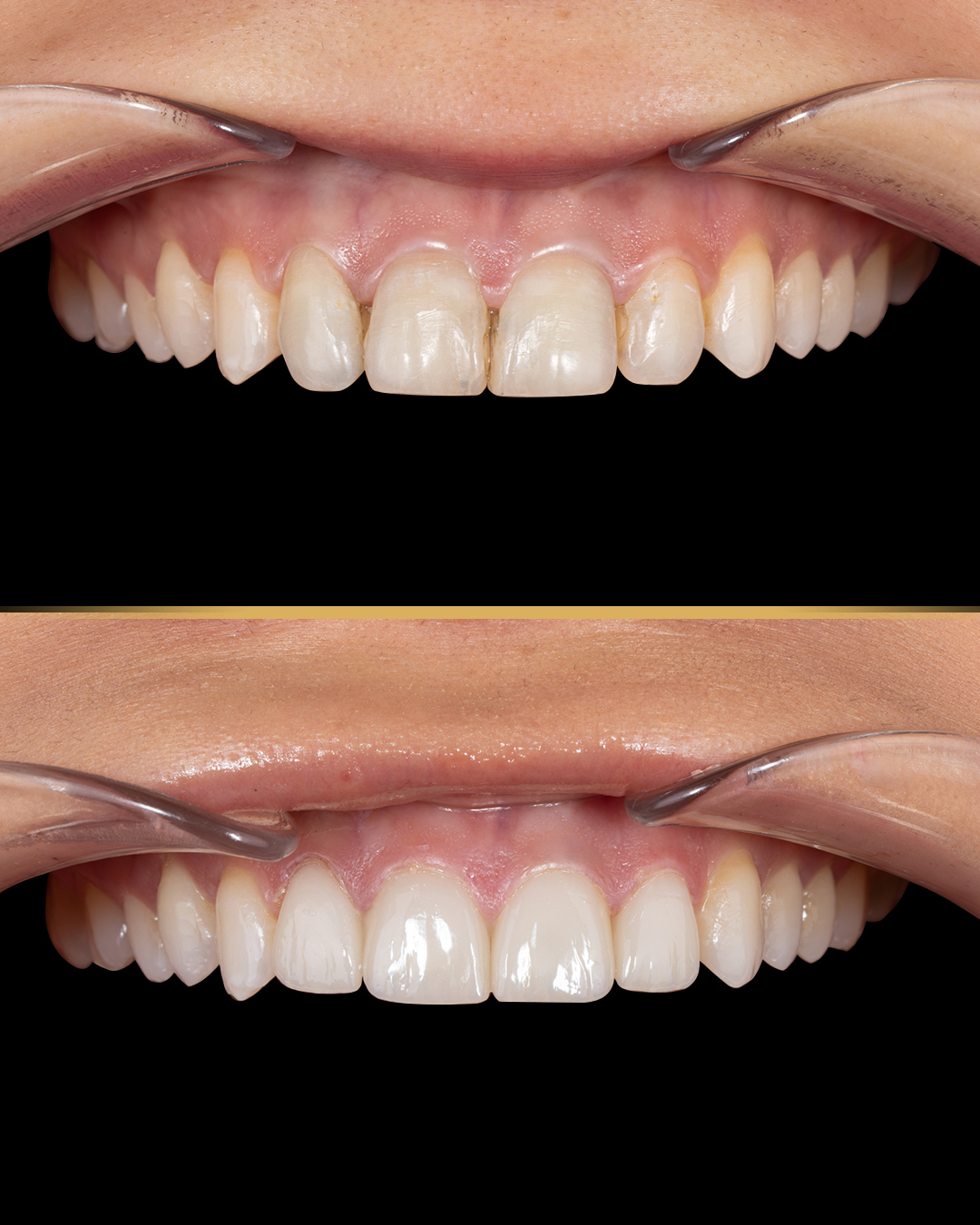
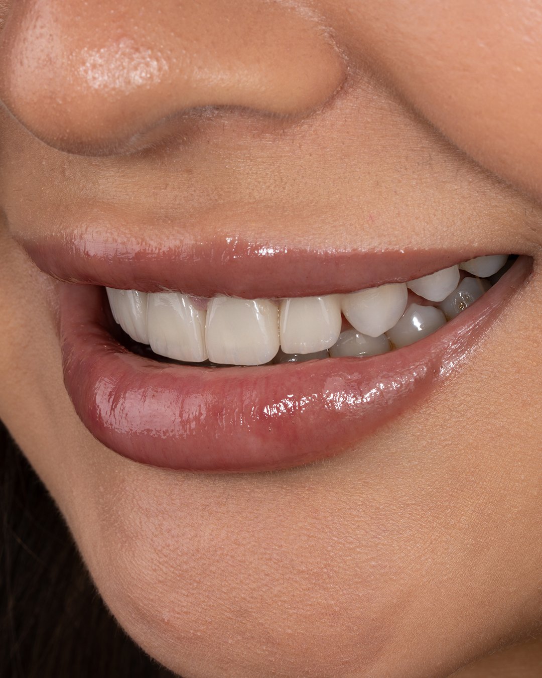
What Are the Alternatives to Tooth Autotransplantation for Young Patients?
Tooth Autotransplantation in Young Patients is one of several ways to manage a missing tooth. Alternatives depend on age, growth stage, the missing tooth, space and bite. Orthodontics can close or hold the gap. Temporary or resin-bonded replacements work during growth. Conventional bridges suit selected cases. Implants can come later once growth slows. Each option has distinct benefits, limits, upkeep and timing. Professional evaluation comes before any definitive choice.
Orthodontic Space Management
Braces or aligners can move neighboring teeth into the gap. Alternatively, they can hold space for later replacement. The choice depends on the missing tooth, profile, bite and growth. Closure can remove the need for future prosthetics. Planning protects nearby roots and developing teeth. Treatment can also be coordinated with later implant placement.
Resin-Bonded or Temporary Tooth Replacement
Interim replacements maintain appearance and hold space during growth. Resin-bonded bridges attach to neighboring teeth with minimal drilling. Removable appliances can adapt as the mouth changes. The dentist reviews bite, enamel, hygiene and eruption. These are often steps toward a later definitive solution. They buy time for further development and planning.
Dental Bridges in Selected Cases
A bridge replaces the visible tooth using neighboring teeth for support. Conventional bridges require trimming healthy teeth, which is often undesirable in youth. Resin-bonded designs are more conservative. Bite, enamel, hygiene and growth all affect suitability. Bridges may need later repair or replacement as the dentition changes. Professional evaluation decides whether a bridge fits.
Delayed Implant Treatment After Growth
When transplantation is unsuitable, an implant can wait until growth slows. Orthodontic or temporary solutions hold space meanwhile. Growth assessment guides timing, not age alone. Bone, gums, bite and neighboring teeth are checked before placement. Additional procedures may occasionally prepare the site. This gives a structured path when early implants are inappropriate.
Understand the effects of Frequent Snacking and Tooth Decay
What Does Tooth Autotransplantation Cost in the US, UK, and Turkey?
Tooth Autotransplantation in Young Patients can cost differently depending on the country, provider, surgical complexity, imaging, and additional treatment. Because dedicated national pricing for autotransplantation is limited, the following figures are approximate treatment estimates, not fixed prices. A straightforward case may cost less, while complex cases requiring orthodontics, endodontics, or surgical preparation can cost more.
Country | Approximate Average Cost | Typical Estimated Range |
🇺🇸 United States | 4,000–6,000 | 3,000–8,000+ |
🇬🇧 United Kingdom | $3,000–$4,500 | $2,000–$6,000+ |
🇹🇷 Turkey | $1,000–$2,000 | $700–$3,000+ |
These figures should be treated as indicative estimates rather than standardized national averages. The final price depends on the donor tooth, recipient site, imaging, surgical complexity, stabilization, follow-up, and whether orthodontic or endodontic treatment is required. Current 2026 dental price comparisons show substantial differences between US, UK, and Turkish dental fees, although autotransplantation-specific prices are not consistently published.
Average Tooth Autotransplantation Cost in the United States
The estimated cost of Tooth Autotransplantation in Young Patients in the United States is approximately 4,000–6,000, with complex cases potentially exceeding $8,000. Costs may include specialist assessment, imaging, donor-tooth removal, recipient-site preparation, transplantation, stabilization, and follow-up. Endodontic or orthodontic treatment may increase the final amount. US dental fees vary significantly by location and provider, so patients should request an itemized treatment estimate before treatment.
Average Tooth Autotransplantation Cost in the United Kingdom
The estimated cost in the UK is approximately $3,000–$4,500, although straightforward or complex cases may fall outside this range. Tooth Autotransplantation in Young Patients may require specialist assessment, imaging, surgery, stabilization, and follow-up. Orthodontic treatment may also be needed to create or maintain space. Private dental fees vary by region and provider. NHS availability depends on local services and clinical eligibility.
Average Tooth Autotransplantation Cost in Turkey
The estimated cost in Turkey is approximately $1,000–$2,000, with complex cases potentially reaching around $3,000 or more. Tooth Autotransplantation in Young Patients may involve digital imaging, surgery, stabilization, follow-up, and additional dental care. Turkish dental treatment prices are generally lower than many private US and UK prices, but individual clinics use different pricing structures. At Vitrin Clinic, the final price should be determined after reviewing the patient's clinical requirements.
What Can Change the Total Treatment Cost?
Several factors can increase or decrease the final cost. Tooth Autotransplantation in Young Patients may require CBCT or other diagnostic imaging, complex donor-tooth removal, recipient-site preparation, stabilization, or endodontic treatment. Orthodontics can add costs when space must be created or maintained. Follow-up requirements also vary between patients. International patients should additionally consider travel, accommodation, and future follow-up expenses. A personalized treatment plan provides a more accurate estimate than an advertised starting price.
3D Imaging and Diagnostic Planning
CBCT adds cost when clinically indicated, not in every case. It shows root anatomy, bone size and nearby structures. Scans may be billed separately from surgery. Ask why each scan is recommended. Vitrin Clinic uses CBCT, digital X-rays and intraoral scanning when clinically appropriate.
Surgical Complexity
Deeply positioned donor teeth need more complex access. Curved roots make removal more demanding. Site preparation, past trauma or limited space add difficulty. Complex cases may need longer appointments or specialist input. Quotes should reflect the anatomy actually found.
Endodontic Treatment
Mature donor teeth often need root canal treatment. Immature teeth may avoid it if the pulp revascularizes. The need may only become clear during follow-up. Root canal care is usually a separate fee. Ask whether possible endodontic care is included.
Orthodontic Treatment
Orthodontics may create space before surgery or refine position afterward. The donor may be chosen within an existing orthodontic plan. Treatment length depends on development and goals. Orthodontic fees are usually separate. Ask whether orthodontic planning is part of the quote.
Follow-Up and Additional Procedures
Monitoring continues well beyond surgery day. Visits may include exams, radiographs, vitality tests and bite checks. Complications may need extra treatment. Reshaping may improve function or appearance. International patients should ask how aftercare works back home.
Why Autotransplantation Prices Are Difficult to Standardize
Each case involves different donor and recipient anatomy. One procedure may be simple, another highly complex. Imaging, orthodontics, root canal care and follow-up vary widely. Location and provider experience affect private fees. Online prices also define what is included differently. Treat any published figure as approximate until assessed.
What We Notice Clinically
Tooth Autotransplantation in Young Patients works best with growth-aware planning. Donor maturity often matters more than chronological age. The recipient site must offer bone, gum tissue and space. A missing tooth does not automatically call for an implant during growth. Preserving a natural tooth can bring biological advantages in selected patients. Evidence is favorable for well-selected developing teeth, though outcomes vary. Careful diagnosis beats any fixed treatment formula. At Vitrin Clinic, examination and digital diagnostics support individual planning when indicated.
Young Patients Often Need Growth-Aware Treatment Planning
A plan that fits today may need adjusting as nearby teeth erupt. A transplanted tooth can take part in ongoing development. An implant cannot. Orthodontics may shift the tooth's final position. Planning considers current anatomy and expected growth together. This reduces conflicts with future orthodontic treatment.
A Missing Tooth Does Not Automatically Mean an Implant Is the First Option
Growing patients still have developing jaws and erupting teeth. A natural donor may offer another path. Orthodontic space management or temporary replacement are also options. Implants can wait until growth slows. Assessment identifies which options remain appropriate. No single replacement suits every young patient.
Donor Tooth Development Can Be More Important Than Chronological Age
Root maturity shapes healing potential. Immature teeth may keep forming roots after transfer. An open apex may also support pulp revascularization. Age does not precisely reflect every tooth's stage. Radiographs show root length and apex development. Studies support development-based, rather than age-based, assessment.
A Healthy Recipient Site Is Essential for Predictable Healing
Healing depends on the local biological environment. Bone should support the root without pressure. Gums should provide good coverage. Infection or inflammation needs treatment first. Space and bite are checked too. Unfavorable site conditions add complexity and risk.
Common Misconceptions About Tooth Transplantation
Many people assume any healthy tooth can be moved. Others believe age alone decides eligibility. Some think follow-up is unnecessary once the tooth feels normal. Each belief is inaccurate. Favorable evidence still does not guarantee individual success. Professional assessment remains essential.
Tooth Autotransplantation Is Not the Same as a Dental Implant
Transplantation moves a living tooth within the same mouth. An implant is an artificial fixture placed in bone. The transplanted tooth keeps its ligament, while an implant relies on osseointegration. The tooth can erupt with growth, but an implant cannot. Donor needs, surgery, upkeep and timing also differ.
Not Every Healthy Tooth Can Be Used as a Donor Tooth
Health alone does not make a good donor. Root stage, shape, size, access and function all matter. Removing a useful tooth may create a new problem. Some root shapes resist gentle removal. A tooth is only a candidate until examination and imaging confirm it.
Clinical Note
Assessment should combine dental development, donor anatomy, site condition and long-term goals. Evidence favors well-managed developing donor teeth. Tooth Autotransplantation in Young Patients should never be planned from statistics alone. Published rates cannot replace individual diagnosis. Imaging or surgical findings may also change the plan.
Dr. Rifat Alsaman, Head of the Medical Team at Vitrin Clinic and cosmetic dentist, on Individualized Assessment
Dr. Rifat Alsaman emphasizes individualized assessment before any complex dental treatment. For young patients, evaluation covers donor tooth, recipient site, development, space and surrounding structures. Digital X-rays, CBCT or 3D intraoral scans support the exam when clinically appropriate. Future orthodontic and restorative needs are also weighed. The aim is understanding each patient's anatomy before choosing a pathway. This approach separates suitable candidates from cases needing alternatives.
What Should Parents and Young Patients Ask Before Treatment?
Tooth Autotransplantation in Young Patients involves decisions families should understand clearly. Ask why transplantation is proposed and what alternatives exist. Cover donor suitability, site health, root stage, imaging, surgery and follow-up. Ask about possible complications and how they are managed. Ask whether orthodontic or root canal care may follow. Clarify full costs, not just the surgical fee. International families should confirm how aftercare will work at home.
Is the Donor Tooth Suitable?
Ask about the donor's root stage, shape, size, gum health and position. Ask whether removing it creates a new problem. Imaging should show it can come out gently. The dentist should explain why this tooth fits the site. Ask whether another donor or option exists.
Is the Recipient Site Ready?
Ask whether there is enough bone, gum tissue and space. Discuss any infection or unfavorable anatomy. Ask whether orthodontics is needed first. Ask how the site may change with growth. Clinical and imaging findings should guide the answer.
How Much Root Development Is Present?
Ask whether the root is immature, partly formed or complete. An open apex may allow continued development. Mature roots may need planned root canal care. Ask about root shape and curvature, too. Root stage is one factor among several.
What Imaging Is Needed?
Ask which scans are recommended and why. X-rays show root stage and tooth position. CBCT adds 3D detail when needed. Intraoral scans measure space and bite. Not every patient needs every scan. Vitrin Clinic uses each method when clinically appropriate.
What Is the Expected Follow-Up Schedule?
Ask how often visits occur and what each one checks. Early visits focus on mobility, gums, bite and symptoms. Later visits track roots, pulp and radiographic changes. Young patients may need years of monitoring. Travelers should arrange aftercare at home. Learn which symptoms need earlier contact.
What Alternatives Should Be Considered?
Ask about orthodontic space management, temporary replacement, resin-bonded bridges and delayed implants. Each has different benefits, limits, upkeep and timing. The dentist should explain why each fits or does not. A second opinion is reasonable for complex decisions.
Tips for Patients
Tooth Autotransplantation in Young Patients depends on good preparation and careful aftercare. Bring previous dental records and X-rays to the consultation. After surgery, follow instructions on eating, brushing, medication and activity. Choose soft foods during early healing. Clean the area exactly as instructed. Avoid biting hard objects or chewing directly on the new tooth. Keep every follow-up visit, even when all feels well. Report unusual pain, swelling, discharge or mobility promptly.
How to Prepare for a Tooth Autotransplantation Consultation
Bring previous X-rays, records and details of past trauma or tooth loss. Note when the tooth was lost, damaged or failed to erupt. Prepare questions on timing, healing, risks and alternatives. Expect a clinical exam and possibly imaging. Vitrin Clinic may use digital diagnostics when clinically appropriate. A complete evaluation supports an individualized plan.
What to Do After the Procedure
Protect the tooth while ligament tissue heals. Avoid pressure on the treated area. Take medication exactly as directed. Follow eating and cleaning instructions closely. Keep all scheduled reviews. Contact the team if pain, swelling, bleeding or mobility worsens. Parents should support younger patients' hygiene.
Foods and Habits to Avoid During Early Healing
Avoid hard, sticky, crunchy or very chewy foods early on. Do not bite directly on the treated tooth. Stop nail biting, pen chewing and opening packages with teeth. Smoking and vaping impair healing and should be discussed with the team. Follow individualized dietary advice from the dentist.
Oral Hygiene During Recovery
Keep brushing, but follow modified instructions near the surgical site. Use prescribed rinses as directed. Avoid aggressive brushing or poking the area. Younger patients may need parental help. Instructions may change as healing progresses. Report unusual bleeding, swelling or discharge.
Why Follow-Up Appointments Should Not Be Skipped
Visits catch problems early, often before symptoms appear. They track root growth, ligament healing and vitality. Schedules depend on root stage and individual risk. Missed visits can delay detecting resorption, ankylosis or pulp issues. Keep appointments even when the tooth feels fine.
Note : All images used are for editorial and illustrative purposes only and may not originate from the original news provider or associated company.
When Should a Young Patient Seek Professional Evaluation?
Seek evaluation after permanent tooth loss, significant trauma, delayed eruption or a congenitally missing tooth. Tooth Autotransplantation in Young Patients may be possible when a suitable donor and site exist. Early review assesses development, space, bone, gums and bite. After trauma, prompt care matters because timing can affect options. Delayed eruption also deserves professional assessment. After transplantation, rising pain, swelling, discharge, fever, mobility or bite changes need prompt review.
After Losing a Permanent Tooth
See a dentist promptly after losing a permanent tooth. Timing affects the site and available options. The dentist evaluates the gap and remaining teeth. Transplantation, orthodontics or temporary replacement may be discussed. Previous records help clarify development and space.
After Dental Trauma
Trauma can damage teeth and tissues even when little looks wrong. An exam finds hidden injuries. Imaging shows the tooth and surrounding bone. Seek care after impact, displacement, fracture or tooth loss. Some complications appear later, so follow-up matters.
When a Permanent Tooth Is Not Erupting Normally
Delayed eruption may stem from crowding, position, retained baby teeth or developmental issues. Imaging shows the unerupted tooth's position and stage. Orthodontics may create space for eruption. Transplantation may suit selected cases with an absent or compromised tooth. Early review widens the options.
When a Child or Teenager Has a Congenitally Missing Tooth
A missing tooth should be assessed within overall development. The dentist reviews space, neighbors, bite, bone and possible donors. Transplantation may work with a suitable developing donor. Orthodontics or temporary replacement are alternatives. Timing matters because growth continues. Discuss both short-term and long-term goals.
Urgent Warning Signs After Transplantation
Rising pain, major swelling, persistent bleeding, discharge, fever or worsening mobility need prompt care. Bite changes also warrant review. Contact the team rather than waiting for the next visit. Emergency care may be needed for suspected infection or new trauma. Follow the clinic's emergency instructions.
Tooth Autotransplantation Assessment at Vitrin Clinic
At Vitrin Clinic, assessment focuses on each patient's anatomy and dental development. Tooth Autotransplantation in Young Patients requires review of donor stage, root shape, site health, space and bite. Neighboring teeth are assessed as well. Clinical examination is paired with imaging when indicated. Digital X-rays or CBCT add anatomical detail, while 3D scanning supports planning. The team also considers whether orthodontic, root canal or restorative care should be coordinated. Dr. Rifat Alsaman supports individualized assessment before treatment decisions. The final recommendation follows clinical findings and long-term needs.
Clinical Assessment for Tooth Autotransplantation
Assessment begins with examining the donor and recipient areas. The team evaluates tooth condition, gums, space, bite and nearby structures. Root development and donor position are reviewed. Past trauma and treatments are considered. Age alone does not decide suitability. Findings are then combined with imaging.
Digital X-Rays and CBCT When Clinically Appropriate
Digital X-rays show tooth position, root stage and bone. CBCT provides 3D detail when plain images fall short. At Vitrin Clinic, CBCT is used when clinically appropriate, not routinely. It helps when donor roots or site dimensions are complex. The dentist chooses the method that answers the clinical question.
3D Intraoral Scanning for Treatment Planning When Indicated
Intraoral scanning captures detailed digital models of teeth and bite. These help measure space and tooth relationships. At Vitrin Clinic, scanning is used when indicated. It complements, but never replaces, examination and radiographs.
Personalized Assessment of Donor and Recipient Teeth
Success depends on how well the donor and site match. The donor is judged on stage, root shape, size and access. The site is judged on space, bone, gums and bite. Findings decide whether transplantation or another path fits best.
Coordinating Autotransplantation With Relevant Endodontic, Orthodontic, or Restorative Care
Orthodontics may create or hold space. Root canal care may be needed if pulp healing fails. Restorative work may reshape the transplanted tooth. Vitrin Clinic reviews these needs alongside the transplantation plan. The sequence follows clinical findings and growth stages.
Internal Link Opportunity: Relevant Vitrin Clinic Treatment Options
Link here to the relevant Vitrin Clinic treatment page, such as orthodontics, implants or restorative dentistry. Use descriptive anchor text naming the specific treatment. Avoid generic phrases such as "click here". The link should present transplantation as one option, not a universal solution.
How Vitrin Clinic Supports Your Treatment Journey
Vitrin Clinic supports patients from consultation through diagnostics, planning and aftercare. Tooth Autotransplantation in Young Patients involves several clinical factors that shape long-term results. The first visit reviews history, imaging, trauma, missing teeth and current concerns. Digital diagnostics support evaluation when clinically appropriate. Related orthodontic, root canal or restorative care can be planned together. International patients receive help coordinating treatment stages and follow-up. Ongoing monitoring then checks healing, stability, vitality and root growth.
Initial Consultation and Review of Dental History
The consultation reviews past trauma, missing teeth, eruption delays and earlier treatments. Previous X-rays and records add useful context. Families can raise expectations and concerns. Further diagnostics may be recommended when needed.
Digital Diagnostic Assessment
Assessment may include digital X-rays, CBCT or 3D intraoral scanning. Vitrin Clinic uses these tools when clinically appropriate. They complement the physical exam. The chosen methods depend on each case.
Individualized Treatment Planning
Plans consider donor tooth, recipient site, growth, bite and long-term goals. The team explains stages, monitoring and alternatives. Orthodontic, root canal or restorative steps are built in when needed. The final recommendation follows examination and diagnostics.
Coordinating Treatment Steps for International Patients
International patients may need several appointments across treatment stages. The team organizes diagnostics, procedures and follow-up around clinical needs. Monitoring continues after the procedure, so continuity matters. Patients should know which visits are essential before traveling. Clear contact instructions help if concerns arise at home.
Follow-Up and Post-Treatment Guidance
Follow-up assesses ligament healing, stability, vitality and root growth. Guidance covers hygiene, eating, activity and review visits. Schedules depend on the patient's condition. Report pain, swelling, mobility, discharge or bite changes promptly. Monitoring continues even when early healing looks good.
What Relief and Medical Tips Can Help During Recovery?
Tooth Autotransplantation in Young Patients may cause temporary tenderness, swelling or discomfort. Follow the dentist's instructions on pain relief, diet, hygiene and activity. Soft foods reduce pressure on the tooth early on. Avoid hard or sticky foods until your dentist advises otherwise. Keep cleaning gently around the area. Pain medication should not mask worsening pain, swelling, discharge, fever or mobility. These symptoms need professional review.
Managing Normal Postoperative Discomfort
Mild tenderness or swelling is common after surgery. Rest and follow postoperative instructions. A cold compress on the cheek may ease early swelling when advised. Avoid pressure on the tooth. Discomfort should improve, not worsen.
Following Prescribed Pain-Relief Instructions
Pain relief should follow your dentist's advice. Take the prescribed dose, timing and duration. Never exceed doses or combine medicines without guidance. Parents should supervise younger patients. Severe pain despite medication needs review.
Protecting the Transplanted Tooth During Early Healing
Shield the tooth from heavy biting while tissues heal. Follow guidance on diet, chewing, brushing and sport. Avoid biting objects or using teeth as tools. Instructions may change as healing progresses.
When Pain Relief Is Not Enough and Professional Review Is Needed
Pain that grows, persists or comes with swelling needs assessment. Fever, discharge or rising mobility also warrant prompt review. Waiting may delay treatment. Emergency care may be needed for severe infection or trauma. The team decides on further examination or imaging.
What Side Effects Can Occur After Tooth Autotransplantation?
Short-term effects include tenderness, mild swelling, sensitivity and temporary mobility. Tooth Autotransplantation in Young Patients can also bring less common, longer-term complications. These include root resorption, ankylosis, pulp necrosis, infection and gum problems. Risk depends on donor stage, site condition, surgery and follow-up. Some complications cause no symptoms early on. Scheduled monitoring therefore matters. Report rising pain, swelling, discharge, bleeding, mobility or bite changes promptly.
Expected Short-Term Effects
Tenderness, mild swelling, sensitivity and cautious chewing are common early. Temporary mobility may occur depending on splinting. These usually ease as healing progresses. Ask your dentist which effects to expect. Persistent or worsening symptoms are not normal.
Symptoms That May Require Dental Review
Rising pain, swelling, discharge, fever, persistent bleeding or worsening mobility need attention. Bite changes or unusual sensitivity also warrant review. Do not self-diagnose. Prompt assessment finds infection, pulp or healing problems early.
Possible Long-Term Complications
Some problems appear months or years later. They include resorption, ankylosis, pulp problems and gum changes. Not every patient develops them. Regular exams and imaging support early detection and management.
Root Resorption
Resorption gradually removes hard tissue from the root surface. Some forms stay limited, while others progress. Radiographs reveal changes before symptoms arise. Management depends on type, extent and progression.
Ankylosis
Ankylosis fuses the root to bone without a ligament. During growth, the tooth may lag behind its neighbors. Monitoring tracks its position over time. Management depends on age, severity and the wider plan.
Pulp-Related Complications
Developing donors often have good pulp-healing potential. Pulp necrosis can still occur and may need root canal treatment. Mature teeth have different endodontic needs. Follow-up guides timely management.
Key Takeaways
Tooth Autotransplantation in Young Patients depends on careful evaluation, not age alone. Donor development, site health, space and gums matter most. A natural tooth can adapt as the jaw grows. Recovery needs protection, hygiene and professional guidance. Long-term follow-up tracks roots, vitality and healing. Seek evaluation after tooth loss, trauma, delayed eruption or congenital absence. Costs vary by country and case. Individual assessment remains essential.
Tooth Autotransplantation in Young Patients Depends on More Than Age
Age gives context but does not decide suitability. Donor stage, site condition, space, bite and development matter more. Timing should respect ongoing growth and eruption.
Donor Tooth Development and Recipient-Site Health Are Major Considerations
Developing donors with favorable roots are often preferred. The site needs space, bone and healthy gums. Neither factor is judged alone. Imaging and examination confirm compatibility.
Natural-Tooth Preservation Can Be an Important Consideration During Growth
A natural tooth keeps its ligament and adapts with growth. This helps when implants are unsuitable. Still, transplantation needs a suitable donor and site. Monitoring continues after treatment.
Relief, Recovery, and Medical Tips Should Follow the Treating Dentist's Instructions
Recovery advice depends on the procedure and healing response. Follow guidance on medication, chewing, brushing and activity. Worsening pain or swelling needs review. Parents should help younger patients comply.
Side Effects and Warning Signs Should Be Monitored During Follow-Up
Resorption, ankylosis, pulp or gum problems may develop silently. Regular clinical and radiographic checks catch them early. Report pain, swelling, discharge, mobility or bite changes promptly.
Professional Evaluation Is Important After Permanent Tooth Loss or Dental Trauma
Prompt assessment protects future options. Transplantation may be one path when a donor exists. Orthodontic and temporary options are also reviewed. Imaging helps clarify the anatomy.
Treatment Cost Varies Between the US, UK, and Turkey and Requires Individual Assessment
No universal price exists across these countries. Imaging, orthodontics, root canal care and restorations change totals. Travel adds cost abroad. A full treatment plan gives a reliable estimate.
At Vitrin Clinic
Our treatment planning begins with an individualized assessment based on each patient’s oral health, dental needs, and treatment goals. For younger patients with missing or developing teeth, the team considers the overall condition of the teeth, gums, bite, available space, and surrounding structures before recommending a suitable treatment approach. Depending on the clinical findings, treatment may involve restorative dentistry, orthodontics, endodontic care, dental implants, or other appropriate procedures. Digital X-rays, CBCT, and 3D scanning may be used when clinically appropriate to support accurate diagnosis and planning. Dr. Rifat Alsaman, Head of the Medical Team at Vitrin Clinic and cosmetic dentist, emphasizes that treatment should be tailored to the individual rather than based on age alone. International patients can also receive coordinated consultations, diagnostics, treatment planning, and follow-up guidance.
Reference
FAQs
Dr. Rifat Alsaman has more than 5 years of clinical experience in dentistry and currently serves as the Head of the Medical Team at Vitrin Clinic. He is dedicated to providing exceptional patient care, overseeing treatment planning, and ensuring the highest clinical standards across the team. His expertise, attention to detail, and commitment to continuous professional development have helped countless patients achieve healthier, more confident smiles.


