
A black spot inside your cheek can have several causes, ranging from harmless pigmentation to lesions requiring professional evaluation. A single dark mark may develop from melanin, dental materials, trauma, inflammation, medication, or tobacco exposure. Rarely, a persistent or changing lesion can represent oral melanoma or another significant oral condition. Appearance alone cannot reliably establish the diagnosis. At Vitrin Clinic, an unusual oral finding can be assessed during an appropriate dental examination.
What Is a Black Spot Inside the Cheek?
A dark mark inside the cheek describes a visible color change affecting the oral lining. The color may appear black, brown, gray, blue, purple, or mixed. Some lesions are flat and smooth, while others become raised, thickened, or nodular. Pigment can arise from melanin within oral tissues. Other colors may reflect blood, trauma, medications, metals, or vascular changes.
What Does a Dark Spot Inside the Mouth Look Like?
A dark spot may appear as a tiny dot or larger irregular patch. Some lesions have uniform color, while others contain several shades. Borders may appear sharply defined or gradually merge with surrounding tissue. The surface can remain completely smooth and flat. Traumatic lesions may look darker initially before changing during healing. Lighting, camera angle, and saliva can all change how dark a mark appears.
Is a Black Dot Inside the Cheek Always Caused by Pigmentation?
No, a dark dot does not always represent melanin pigmentation. Blood can create a purple, blue, brown, or black appearance after trauma. A hematoma can resemble a pigmented macule during early healing. Vascular lesions may also appear dark under certain conditions. Dental materials can deposit microscopic particles within oral connective tissues. Some medications can increase melanin production within mucosal tissues.
Why Can the Inside of the Cheek Become Darker?
The inner cheek can darken because of increased pigment production or pigment deposition. Repeated irritation may stimulate pigmentation in susceptible tissues. Tobacco exposure can increase oral melanin production and produce multifocal pigmentation. Previous dental procedures can leave embedded restorative material. Inflammation may produce post-inflammatory hyperpigmentation after tissue injury. Certain medications and systemic disorders can also affect oral pigmentation.
Find out how CT Scans Detect Medication-Related Osteonecrosis of the Jaw
What Are the Common Causes of a Black Spot Inside Your Cheek?
Common causes include melanotic macules, nevi, amalgam tattoos, smoker's melanosis, trauma, and post-inflammatory pigmentation. Medication-related and systemic causes are less frequent but remain clinically relevant. Large studies demonstrate considerable variation in the frequency of different causes. One study identified oral pigmentation in 30.2% of 1,278 examined patients. That study found racial pigmentation and smoker's melanosis among the leading causes. Understanding the common causes helps put a black spot inside your cheek into a realistic perspective.
Oral Melanotic Macule
An oral melanotic macule is usually a localized area of increased melanin. It commonly appears as a small, flat, brown-to-black lesion. The color may be uniform or slightly variable. Many lesions remain asymptomatic and stable over long periods. Melanotic macules can occur on the lip, palate, gingiva, and buccal mucosa. A review notes that definitive diagnosis may require histopathologic evaluation. When a black spot inside your cheek matches this description, documentation is often the first step. At Vitrin Clinic, clinicians can document the lesion's appearance during an appropriate examination.
Oral Nevi and Other Benign Pigmented Lesions
Oral nevi are benign melanocytic proliferations that can create localized pigmentation. They may appear as brown, blue, gray, or dark lesions. Some are flat, while others show slight elevation. Blue nevi represents one recognized benign subtype within oral melanocytic lesions. Oral nevi are less common than skin moles, which surprises many patients.
Amalgam Tattoo and Dental Material-Related Pigmentation
An amalgam tattoo occurs when restorative material becomes embedded within oral tissues. It typically presents as a flat blue-gray, gray, or black area. The lesion often develops near previous dental restorations or extraction sites. Particles may enter the tissue during drilling, polishing, or tooth removal. Its color may remain stable for years. The lesion can resemble a melanocytic lesion clinically. At Vitrin Clinic, previous restorative history can be reviewed alongside clinical findings.
Smoker’s Melanosis and Tobacco-Related Pigmentation
Smoker's melanosis involves increased melanin pigmentation associated with tobacco exposure. It commonly produces brown macules affecting multiple oral sites. The gingiva is a frequent location, although other mucosal areas can be involved. Pipe smokers may show pigmentation on the cheek lining and palate more often. A review estimates that smoker's melanosis occurs in approximately 25% to 31% of tobacco users. A smoker can still develop a black spot inside your cheek from an unrelated cause.
Blood Blister, Bruising, or Minor Trauma
Trauma can create dark areas without involving abnormal melanin production. Cheek biting, sharp teeth, hard foods, or accidental injury can damage small blood vessels. The resulting blood collection may appear red, purple, blue, brown, or black. A blood blister can develop quickly after localized trauma. A bruise generally changes color while the blood is gradually resorbed. At Vitrin Clinic, examination can help determine whether a local dental factor may contribute.
Post-Inflammatory Oral Hyperpigmentation
Inflammation can sometimes leave increased pigmentation after oral tissues heal. This process is called post-inflammatory hyperpigmentation. It may follow irritation, injury, inflammatory oral disease, or repeated tissue trauma. Conditions such as oral lichen planus are sometimes linked with this type of pigmentation. The resulting pigment can persist after the original symptoms disappear. Clinical history can therefore be especially helpful.
Medication-Related or Systemic Causes of Oral Pigmentation
Some medications can cause increased oral pigmentation through melanin-related mechanisms. Reported examples in the literature include certain antimalarials, minocycline, and some chemotherapy agents. Certain systemic diseases can also produce diffuse or multifocal pigmentation. Examples discussed in oral pathology literature include endocrine disorders and inherited syndromes. Medication history should include recently started drugs and long-term treatments.
When Can Multiple Dark Spots Suggest a Systemic Condition?
Multiple dark spots can sometimes occur with systemic disorders or inherited syndromes. The pattern becomes more informative when pigmentation appears across several oral sites. Skin findings, gastrointestinal symptoms, endocrine symptoms, or other changes may provide additional clues. Examples in published reviews include Addison's disease and Peutz-Jeghers syndrome. Addison's disease may cause fatigue, weight loss, and darkening of skin creases.
Can Normal Oral Pigmentation Look Black or Dark Brown?
Yes, normal physiologic pigmentation can appear brown, dark brown, gray, or nearly black. Melanin is a major contributor to normal oral mucosal color. The amount and distribution of pigment vary naturally between individuals. People with darker skin tones often have more visible oral pigmentation. Physiologic pigmentation is often more widespread than a solitary acquired lesion. At the Vitrin Clinic, comparison with surrounding tissues can support the initial assessment.
Read about Vitamin B1 Deficiency and Tooth Damage
What Does the Location of the Dark Spot Inside Your Mouth Tell You?
Location provides important information but does not establish a diagnosis alone. Different pigmented lesions show preferences for particular oral sites. Cheek mucosa, gingiva, palate, lips, and tongue can all develop pigmentation. A study of histopathological specimens found cheek mucosa involved in 21.0% of cases. Gingiva and buccal mucosa were also common locations in another clinical study. For a black spot inside your cheek, the exact position along the bite line matters.
Black Spot on the Inner Cheek
A dark lesion on the inner cheek may represent pigmentation, trauma, or dental-material deposition. Cheek mucosa is also exposed to biting, friction, and contact with restorations. The clinical history can therefore be especially informative. A lesion near a filling may raise suspicion for an amalgam tattoo. A lesion following repeated cheek biting may support traumatic discoloration. A dark mark along the bite line may relate to biting habits. At Vitrin Clinic, the area can be documented during an oral examination.
Dark Spot Near a Dental Filling or Crown
A dark mark near a restoration may have several explanations. Amalgam particles can enter nearby tissues and create a stable blue-gray discoloration. Trauma from an uneven restoration can also irritate the cheek. A crown or filling may change local friction patterns. A chipped or rough edge can repeatedly rub the same small area. Vitrin Clinic can review dental treatment history alongside clinical findings.
Dark Spot on the Gums, Palate, Lip, or Tongue
Pigmentation can occur on many oral surfaces with different clinical patterns. Gingival pigmentation may be physiologic or related to tobacco exposure. Palatal pigmentation requires careful evaluation because palate lesions appear among important differentials. Lip lesions can include melanotic macules and other pigmented conditions. Tongue discoloration has a broader differential that includes vascular and traumatic causes.
Why Do Color, Shape, and Location Matter in Oral Lesion Diagnosis?
Color can indicate melanin, blood, foreign material, or vascular changes. Shape and border characteristics provide information about lesion organization. Location helps narrow the differential based on common clinical patterns. Duration and change add another important dimension. A stable, uniformly colored macule differs from a rapidly changing irregular lesion. No single feature reliably establishes malignancy.
Learn How to Stop Acid Erosion on Teeth
What We Notice Clinically
Clinicians usually begin by describing the lesion before assigning a diagnosis. They assess size, color, surface, border, location, distribution, and surrounding tissues. They may ask about trauma, dental procedures, smoking, medications, and previous lesions. Photographs can help establish whether the lesion is changing. The oral cavity should be examined systematically rather than focusing only on the visible spot. At Vitrin Clinic, examination can include digital documentation when clinically appropriate. When a patient reports a black spot inside your cheek, the whole mouth is still examined.
Why One Small Dark Spot Can Have Several Different Causes
A tiny dark spot can reflect several completely different biological processes. Melanin may collect within the epithelium or deeper connective tissues. Blood may temporarily darken following trauma. Metal fragments can remain permanently within oral tissues. Medication and inflammation can also alter pigmentation. These processes may produce similar colors during visual inspection. Research describes oral pigmentation as a diagnostically challenging spectrum. A very small mark still deserves the same careful approach.
Clinical Differences Between Pigmentation, Bruising, and a Blood Blister
Pigmented lesions often remain stable and appear flat. Bruising usually follows recent trauma and changes color during healing. Blood blisters often appear suddenly and may have a raised or tense surface. Traumatic lesions can be tender or associated with local irritation. Melanin-related lesions are commonly asymptomatic. However, symptoms overlap and cannot establish diagnosis independently. Persistent uncertainty may lead to biopsy.
Why a New or Changing Pigmented Oral Lesion Deserves Attention
A new lesion represents a change from the person's established oral appearance. Change may result from benign causes, but it deserves proper documentation. Clinicians monitor changes in size, shape, border, surface, and color. Rapid enlargement can be particularly important when accompanied by other abnormalities. Ulceration, bleeding, pain, or a persistent lump may provide additional clinical clues. A newly noticed black spot inside your cheek is best checked rather than guessed about. At Vitrin Clinic, documenting baseline findings can support follow-up decisions.
Common Patient Misconceptions About Dark Spots in the Mouth
One misconception is that every dark mark represents oral cancer. Most oral pigmented lesions are benign, according to contemporary reviews. Another misconception is that every black mark comes from smoking. Dental materials, trauma, pigmentation disorders, and melanocytic lesions have different causes. Some patients also assume painless lesions never require assessment. However, oral melanoma can occur without pain.
Clinical Note
A visual diagnosis is sometimes possible, but uncertainty changes the management approach. Biopsy is used when tissue diagnosis is needed for clarification. Published reviews recommend biopsy when the clinical diagnosis cannot be established with sufficient certainty. The decision depends on lesion characteristics and clinician judgment. Not every stable benign-looking lesion automatically requires surgery. Clinical documentation supports safer follow-up.
Dr. Rifat Alsaman’s Clinical Perspective on Evaluating Oral Pigmentation
Dr. Rifat Alsaman, Head of the Medical Team at Vitrin Clinic and cosmetic dentist, emphasizes structured assessment. The first step is understanding the lesion's appearance and clinical history. Examination includes the surrounding teeth, gums, oral tissues, and possible local irritants. Digital X-rays or CBCT may be considered when clinically appropriate. Three-dimensional intraoral scanning can support digital records when indicated. A black spot inside your cheek should be described clearly before anyone names a cause.
Understand the link in Sinonasal Disease and Dental Health
What Symptoms Can Occur With a Black Spot Inside Your Cheek?
Many oral pigmented lesions cause no pain or other symptoms. Some patients notice the mark only while brushing or looking in a mirror. Traumatic lesions are more likely to produce tenderness or localized soreness. Blood blisters may also develop suddenly after accidental injury. Other lesions can become ulcerated, swollen, or raised. Oral melanoma can sometimes remain painless during early development. At Vitrin Clinic, symptom history can be reviewed during dental assessment.
Can a Benign Oral Pigmented Lesion Be Completely Painless?
Yes, many benign pigmented lesions are asymptomatic. Melanotic macules commonly appear as painless, flat areas of pigmentation. Oral nevi can also remain symptom-free for long periods. Physiologic pigmentation usually causes no discomfort. Amalgam tattoos are typically painless and may go unnoticed for years. The absence of pain does not confirm that a lesion is benign. A painless black spot inside your cheek should still be mentioned at your next visit.
What Symptoms May Occur With Traumatic or Blood-Related Spots?
Traumatic dark spots can cause tenderness, sensitivity, or mild swelling. A blood blister may feel raised or tense before gradually resolving. Bruising can change color as blood products break down. Patients may remember cheek biting or pressure from food. Sharp teeth, rough fillings, or orthodontic appliances can contribute to repeated trauma. Repeated irritation can prevent normal healing.
Can Oral Pigmentation Cause Swelling, Bleeding, or Soreness?
Pigmentation itself is often asymptomatic and does not cause significant inflammation. Swelling, bleeding, or soreness can indicate additional tissue changes. Those symptoms can arise from trauma, infection, ulceration, or another condition. They are not specific to malignancy. However, persistent symptoms increase the importance of professional evaluation. A clinician may examine surrounding tissue for ulceration or induration. Vitrin Clinic can document symptoms and clinical findings during an appropriate assessment.
What Changes Should You Monitor Over Time?
Monitor changes in size, shape, border, color, surface, and elevation. Record whether the lesion becomes painful, swollen, ulcerated, or prone to bleeding. Photographs can provide a useful visual comparison between dates. Try using consistent lighting and similar camera distance. Avoid repeatedly touching the lesion because irritation can alter its appearance. Note any recent dental treatment near the affected area. Monitoring a black spot inside your cheek works best alongside regular dental check-ups.
Check the effects of Secondhand Smoke and Tooth Enamel Development
When Could a Black Spot Inside the Cheek Be More Concerning?
Concerning features include progressive enlargement, irregular borders, uneven color, ulceration, bleeding, or persistent elevation. Numbness and unexplained tooth mobility can also warrant prompt evaluation. These signs are not diagnostic by themselves. They simply increase the need for appropriate clinical assessment. Oral melanoma is rare but important because delayed recognition can affect management. Many benign lesions can share some concerning features. Knowing these features helps you decide when a black spot inside your cheek needs faster attention.
What Are the Warning Signs of an Abnormal Pigmented Oral Lesion?
Warning signs include a newly appearing lesion without an obvious explanation. A lesion that grows or changes shape also deserves assessment. Irregular borders or multiple colors may increase diagnostic uncertainty. Surface ulceration, bleeding, or persistent swelling can provide additional concern. Persistent lumps should be examined rather than observed indefinitely. Numbness can indicate deeper tissue involvement in some oral conditions.
Asymmetry, Irregular Borders, and Uneven Color
Asymmetry means one area does not mirror another part of the lesion. Irregular borders may make the lesion appear poorly defined or uneven. Uneven color can involve brown, black, gray, red, or other shades. These features can occur in both benign and malignant conditions. Their significance depends on the complete clinical context. A magnified examination can provide additional visual information. A dark mark with mixed colors should be shown to a clinician.
Enlargement or Progressive Change
Growth means the lesion becomes larger or changes its overall pattern. Progressive change can occur over weeks, months, or longer periods. Some benign lesions can also change, so growth is not diagnostic. Clinicians compare current findings with previous photographs or measurements. Rapid changes usually deserve earlier assessment. The clinician may ask when the patient first noticed the lesion.
Ulceration, Bleeding, Pain, or a Persistent Lump
Ulceration creates a break in the oral surface. Bleeding may result from trauma, inflammation, vascular lesions, or another cause. Pain can arise from ulceration or local tissue irritation. A persistent lump may represent a deeper tissue change. These features do not identify one specific diagnosis. However, persistent combinations should receive professional assessment. Further diagnostic testing depends on the clinical findings. A black spot inside your cheek that also bleeds or ulcerates deserves prompt review.
Associated Tooth Mobility, Numbness, or Other Unusual Changes
Unexplained tooth mobility can signal changes involving supporting oral structures. Numbness may indicate involvement of local nerves or surrounding tissues. Neither finding automatically indicates malignancy. Both warrant appropriate clinical evaluation when unexplained or persistent. A dentist may assess adjacent teeth, mucosa, bone, and nerve-related symptoms. Imaging may be appropriate if deeper structures are suspected.
What Does Oral Melanoma Look Like?
Oral melanoma is a rare malignant tumor arising from melanocytes within oral mucosa. It may present as a dark macule, patch, plaque, or mass. Color can vary and may include brown, black, gray, red, or mixed areas. Some lesions have irregular borders or progressive changes. Commonly described sites include the palate and maxillary gingiva. A black spot inside your cheek is far more likely to have a benign cause.
Can Oral Melanoma Develop Without Pain?
Yes, oral melanoma can initially develop without significant pain. Published reviews describe pigmented lesions that may remain unnoticed for extended periods. This is one reason painless pigmentation should not automatically be ignored. However, painless does not mean malignant. Many benign oral pigmented lesions are also completely asymptomatic. Clinicians therefore evaluate appearance, duration, change, and associated findings together.
Why Should Oral Melanoma Not Be Diagnosed From Appearance Alone?
Oral melanoma can closely resemble benign pigmented lesions. Amalgam tattoos, melanotic macules, nevi, and physiologic pigmentation can appear similarly dark. Traumatic blood collections can create another visual mimic. Clinical photographs cannot determine microscopic cellular behavior. Histopathology allows a pathologist to examine tissue architecture and melanocytic changes. A biopsy may therefore be essential when clinical certainty is insufficient.
Compare treatments in Professional Teeth Whitening vs Whitening Strips
Who May Be More Likely to Develop Oral Pigmentation?
Risk and likelihood depend on the type of pigmentation involved. Tobacco users have an increased risk of smoker's melanosis. People with previous dental amalgam may develop localized amalgam tattoos. Certain medications can produce oral hyperpigmentation. Individuals with physiologic pigmentation may naturally have darker oral mucosa. Some inherited or systemic conditions cause multiple pigmented areas. A full history remains essential. Anyone can develop a black spot inside your cheek, regardless of risk factors.
How Can Smoking Affect Oral Pigmentation?
Smoking can stimulate melanin production in oral mucosal tissues. Researchers believe melanin may help bind some harmful substances found in smoke. The resulting smoker's melanosis often appears as multiple brown macules. The anterior gingiva is frequently involved. Other sites may become affected with different smoking patterns. A review reports smoker's melanosis in approximately 25% to 31% of tobacco users.
Can Previous Dental Work Cause a Dark Spot?
Yes, previous dental work can sometimes contribute to localized oral discoloration. Amalgam tattoos are a recognized example. They result from embedded dental restorative material within nearby tissues. A lesion may appear near an old amalgam restoration or previous extraction site. The pigmentation can remain stable for years. Other dental procedures can also cause localized trauma or irritation. At Vitrin Clinic, prior dental procedures can be reviewed during assessment. Old dental records can help link a black spot inside your cheek to past treatment.
Can Age, Trauma, Medication, or Local Irritation Influence Pigmentation?
Yes, several factors can influence oral pigmentation and its appearance. Trauma can cause bruising or post-inflammatory pigmentation. Local irritation may affect tissue color over time. Some medications are associated with oral hyperpigmentation. Age may influence the timing and recognition of acquired lesions. However, age alone does not identify the cause. Clinicians may compare older dental records or photographs where available.
Does a Family or Personal History Affect the Evaluation?
Yes, family and personal history can influence the diagnostic assessment. Some syndromic conditions can produce multiple oral pigmented lesions. A personal history of oral lesions may also provide useful context. Previous biopsies can reveal whether a similar lesion was already evaluated. Medication history can identify possible drug-related pigmentation. Smoking history can support or weaken a clinical hypothesis.
Explore the difference between Hollywood Smile vs Veneers
How Is a Black Spot Inside the Cheek Diagnosed?
Diagnosis begins with a detailed oral examination and clinical history. The clinician evaluates color, size, borders, surface, location, distribution, and duration. Dental treatment, trauma, tobacco use, and medications may also be reviewed. Photographs can establish a baseline for future comparison. Imaging may provide useful information when foreign material or deeper structures are involved. At Vitrin Clinic, examination and digital records can support appropriate treatment planning. The pathway for a black spot inside your cheek usually moves from simple to detailed steps.
What Does a Dentist or Oral Medicine Specialist Check?
The clinician checks the lesion's exact size, shape, color, surface, and location. They examine whether the lesion is flat, raised, nodular, or ulcerated. Surrounding mucosa is inspected for additional pigmentation or inflammation. Nearby teeth and restorations are assessed for trauma or material-related causes. The clinician may examine the entire mouth rather than only one area.
Which Questions About Your Medical and Dental History Matter?
Clinicians may ask when the lesion was first noticed. They may ask whether it has changed in size or color. Recent cheek biting or trauma can be important. Previous fillings, crowns, extractions, or other procedures may provide clues. Smoking and tobacco exposure should be disclosed. Current and previous medications may also be relevant. Other unexplained symptoms should also be mentioned. For a black spot inside your cheek, honest history often saves unnecessary tests.
How Do Color, Size, Location, Duration, and Changes Help Diagnosis?
Color helps clinicians consider melanin, blood, metals, and other causes. Size indicates whether the lesion is tiny, broad, focal, or expanding. Location can make particular diagnoses more or less plausible. Duration distinguishes longstanding findings from recent changes. Progressive changes can increase diagnostic concern. These variables are consistently highlighted in reviews of oral pigmentation. None of them independently proves malignancy.
When Are Dental X-Rays or Digital Imaging Useful?
Imaging is not required for every superficial pigmented lesion. It becomes useful when the clinician suspects underlying dental or bony involvement. Radiographs may support evaluation of suspected amalgam deposits in selected cases. Larger metallic fragments can appear as small bright specks on an X-ray. CBCT can provide three-dimensional information when deeper structures require assessment. At Vitrin Clinic, digital X-rays, CBCT, or intraoral scanning may be used when clinically appropriate.
When Is a Biopsy for Oral Pigmentation Recommended?
Biopsy is considered when clinical findings cannot establish a confident diagnosis. It may also be recommended when a lesion has suspicious or changing characteristics. Published reviews support biopsy for uncertain pigmented oral lesions. A biopsy removes tissue for microscopic examination. The appropriate technique depends on lesion size and clinical circumstances. Some small lesions can be removed completely. Biopsy can turn uncertainty about a black spot inside your cheek into a clear answer.
What Happens During an Oral Biopsy?
Most oral biopsies use local anesthesia to numb the area. The clinician then removes all or part of the lesion. The specimen is placed in a suitable container for pathology processing. Stitches may be placed to support healing. University hospital guidance states that the procedure itself is generally painless after local anesthesia. Post-procedure discomfort is generally manageable with appropriate advice.
Incisional Biopsy vs. Excisional Biopsy
An incisional biopsy removes only part of the lesion. It is useful when the lesion is larger or diagnosis requires sampling first. An excisional biopsy removes the entire lesion. It is generally more suitable for selected small lesions. Cambridge University Hospitals describes these as two common biopsy approaches. The clinician chooses the approach according to size, location, and diagnostic goals. A small mark may sometimes be removed entirely in one step.
How Is the Tissue Examined After Biopsy?
The tissue is processed and examined under a microscope. A pathologist evaluates cellular and architectural features. Special stains or additional tests may be used when necessary. Histopathology can distinguish pigment type, cellular patterns, and abnormal changes. This information can separate benign lesions from malignant conditions. It can also show whether dark particles are metal rather than melanin.
How Long Can Histopathology Results Take?
Timing varies between laboratories, lesion complexity, and additional testing needs. Some private services report results within approximately seven to ten working days. Hospital pathways may take longer depending on workload and specialist review. Additional stains or specialist opinions can extend the timeline. Patients should ask the treating provider about the expected reporting period.
Why Can Similar-Looking Pigmented Oral Lesions Require Different Diagnoses?
Different biological processes can produce almost identical visible colors. Melanin, blood, metal particles, and vascular changes can all darken oral tissue. Benign melanocytic lesions can also resemble malignant melanoma clinically. This visual overlap explains why biopsy may be needed. A diagnosis based only on color can be unreliable. Clinical history improves accuracy but may still leave uncertainty.

What Is the Differential Diagnosis for a Black Spot in the Mouth?
The differential diagnosis includes physiologic pigmentation, melanotic macules, nevi, and amalgam tattoos. It also includes smoker's melanosis, post-inflammatory pigmentation, and drug-related pigmentation. Vascular mimics and melanoma complete the main list. The list changes according to lesion pattern and patient history. A solitary lesion creates a different differential from diffuse pigmentation. Trauma can also mimic pigmented disease. For a black spot inside your cheek, that list guides which tests are worthwhile.
How Do Dentists Distinguish an Oral Melanotic Macule From an Oral Nevus?
Both may appear as localized dark macules. Clinical examination considers color, surface, border, elevation, and duration. An oral nevus may be slightly raised more often than a melanotic macule. However, clinical distinctions may remain uncertain. Histopathology can identify characteristic melanocytic patterns. A melanotic macule mainly shows extra melanin, while a nevus shows clustered melanocytes.
How Can an Amalgam Tattoo Be Distinguished From Melanin Pigmentation?
An amalgam tattoo often appears blue-gray, gray, or black. It commonly occurs near previous amalgam restorations or extraction sites. A history of dental work can provide a strong clue. Radiographs may show retained metallic fragments in selected cases. However, not every amalgam tattoo is radiographically obvious. Melanin-related lesions may have different borders, distributions, or clinical histories. If a black spot inside your cheek sits beside an old silver filling, mention it.
How Can Smoker’s Melanosis Differ From a Solitary Pigmented Lesion?
Smoker's melanosis commonly produces multiple or diffuse areas of pigmentation. The gingiva is a frequent site. A solitary focal lesion may therefore require a different differential. Smoking history strengthens consideration of smoker's melanosis but does not prove it. A new isolated lesion should not automatically be attributed to tobacco. Distribution and clinical patterns help distinguish these possibilities.
How Can a Blood Blister or Hematoma Mimic a Dark Pigmented Lesion?
Blood within tissues can appear dark enough to resemble melanin pigmentation. The color may shift as the blood breaks down. A blood blister may develop quickly and look raised. A hematoma usually has a recent traumatic history. Tenderness can provide another clue. Resolution over time can support a traumatic explanation. However, persistence or progressive change should trigger reassessment. A fading black spot inside your cheek after an injury often points toward trauma.
How Is Oral Melanoma Distinguished From Other Pigmented Oral Lesions?
Clinical evaluation identifies features that increase suspicion but cannot establish melanoma alone. Microscopic examination is used when tissue diagnosis is necessary. Pathologists assess melanocytic architecture, cellular atypia, and invasion. Special immunohistochemical stains can help confirm melanocytic origin. Clinical history and lesion location are supplied with the specimen. Oral melanoma remains a rare diagnosis compared with benign pigmentation.
See what works best for you with Fixed Retainers vs Removable Retainers
Does a Black Spot Inside Your Cheek Need Treatment?
Treatment depends entirely on the underlying diagnosis. A benign stable lesion may require no active treatment. A traumatic mark may resolve after the source of irritation is removed. A dental-material tattoo usually does not require removal when diagnosis is secure. Some lesions may be surgically removed for diagnosis or other clinical reasons. Malignant conditions require specialist management. At Vitrin Clinic, assessment can help determine appropriate next steps.
When Does a Benign Pigmented Lesion Need No Treatment?
Some benign lesions require only observation and documentation. This may apply when the diagnosis is secure and the lesion remains stable. The clinician may recommend routine monitoring during dental visits. Photographs can support comparison over time. No treatment is needed simply because a lesion is dark. Patients should still report new changes. The follow-up schedule depends on the individual diagnosis. A diagnosed benign black spot inside your cheek can often simply be left alone.
When Might Surgical Removal Be Considered?
Surgical removal may be considered when complete excision provides diagnostic or therapeutic value. Small lesions can sometimes be completely removed during an excisional biopsy. Removal may also be considered for selected confirmed benign lesions. The clinician weighs location, size, appearance, symptoms, and recurrence risk. Removal is not automatically necessary for every pigmented lesion.
When Is Treatment Focused on the Underlying Cause?
Treatment should address the cause when one is identifiable. Removing repeated trauma can help a traumatic lesion heal. Smoothing a sharp tooth edge or adjusting a filling may be enough. Stopping tobacco exposure can reduce smoker's melanosis over time. Medication-related pigmentation requires discussion with the prescribing clinician. A dental-material-related lesion may only need documentation when diagnosis is secure. Treating the cause often prevents a black spot inside your cheek from returning.
What Treatment Is Used When a Biopsy Identifies a Serious Condition?
Treatment depends on the pathology diagnosis and disease stage. A patient with oral melanoma may require coordinated specialist oncology and surgical care. Management can include surgery and additional treatments depending on disease characteristics. Additional imaging may be used to understand the extent of the disease. The treatment plan is not determined from appearance alone.
What Relief and Medical Tips Can Help With a Dark Spot Inside the Cheek?
There is no universal home treatment for unexplained oral pigmentation. The safest approach is avoiding further irritation while arranging appropriate assessment. Do not scrape, burn, bleach, or chemically treat the lesion yourself. Avoid behaviors that repeatedly injure the area. Continue routine oral hygiene unless brushing directly irritates the lesion. Smoking and tobacco avoidance can reduce ongoing mucosal irritation. At Vitrin Clinic, patients can discuss clinical concerns during an appropriate dental evaluation.
What Can You Safely Do While Waiting for a Dental Evaluation?
Keep the mouth clean with gentle routine oral hygiene. Avoid picking, scratching, or repeatedly touching the area. Choose softer foods if chewing causes irritation. Avoid very hot foods when the tissue feels tender. Spicy or acidic foods may also sting a sore area. Document the lesion with clear photographs. Note recent trauma, dental treatment, or medication changes. A painless mark usually needs no special home care.
Should You Stop Irritating or Biting the Area?
Yes, reducing repeated trauma is sensible while the area is being evaluated. Cheek biting can cause persistent inflammation and repeated injury. Sharp tooth edges can create friction against mucosal tissue. Poorly fitting appliances may also contribute to irritation. Avoid deliberately pressing the lesion to test its sensitivity. Stress-related biting habits are common and often happen without awareness.
Should You Avoid Smoking and Tobacco Products?
Avoiding tobacco is advisable for overall oral health. Smoking is associated with smoker's melanosis and other oral health risks. Pigmentation associated with smoking may decrease gradually after cessation. Stopping tobacco also reduces continued mucosal irritation. It lowers the risk of gum disease, tooth loss, and oral cancer. However, cessation does not eliminate the need to evaluate an unexplained lesion.
Should You Try Whitening Products or Home Remedies on Oral Pigmentation?
No, unexplained oral lesions should not be treated with whitening products. Whitening materials are designed for teeth rather than diagnostic evaluation of mucosal lesions. Acids, abrasives, bleaching agents, and household remedies can injure oral tissues. Lemon juice, baking soda pastes, and peroxide rinses can all burn the lining. Chemical irritation may also alter the lesion's appearance. Trying to lighten a black spot inside your cheek at home may hide important changes.
What Information Should You Record Before Your Appointment?
Record when you first noticed the lesion. Note whether its size or color has changed. Write down recent cheek biting or oral trauma. Include previous fillings, crowns, extractions, or dental procedures nearby. List medications and tobacco exposure when relevant. Take clear photographs using similar lighting and distance. Record symptoms such as pain, bleeding, swelling, or numbness.
What Side Effects Can Occur After an Oral Biopsy?
Most oral biopsies are straightforward, but temporary side effects can occur. Mild pain, tenderness, swelling, and limited bleeding are possible. Stitches may be placed and are often dissolvable. The area can feel uncomfortable after local anesthesia wears off. Infection is possible but uncommon. A small scar may form, though scars inside the mouth usually fade well.
Is Mild Pain or Tenderness Normal After a Mouth Biopsy?
Yes, mild discomfort can occur once local anesthesia wears off. Hospital guidance describes soreness or tenderness as a common temporary effect. The intensity depends on biopsy location and size. Tongue procedures can sometimes feel more uncomfortable than other oral sites. Cheek biopsies are often well tolerated, though chewing may feel awkward briefly. Patients should follow their clinician's instructions regarding medications.
Can Swelling or Minor Bleeding Occur?
Yes, mild swelling and limited bleeding can occur after biopsy. Small amounts of bleeding often stop quickly. Some swelling may last for several days. Pressure can often help control minor postoperative bleeding. Patients should follow the specific pressure instructions provided by their clinician. Pink-tinged saliva is common and usually not a cause for concern.
How Should the Biopsy Area Be Cared For During Healing?
Follow the treating clinician's exact aftercare instructions. Avoid vigorous rinsing immediately after the procedure when advised. Soft foods may be more comfortable during early healing. Be careful with hot foods while numbness remains. Avoid biting the anesthetized area. Hospital guidance commonly recommends gentle oral care and attention to bleeding. Gentle warm salt-water rinses are often advised after the first day.
When Should You Contact Your Dentist After a Biopsy?
Contact the provider if bleeding does not stop with recommended pressure. Worsening pain can also require reassessment. Persistent swelling, redness, fever, or discharge may indicate a complication. Difficulty swallowing or breathing requires urgent medical attention. Patients should follow the facility's specific emergency instructions. Do not wait for routine follow-up when severe symptoms occur.
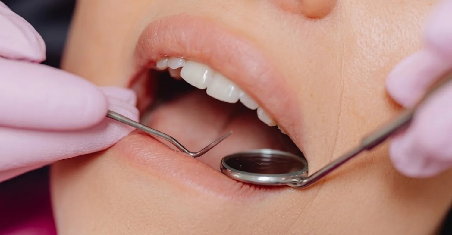
Note : All images used are for editorial and illustrative purposes only and may not originate from the original news provider or associated company.
When Should You See a Dentist or Doctor About a Black Spot Inside Your Cheek?
A new unexplained lesion should be professionally evaluated. Prompt assessment becomes more important when the lesion changes or persists. Bleeding, ulceration, pain, swelling, numbness, or a persistent lump deserve attention. A specialist referral may be appropriate when diagnosis remains uncertain. The goal is identifying the cause before deciding whether treatment is necessary.
When Should a New Dark Spot Be Evaluated?
A new dark spot should be mentioned to a dentist or doctor. Evaluation is especially relevant when no obvious cause exists. A recent injury can explain some lesions, but persistence changes the situation. If a mark after biting has not faded within about two weeks, arrange a review. The clinician may document the lesion and monitor it. A black spot inside your cheek with no clear cause should not wait months.
When Does a Persistent or Changing Lesion Need Prompt Assessment?
Persistence becomes more important when the cause remains unexplained. Progressive enlargement or color changes also increase the need for reassessment. A lesion that repeatedly returns should not be ignored. Changes in border or surface can provide additional clues. Prompt evaluation helps determine whether biopsy or specialist referral is warranted. Published reviews emphasize careful management of uncertain pigmented lesions.
When Should Bleeding, Ulceration, Pain, or Numbness Be Evaluated?
Persistent unexplained bleeding should be assessed professionally. Ulceration that does not heal should also receive clinical attention. Persistent pain can indicate irritation, inflammation, trauma, or another process. Numbness may require evaluation of deeper structures. These symptoms are not specific to cancer. They simply indicate that further assessment may be appropriate. A black spot inside your cheek combined with these symptoms deserves timely review.
When Should a Dentist Refer You to an Oral Medicine or Oral and Maxillofacial Specialist?
Referral may be appropriate when the diagnosis remains uncertain. It can also help when a lesion has unusual characteristics or requires biopsy. Oral medicine specialists manage a wide range of mucosal disorders. Oral and maxillofacial specialists can assess lesions requiring surgical diagnosis or management. Referral decisions depend on the clinician's findings. At Vitrin Clinic, additional specialist coordination may be considered when clinically appropriate.
How Much Does It Cost to Evaluate or Biopsy a Black Spot Inside the Cheek?
Costs vary substantially by country, provider, procedure complexity, and pathology requirements. Consultation, biopsy, histopathology, imaging, and follow-up may be billed separately. Some providers publish bundled prices, while others separate each component. In the United States, MDsave currently lists mouth biopsy prices from $440 to $1,222. That is a marketplace range rather than a universal national clinical fee.
Average Cost in the United States: Approximately 440–1,222 for a Mouth Biopsy
MDsave currently lists mouth biopsy prices ranging from $440 to $1,222. The listed MDsave national average is different from that range. Therefore, the 440–1,222 figure is best understood as a published price range. It is not a universal national average. Hospital or specialist charges can differ substantially. Professional fees and pathology charges may also be separate.
Average Cost in the United Kingdom: Approximately £290–£511 for Private Oral Biopsy Services
Private UK prices vary according to provider and what the package includes. One current private provider lists oral biopsy under local anesthesia from £290. That published price includes procedure, histology, and written reporting. A reliable national average of £290–£511 is not established by that source. Therefore, this range is best treated as an approximate private-market reference.
Average Cost in Turkey: Why Private Clinic Prices Vary by Consultation, Biopsy, and Histopathology
Turkey has substantial variation between private dental and medical providers. There is no single national private fee for evaluating an oral pigmented lesion. Costs depend on consultation type, biopsy technique, laboratory pathology, and specialist involvement. Imaging may add a separate charge when clinically indicated. Excision can cost more than a simple incisional biopsy. At Vitrin Clinic, the diagnostic pathway can be discussed according to clinical findings.
What Can Change the Final Cost of Oral Pigmentation Diagnosis?
The final cost depends on the diagnostic steps required. Some patients only need clinical evaluation and monitoring. Others require biopsy, histopathology, imaging, or specialist referral. Large or difficult lesions can require more complex procedures. Follow-up appointments may also create additional charges. International patients should clarify what their package includes. A written quotation provides greater cost transparency. The cost of checking a black spot inside your cheek is often lower than people fear.
Consultation and Specialist Examination
The initial consultation may be charged separately. Specialist oral medicine or oral surgery assessment can have different fees. The consultation may include examination, photographs, measurements, and treatment planning. Complex cases may require longer appointments. Patients should confirm whether follow-up is included. Some clinics include a basic oral lesion check within a routine dental examination.
Biopsy and Histopathology
Biopsy fees may include local anesthesia, tissue removal, and wound closure. Histopathology may be billed separately or included within a package. Additional laboratory stains can sometimes create further charges. The final pathology report is essential when microscopic diagnosis is required. Always ask whether pathology is included in the quoted price.
Imaging or Additional Laboratory Testing
Imaging is only needed when clinically indicated. Radiographs can assist with suspected dental-material deposits or nearby structural changes. CBCT may be considered when three-dimensional information is necessary. Laboratory testing may be appropriate when systemic causes are suspected. Each additional test can affect the total cost. Blood tests, for example, may be ordered through a medical doctor.
Excision or Further Treatment When Required
Complete excision may cost more than an incisional biopsy. Further treatment depends entirely on the pathology diagnosis. Benign lesions may require no additional treatment. Serious diagnoses can require coordinated specialist care. The initial biopsy does not always represent the total treatment pathway. Ask the clinician to explain likely next steps before committing to any plan.
Tips for Patients
Good preparation can make oral lesion assessment more efficient. Bring details about onset, changes, dental treatment, trauma, medication, and tobacco exposure. Take clear photographs before the appointment when possible. Do not apply whitening agents or household remedies. Avoid repeatedly biting or irritating the area. Continue normal oral hygiene unless it directly causes discomfort. Ask how the diagnosis will be established. At Vitrin Clinic, patients can discuss clinical findings and possible next steps during assessment.
What Should You Tell Your Dentist About a Dark Spot in Your Mouth?
Tell your dentist when you first noticed it. Explain whether its size or color has changed. Mention trauma, cheek biting, burns, or repeated irritation. List nearby dental procedures and restorations. Tell the clinician about tobacco exposure. Provide current and recent medications. Mention bleeding, pain, swelling, ulceration, or numbness. Previous photographs can also be helpful.
How Can You Photograph and Monitor a Changing Oral Spot?
Use bright, consistent lighting whenever possible. Take photographs from similar distances each time. Include a wider photograph showing the lesion's location. Use a close photograph for surface and border details. Avoid stretching the tissue excessively. Record the date beside each image. A clean finger or spoon handle can gently hold the cheek aside. Photographing a dark oral spot is easier with a phone flash.
What Should You Avoid Before Professional Evaluation?
Avoid scraping or cutting the lesion. Do not apply bleach, acids, or whitening products. Avoid irritating foods when the area is sore. Reduce repeated biting or rubbing. Do not stop prescribed medications without medical guidance. Avoid assuming smoking is the only explanation. Keep useful dental records and photographs available. Preserving the lesion's natural appearance can help clinical assessment.
What Questions Should You Ask About Oral Lesion Diagnosis and Biopsy?
Ask what diagnoses are being considered. Ask whether the lesion can be diagnosed clinically. Ask whether monitoring or biopsy is recommended. Ask which biopsy technique would be used and why. Ask whether pathology is included in the quoted cost. Ask how results will be communicated. Ask whether referral to a specialist is appropriate.
Oral Pigmentation Assessment at Vitrin Clinic
Vitrin Clinic can incorporate an unusual oral finding into an appropriate dental assessment. The clinical evaluation begins with examination of the lesion and surrounding oral tissues. Relevant dental history and previous treatments are considered. Digital records may support documentation when clinically appropriate. Digital X-rays, CBCT, or three-dimensional scanning may be used when indicated. A black spot inside your cheek is assessed within the context of your whole mouth.
How Vitrin Clinic Evaluates an Unusual Dark Spot in the Mouth
Vitrin Clinic can begin with a structured clinical examination. The clinician reviews the lesion's size, color, surface, location, and duration. Nearby teeth and restorations are also assessed. Previous trauma and dental procedures are considered when relevant. Digital records can support documentation and later comparison. Further diagnostic steps depend on the findings. Patients are encouraged to ask questions at every stage.
Clinical Examination and Digital Diagnostics When Clinically Appropriate
Clinical examination remains the foundation of oral lesion assessment. Digital imaging can provide additional information when the clinical situation requires it. Digital X-rays may help evaluate teeth and surrounding structures. CBCT can provide three-dimensional information for selected indications. Three-dimensional intraoral scanning can support digital records. At Vitrin Clinic, these technologies are selected according to clinical need.
When Additional Specialist Assessment May Be Needed
Specialist assessment may be appropriate when clinical diagnosis remains uncertain. It may also be considered for suspicious, changing, or complex lesions. Oral medicine specialists focus on mucosal diseases and diagnostic challenges. Oral and maxillofacial specialists can manage lesions requiring surgical assessment. Pathologists provide microscopic interpretation when tissue is sampled. Vitrin Clinic can support coordination according to the clinical pathway.
Internal Link Opportunity: Dental Check-Up and Relevant Oral Care Services at Vitrin Clinic
A useful internal link is the Dental Check-Up page at Vitrin Clinic. This supports users who are searching for evaluation of unexplained oral changes. The link can guide patients toward routine examination and appropriate dental assessment. Related oral health articles can provide additional educational context. The internal link should use descriptive anchor text relevant to the page. Readers concerned about an oral dark spot benefit from a direct path to booking.
How Vitrin Clinic Supports Your Treatment Journey
Vitrin Clinic can support patients through assessment, documentation, treatment planning, and follow-up discussions. The process should remain individualized because oral lesions have different causes. Clinical findings determine whether monitoring, imaging, referral, or biopsy is appropriate. Digital records may support communication and planning when clinically indicated. International patients can also discuss the sequence of appointments before treatment.
Consultation and Individualized Assessment
The consultation begins with understanding the patient's concern and clinical history. The lesion is assessed alongside the surrounding oral tissues. Previous dental work and local irritation are considered. The patient's symptoms and timeline are documented. Further testing depends on the clinical findings. Vitrin Clinic can use digital documentation when appropriate. Patients are welcome to bring photos, records, and written questions.
Coordinating Appropriate Diagnostic Evaluation
Some findings can be evaluated clinically. Others require additional imaging or tissue diagnosis. The appropriate pathway depends on lesion characteristics and diagnostic certainty. When necessary, specialist assessment can be coordinated. The purpose is obtaining an accurate diagnosis before treatment. Patients should understand why each diagnostic step is recommended. Good coordination reduces repeated visits and unnecessary waiting. For a dark oral mark, the simplest adequate pathway is preferred.
Using Digital Records to Support Treatment Planning When Appropriate
Digital records can preserve images and clinical observations. These records can help compare findings during follow-up. Digital X-rays can provide relevant dental information when indicated. CBCT can provide three-dimensional views when clinically necessary. Intraoral scanning can support digital documentation of oral structures. At Vitrin Clinic, the chosen technology depends on clinical need.
Discussing Findings, Next Steps, and Follow-Up
Patients should receive a clear explanation of the clinical findings. The clinician can discuss whether monitoring, biopsy, referral, or treatment is appropriate. Follow-up timing depends on the diagnosis and lesion characteristics. Patients should know which changes require earlier contact. Vitrin Clinic can support communication about the next stage of care. Clear instructions can reduce unnecessary uncertainty between visits.
Internal Link Opportunity: Vitrin Clinic Treatments and Related Oral Health Articles
Internal links can connect this article with relevant Vitrin Clinic treatment pages. Useful links may include dental check-ups, implants, crowns, veneers, or other applicable services. The chosen links should match the patient's actual clinical needs. Educational articles can also provide supporting information about oral examination. Avoid linking unrelated cosmetic treatments simply because the article discusses oral pigmentation.
How Long Can a Dark Spot Inside the Cheek Last?
The duration depends on the underlying cause. A blood blister or bruise may improve over days or weeks. Post-inflammatory pigmentation can persist longer before gradually fading. Amalgam tattoos can remain stable for many years. Physiologic pigmentation can remain unchanged throughout life. Some benign lesions are also long-lasting and stable. Duration alone does not determine whether a lesion is dangerous. Knowing the expected timeline helps you judge when a black spot inside your cheek needs review.
How Long Can a Traumatic Dark Spot Take to Fade?
Traumatic discoloration usually changes as the injured tissue heals. Blood pigments can shift through different shades during resolution. The exact timeframe depends on injury severity and repeated irritation. Small blood blisters often settle within one to two weeks. Continued biting can prolong healing. A clinician should reassess a lesion that does not behave as expected.
Can Oral Pigmentation Remain Stable for Years?
Yes, some benign pigmented lesions remain stable for many years. Amalgam tattoos are a common example of long-lasting localized discoloration. Physiologic pigmentation can also remain stable. A stable pattern is clinically useful but does not eliminate examination. Changes from baseline are often more informative than color alone. Photographic documentation can assist with long-term monitoring. A long-standing mark still deserves a baseline record.
Key Takeaways
A dark oral spot has many possible causes. Some are harmless, while others require additional diagnostic evaluation. Clinical appearance alone cannot establish the exact diagnosis. Professional assessment considers history, location, color, shape, duration, and change. Biopsy may be necessary when uncertainty remains. At Vitrin Clinic, clinical examination and digital diagnostics can support appropriate assessment.
The Most Common Causes of a Black Spot Inside Your Cheek
Common possibilities include physiologic pigmentation, melanotic macules, amalgam tattoos, smoker's melanosis, nevi, and trauma. Post-inflammatory and medication-related pigmentation are also recognized causes. The frequency of each cause varies by study population. One study found smoker's melanosis, racial pigmentation, and amalgam tattoos among frequent causes. Another pathology series found amalgam tattoo, melanotic macule, and nevus commonly represented.
Why Appearance Alone Cannot Confirm the Diagnosis
Different lesions can share the same dark color. Melanin, blood, foreign material, and vascular changes may look similar. Benign lesions can also resemble malignant melanoma. Clinical examination improves diagnostic accuracy but may not provide certainty. Histopathology can provide definitive evidence when biopsy is required. Therefore, color alone should never determine treatment. Photos and online comparisons cannot replace an in-person examination.
Warning Signs That Need Professional Evaluation
Important changes include growth, irregular borders, uneven coloration, and ulceration. Bleeding, persistent swelling, and numbness are also important changes. Persistent or unexplained lesions also deserve professional attention. These signs do not diagnose cancer. They indicate that further assessment may be appropriate. A dentist can determine whether monitoring, imaging, referral, or biopsy is justified.
Relief and Medical Tips While Waiting for Assessment
Avoid cheek biting and repeated irritation. Do not scrape, bleach, or chemically treat the lesion. Continue gentle oral hygiene. Take photographs and document changes. Note recent trauma, dental treatment, smoking, and medication changes. Arrange professional assessment when the cause remains unexplained. Choose soft, mild foods if the area feels sore.
Possible Side Effects After Diagnostic Procedures
Oral biopsy can cause mild pain, tenderness, swelling, or limited bleeding. Stitches may also be used. Most complications are minor, but persistent symptoms should be reported. Follow the clinic's specific postoperative instructions. Seek urgent advice for heavy bleeding, severe swelling, fever, or worsening symptoms. Healing inside the mouth is usually quick because the tissue has a rich blood supply.
When to Arrange a Dental or Medical Evaluation
Arrange evaluation for any new unexplained oral pigmentation. Earlier assessment is appropriate for changing lesions or concerning symptoms. Specialist referral may be needed when clinical diagnosis remains uncertain. Biopsy can provide tissue confirmation when required. The aim is accurate diagnosis rather than unnecessary treatment. A quick check of a black spot inside your cheek often brings fast reassurance.
At Vitrin Clinic
At Vitrin Clinic, oral findings are assessed before recommending any dental treatment. Our treatments include dental implants, All-on-4, All-on-6, All-on-8, zirconia crowns, E-max crowns, veneers, and orthodontic care. We also provide gum laser treatment, teeth whitening, and other restorative and cosmetic dental treatments. If a Black Spot Inside Your Cheek is noticed, our team first evaluates the area and surrounding oral tissues.
Digital X-rays, CBCT, and 3D intraoral scanning may be used when clinically appropriate. Once the cause is understood, our dentists can determine whether any of our treatments are relevant to your needs. Treatment recommendations are always based on individual clinical findings and oral health requirements.
Reference
FAQs

Dr. Rifat Alsaman has more than 5 years of clinical experience in dentistry and currently serves as the Head of the Medical Team at Vitrin Clinic. He is dedicated to providing exceptional patient care, overseeing treatment planning, and ensuring the highest clinical standards across the team. His expertise, attention to detail, and commitment to continuous professional development have helped countless patients achieve healthier, more confident smiles.


.webp&w=3840&q=75)