General Dentistry
September 1, 2026
Tooth surfaces: What Are the Different Surfaces of a Tooth?
 Dr. Rifat Alsaman
.webp&w=3840&q=75)
Understanding Tooth Surfaces helps patients recognize how dentists describe different areas of every tooth. Each surface has a specific anatomical name based on its location and function. Knowing these terms can make dental explanations, treatment plans, and charting easier to understand. Dentists use surface terminology when discussing cavities, plaque accumulation, restorations, tooth wear, and gum-related concerns. The five primary surfaces include mesial, distal, buccal, lingual, and occlusal areas. Front teeth also have an incisal edge instead of an occlusal surface. Root surfaces require separate consideration because they differ from enamel-covered crown surfaces. Statistics from dental research consistently show that disease risk varies according to tooth location and cleaning access. Vitrin Clinic uses detailed examinations to assess each area during routine dental evaluations. Understanding these anatomical terms can help patients communicate more confidently with their dental team. It can also make treatment recommendations easier to follow.
What Are Tooth Surfaces?
A tooth surface refers to a specific anatomical area of a tooth. Dentists divide each tooth into recognizable surfaces for diagnosis and treatment. These areas help clinicians describe exactly where a cavity, fracture, stain, restoration, or other finding occurs. The terminology also supports consistent dental charting between different professionals. Surface names are based mainly on direction, neighboring structures, and tooth function. For example, mesial means toward the dental midline. Distal means away from that midline. Buccal describes the cheek-facing side of posterior teeth. Lingual describes the tongue-facing side. Occlusal describes the chewing surface of premolars and molars. Incisal describes the cutting edge of front teeth. These distinctions are important during examinations and restorative procedures. They also help patients understand explanations given during consultations. At Vitrin Clinic, precise anatomical descriptions support clearer communication between clinicians and patients. Browse the article about Ai Dental Charting
What Are the 5 Surfaces of the Tooth?
The five commonly taught surfaces are mesial, distal, buccal, lingual, and occlusal. These terms describe the principal areas of many posterior teeth. Mesial identifies the surface facing the dental midline. Distal identifies the surface facing away from the midline. Buccal identifies the cheek-facing surface. Lingual identifies the tongue-facing surface. Occlusal identifies the chewing surface of premolars and molars. Front teeth require an additional terminology distinction. Their cutting edge is called the incisal edge rather than an occlusal surface. Some dental descriptions also use facial as a broader term. Facial can describe either buccal or labial surfaces depending on the tooth. These anatomical distinctions help dentists identify disease locations precisely. They also help explain restorative treatment and hygiene recommendations. Understanding these five surfaces provides a useful foundation for learning dental anatomy.
Mesial Surface of a Tooth
The mesial surface is the side of a tooth closest to the mouth's midline. The midline runs vertically between the two central incisors. Therefore, the mesial direction always points toward that central reference point. Mesial surfaces contact neighboring teeth in normal dentition. They form part of the proximal area between adjacent teeth. These areas can collect plaque when cleaning is inadequate. Flossing or interdental cleaning helps remove deposits from these contact regions. Dentists frequently identify cavities according to their mesial location. A restoration may therefore be described as involving a mesial surface. The term applies to both front and back teeth. However, the exact shape differs between tooth types. Understanding direction is more important than memorizing individual shapes. A simple rule helps patients remember the term. Mesial means moving toward the middle of the dental arch.
Distal Surface of a Tooth
The distal surface is the side of a tooth positioned away from the dental midline. It therefore represents the opposite direction from the mesial surface. Distal surfaces usually contact the neighboring tooth located farther from the center. They form part of the proximal surfaces between adjacent teeth. Plaque can accumulate around these contact areas when cleaning access is limited. Dentists may document cavities, fractures, or restorations using the term distal. For example, a restoration may involve the distal surface of a molar. The term is useful because it provides precise anatomical information. Distal direction remains consistent throughout the dental arch. The surface does not simply mean the back of every tooth. Its meaning depends on distance from the midline. Remembering this distinction makes dental terminology easier to understand. Patients can use the concept when interpreting dental charts or treatment explanations.
Buccal Surface of a Tooth
The buccal surface faces the cheek and is most commonly used for posterior teeth. Premolars and molars have buccal surfaces because they sit beside the cheeks. The corresponding tongue-facing area is called the lingual surface. Buccal surfaces can accumulate plaque around grooves, restorations, and the gumline. Brushing the outer surfaces thoroughly helps reduce bacterial deposits. Some cavities develop near the cervical region where enamel meets the gumline. Dentists inspect these areas during routine examinations. Buccal terminology can also appear in dental charting and treatment plans. The broader term facial may sometimes describe the same general direction. However, buccal is more anatomically specific for posterior teeth. Patients may notice this surface when looking at their teeth in a mirror. Regular brushing should cover the entire cheek-facing area. Vitrin Clinic examinations include assessment of visible outer surfaces for plaque, staining, wear, and restorative concerns.
Lingual Surface of a Tooth
The lingual surface faces the tongue and occurs on teeth throughout the dental arches. It is particularly important when discussing plaque accumulation and tartar formation. The lower front teeth commonly develop calculus behind the incisors. Salivary gland openings contribute to mineral deposition in this region. The lingual surface differs from the labial surface found on anterior teeth. Both terms describe surfaces facing toward the oral cavity. However, lingual specifically indicates the tongue-facing direction. Dentists inspect these areas for caries, erosion, calculus, staining, and periodontal concerns. Effective brushing requires angling the toothbrush toward these less visible surfaces. Patients sometimes overlook them because they cannot easily see them. Interdental cleaning is also important around neighboring contacts. Regular professional examinations can identify problems before symptoms become noticeable. Vitrin Clinic clinicians evaluate both visible and less accessible areas during comprehensive dental assessments.
Occlusal Surface of a Tooth
The occlusal surface is the chewing surface of premolars and molars. It contains anatomical features including cusps, grooves, fossae, and developmental depressions. These structures allow posterior teeth to process food efficiently. They can also create areas where plaque becomes difficult to remove. Deep pits and fissures may increase susceptibility to dental caries. Research consistently identifies pits and fissures as important sites for occlusal decay. Sealants can sometimes protect susceptible grooves in appropriate patients. Dentists evaluate these areas using visual examination and other diagnostic methods. Occlusal surfaces also experience substantial mechanical forces during chewing. Bruxism and other habits may contribute to wear or cracking. A damaged chewing surface can affect bite function and sensitivity. Proper diagnosis helps determine whether preventive, restorative, or protective treatment is appropriate. Patients should brush posterior chewing surfaces carefully and consistently. Follow the latest on How Sugar And Acidic Foods Damage Tooth Enamel
Tooth Surfaces Chart: All Tooth Surface Names
Dental terminology becomes easier when surface names are organized systematically. A chart can show the relationship between anatomical direction and tooth location. Mesial and distal describe horizontal directions within the dental arch. Buccal and lingual describe outer and inner directions. Labial is used specifically for the lip-facing surface of anterior teeth. Occlusal identifies chewing surfaces on posterior teeth. Incisal identifies cutting edges on anterior teeth. Proximal describes surfaces that face adjacent teeth. Facial can serve as a general term for outward-facing surfaces. These terms allow dentists to describe findings accurately without lengthy explanations. They are especially useful when documenting restorations or various lesions. Dental students also use these terms when studying morphology. Patients can use the same terminology to understand treatment recommendations. A clear chart therefore improves communication between patients, dentists, hygienists, and dental laboratories.
Tooth Surfaces Chart
Surface | Meaning | Common Teeth |
Mesial | Toward the midline | All teeth |
Distal | Away from the midline | All teeth |
Buccal | Toward the cheek | Premolars and molars |
Labial | Toward the lips | Incisors and canines |
Lingual | Toward the tongue | All teeth |
Palatal | Toward the palate | Upper teeth |
Occlusal | Chewing surface | Premolars and molars |
Incisal | Cutting edge | Incisors and canines |
Proximal | Toward adjacent teeth | Mesial and distal |
This terminology provides a practical framework for understanding dental descriptions. The same tooth may have several surfaces involved simultaneously. For example, a cavity can affect mesial and occlusal areas together. Dentists may describe that lesion using abbreviated notation. Learning these terms can make dental records less confusing. It also helps patients understand why certain areas receive specific preventive advice.
Tooth Chart With Numbers and Surfaces
Dentists combine tooth numbers with surface terminology when recording findings. Different numbering systems exist internationally, including the Universal and FDI systems. In the Universal system, permanent teeth receive numbers from 1 through 32. The FDI system uses two-digit designations based on quadrant and tooth position. Surface abbreviations then identify the affected area. M commonly represents mesial, while D represents distal. B can represent buccal, while L represents lingual. O commonly represents occlusal, while I represents incisal. P may represent palatal depending on the charting system. A tooth described as 19-MO, for example, involves mesial and occlusal areas. Exact notation conventions can vary between practices and software systems. Understanding the basic concept is more important than memorizing every code. Vitrin Clinic can explain chart findings during consultations when patients need clarification. Facts that matter about Worst Foods And Drinks For Your Teeth
Buccal vs Labial Surface of a Tooth
Buccal and labial both describe outward-facing surfaces, but dentists use them for different groups of teeth. Buccal generally describes the cheek-facing surface of premolars and molars, while labial describes the lip-facing surface of incisors and canines. Both belong to the broader facial direction. This distinction is important when discussing Tooth Surfaces, dental charting, and treatment planning. For example, a stain on a front tooth may be described as labial, while a cavity near the cheek side of a molar may be described as buccal. The anatomical difference is based primarily on the nearby soft tissues. Understanding these terms can help patients interpret professional descriptions. Brushing should cover both areas thoroughly because their locations can influence visibility, plaque accumulation, and cosmetic concerns. Vitrin Clinic clinicians consider these differences during comprehensive examinations and aesthetic treatment planning.
What Is the Labial Surface of a Tooth?
The labial surface faces the lips and occurs primarily on anterior teeth. Incisors and canines therefore have labial surfaces. These areas are highly visible when speaking, smiling, or laughing. Cosmetic concerns frequently involve changes affecting the labial enamel. Discoloration, enamel defects, fractures, and restorations may become noticeable in this region. Proper brushing helps remove plaque without causing unnecessary abrasion. Aggressive brushing can contribute to cervical wear or gum recession, so dentists consider both oral hygiene technique and cosmetic appearance. Labial surfaces are also important during veneer planning because veneers commonly cover the visible facial surface of selected anterior teeth. However, treatment decisions require assessment of enamel thickness, bite, gum health, and tooth structure. Understanding Tooth Surfaces helps clinicians evaluate aesthetic concerns while maintaining appropriate function and preserving healthy dental tissue.
Buccal Surface vs Labial Surface
The key difference involves the soft tissue located beside the tooth. Buccal means facing the cheek, while labial means facing the lips. Buccal terminology commonly applies to posterior teeth, whereas labial terminology usually applies to anterior teeth. Both areas face outward from the dental arch, and dentists may use facial as a broader term covering both directions. This distinction helps clinicians describe plaque, staining, restorations, or lesions accurately. For patients, remembering the neighboring tissue provides an easy method: think of lips when hearing labial and cheeks when hearing buccal. This simple association reduces confusion during dental conversations. Understanding these Tooth Surfaces becomes especially useful when multiple areas require treatment because accurate terminology prevents ambiguity in clinical records and supports communication among dentists, hygienists, orthodontists, and dental laboratories.
Clinical Note
Clinically, outward-facing surfaces are examined for plaque, calculus, cervical caries, erosion, abrasion, discoloration, and defective restorations. Risk varies according to oral hygiene, diet, saliva, anatomy, and patient habits. Surface-specific risk can differ substantially between individuals. Patients with frequent acidic exposure may develop erosive changes, while patients with gum recession may have greater root-surface vulnerability. A clinical examination therefore considers more than visible discoloration. Dentists evaluate texture, location, symmetry, and surrounding tissues to distinguish harmless staining from structural disease. Assessment of these Tooth Surfaces forms part of comprehensive dental evaluation at Vitrin Clinic. Early identification can support conservative management and preventive care. Patients should also discuss persistent sensitivity, roughness, or visible changes with a dental professional. How to manage Daily Oral Hygiene Routine
Mesial vs Distal Surface of a Tooth
Mesial and distal describe opposite horizontal directions based on the dental midline rather than the patient's perspective. Mesial means toward the midline, while distal means away from it. Every tooth has both a mesial and distal surface, and these areas generally form part of the proximal region between neighboring teeth. Plaque can accumulate around these contacts without effective interdental cleaning. Cavities involving these surfaces may remain difficult to detect visually, so dentists may use bitewing radiographs when clinically appropriate. Accurate terminology helps document the exact location of a lesion and can influence restorative access and treatment design. Understanding these Tooth Surfaces makes dental terminology easier to follow. Patients can remember the difference by associating mesial with the middle and distal with a direction farther from the middle.
What Does Mesial Mean in Dentistry?
Mesial means positioned closer to the dental midline. The midline lies between the upper or lower central incisors, and every tooth has a mesial direction pointing toward that center. This concept remains consistent regardless of which side of the mouth contains the tooth. For example, the mesial surface of a right molar faces toward the center of the dental arch, and the same directional principle applies on the left. Mesial surfaces often contact neighboring teeth within the dental arch. Dentists use the term when describing cavities, fractures, restorations, and periodontal findings. It also appears frequently in dental charting. Understanding Tooth Surfaces requires recognizing that mesial does not simply mean the front of a tooth. Tooth position determines the direction, making this distinction especially important when discussing posterior teeth.
What Does Distal Mean in Dentistry?
Distal means positioned farther from the dental midline and represents the direction opposite to mesial. Every tooth has a distal surface directed away from the center. Distal surfaces generally contact neighboring teeth positioned farther from the midline. The word does not simply mean the back of a tooth because its meaning depends specifically on anatomical distance from the midline. A molar can have both mesial and distal surfaces even though both are located toward the back of the mouth. The mesial side remains closer to the midline, while the distal side is farther away. Understanding these Tooth Surfaces makes dental charting easier because the same directional rule applies throughout the dental arches. Patients can use the midline as a reliable reference when interpreting treatment descriptions.
Mesial vs Distal: Easy Way to Remember
The easiest method is to identify the dental midline first. Mesial means moving toward that center, while distal means moving away from it. Imagine an invisible vertical line between the central incisors. Every tooth has one side pointing toward that line, and that side is mesial. The opposite side points away and is distal. This rule works for both upper and lower arches and on both the right and left sides. Patients can become confused when using front-versus-back descriptions because perspective changes. Anatomical terminology avoids this problem. When identifying Tooth Surfaces, focus on the midline rather than the viewing angle. Once this concept becomes familiar, mesial and distal terminology becomes easier to understand during consultations, dental charting, and treatment planning.
Clinical Note
Proximal surfaces deserve careful clinical attention because they can be difficult to inspect directly. Contacts between neighboring teeth can hide early carious lesions. Bitewing radiographs may help identify interproximal changes when indicated. Flossing and interdental brushes can improve plaque removal around these regions. However, disease development depends on multiple factors, including saliva, fluoride exposure, diet, bacterial activity, and previous disease history. Dentists therefore assess individual risk rather than relying on location alone. A mesial or distal lesion may require minimally invasive treatment when detected early, while larger lesions can require restorative procedures. Careful examination of these Tooth Surfaces supports earlier detection and individualized treatment. Vitrin Clinic emphasizes assessing the patient's overall oral condition rather than assuming every surface carries identical risk. Causes and impacts of What To Do If A Tooth Is Knocked Out During Sports
What Is the Proximal Surface of a Tooth?
The proximal surface refers to a surface that faces an adjacent tooth and includes both mesial and distal surfaces. Therefore, proximal is a broader descriptive term rather than a separate seventh surface. These areas form contact relationships within the dental arch and contribute to tooth stability and normal function. Food and plaque can become trapped when contacts or tooth alignment create difficult cleaning areas. Interdental cleaning is therefore essential for maintaining these regions. Dentists evaluate proximal areas during routine examinations, and radiographs may be used when clinically indicated to detect hidden caries. Proximal cavities can progress without obvious symptoms. Understanding Tooth Surfaces helps patients recognize why flossing and other interdental cleaning methods are important. Regular preventive care can support early detection and help protect the health of neighboring teeth.
Proximal Surface Definition
A proximal surface is a tooth surface that faces another tooth. The term includes both mesial and distal surfaces. These surfaces are located between neighboring teeth within the dental arch. Healthy proximal contacts allow teeth to function as an organized unit. They also influence food movement and periodontal tissue protection. Problems can develop when plaque remains around these areas. Cavities may begin beneath or adjacent to contact points. Gum inflammation can also occur when plaque accumulates interproximally. Dental professionals inspect these areas during routine examinations. Floss, interdental brushes, and other cleaning aids can access these spaces. The most appropriate device depends on contact size and individual anatomy. Patients with bridges or orthodontic appliances may require specialized cleaning tools. Professional guidance can improve technique and reduce unnecessary tissue trauma.
Mesial and Distal as Proximal Surfaces
Mesial and distal surfaces together make up the proximal surfaces. Mesial points toward the dental midline. Distal points away from the dental midline. Both surfaces typically participate in contact with neighboring teeth. The exact contact pattern depends on tooth position and dental anatomy. Central incisors have distinctive proximal relationships near the midline. Posterior teeth usually contact several neighboring structures through their proximal areas. These relationships help maintain arch stability and efficient chewing. Changes caused by tooth loss can alter adjacent contacts. Orthodontic movement can also modify proximal relationships. Restorative treatment must therefore preserve appropriate contact anatomy whenever possible. Poorly shaped contacts may contribute to food impaction. Dentists assess contact points carefully during restorative procedures. Understanding these relationships helps explain why seemingly small fillings require precise shaping.
Proximal Surfaces and Tooth Contact
Proximal surfaces create contact areas between neighboring teeth. These contacts help prevent excessive food impaction during normal chewing. They also contribute to the stability of the dental arch. Contact strength and shape vary according to tooth type and position. Restorations must reproduce appropriate contact relationships whenever possible. An excessively open contact can allow food to lodge repeatedly. An overly tight contact can make cleaning difficult. Both situations may create discomfort or hygiene challenges. Dentists use specialized instruments to assess contacts after restorative treatment. Patients may notice changes when floss suddenly becomes difficult or excessively loose. Such changes should be discussed with a dental professional. Proximal health also depends on effective interdental plaque removal. Research supports interdental cleaning as an important component of oral hygiene. Individual recommendations should reflect gum health and tooth anatomy.
Clinical Note
Proximal areas frequently require additional diagnostic attention because direct visual access is limited. Dentists may combine clinical inspection with radiographic assessment. Bitewing radiographs are commonly used when appropriate for detecting proximal caries. The American Dental Association emphasizes individualized radiographic decisions based on patient risk. Routine imaging intervals should therefore not be identical for every patient. Clinical findings, age, previous disease, and risk factors influence recommendations. Proximal plaque control remains essential regardless of imaging frequency. Floss can clean tight contacts, while interdental brushes may suit larger spaces. Patients should avoid forcing cleaning devices aggressively into healthy tissues. Gentle, controlled movement is generally preferable. Professional instruction can help identify the most suitable method. At Vitrin Clinic, preventive recommendations are tailored to the patient's anatomy and clinical findings. Direct effect of Dental Treatment And Heart Disease
Lingual vs Palatal Surface of a Tooth
Lingual and palatal describe inward-facing surfaces, but their use differs by location. Lingual refers generally to surfaces facing the tongue. Palatal specifically describes surfaces of upper teeth facing the palate. Therefore, every palatal surface is lingual in a broad directional sense. However, dentists often use palatal for maxillary teeth. This distinction is useful when describing restorations or anatomical findings. Lower teeth usually receive lingual terminology because they face the tongue directly. Upper posterior teeth can be described as palatal because they face the hard palate. These surfaces can accumulate plaque and calculus. Lower anterior lingual surfaces are particularly prone to calculus formation. Good brushing technique should reach these areas carefully. Dentists inspect them for caries, erosion, wear, calculus, and periodontal concerns. Understanding the terminology makes dental explanations easier to follow. It also supports more accurate interpretation of treatment records.
What Is the Lingual Surface?
The lingual surface is the side of a tooth facing the tongue. The term can describe teeth in both dental arches. It is especially common when describing mandibular teeth. Lingual surfaces may contain grooves, ridges, pits, or other anatomical features. These structures can influence plaque retention and cleaning difficulty. Lower front teeth often develop calculus along their lingual surfaces. Saliva from nearby glands contributes minerals that can harden plaque. Careful brushing helps reduce accumulation. However, hardened calculus requires professional removal. Lingual surfaces can also develop caries, erosion, abrasion, or restorations. Their position makes them less visible to patients. A mirror can help patients inspect these areas during home care. Regular dental examinations remain important because hidden problems may progress without symptoms. Consistent hygiene protects both tooth structure and surrounding gum tissues.
What Is the Palatal Surface?
The palatal surface is the side of an upper tooth facing the palate. The palate forms the roof of the mouth. Dentists commonly use palatal terminology for maxillary teeth. These surfaces include anatomical features that vary between incisors, canines, premolars, and molars. Upper front teeth often have prominent palatal anatomy. Some areas can retain plaque if brushing technique is incomplete. Palatal surfaces can develop caries, wear, erosion, and restorative defects. They also experience functional contact during certain jaw movements. Dentists consider these contacts when evaluating restorations or cosmetic treatments. Patients may find palatal surfaces difficult to see directly. Regular professional examinations can identify changes early. Brushing should reach the entire inner surface without excessive pressure. Proper technique helps protect enamel and gingival tissues.
Lingual vs Palatal: What Is the Difference?
The difference mainly concerns the anatomical structure beside the tooth. Lingual means facing the tongue. Palatal means facing the palate. Palatal therefore applies specifically to upper teeth. Linguistics can apply to either upper or lower teeth. Dentists may use lingual as a general term when discussing inward-facing surfaces. However, palatal provides greater anatomical specificity for maxillary teeth. The distinction becomes important in dental charting. It can also clarify treatment discussions involving restorations or tooth wear. Patients can remember the difference using a simple association. Tongue means lingual. Palate means palatal. This memory technique makes the terminology easier to retain. Both surfaces require thorough cleaning because they can collect plaque. Professional examinations help detect changes that patients may not notice themselves.
Clinical Note
Inner tooth surfaces can present unique hygiene challenges. Their position makes them harder to see and reach effectively. Lower anterior lingual surfaces commonly accumulate calculus because of nearby salivary ducts. Upper palatal areas can also retain plaque when brushing coverage is incomplete. Clinical studies consistently demonstrate the importance of effective plaque control for periodontal health. However, calculus cannot be removed reliably through ordinary brushing. Professional scaling may therefore be necessary when mineralized deposits develop. Dentists also evaluate these surfaces for erosive wear. Frequent acidic exposure can soften enamel and increase susceptibility to mechanical wear. Dietary counseling may be appropriate when erosion risk is identified. Vitrin Clinic incorporates preventive education into individualized oral examinations. Patients should report sensitivity, roughness, or unusual changes on inner surfaces. The link between Dental Care For Athletes Protecting Your Smile In Sports
Occlusal vs Incisal Surfaces
Occlusal and incisal describe functional areas of different tooth groups. Occlusal surfaces occur on premolars and molars. Incisal surfaces refer to the cutting edges of incisors. Canines also have incisal edges rather than broad occlusal tables. Posterior teeth grind and crush food using cusps and fossae. Front teeth cut and guide food using their incisal edges. These areas experience different mechanical forces. Their anatomical shapes reflect their different functions. Occlusal surfaces contain grooves and pits that can retain plaque. Incisal edges can experience chipping or wear from functional and parafunctional forces. Dentists assess both areas during examinations. Treatment planning considers anatomy, bite relationships, tooth strength, and remaining structure. Understanding the terminology helps patients understand descriptions of fractures and restorations. It also clarifies why different teeth receive different preventive recommendations.
What Is the Occlusal Surface?
The occlusal surface is the chewing surface of posterior teeth. Premolars and molars contain these broad functional surfaces. Their anatomy includes cusps, ridges, grooves, and fossae. These structures work together during mastication. Deep grooves can sometimes retain plaque despite regular brushing. Occlusal caries is therefore an important clinical concern. Fluoride exposure and sealants can reduce risk in appropriate circumstances. Dentists evaluate occlusal surfaces for decay, cracks, wear, and defective restorations. Bite forces also affect the condition of these surfaces. Bruxism can produce flattened cusps and visible wear facets. Cracked teeth may cause intermittent pain during chewing. Early assessment can prevent minor structural problems from progressing. Patients should spend adequate brushing time on posterior chewing surfaces. Professional examinations remain important because early lesions may produce no symptoms.
What Is the Incisal Surface?
The incisal surface refers to the cutting edge of anterior teeth. Incisors primarily use their incisal edges to cut food. Canines also have pointed functional edges that support tearing. Unlike molars, anterior teeth do not have broad occlusal tables. Incisal edges can experience wear from normal function and parafunctional habits. Teeth grinding may accelerate flattening or chipping. Trauma can also fracture these exposed edges. Dentists evaluate incisal anatomy during restorative and cosmetic consultations. The relationship between upper and lower incisal edges affects bite function. Restorations should preserve appropriate proportions and functional contacts. Excessive reshaping can alter speech or chewing patterns. Patients should avoid using front teeth to open packages or bite hard objects. Such habits can increase fracture risk. Vitrin Clinic considers function when evaluating aesthetic changes involving anterior teeth.
Occlusal Surface of Premolars and Molars
Premolars and molars have complex chewing anatomy. Their occlusal surfaces contain multiple structures that distribute chewing forces. Cusps contact opposing teeth during specific jaw movements. Grooves guide food and separate anatomical elevations. Fissures may become plaque-retentive when they are narrow or deep. This anatomy partly explains why posterior teeth can develop occlusal caries. Fluoride toothpaste supports enamel resistance against acid attacks. Sealants may be considered for selected high-risk grooves. Dentists assess individual caries risk before recommending preventive treatment. Occlusal wear can also indicate functional or parafunctional loading. Severe wear may affect bite relationships and tooth sensitivity. Regular examinations allow dentists to monitor changes over time. Patients should clean chewing surfaces carefully using small, controlled movements. Proper hygiene reduces plaque retention around complex posterior anatomy.
Incisal Surface of Front Teeth
Front teeth use their incisal edges for cutting rather than grinding. These edges are visible during smiling and speaking. Their shape contributes significantly to dental appearance. Normal wear gradually changes incisal contours over time. Accelerated wear may occur with bruxism, abrasive habits, or certain dietary factors. Chipping can result from trauma or excessive biting forces. Dentists evaluate symmetry, thickness, translucency, and functional contacts. Cosmetic procedures should preserve appropriate tooth structure whenever possible. Excessive reduction may weaken remaining enamel and dentin. Patients should avoid biting hard objects using anterior teeth. Nail biting and package opening can create unnecessary mechanical stress. A balanced bite can also reduce uneven loading. Vitrin Clinic evaluates both aesthetics and function when planning anterior restorative treatment.
Clinical Note
Functional surfaces provide important information about chewing and oral habits. Dentists examine wear patterns to understand how teeth interact during function. Localized flattening can suggest repeated mechanical contact. Generalized wear may have multiple contributing factors. Erosion can soften enamel before mechanical forces remove the weakened structure. Attrition involves tooth-to-tooth contact, while abrasion involves external mechanical forces. These processes can occur together. Clinical evaluation considers diet, habits, medications, saliva, and bite relationships. Statistical research indicates that tooth wear is increasingly recognized as an important oral health concern. Management depends on identifying the underlying cause. Preventive strategies may include dietary changes, protective appliances, or restorative treatment. Early intervention can help preserve remaining tooth structure. Behind the scenes of Does Not Brushing Teeth Cause Tonsillitis
How to Identify the Surface of Any Tooth
Identifying a surface becomes easier when directional landmarks are used. First, determine whether the tooth is anterior or posterior. Then identify the dental midline. Next, determine which side faces the lips, cheeks, tongue, or palate. Finally, identify whether the surface points toward or away from the midline. The functional edge can then be classified as incisal or occlusal. This systematic approach reduces confusion between similar anatomical terms. Tooth diagrams can help patients practice these relationships. Dentists use the same directional principles when documenting clinical findings. The terminology remains consistent even when tooth position changes. Learning these concepts is more useful than memorizing isolated definitions. Patients can apply the method to dental charts and treatment explanations. Understanding anatomical direction also makes conversations with dental professionals easier.
Identify the Front and Back of the Mouth
Begin by separating anterior and posterior teeth. Incisors and canines are considered anterior teeth, while premolars and molars are considered posterior teeth. Anterior teeth generally have incisal edges, whereas posterior teeth generally have occlusal surfaces. This distinction immediately narrows the possible surface terms. Next, identify whether the tooth faces the lips or cheeks. Anterior outward surfaces are usually labial, while posterior outward surfaces are usually buccal. The inward side faces the tongue or palate. These landmarks provide a useful starting point for identifying Tooth Surfaces. Tooth morphology can vary between individuals, so diagrams are helpful. Patients do not need to memorize every anatomical feature. Learning the major directional terms provides sufficient understanding for most dental conversations.
Find the Midline
The dental midline provides the reference point for mesial and distal directions. It generally passes between the upper central incisors, with a similar midline between the lower central incisors. Once identified, imagine a vertical line extending through the center. The surface closer to that line is mesial, while the surface farther from it is distal. This rule works regardless of whether the tooth is on the right or left. It also prevents confusion caused by viewing the mouth from different directions. Dentists rely on anatomical orientation rather than simple front-versus-back descriptions. Patients can practice this concept using a labeled diagram of Tooth Surfaces. Once the midline becomes familiar, mesial and distal become much easier to understand when reading dental treatment plans.
Identify the Buccal, Labial, and Lingual Sides
Look at the tissue located beside the tooth. If the tooth faces the lips, the surface is labial. If a posterior tooth faces the cheek, the surface is buccal. If the tooth faces the tongue, the surface is lingual. Upper teeth facing the palate can specifically be called palatal. These terms describe direction rather than disease. The same surface may appear healthy or affected depending on clinical findings. Patients can use nearby soft tissues as memory cues: lips correspond with labial, cheeks with buccal, tongue with lingual, and palate with palatal. These associations make Tooth Surfaces terminology easier to understand. Professional diagrams can further reinforce these concepts during dental consultations.
Identify Mesial and Distal
After finding the midline, identify the two proximal directions. The surface pointing toward the midline is mesial, while the surface pointing away is distal. Every tooth has both directions, and these surfaces usually face adjacent teeth. Therefore, they are also considered proximal surfaces. This relationship explains why dental professionals may use several terms together. A cavity can be described as mesial and proximal simultaneously. The terminology becomes clearer once directional concepts are combined. Patients should focus on the reference point rather than tooth orientation to avoid mistakes when looking at diagrams or photographs. Understanding Tooth Surfaces can also improve interpretation of dental chart abbreviations and treatment descriptions.
Identify Occlusal or Incisal Surfaces
Finally, identify the functional edge or chewing area. Premolars and molars have occlusal surfaces, while incisors and canines have incisal edges. Posterior teeth use broad surfaces for crushing and grinding, whereas anterior teeth use thinner edges for cutting. This functional difference explains the terminology and the differences in anatomy and wear patterns. Look for cusps and grooves when identifying an occlusal surface and the cutting edge when identifying an incisal area. Clinical findings may involve these functional regions, including caries, cracks, wear, and restorations. Understanding these Tooth Surfaces becomes easier once patients recognize the different functions of anterior and posterior teeth. Complete coverage of Black Tongue Treatment
Tooth Surfaces and Common Dental Problems
Different anatomical areas can experience different dental problems. Risk depends on plaque retention, access to cleaning, tooth morphology, diet, saliva, fluoride exposure, and previous disease. Posterior grooves can retain plaque and support caries development, while proximal areas can hide early lesions between neighboring teeth. Gum recession can expose root surfaces that are less mineralized than enamel. Functional surfaces can experience wear from chewing and grinding. Outer surfaces can develop staining or cervical lesions, while inner surfaces can accumulate calculus near salivary ducts. These differences explain why dentists inspect the mouth systematically. No single area should automatically be considered diseased. Individual risk assessment remains essential. Preventive care should address the patient's specific anatomy and habits. Vitrin Clinic uses comprehensive examinations to identify risk patterns and recommend appropriate care.
Which Tooth Surfaces Are Most Prone to Cavities?
Cavity risk varies according to tooth anatomy and individual behavior. Occlusal grooves can be vulnerable because their narrow anatomy may retain plaque. Proximal surfaces can also develop cavities because direct cleaning is more difficult. Root surfaces become susceptible when gum recession exposes them. Smooth surfaces may develop caries when plaque remains undisturbed for long periods. Saliva, fluoride exposure, diet, bacterial activity, and previous caries history influence individual risk. Tooth Surfaces therefore do not all carry the same level of cavity risk. Dentists use risk-based prevention rather than one universal approach. Fluoride toothpaste remains a central preventive measure for most patients, while regular dental visits support early identification of suspicious changes.
Plaque Buildup on Different Tooth Surfaces
Plaque can accumulate on virtually any area of a tooth, but some locations are harder to clean effectively. Proximal contacts can retain plaque between neighboring teeth, gingival margins can collect deposits near the gumline, and occlusal grooves can trap bacterial biofilm in narrow depressions. Lingual surfaces can also be difficult to visualize during brushing. Orthodontic appliances and crowded teeth may create additional retention areas. Plaque contains complex microbial communities embedded within a protective matrix. When left undisturbed, these communities can contribute to caries and periodontal inflammation. Daily mechanical removal is fundamental to oral health. Effective cleaning should address all Tooth Surfaces, with interdental cleaning complementing brushing. Professional cleaning may be needed when hardened calculus develops.
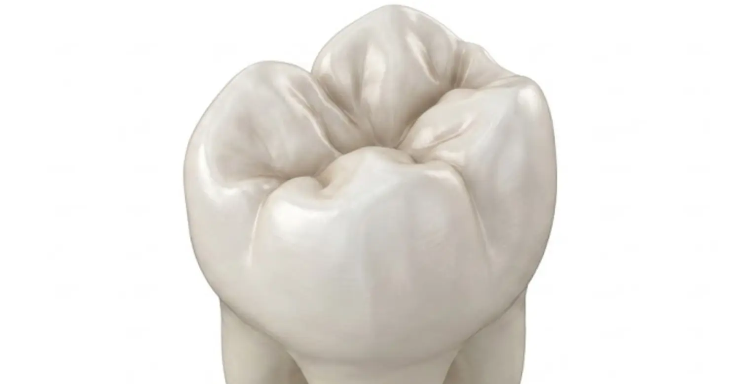
Tooth Wear and Surface Damage
Tooth wear involves progressive loss of dental hard tissue. Several mechanisms can contribute, including attrition, abrasion, and erosion. Attrition results primarily from tooth-to-tooth contact, abrasion results from external mechanical forces, and erosion involves chemical dissolution from acids. These processes often interact rather than occurring independently. For example, acid-softened enamel may become more vulnerable to brushing. Bruxism can accelerate wear on functional areas, while dietary acids may contribute to erosive changes. Dentists assess the location, shape, symmetry, and progression of affected Tooth Surfaces. Treatment focuses on controlling underlying causes before replacing lost structure. Protective appliances may be considered for selected patients. Early recognition helps preserve remaining tooth tissue. Vitrin Clinic can evaluate wear patterns and develop individualized preventive strategies.
Tooth Surface Decay
Tooth decay begins when bacterial activity produces acids that challenge mineralized tooth structure. Repeated acid attacks can cause progressive mineral loss. Early lesions may appear as subtle changes before cavitation develops. Fluoride can support remineralization and strengthen enamel against future acid attacks, while saliva contributes to natural buffering and mineral supply. Frequent sugar exposure can increase opportunities for acid production, and plaque retention can make certain locations more vulnerable. Dentists assess lesion activity, depth, location, and progression. Not every early lesion requires immediate drilling; preventive management may be appropriate for selected noncavitated lesions. Cavitated lesions may require restorative treatment. Understanding how decay affects different Tooth Surfaces can help patients recognize why prevention and early assessment are important.
Exposed Root Surfaces
Root surfaces can become exposed when gum tissue recedes. Unlike the crown, roots are covered primarily by cementum rather than enamel. Cementum is thinner and more vulnerable to mechanical and chemical challenges. Root exposure can therefore increase sensitivity and root caries risk. Common contributing factors include periodontal disease, traumatic brushing, aging-related changes, and anatomical factors. Not every recession pattern has the same cause. Dentists evaluate gum thickness, inflammation, attachment levels, and cleaning technique. Gentle brushing with an appropriate toothbrush can help prevent further mechanical trauma. Fluoride toothpaste can support protection against root caries. Desensitizing products may help selected patients with sensitivity. Professional monitoring is important when exposed roots are present. Treatment depends on symptoms, progression, aesthetics, and periodontal health. Medical review of Lung Cancer Mouth Symptoms
How Dentists Describe Tooth Surfaces
Dental professionals use standardized terminology to communicate findings efficiently. Surface names identify the location of cavities, restorations, fractures, wear, and other conditions. Tooth numbers identify the individual tooth. Surface abbreviations then identify the affected areas. This combination allows detailed information to fit within compact clinical records. Different countries and dental systems may use different tooth numbering methods. Universal numbering is common in the United States. FDI notation is widely used internationally. Dental software may also display visual tooth charts. Understanding basic notation can help patients read treatment plans. However, abbreviations can vary slightly between practices. Patients should ask their dentist when an unfamiliar code appears. Clear explanations can prevent unnecessary confusion. Accurate charting also supports communication between clinicians and dental laboratories.
Tooth Number and Surface Notation
Dental charting combines tooth identification with surface notation. The numbering system first identifies the specific tooth. Surface abbreviations then identify where the finding occurs. Common abbreviations include M for mesial and D for distal. B commonly means buccal. L commonly means lingual. O commonly means occlusal. I commonly mean incisal. P may indicate palatal depending on the notation system. Multiple letters can describe one restoration involving several surfaces. For example, MO can indicate mesial and occlusal involvement. MOD can indicate mesial, occlusal, and distal involvement. These codes help dentists document treatment efficiently. They also support communication with insurance providers and laboratories. Patients can request explanations whenever charting terminology is unclear.
Common Dental Surface Abbreviations
Common surface abbreviations make dental records concise. M usually represents mesial. D represents distal. B represents buccal. L represents lingual. O represents occlusal. I represent Incisal. P can represent palatal in some systems. F may represent facial features in certain charting conventions. Abbreviations can differ between dental software platforms. Therefore, patients should not assume every letter has identical meaning everywhere. Context usually clarifies the intended surface. Multiple letters indicate several involved surfaces. For example, MOD commonly represents mesial, occlusal, and distal. An anterior restoration may use an incisal abbreviation. Understanding these basic codes makes treatment plans easier to interpret. Dentists can explain any unfamiliar notation during consultations.
Examples of Dental Charting
A dentist might document a cavity using both a tooth number and surface abbreviation. For example, a molar may have an occlusal lesion. The record could identify that tooth using the practice's numbering system. The abbreviation O would then indicate the affected chewing surface. A lesion involving mesial and occlusal areas may receive MO notation. A larger restoration could involve mesial, occlusal, and distal areas. An anterior restoration may involve facial and incisal areas. These descriptions provide precise information without lengthy written explanations. Digital dental charts often display the information visually. Radiographs may supplement charting when lesions are difficult to see. Patients should ask for clarification if terminology appears confusing. Clear communication helps patients understand proposed treatments and their purpose.
Clinical Note
Dental charting is a communication system rather than a diagnosis by itself. Surface notation describes location. The clinical diagnosis explains what is happening in that location. For example, an occlusal notation does not automatically mean decay. It may describe a restoration, sealant, fracture, or other finding. Dentists combine location with clinical evidence before recommending treatment. This distinction prevents patients from misinterpreting abbreviations. Modern dental records may include photographs, radiographs, periodontal measurements, and digital scans. These records create a more complete diagnostic picture. Evidence-based care emphasizes individualized assessment rather than treatment based on terminology alone. Vitrin Clinic can explain charting findings in patient-friendly language. Understanding the record can help patients participate more confidently in treatment decisions.
What We Notice Clinically
From a clinical perspective, surface anatomy is important because location influences diagnosis and treatment. Dr. Rifat Alsaman emphasizes evaluating the entire tooth rather than focusing on one visible area. A small lesion may reflect broader hygiene, dietary, functional, or structural factors. Clinicians should therefore examine neighboring surfaces and supporting tissues. Proximal findings require particular attention because they can remain hidden from direct vision. Occlusal anatomy can also influence plaque retention and caries susceptibility. Root exposure changes the type of tissue being evaluated. Functional wear may provide clues about bite forces or parafunctional habits. Treatment should preserve healthy structure whenever possible. Preventive care remains essential after restorative treatment. Vitrin Clinic combines clinical examination with individualized treatment planning. This approach supports accurate diagnosis and conservative decision-making when clinically appropriate. What you should know about Unbearable Sinus Tooth Pain
Tips for Patients: How to Care for Different Tooth Surfaces
Effective home care requires more than brushing the front of visible teeth. Every tooth has multiple surfaces that need consistent attention. Outer surfaces require controlled brushing along the gumline. Inner surfaces need careful coverage despite being harder to see. Proximal areas require interdental cleaning because toothbrush bristles may not reach contacts effectively. Occlusal grooves need focused brushing because their anatomy can retain plaque. Exposed roots require gentle cleaning and appropriate fluoride protection. Patients should avoid excessive brushing pressure because it can contribute to cervical damage. Fluoride toothpaste remains a key component of daily preventive care. Diet also influences caries and erosion risk. Regular dental examinations help identify areas that need additional attention. Vitrin Clinic can provide personalized hygiene recommendations based on individual findings.
Cleaning the Outer and Inner Surfaces
Brush the outer surfaces using gentle, systematic movements. Cover every tooth rather than focusing only on visible front teeth. Move gradually around the entire dental arch. Then clean the inner surfaces facing the tongue or palate. Angle the brush appropriately toward the gumline. Excessive pressure is unnecessary and may contribute to abrasion or recession. A soft-bristled toothbrush is commonly recommended for routine cleaning. Fluoride toothpaste provides important protection against dental caries. Patients should follow the fluoride recommendations appropriate for their age and circumstances. Electric toothbrushes can help some patients improve consistency and plaque removal. However, technique remains important regardless of brush type. Replace toothbrushes when bristles become significantly worn. Professional advice can help patients adjust technique when persistent plaque remains.
Cleaning Proximal Surfaces
Brushing alone may not adequately clean proximal areas. Interdental cleaning reaches spaces between neighboring teeth. Dental floss can clean relatively tight contacts effectively. Interdental brushes may work better in larger spaces. The appropriate size should allow cleaning without forcing the brush. Water flossers can provide another option for selected patients. However, individual circumstances influence which method works best. Patients with braces, bridges, implants, or periodontal spaces may need specialized approaches. Bleeding during cleaning can indicate inflammation rather than a reason to stop. Persistent bleeding should be evaluated by a dental professional. Gentle technique reduces unnecessary tissue trauma. Daily interdental cleaning supports plaque control around neighboring teeth. Vitrin Clinic can demonstrate suitable methods during preventive appointments.
Cleaning Occlusal Surfaces
Posterior chewing surfaces contain anatomical grooves that require careful brushing. Position the toothbrush so bristles reach the chewing anatomy. Use small controlled movements rather than aggressive scrubbing. Spend enough time on each posterior tooth. Fluoride toothpaste helps strengthen enamel against acid-related mineral loss. Children and high-risk patients may benefit from professional sealant evaluation. Adults can also have susceptible pits and fissures. Dentists determine sealant suitability based on anatomy and caries risk. Persistent food trapping should be discussed during examinations. A cracked or painful chewing surface may require prompt evaluation. Do not ignore intermittent chewing pain because symptoms can fluctuate. Regular dental examinations allow monitoring of changes over time. Consistent home care remains the foundation of prevention.
Protecting Exposed Root Surfaces
Exposed roots require careful cleaning because their surfaces differ from enamel. Use a soft toothbrush and avoid excessive pressure. Fluoride toothpaste can help reduce root caries risk. Patients with sensitivity may benefit from desensitizing toothpaste. Frequent acidic foods and drinks can increase vulnerability. Rinsing with water after acidic exposure may help reduce prolonged acid contact. Patients should avoid brushing immediately after strong acidic exposure. Professional assessment is important when recession progresses or sensitivity persists. Dentists can evaluate whether periodontal treatment or restorative options are appropriate. Root exposure does not always require surgery. Treatment depends on cause, severity, symptoms, progression, and patient goals. Preventive monitoring can help protect exposed tissues over time.
Clinical Note
Home-care recommendations should be personalized rather than identical for every patient. Tooth alignment, restorations, gum health, orthodontic appliances, and root exposure affect cleaning needs. Caries risk also changes the intensity of preventive recommendations. Patients with high risk may require additional fluoride strategies under professional guidance. Periodontal patients may need more detailed interdental cleaning instruction. Individuals with sensitive teeth may need gentler techniques. Statistical evidence consistently supports regular fluoride exposure and effective plaque control. However, technique quality can matter as much as frequency. Vitrin Clinic emphasizes practical instructions that patients can maintain consistently. The goal is effective cleaning without unnecessary abrasion or tissue trauma. Patients should report persistent bleeding, sensitivity, pain, or changes in tooth shape.
How Tooth Surfaces Affect Dental Treatment
Surface anatomy directly influences dental treatment decisions. Fillings must restore affected areas while preserving healthy tooth structure. Crowns cover substantial portions of damaged teeth and must respect functional anatomy. Veneers primarily involve visible facial surfaces of selected anterior teeth. Root canal treatment depends on internal anatomy rather than external surface terminology alone. Implant treatment requires assessment of neighboring teeth and supporting structures. Dentists consider surface location when determining access, restoration design, and functional contacts. Treatment also depends on lesion depth and remaining tooth structure. A small surface defect may require preventive monitoring rather than restoration. Larger structural damage may require more extensive treatment. Modern dentistry increasingly emphasizes minimally invasive approaches when appropriate. Accurate diagnosis remains essential before choosing a procedure. Vitrin Clinic integrates clinical findings with digital planning when suitable.
Fillings and Tooth Surfaces
Dental fillings restore tooth structure affected by decay or other defects. The surfaces involved influence the restoration's shape and access. An occlusal filling must reproduce functional grooves and contacts. A proximal filling must recreate an appropriate contact with the neighboring tooth. An anterior filling may involve facial, lingual, or incisal areas. Dentists select restorative materials according to location and clinical requirements. Composite resin is commonly used for many aesthetic restorations. Posterior restorations must withstand substantial chewing forces. Proper isolation and bonding influence restoration longevity. Patients should maintain excellent hygiene around restoration margins. A filling does not make the surrounding tooth immune to future decay. Regular examinations can identify marginal changes or recurrent disease. Vitrin Clinic evaluates both the affected area and surrounding tooth structure before treatment.
Dental Crowns and Tooth Surfaces
Dental crowns cover a substantial portion of a prepared tooth. They restore shape, function, and structural integrity when indicated. Crown preparation involves multiple external tooth areas. Dentists must preserve sufficient healthy structure while creating appropriate restorative space. The crown must reproduce functional contacts with neighboring and opposing teeth. Poorly designed contours can complicate cleaning around the gumline. Bite relationships also influence crown design. Material selection depends on location, aesthetics, strength, and individual requirements. Zirconia, ceramic, and other materials may be appropriate in different situations. The underlying tooth must also be evaluated for decay and structural damage. Good oral hygiene remains essential after crown placement. Professional follow-up helps monitor margins, gums, and function.
Dental Veneers and Labial Surfaces
Dental veneers commonly cover the visible facial surface of anterior teeth. Their primary goal may involve improving color, shape, symmetry, or minor structural imperfections. Veneer planning requires careful evaluation of enamel availability. The bite must also be assessed before treatment. Labial surface anatomy influences veneer thickness and final appearance. Dentists consider tooth proportions, gum contours, facial characteristics, and smile dynamics. Preserving healthy enamel is important for predictable bonding. Not every cosmetic concern requires veneers. Whitening, bonding, orthodontics, or other conservative options may sometimes be appropriate. Patients should receive an individualized assessment before choosing treatment. At Vitrin Clinic, cosmetic planning considers function alongside aesthetics. Proper maintenance and professional monitoring help protect the underlying teeth.
Root Canal Treatment and Tooth Anatomy
Root canal treatment addresses disease or injury involving the dental pulp. External surface terminology helps identify the tooth but does not describe its internal canals. Root canal anatomy varies considerably between tooth types and individuals. Dentists use clinical tests and imaging to evaluate suspected pulpal disease. Treatment requires access through the crown to reach the canal system. The access location depends on tooth anatomy and restorative considerations. Cleaning and shaping remove infected or damaged tissue from the canal system. The canals are then filled using appropriate materials. A final restoration protects the treated tooth from structural failure. Posterior teeth often require substantial restoration because they experience high chewing forces. Follow-up is important after endodontic treatment.
Dental Implants and Adjacent Tooth Surfaces
Dental implants replace missing tooth roots using biocompatible implant components. Planning requires assessment of the implant site and neighboring teeth. Adjacent tooth anatomy can influence available space and implant positioning. Digital imaging may help evaluate bone dimensions and anatomical structures. Dentists also assess neighboring roots and periodontal conditions. Implant placement should avoid damaging adjacent teeth whenever possible. Restoration design must provide appropriate contacts with neighboring teeth. Excessively tight or open contacts can create hygiene or food-retention problems. Long-term implant health also depends on plaque control. Patients need regular professional maintenance after implant restoration. Individual treatment planning remains essential because bone and soft tissue conditions vary. Vitrin Clinic can evaluate the complete restorative environment before implant treatment.
Why Choose Vitrin Clinic for Dental Care?
Choosing a dental clinic involves more than selecting a treatment procedure. Comprehensive diagnosis, communication, technology, and clinical experience all influence patient care. Vitrin Clinic focuses on individualized dental assessment and treatment planning, considering Tooth Surfaces alongside gums, bite relationships, tooth structure, and overall oral health. This approach helps clinicians understand the complete clinical situation before recommending treatment. Patients can receive clear explanations about findings and available options. Modern diagnostic tools may support more detailed planning when clinically appropriate. Preventive recommendations are also adapted to individual needs. Cosmetic and restorative procedures require careful evaluation to balance aesthetic goals with long-term oral function. Clear communication can make complex dental terminology easier to understand. Vitrin Clinic aims to provide a patient-centered environment throughout the treatment process.
Personalized Diagnosis and Treatment Planning
Every patient has different anatomy, risk factors, previous treatment, and oral health goals. Personalized diagnosis helps identify which areas require immediate attention. Dentists assess teeth, gums, bite relationships, restorations, and supporting structures. Treatment plans should reflect clinical findings rather than generic recommendations. Conservative options may be considered when appropriate, while more extensive treatment may be necessary when structural damage is significant. Tooth Surfaces are evaluated according to their location, condition, function, and relationship with surrounding structures. Patients should understand the purpose, benefits, limitations, and alternatives of proposed procedures. Vitrin Clinic develops treatment plans around individual clinical needs, with follow-up recommendations adapted according to risk. This approach helps patients understand why certain areas receive greater attention while supporting long-term oral health.
Advanced Dental Technology
Modern dental technology can improve diagnostic visualization and treatment planning. Digital radiography can provide detailed images, while intraoral scanners can capture digital representations of dental structures. Digital photographs can document aesthetic and clinical changes, and computer-assisted planning can support certain restorative and implant procedures. Technology does not replace clinical judgment; instead, it provides additional information for evidence-based decision-making. Dentists combine digital information with clinical examination and patient history to understand Tooth Surfaces and surrounding structures more accurately. Vitrin Clinic uses appropriate technology to support individualized treatment workflows. The exact tools used depend on each patient's clinical needs. Imaging should be recommended when clinically justified and when it provides meaningful diagnostic information.
Experienced Dental Team
Dental care often involves multiple areas of expertise, including restorative dentistry, prosthodontics, periodontics, endodontics, orthodontics, and implant dentistry. An experienced team can coordinate these perspectives when complex treatment requires multiple disciplines. Surface anatomy provides a common language across dental specialties, helping clinicians communicate findings precisely. Consistent communication can improve treatment planning and continuity of care. Patients also benefit when clinicians explain procedures in understandable terms. Experience should always be combined with evidence-based clinical decision-making. Vitrin Clinic's dental team focuses on comprehensive evaluation and individualized treatment. Tooth Surfaces are considered as part of the broader clinical picture rather than evaluated in isolation. Follow-up care remains important after restorative or cosmetic procedures, and patients should feel comfortable asking questions throughout treatment.
Understanding Tooth Surfaces at Vitrin Clinic
Understanding dental anatomy can make consultations much easier. Patients can better follow explanations when they know basic directional terminology and understand how dentists identify different areas. Vitrin Clinic incorporates anatomical assessment into comprehensive dental examinations. Clinicians evaluate visible and less accessible areas of each tooth, including Tooth Surfaces, surrounding gums, bite relationships, and supporting structures. Digital records can help document changes over time, while photographs and imaging may support diagnosis when clinically indicated. Treatment planning considers tooth structure, gum health, function, and aesthetic goals. Preventive advice can then target areas that require greater attention. Patients are encouraged to understand their findings before proceeding with treatment. This approach supports shared decision-making and makes restorative recommendations easier to understand.
Comprehensive Dental Examination
A comprehensive examination evaluates more than obvious tooth discoloration or pain. Dentists inspect tooth structure, gums, restorations, bite relationships, and oral tissues. Each tooth is assessed systematically according to location and clinical condition. Surface-specific findings can identify areas requiring preventive monitoring or treatment. Tooth Surfaces are examined for signs such as decay, wear, fractures, discoloration, plaque accumulation, and defective restorations. Periodontal measurements may be taken when indicated, while radiographs can provide additional information about hidden structures. Photographs may document visible findings and support treatment planning. The examination also includes discussion of symptoms and relevant habits. A thorough evaluation creates a stronger basis for diagnosis. Vitrin Clinic uses comprehensive assessment to develop individualized care recommendations based on the patient's clinical needs.
Digital Imaging and Treatment Planning
Digital imaging can provide detailed information that cannot always be obtained through visual examination alone. Radiographs can reveal proximal lesions, bone levels, roots, and other hidden structures. Three-dimensional imaging may be appropriate for selected implant or complex cases. Imaging decisions should consider clinical need and diagnostic value, and not every patient requires advanced imaging. Digital treatment planning can integrate clinical photographs, scans, and radiographic findings to provide a more complete view of Tooth Surfaces and surrounding anatomy. This information can improve communication between clinicians and patients and may support laboratory communication for certain restorative procedures. Vitrin Clinic uses digital tools when they provide meaningful clinical benefits. Technology remains one component of a broader diagnostic process rather than a replacement for clinical judgment.
Patient-Centered Dental Care
Patient-centered care considers clinical findings alongside individual expectations and circumstances. Treatment decisions should involve clear explanations and realistic expectations. Patients deserve to understand why a procedure is recommended and what potential alternatives may be available. Preventive advice should also be practical enough to follow consistently. Communication becomes especially important when dental terminology is complex. Explaining the location and condition of Tooth Surfaces can make treatment discussions easier to understand. Vitrin Clinic focuses on creating clear and understandable treatment conversations. Patients can ask questions about diagnosis, treatment sequence, maintenance, and expected outcomes. Long-term oral health remains a central treatment objective. Cosmetic goals can be addressed while protecting function, healthy tooth structure, and overall oral health.
Dr. Rifat Alsaman’s Approach
Dr. Rifat Alsaman's clinical approach emphasizes comprehensive assessment before treatment. A visible dental problem may involve contributing factors elsewhere in the mouth, so clinicians should evaluate surrounding teeth, gums, bite, and oral hygiene. Surface anatomy helps identify where disease or structural changes occur and supports precise treatment planning. Evaluating Tooth Surfaces together with surrounding tissues can provide a more complete understanding of the patient's oral condition. Conservative care is preferred when it can achieve appropriate clinical outcomes. Preventive maintenance remains important after restorative or cosmetic treatment. Patients should understand the reasons behind recommendations rather than simply receiving procedures. Vitrin Clinic incorporates this patient-focused philosophy into dental consultations. Clinical decisions remain individualized according to examination findings and treatment goals, with ongoing monitoring used to assess stability after treatment.
Conclusion
Understanding tooth surfaces makes dental terminology, prevention, and treatment discussions much easier. Terms such as buccal, labial, mesial, distal, proximal, lingual, occlusal, and incisal describe specific anatomical directions and help dentists identify exactly where a problem occurs. Knowing these surfaces can also help patients understand areas prone to plaque buildup, cavities, staining, and tooth wear. Regular brushing, interdental cleaning, routine examinations, and personalized preventive care remain essential for maintaining oral health. At Vitrin Clinic, comprehensive examinations consider tooth anatomy, gum health, function, and individual treatment needs to support accurate diagnosis and appropriate care.
Reference
FAQs
Dr. Rifat Alsaman has more than 5 years of clinical experience in dentistry and currently serves as the Head of the Medical Team at Vitrin Clinic. He is dedicated to providing exceptional patient care, overseeing treatment planning, and ensuring the highest clinical standards across the team. His expertise, attention to detail, and commitment to continuous professional development have helped countless patients achieve healthier, more confident smiles.


