
Understanding the visual markers of oral infections is crucial for timely intervention. Many patients frequently ask: what does periodontal disease look like? Periodontal disease, commonly known as gum disease, is a progressive bacterial infection that destroys the supporting structures of your teeth. According to the CDC, an estimated 47.2 percent of American adults — roughly 64.7 million people — have some degree of periodontitis. This makes it one of the most common chronic conditions affecting adult mouths.
The disease often develops silently and without pain in its early stages, a pattern confirmed by both Cleveland Clinic and the NIDCR. This is exactly why visual recognition matters so much. Knowing what to look for can mean the difference between saving your natural smile and facing irreversible bone and tooth loss. Many patients search for gum disease pictures online for this reason. Seeing the signs of gum disease side by side makes them easier to recognize in the mirror.
What does periodontal disease look like?
To fully answer the question, what does periodontal disease look like, we can look at the condition as an evolving visual timeline. Healthy gums provide a useful baseline. They are firm, resilient, and pale pink or coral-colored, fitting snugly around the necks of the teeth like a tight collar. They do not bleed when you brush or floss. The small triangular pieces of gum tissue between your teeth, known as papillae, are sharp and clean. When periodontal disease sets in, this healthy visual presentation transforms completely across several distinct phases. Related read: what periodontal maintenance involves.
What Does Periodontal Disease Look Like?
Comparing all three stages side by side makes the visual timeline easier to recognize at a glance. This healthy gums vs gum disease breakdown, along with a closer look at gingivitis vs periodontitis specifically, is one of the fastest ways to tell where you currently stand:
Feature | Healthy Gums | Gingivitis | Periodontitis |
Color | Pale pink or coral | Bright red / reddish-purple | Dark red, purplish, bulbous |
Texture | Firm, tight, stippled | Puffy, swollen, rolled edges | Detached, receded, or ulcerated |
Bleeding | None on brushing/flossing | Bleeds easily | Bleeds and may ooze pus |
Pocket depth | 1–3mm | 1–3mm (no attachment loss yet) | 4mm and deeper |
Tooth appearance | Normal length, tight papillae | Normal length | Elongated, black triangles, mobility |
A pocket depth of 1–3mm is considered the healthy baseline, per Cleveland Clinic's diagnostic criteria. Anything measuring deeper signals that disease is present and progressing. Periodontal pocket depth is one of the clearest, most quantifiable ways to answer what does periodontal disease look like, beyond color and bleeding alone. It is often assessed at Vitrin Clinic during a periodontal evaluation, before Dr. Rifat Alsaman, Head of the Medical Team at Vitrin Clinic, recommends a treatment path. Age is a major factor in this progression. CDC data shows prevalence climbs from roughly 47 percent in the general adult population to 70.1 percent among adults 65 and older. This is why visual vigilance becomes increasingly important later in life.
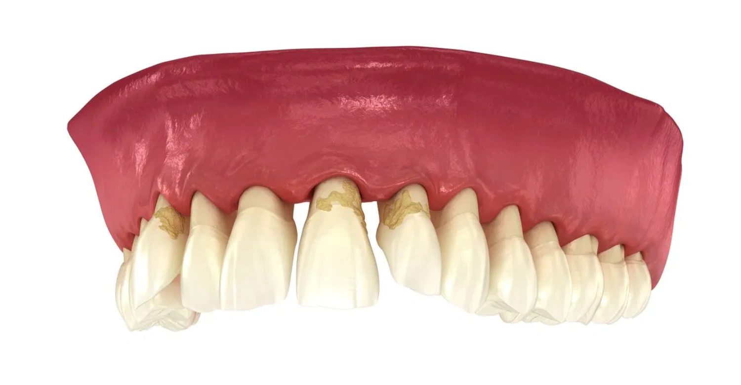
What Does Periodontal Disease Look Like?
In its earliest form, known as gingivitis, the visual changes are subtle but distinct. So what does gingivitis look like in practice? If you look closely in the mirror, you will notice that the edges of the gums, right where they meet the teeth, lose their pale pink hue. They turn a bright red or deep reddish-purple. This discoloration indicates localized inflammation as blood rushes to the area to fight accumulating bacterial plaque.
Beyond color changes, inflamed gums appear puffy, swollen, and slightly rolled at the borders rather than flat and tight. Another undeniable visual cue is bleeding. If you spit out pink-tinged foam or notice bright red blood on your toothbrush or dental floss, you are looking at a classic manifestation of early periodontal disease. Early stage gum disease pictures typically show exactly this combination: redness, puffiness, and light bleeding, without any recession or tooth movement yet. At this point, the underlying bone is still intact. This means the visual damage is completely reversible with professional cleaning and improved daily care.
What Does Periodontal Disease Look Like
Small daily habits make it easier to catch periodontal disease early, before the visual signs progress:
Check your gums in a well-lit mirror weekly for color changes, puffiness, or bleeding.
Watch for teeth that appear to be lengthening or new gaps forming near the gumline, both signs of recession.
Avoid smoking, since it can mask early visual symptoms even while the disease progresses underneath.
Book a professional pocket-depth check rather than relying on mirror checks alone, especially if you smoke.
Seek a periodontal evaluation promptly if you notice bleeding, pus, or any looseness in your teeth.
What Does Periodontal Disease Look Like?
When plaque hardens into tartar, the disease progresses below the gumline into periodontitis. The visual indicators become much more dramatic. Read also: is periodontal disease fatal.
Bacteria destroy the connective fibers holding tissue to the tooth root. The gums begin to detach and migrate downward. This process is called gum recession. It fundamentally alters the appearance of your smile. Gum recession pictures typically show this exact transition. Your teeth will look much longer than they used to. This happens because the darker, yellowish root surfaces are now exposed.
As the gums pull away, dark empty spaces form between the teeth near the gumline. Dentists call these black triangles teeth, or simply "black triangles." They occur because the healthy tissue that once filled those gaps has degraded. You may also see visible deposits of hardened tartar. Unlike soft, whitish plaque, tartar often presents as a stubborn, chalky crust. It can appear white, yellow, or dark brown. It cements tightly around the base of the teeth.
Clinical Note
Visual self-assessment is a valuable early warning tool. But it cannot replace a professional periodontal exam. Dr. Rifat Alsaman, Head of the Medical Team at Vitrin Clinic and a cosmetic dentist, advises that pocket-depth measurements, digital imaging, and a full staging and grading assessment are needed. Only then should any treatment plan be recommended.
The Advanced Visual Signs: Advanced Periodontitis
In the late stages, the visual presentation shifts from subtle warning signs to structural failure. The infection penetrates deeply into the jawbone, causing severe destruction.
At this juncture, the answer to what does periodontal disease look like includes watching your teeth physically shift out of position. The underlying bone foundation is melting away. Teeth may develop wide new gaps. They can also flare outward or twist unnaturally. When you bite down, your upper and lower teeth may no longer align the way they used to. Learn more: can periodontal disease be cured.
The gums themselves often look dark red, purplish, and noticeably bulbous or detached from the enamel. A particularly distressing symptom is the presence of pus. If you apply gentle pressure to the swollen gum tissue, you may see a thick, yellowish-white fluid ooze out. This fluid comes from the deep pockets surrounding the tooth. At this severe stage, the teeth may visibly wobble or move when touched by the tongue. This signals imminent tooth loss unless specialist surgical care is sought immediately.
Why Smoking Can Hide the Visual Signs
Smoking is the most significant risk factor for periodontal disease, a finding confirmed by the NIDCR. It complicates visual self-assessment in a dangerous way. Gum disease in smokers behaves differently at a visual level. Nicotine restricts blood flow to the gum tissue. This can suppress the redness, swelling, and bleeding that usually signal inflammation. As a result, a smoker's gums may look deceptively pale, firm, and healthy. Yet periodontitis may be actively destroying bone and attachment beneath the surface.
This masking effect means smokers often present with more advanced disease than their gums visually suggest. Regular professional pocket-depth checks are especially important. Relying on mirror checks alone is not enough. The numbers bear this out. CDC data finds periodontitis in 64.2 percent of current smokers. The disease is also more common in men (56.4 percent) than in women (38.4 percent). Researchers partly attribute this gap to differences in smoking rates and oral hygiene habits.
Beyond Smoking: Other Risk Factors to Consider
Smoking is not the only risk factor. Several other factors can increase your susceptibility. Diabetes is one. People with diabetes, especially those with poorly controlled blood sugar, are at higher risk for infections, including gum disease. The relationship is bidirectional. Periodontal disease can also make it harder to control blood sugar.
Genetics also play a role. Some individuals are genetically predisposed to periodontal disease, even with good oral hygiene. If you have a family history of gum disease, you may need to be extra vigilant. Poor oral hygiene is another key factor. Inadequate brushing and flossing allow plaque to build up. This can harden into tartar and lead to gum inflammation and infection. Other factors include stress, certain medications, teeth grinding, and a diet low in vitamin C.
What Happens If Periodontal Disease Is Left Untreated?
Ignoring periodontal disease can lead to serious complications beyond tooth loss. The bacteria from infected gums can enter your bloodstream. This can contribute to systemic health issues. Studies have linked periodontal disease to an increased risk of heart disease and stroke. The inflammation and bacteria may affect the heart and blood vessels.
Periodontal disease can also make it harder to control blood sugar. This creates a vicious cycle for people with diabetes. Bacteria from the mouth can be inhaled into the lungs. This may cause pneumonia or worsen chronic respiratory conditions like COPD. Pregnant women with periodontal disease may be at higher risk for preterm birth and low birth weight. Untreated gum disease can also lead to painful abscesses, bone loss, and tooth loss. This can affect your ability to eat and speak properly, and impact your self-esteem.
How Dentists Stage and Grade Periodontitis
Beyond visual inspection, periodontitis is formally classified using a staging and grading system. This system was established by the American Academy of Periodontology. Periodontal disease staging and grading works on two axes. Staging (I through IV) reflects the severity of tissue and bone loss already present. Grading (A, B, or C) estimates how quickly the disease is likely to progress. It is based on factors like smoking history. This system clarifies that what a patient sees in the mirror is only a starting point. The stage and grade determined during a clinical exam at Vitrin Clinic guide the actual treatment plan. This can range from non-surgical deep cleaning to advanced periodontal surgery.
What We Notice Clinically
Dr. Rifat Alsaman, Head of the Medical Team at Vitrin Clinic and a cosmetic dentist, notes that patients often underestimate gingivitis. Bleeding gums are dismissed as normal. They are not treated as a warning sign. In his clinical experience, smokers are often surprised by their pocket depths. These depths may already be in the periodontitis range. This happens even when gums look calm in the mirror. Routine pocket-depth measurement is more reliable than visual inspection alone. It is key to catching periodontal disease before bone loss or tooth mobility develops.
Seeking Care: Periodontal Treatment at Vitrin Clinic
Recognizing what does periodontal disease look like is the first step. Taking action requires professional intervention. For gum recession, bone deterioration, or aesthetic imbalances, specialized centers like Vitrin Clinic offer solutions. Located in Istanbul, Turkey, this clinic specializes in advanced periodontics, laser gum therapies, and gingival aesthetics. Its goal is to restore both the health and framework of your mouth. Explore: preventing periodontal disease.
When estimating the financial aspect of managing these conditions abroad, the average cost of Vitrin Clinic treatments varies. It depends on the severity of the damage. For early-to-intermediate issues requiring tissue reshaping, a gingivoplasty or gum contouring procedure averages around 480 USD. For more extensive restorative needs resulting from advanced periodontal bone loss, such as high-quality zirconia crowns to restore damaged teeth, prices average between 240 USD and 2,050 USD. The exact cost depends on the specific case scope and material choice. These therapies incorporate state-of-the-art digital imaging and painless laser technology. They allow patients to manage the visual and structural impacts of the disease at a fraction of the cost of traditional private clinics in Western countries.
Ultimately, paying close attention to changes in gum color, tooth length, and the appearance of spaces in your mouth can help. It ensures you catch periodontal disease before it compromises your long-term health and wellness. Find out more: how to treat periodontitis.
About Vitrin Clinic
Vitrin Clinic is an international dental clinic based in Istanbul, Turkey. It serves patients traveling from abroad for cosmetic and restorative dental care. The clinic is led by Dr. Rifat Alsaman, Head of the Medical Team at Vitrin Clinic and a cosmetic dentist. It combines advanced periodontics, laser gum therapy, and full-mouth restoration with multilingual patient support. This allows international patients to access specialist-level treatment for conditions like periodontal disease, at a fraction of typical Western pricing. Patients don't have to compromise on diagnostic precision or aftercare.
Resources & References
American Academy of Periodontology — Staging and Grading Periodontitis — the staging and grading diagnostic framework for periodontitis
NIDCR — Periodontal (Gum) Disease — general gum disease progression and smoking as a risk factor
Cleveland Clinic — Periodontitis — healthy pocket-depth ranges and clinical diagnostic criteria
FAQs

Dr. Rifat Alsaman has more than 5 years of clinical experience in dentistry and currently serves as the Head of the Medical Team at Vitrin Clinic. He is dedicated to providing exceptional patient care, overseeing treatment planning, and ensuring the highest clinical standards across the team. His expertise, attention to detail, and commitment to continuous professional development have helped countless patients achieve healthier, more confident smiles.


