
Orthodontics has evolved dramatically over the last few decades, moving away from conventional, two-dimensional diagnostics toward highly accurate, three-dimensional digital mapping. Central to this technological paradigm shift is Cone Beam Computed Tomography (CBCT). When analyzing how 3d cbct improves orthodontic treatment planning, it becomes evident that this diagnostic tool provides unparalleled visualization of the craniofacial complex, completely transforming how orthodontists assess, simulate, and execute tooth movement.
At Vitrin Clinic, a leading luxury dental health center located in Istanbul, Turkey, the integration of advanced 3D diagnostics has redefined the standards of orthodontic care. By utilizing state-of-the-art CBCT imaging, the medical team at Vitrin Clinic can design highly precise, predictable, and individualized treatment journeys for both local and international patients.
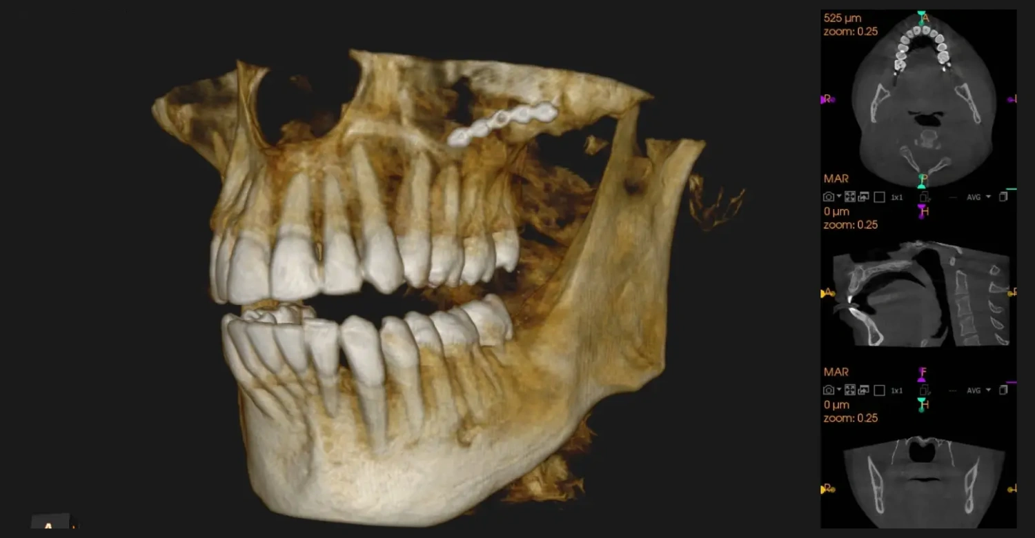
1. True Three-Dimensional Anatomical Visualization
Traditional orthodontic diagnostics rely heavily on panoramic and cephalometric X-rays. While useful, these modalities compress complex, multi-layered three-dimensional facial structures into a flat, two-dimensional image. This inevitably leads to anatomical distortion, magnification errors, and structural overlapping.
In exploring how 3d cbct improves orthodontic treatment planning, the primary benefit is the elimination of these overlapping artifacts. A single low-dose CBCT scan captures a comprehensive volume of data, enabling clinicians to view the skull, jawbones, and dentition from any angle. The precise rendering allows for exact measurements of bone height, bone width, and the true spatial relationship of the teeth relative to their supporting structures. Since untreated bruxism is the main contraindication mentioned here, our guide to crowns for teeth grinding explains how heavy bite forces are managed before any thin ceramic restoration is placed
2. Accurate Assessment of Impacted and Ectopic Teeth
Managing impacted teeth particularly maxillary canines presents a significant clinical challenge in orthodontics. Two-dimensional imaging can reveal that a tooth is impacted, but it cannot accurately determine whether the tooth lies palatally (toward the roof of the mouth) or buccally (toward the cheek).
The adoption of 3D CBCT completely resolves this ambiguity. It allows specialists to pinpoint the exact three-dimensional position of the impacted crown and root. Furthermore, it reveals whether the impacted tooth is causing root resorption on neighboring healthy teeth. Having this precise data beforehand alters the biomechanical approach, enabling the creation of safer, direct traction pathways that minimize risk to adjacent structures.
3. Evaluation of Alveolar Bone Quality and Volume
Moving teeth through bone requires a thorough understanding of the biological boundaries of the alveolar ridge. Forcing a tooth beyond the cortical bone plate can result in severe clinical complications, such as bone fenestration, dehiscence, or irreversible root resorption.
Understanding how 3d cbct improves orthodontic treatment planning highlights its capacity to map out cortical and cancellous bone thicknesses prior to initializing tooth movement. This is particularly crucial for adult patients, who often present with reduced bone volume or pre-existing periodontal compromises. By visualizing the true boundaries of the bone, orthodontists can customize alignment boundaries safely.
4. Enhanced Airway Analysis and Skeletal Symmetry
Modern orthodontics looks beyond just straight teeth; it encompasses overall facial harmony, airway health, and temporomandibular joint (TMJ) function.
CBCT technology generates clear cross-sections of the upper airway, helping clinicians identify pharyngeal airway restrictions that could contribute to obstructive sleep apnea (OSA) or mouth breathing. Additionally, 3D imaging allows for a meticulous volumetric analysis of the condyles within the TMJ. This assists in identifying subtle skeletal asymmetries or structural wear that traditional radiographs easily miss, creating a more comprehensive foundation for orthognathic surgical cases or functional appliance therapy. Patients weighing that exact decision can read our comparison of veneers vs. crowns, which explains which structural situations call for each.
5. Precise Digital Integration for Clear Aligners and Temporary Anchorage Devices (TADs)
The data obtained from a 3D CBCT scan integrates seamlessly with computer-aided design and manufacturing (CAD/CAM) workflows. When planning for clear aligners, such as Invisalign, combining intraoral surface scans with root data from a CBCT yields an incredibly accurate digital patient model.
Moreover, when a treatment requires the use of Temporary Anchorage Devices (TADs) or orthodontic mini-screws, CBCT is invaluable. It provides a detailed map of the interradicular space (the area between tooth roots) and bone density, ensuring that the clinician places the mini-screw safely without damaging roots or neurovascular bundles.
.jpg)
The Average Cost of Orthodontic Treatments at Vitrin Clinic
For patients seeking world-class orthodontic and cosmetic enhancements, Vitrin Clinic provides premium, luxury dental packages at highly competitive rates compared to Western Europe and North America.
While diagnostic CBCT scans are often included as part of the complimentary initial consultation and digital workflow design for international patients, the average cost for advanced orthodontic treatments at Vitrin Clinic reflects its accessible luxury pricing. For instance, a full-case premium clear aligner system (such as American Invisalign) at Vitrin Clinic averages around $4,100. This all-inclusive pricing structure typically covers premium digital planning, localized transfers, translation services, and elite medical monitoring throughout the treatment duration in Istanbul.
Clinical Insights from Dr. Rifat Alsaman
When assessing the practical execution of these advanced imaging systems, the clinical expertise behind the technology is paramount. Dr. Rifat Alsaman, the Head of the Medical team at Vitrin Clinic, emphasizes that 3D imaging has shifted orthodontic planning from an observational science to an exact digital methodology. As a premier cosmetics dentist, Dr. Rifat Alsaman recognizes that a beautiful smile design must always be built upon a healthy, structurally sound anatomical foundation.
According to Dr. Rifat Alsaman, utilizing 3D CBCT allows his team to visualize the patient's entire maxillofacial architecture down to the millimeter before a single bracket or aligner is placed. Dr. Rifat Alsaman, Head of the Medical team at Vitrin Clinic, explains that this advanced visualization is crucial for predicting how soft tissues and lip profiles will respond to changes in tooth positioning.
As an elite cosmetics dentist, Dr. Rifat Alsaman notes that incorporating CBCT into the workflow at Vitrin Clinic dramatically accelerates treatment timelines and increases safety. By understanding the precise bone densities and root trajectories from day one, the team can avoid the trial-and-error approach often associated with old 2D planning. Dr. Rifat Alsaman maintains that the combination of cosmetic vision and 3D diagnostic precision ensures that every patient at Vitrin Clinic receives a structurally stable, balanced, and aesthetically flawless smile. Since the digital preview is central to this treatment, our smile design page explains how 3D simulation and facial mapping let you approve the result before fabrication begins.
References:
FAQs

Dr. Rifat Alsaman has more than 5 years of clinical experience in dentistry and currently serves as the Head of the Medical Team at Vitrin Clinic. He is dedicated to providing exceptional patient care, overseeing treatment planning, and ensuring the highest clinical standards across the team. His expertise, attention to detail, and commitment to continuous professional development have helped countless patients achieve healthier, more confident smiles.


