Oral Surgery
June 23, 2026
What is Sinus Perforation and How is it Recognized?

Sinus perforation is a dental complication in which an unintended opening forms between the oral cavity and the maxillary sinus the air-filled space located directly above the upper back teeth. It most commonly occurs following tooth extraction, implant placement, or sinus lift procedures in the posterior upper jaw. Recognizing Sinus Membrane Perforation early is critical: patients may notice air passing through the extraction socket, unilateral nasal congestion, or fluid regurgitating into the nose during eating. Prompt clinical evaluation ensures timely treatment and prevents serious complications.
Introduction
Dental procedures involving the upper jaw carry a unique anatomical challenge: the maxillary sinus sits in close proximity to upper molar roots. As dental technology continues to advance, patients benefit from safer, more predictable outcomes than ever before. Yet even within modern clinical environments, certain complications remain possible. Sinus perforation is one such complication uncommon, but important to understand. This blog explores what it is, how it is recognized, and what steps patients should take when symptoms arise.
Modern dental technology and improved patient safety
Today's dental landscape is defined by digital precision. Cone beam CT imaging, computer-guided implant placement, and advanced surgical instruments have dramatically reduced the rate of intraoperative complications. Patients undergoing upper jaw procedures at facilities that invest in this technology experience significantly improved safety profiles. Modern dental technology allows clinicians to visualize sinus anatomy before making a single incision, ensuring that procedures are planned around individual anatomy rather than generalized estimates. This represents a substantial leap forward in protecting patients from avoidable complications including sinus perforation.
Why complications can still occur in upper jaw procedures
Despite technological progress, the maxillary sinus remains anatomically variable. Some patients have sinus floors that dip unusually low, placing them just millimeters above upper molar roots. In these cases, even a carefully performed tooth extraction, implant placement, or sinus lift procedure carries inherent risk. Bone density, root curvature, pre-existing sinus conditions, and prior dental history all influence surgical complexity. Understanding that complications like sinus perforation can occur even in experienced hands helps patients approach their care with realistic expectations and readiness to recognize early warning signs.
Importance of early recognition by patients
Patients play a vital role in post-procedural safety. A sinus perforation that is identified within the first 24 to 48 hours after a dental procedure is significantly easier to manage than one that goes unnoticed for days or weeks. Patients who understand what Sinus Membrane Perforation symptoms look like are better positioned to contact their dental team promptly, avoid behaviors that worsen the condition, and begin appropriate treatment before complications develop. Patient awareness is not a substitute for clinical oversight, it is a complement to it.
What Is Sinus Perforation?
Sinus perforation refers to the creation of an unintended opening between the oral cavity and the maxillary sinus, the air-filled space located above the upper back teeth. This opening, sometimes described as an accidental hole in sinus cavity tissue, disrupts the natural barrier that separates the mouth from the sinus. When this barrier is breached, air, bacteria, and fluid can pass between the two spaces, creating conditions that may lead to infection, chronic discomfort, and delayed healing. Sinus Membrane Perforation is a recognized complication within oral and maxillofacial surgery.
Clinical definition
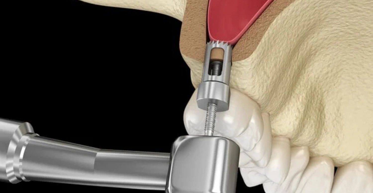
Clinically, sinus perforation also referred to as oroantral communication describes a pathological opening between the maxillary sinus and the oral cavity. This typically occurs at the site of an extracted upper molar or premolar, or at an implant site in the posterior maxilla. The perforation may range in size from a small pin-point opening to a larger defect several millimeters in diameter. Size directly influences the likelihood of spontaneous closure and the type of treatment required. Smaller perforations may heal independently under the right conditions; larger ones almost always require surgical closure.
Common dental causes
The most frequent cause of sinus perforation is tooth extraction sinus complication following the removal of upper molar or premolar teeth whose roots extend close to or into the sinus floor. Additional causes include dental implant placement in the posterior upper jaw, sinus lift procedures performed to augment bone volume, removal of large cysts or granulomas near the sinus, and aggressive curettage during root canal procedures. Patients with low sinus floors, thin cortical bone, or history of periodontal disease are at elevated risk. Pre-surgical CBCT imaging is the most effective preventive tool available.
Early warning symptoms
Sinus perforation symptoms in the immediate post-procedural period can be subtle. Patients may notice air passing through the extraction socket when blowing their nose, a feeling of nasal congestion on one side, or a slightly altered taste or smell. Some experience a sense of pressure around the cheekbone or under the eye. In the absence of infection, these symptoms may not be accompanied by significant Sinus Membrane Perforation pain. This is precisely why patients must be informed of what to watch for; the absence of severe pain does not mean the complication is absent or minor.
How Is Sinus Perforation Recognized?
How is sinus perforation recognized in a clinical setting, and what role does patient self-reporting play in early detection? Recognition of Sinus Membrane Perforation relies on a combination of patient-reported observations, clinical examination findings, and diagnostic imaging. No single factor is sufficient on its own. A patient who reports air moving through a tooth socket after blowing their nose is providing a highly specific symptom that should prompt immediate clinical evaluation. Similarly, a dentist performing a post-extraction review should actively assess for sinus involvement using established clinical tests. Early recognition is the foundation of effective management.
Patient-reported symptoms
Sinus perforation symptoms reported by patients include nasal regurgitation of fluids during eating or drinking, unilateral nasal congestion, a whistling or air-passing sensation at the extraction site when breathing through the nose, and a feeling of pressure beneath the eye or cheek. Some patients describe a muffled or echo-like quality to sounds in the affected area. Bloody nasal discharge, particularly within the first 24 hours, may also indicate sinus involvement. Patients should not dismiss these signs as normal post-extraction discomfort each warrants a clinical evaluation to rule out or confirm Sinus Membrane Perforation.
Clinical indicators
Clinically, how is sinus perforation recognized involves a straightforward examination technique called the Valsalva test. The patient is asked to gently close their nostrils and attempt to exhale through the nose. If a perforation is present, air will pass through the opening into the oral cavity, producing a visible bubbling or rushing sensation at the extraction site. Clinicians also assess for probe depth at the socket, the presence of sinus fluid on suction, and mucosal color changes. Unilateral rhinorrhea following an upper jaw procedure is another reliable clinical indicator that should always be investigated.
Diagnostic confirmation tools
When clinical examination raises suspicion, diagnostic imaging provides confirmation. Panoramic radiographs offer an initial overview of the maxillary sinus and adjacent tooth roots, helping identify whether root tips extended into sinus tissue. Cone beam CT, the gold standard in this context, delivers three-dimensional visualization of the sinus floor, any bony defect present, and the degree of mucosal involvement. CBCT imaging also assists in planning surgical repair if required. Water's view radiograph, though older, may also reveal sinus opacification consistent with sinusitis after dental procedure, supporting the diagnosis of sinus perforation.
What to Do When You Notice Symptoms of Sinus Perforation
If a patient suspects sinus perforation following a dental procedure, their first response must be calm, immediate, and structured. The steps taken in the first hours after noticing symptoms can meaningfully influence the healing trajectory. Above all, patients should avoid behaviors that increase sinus pressure, as these actions can enlarge a small perforation or push bacteria from the oral cavity into the sinus cavity. Contacting a dental professional immediately remains the single most important step. The following sections outline what patients should and should not do when Sinus Membrane Perforation symptoms appear.
Immediate steps
Upon noticing sinus perforation symptoms, contact your dental provider without delay. Report the specific symptoms you are experiencing, including any sensation of air moving through the socket, nasal fluid discharge, or pressure around the cheek. Do not blow your nose, as this action forces air and bacteria through any perforation present, widening the defect and increasing infection risk. If prescribed antibiotics have been provided, ensure they are being taken as directed. Do not probe the socket with your tongue or any instrument. Keep the area clean and undisturbed. Prompt clinical assessment is the only appropriate response to suspected sinus perforation.
Daily precautions
During the period of suspected or confirmed sinus perforation, patients must observe strict daily precautions. Avoid sneezing with the mouth closed, as this dramatically increases intraoral pressure. Sleep with your head slightly elevated to reduce sinus congestion and fluid pooling. Consume soft foods and avoid chewing on the affected side to prevent disturbing the socket. Do not smoke under any circumstances smoking impairs tissue healing, increases infection risk, and creates negative pressure in the oral cavity that can disrupt clot formation. Refrain from drinking through straws for the same reason. These measures support healing and prevent complication progression.
Symptom control
Mild sinus perforation pain can typically be managed with over-the-counter analgesics such as ibuprofen or paracetamol, taken according to standard dosing guidelines. Saline nasal rinses administered gently without the use of pressure devices may help maintain nasal hygiene and reduce congestion associated with sinusitis after dental procedure. Cold compresses applied externally to the cheek can reduce swelling during the first 48 hours. Patients should not use medicated nasal sprays without clinical guidance, as some preparations contain vasoconstrictors that interfere with mucosal healing. All symptom management approaches should be discussed with a dental professional before use.
What Happens If Sinus Perforation Is Not Treated?
Untreated sinus perforation risks are serious and progressive. A perforation left unaddressed does not simply remain stable; it creates a persistent communication between two environments that are designed to remain separate. The oral cavity contains a high bacterial load, and continuous contamination of the maxillary sinus through an unhealed perforation leads to infection, mucosal thickening, and eventually chronic sinusitis. Patients who delay treatment often find that what began as a manageable complication has evolved into a complex surgical problem requiring more invasive intervention than would have been necessary with early treatment.
Early complications
In the first days following an unrecognized or untreated sinus perforation, early complications begin to develop. The blood clot that forms in the socket a critical element of the healing process can be dislodged by air pressure moving through the perforation. Without clot protection, the socket is exposed to oral bacteria. These bacteria migrate upward into the sinus, triggering an acute inflammatory response. Patients begin to experience unilateral nasal congestion, foul-tasting discharge, and increasingly pronounced Sinus Membrane Perforation pain around the cheek and under the eye. Fever may develop as the body responds to infection. These symptoms indicate that sinusitis after dental procedure has begun.
Oral-nasal communication issues
One of the more debilitating consequences of untreated sinus perforation is the development of a persistent oroantral fistula, a fully epithelialized tract between the mouth and the sinus that allows continuous communication between the two spaces. Once this occurs, patients experience regurgitation of food and fluids through the nose, persistent nasal voice quality, and recurring episodes of sinusitis. An accidental hole in sinus cavity tissue that has epithelialized cannot close on its own and requires surgical excision and formal closure. This condition represents a significantly more complex treatment scenario than simple early-stage Sinus Membrane Perforation management.
Long-term consequences
Among the most serious long-term untreated sinus perforation risks is the development of chronic maxillary sinusitis, a persistent inflammatory condition of the sinus lining. This may require functional endoscopic sinus surgery (FESS) in addition to oral surgical closure of the perforation itself. In severe cases, infection can spread to adjacent sinuses or nearby anatomical structures. Patients with compromised immune systems face heightened risk of systemic infection. Dental implant failure in the affected region is also likely if chronic infection persists. The longer Sinus Membrane Perforation remains untreated, the more complex, costly, and lengthy the recovery process becomes.
Symptoms and Risks of Untreated Sinus Perforation
Understanding the symptoms and untreated sinus perforation risks in detail helps patients recognize when their condition requires urgent escalation. While early Sinus Membrane Perforation can present with mild or non-specific symptoms, untreated cases evolve into identifiable clinical syndromes with significant quality-of-life impact. Patients who are aware of the trajectory of untreated Sinus Membrane Perforation are more likely to seek timely care. The following sections examine chronic pain patterns, the mechanics of infection spread, and the specific ways in which healing failure manifests in the context of unmanaged Sinus Membrane Perforation.
Chronic pain and discomfort
Sinus perforation pain in untreated cases typically evolves from intermittent post-procedural discomfort into persistent, daily facial pain. Patients describe a deep ache beneath the cheekbone, increased pressure when bending forward or changing head position, and sensitivity around the affected upper teeth. Headaches localizing to the forehead and temple are common as sinusitis becomes established. Pain may worsen at night when lying flat. Over time, patients may also develop referred pain to the upper teeth, making it difficult to distinguish between ongoing Sinus Membrane Perforation-related discomfort and dental pathology. This chronic pain pattern significantly impacts daily functioning and quality of life.
Infection spread risks
Untreated sinus perforation risks include infection pathways that extend beyond the maxillary sinus. The maxillary sinus connects to the nasal cavity and communicates with adjacent ethmoid and frontal sinuses. Bacterial contamination introduced through an unhealed Sinus Membrane Perforation can ascend through these pathways, resulting in pansinusitis infection affecting multiple sinuses simultaneously. In immunocompromised patients or those with pre-existing inflammatory sinus disease, infection can progress rapidly. Orbital complications, including periorbital swelling and visual disturbance, represent the most serious regional spread. Although rare, these outcomes underscore the importance of not dismissing tooth extraction sinus complication symptoms in the early post-operative period.
Healing failure risks
Sinus perforation healing time is highly dependent on the size of the defect, the patient's general health, and whether appropriate management was initiated promptly. When treatment is delayed, the physiological window for spontaneous closure passes. The socket lining undergoes epithelialization, the same process that forms protective skin over wounds but in this case it forms a permanent mucous membrane-lined tract rather than normal healing tissue. Once this occurs, Sinus Membrane Perforation can no longer heal on its own regardless of time elapsed. Healing failure is not an abstract risk; it is the predictable outcome of untreated Sinus Membrane Perforation beyond a critical early management window.
Advanced Complication Management at Vitrin Clinic
At Vitrin Clinic, the management of dental complications including sinus perforation follows internationally recognized clinical protocols supported by advanced digital technology. The clinic's approach integrates diagnostic precision, specialist surgical expertise, and structured patient follow-up pathways to ensure that complications are identified early, managed appropriately, and resolved with minimal impact on long-term oral health. Patients traveling from outside Turkey for dental treatment at Vitrin Clinic benefit from a clinical environment that prioritizes transparent communication, individualized care planning, and outcomes-focused surgical management for both routine and complex cases.
A global standard in surgical safety and precision
Vitrin Clinic operates at a level of surgical safety and precision consistent with leading international dental institutions. Complication management protocols are developed in alignment with evidence-based guidelines from recognized oral and maxillofacial surgery bodies. Every patient undergoing upper jaw procedures receives pre-surgical risk assessment specifically addressing sinus proximity and perforation risk. Intraoperative monitoring, careful tissue handling, and systematic post-operative follow-up are embedded in every upper jaw surgical pathway. When sinus perforation does occur, the clinical team responds according to a clearly defined protocol ensuring that the patient receives the appropriate level of care without delay.
CBCT-guided digital treatment planning
Cone beam CT imaging is standard practice at Vitrin Clinic for all upper jaw surgical procedures. CBCT provides three-dimensional visualization of the maxillary sinus floor, root morphology, and bone volume allowing precise measurement of the distance between planned surgical sites and sinus tissue. This level of detail dramatically reduces the risk of accidental hole in sinus cavity formation during implant placement or tooth extraction. When sinus perforation does occur, CBCT imaging is also used to characterize the size and location of the defect, assess mucosal involvement, and plan surgical closure with anatomical accuracy unavailable through conventional two-dimensional radiography.
Specialist-led oral surgery protocols
Oral surgical procedures at Vitrin Clinic are performed by specialist surgeons with specific training in maxillofacial and implant surgery. When a case presents elevated sinus risk, specialist-to-specialist consultation occurs prior to the procedure. Patients with anatomically challenging sinus anatomy receive modified surgical protocols designed to minimize sinus perforation risk without compromising the primary treatment objective. In cases where sinus perforation is identified intraoperatively, immediate management is initiated including primary closure where appropriate and antibiotic prophylaxis. Post-operative monitoring is structured and consistent, with clear escalation pathways if Sinus Membrane Perforation symptoms emerge following discharge.
About Vitrin Clinic
Vitrin Clinic is an international dental center based in Istanbul, Turkey, specializing in advanced restorative and surgical dentistry for patients traveling from across the world. The clinic combines digital dentistry technology, specialist surgeons, and modern clinical protocols to deliver comprehensive care across disciplines including implantology, oral surgery, and aesthetic dentistry. Vitrin Clinic is recognized for comprehensive care pathways designed around safety, precision, and predictable outcomes. Patients receive individualized treatment planning, transparent clinical communication, and structured follow-up regardless of their country of origin. The clinic's approach ensures that even complex complications such as sinus perforation are managed within a framework of clinical excellence.
FAQs

Dr. Rifat Alsaman has more than 5 years of clinical experience in dentistry and currently serves as the Head of the Medical Team at Vitrin Clinic. He is dedicated to providing exceptional patient care, overseeing treatment planning, and ensuring the highest clinical standards across the team. His expertise, attention to detail, and commitment to continuous professional development have helped countless patients achieve healthier, more confident smiles.


