Dental Implant
October 14, 2026
What Is a Dental Implant Emergence Profile, and Why Does It Matter?
 Dr. Rifat Alsaman
A Dental Implant Emergence Profile is the shape of the restoration as it rises from the implant through the gum. This contour affects how the crown looks, how the gum heals, and how easily the area is cleaned. One US analysis projected implant use could reach 23% of adults with missing teeth by 2026. Large reviews often report ten-year implant survival above 95%. However, a surviving implant is not always a healthy or natural-looking one. This guide covers design, healing, complications, correction, and costs in the US, UK, and Turkey.
At a glance
• What it is: The transition zone between the implant and the visible crown.
• Why it matters: Contour influences gum health, plaque control, and gumline appearance.
• Soft-tissue adaptation: Usually several weeks to a few months.
• Implant, abutment, and crown: About 3,000–6,000 (US), 1,800–3,500 (UK), 450–1,100 (Turkey).
Ready to transform your smile?
Talk to our team and get a personal FREE treatment plan.
.jpg)
.jpg)
.jpg)
.jpg)
What Is a Dental Implant Emergence Profile?
The Dental Implant Emergence Profile describes how the restoration “emerges” from the implant platform to the gumline. An implant fixture is usually 3.5–5 mm wide, while a natural molar can measure over 10 mm. The restoration must widen gradually across this gap. That widening happens inside a soft-tissue tunnel roughly 3–4 mm deep. How the dentist shapes this tunnel strongly influences comfort, appearance, and long-term gum health.
Understanding the Shape Where an Implant Crown Meets the Gum
Imagine the implant as a narrow root and the crown as a wider tooth. The emergence profile is the bridge between the two. It begins at the implant platform, below the gum, and ends where the crown appears in the mouth. A smooth, gradual transition supports the gum like a natural tooth. An abrupt or bulky transition can push tissue, trap plaque, and look unnatural.
How the Implant Fixture, Abutment, Crown, and Gingival Tissue Work Together
Four parts form one functional unit. The fixture is the titanium or zirconia post that fuses with the jawbone. The abutment connects the fixture to the crown and passes through the gum. The crown is the visible tooth that restores chewing and appearance. The gingival tissue seals around the abutment, forming a protective barrier. Research describes this peri-implant seal as roughly 3–4 mm high, combining connective tissue and epithelium. Each part must fit the others precisely for the seal to stay healthy.
Dental Implant Emergence Profile vs. Abutment Contour: What Is the Difference?
Abutment contour refers to the shape of one component only. The emergence profile is the overall shape created by the abutment and crown together. A stock abutment can have a fixed contour that suits some patients but not others. Custom abutments let the laboratory shape the transmucosal section for each site. In other words, abutment contour is one ingredient, while the emergence profile is the finished result.
Why Emergence Profile Design Matters for Function, Appearance, and Oral Hygiene
Design affects three everyday outcomes. Functionally, a well-shaped crown distributes chewing forces and prevents food packing. Aesthetically, it supports the gum margin and papillae, avoiding dark gaps or gray shadows. Hygienically, it lets floss, brushes, and interdental cleaners reach the junction. This matters because a 2015 meta-analysis estimated peri-implant mucositis affects about 43% of implants. The same review estimated peri-implantitis at roughly 22%. A cleansable Dental Implant Emergence Profile is one practical way to reduce plaque-related risk.
What Is the Ideal Emergence Profile for Dental Implants?
The ideal profile is the one that supports healthy tissue, natural appearance, and easy cleaning for that specific site. It is not a fixed angle or a standard shape. Dentists adapt it to implant depth, gum thickness, tooth type, and neighboring teeth. Current literature favors restrained, gradual contours over bulky ones, especially near the bone.
What Determines the Ideal Shape of an Implant Crown?
Several measurable factors guide the shape. Implant depth sets how much vertical space exists for the transition. Implant diameter and the planned tooth width determine how much the contour must widen. Gum thickness affects how much pressure the tissue tolerates. The position of adjacent teeth and bone peaks guides papilla support. Finally, the patient’s dexterity influences how cleansable the design must be. A shape that works for a lower molar may fail on an upper incisor.
Concave, Straight, and Convex Implant Emergence Profiles
Clinicians usually describe three basic contours in cross-section. A concave profile curves inward, leaving more room for soft tissue. A straight profile rises in a relatively direct line from platform to crown. A convex profile bulges outward and pushes against the surrounding gum. Many restorations combine shapes at different levels. Researchers often divide the area into a deeper “critical” zone and a shallower “subcritical” zone.
How Concave Contours Can Support Soft-Tissue Space and Cleaning Access
A concave or narrow contour near the implant platform leaves space for thicker connective tissue. Thicker tissue tends to form a more stable seal and resists recession better. The undercut shape also gives floss and brushes a clearer path. Many clinicians favor a slim, concave design in the deeper critical zone. This approach aims to protect bone while allowing the gum to fill the space naturally.
When Convex Contours May Require Greater Clinical Attention
Convex contours are not always wrong, but they need careful judgment. A gentle convexity in the subcritical zone can support the gingival margin and papillae. Excessive convexity near the bone, however, may compress tissue and hide plaque. One 2018 radiographic study linked convex profiles with steep angles to higher peri-implantitis prevalence. Dentists therefore reserve convexity for specific aesthetic purposes and monitor these sites closely.
Emergence Angle, Crown Width, and the Natural Tooth Transition
The emergence angle is measured between the implant’s long axis and a line along the restoration contour. Several studies have used 30 degrees as a reference threshold. Wider crowns on narrow implants often need steeper angles, unless implant depth provides extra vertical room. The aim is a transition that resembles a natural root rising into enamel. Crown width must match the gap so contacts and embrasures look and work naturally.
How Dentists Balance Gum Support, Aesthetics, and Cleansability
Every design involves trade-offs. More contour can lift the gum margin and support papillae in the front. Less contour often improves cleaning access and reduces tissue pressure. Back teeth usually prioritize cleansability and food control, while front teeth demand careful aesthetic support. A good Dental Implant Emergence Profile finds the balance point for each site. That balance is planned, tested with a provisional crown, and then refined.
Why There Is No Single Ideal Emergence Profile for Every Patient
Mouths differ in bone level, gum biotype, smile line, and tooth shape. Studies of smile lines suggest about 10–11% of people show a high gum display. For these patients, small contour errors become highly visible. Others rarely show the gumline, so cleansability may outweigh fine aesthetic detail. Implant systems and connection types also behave differently. For these reasons, ideal design remains an individual clinical decision rather than a universal formula.
How Is Dental Implant Emergence Profile Design Planned?
Planning starts before surgery, not when the final crown is made. The dentist considers the future tooth first, then positions the implant to support it. This approach is often called restoration-driven or prosthetically guided planning. Scans, X-rays, photographs, and provisional crowns all feed into the final contour.
Factors That Influence Customized Dental Implant Emergence Profile Design
No single factor decides the outcome. Instead, surgical position, tissue quality, crown space, and component choice interact. A weakness in one area often limits what the others can achieve. The sections below explain each factor in turn.
Implant Position, Depth, and Three-Dimensional Placement
Implant position is measured in three dimensions: mesiodistal, buccolingual, and vertical. Classic research suggests keeping about 1.5 mm between an implant and a natural tooth. Around 3 mm is commonly recommended between two adjacent implants. Many protocols place the platform roughly 3–4 mm below the planned gingival margin in aesthetic sites. This depth allows the restoration room to widen gradually. An implant placed too shallow or too far facially leaves little room for correction.
Gum Thickness, Soft-Tissue Volume, and Bone Support
Tissue thickness strongly influences stability. Linkevicius and colleagues linked thin mucosa, about 2 mm or less, with more early crestal bone loss. Many clinicians also aim for at least 2 mm of keratinized tissue around implants. Facial bone thickness of about 2 mm is often cited as protective against recession. When these targets are not met, grafting may be planned before or during implant placement.
Crown Shape, Adjacent Teeth, and Available Interdental Space
The crown must harmonize with neighboring teeth in width, length, and contour. Contact point position affects whether the papilla can fill the space. Tarnow’s 1992 study found papillae were present in nearly all sites when contact-to-bone distance was 5 mm or less. At 6 mm, the papilla was present in only about 56% of sites. At 7 mm, this fell to roughly 27%.
Implant Abutment Material, Height, and Connection Design
Titanium abutments are strong and well documented. Zirconia abutments can reduce gray show-through in thin tissue. Abutment height also matters. Research by Galindo-Moreno and colleagues reported more marginal bone loss with abutments shorter than 2 mm. Connection type, such as internal conical or platform-switched designs, influences how contour can be developed. The chosen system must suit the planned Dental Implant Emergence Profile.
How Provisional Implant Crowns Help Shape the Surrounding Gum
A provisional crown acts like a sculpting tool for soft tissue. The dentist adds or removes material in small steps over several visits. Each change gently guides the gum toward the desired scallop and papilla height. This process is sometimes called dynamic compression. It lets the patient preview appearance and hygiene before the definitive crown is made. Provisional shaping commonly takes about 6–12 weeks in aesthetic cases.
How Digital Scans and CAD/CAM Technology Assist Crown Contouring
Intraoral scanners record the implant position and the shaped soft tissue without traditional impression trays. Scan bodies transfer the exact implant location into design software. Technicians then design custom abutments and crowns with computer-aided design. Milling machines or printers produce parts with high consistency. Digital libraries also allow designers to copy the contour of a successful provisional crown. These tools improve predictability, although clinical judgment remains essential.
How Dentists Transfer a Successful Temporary Contour to the Final Crown
Once the gum is shaped, it can collapse within minutes if the provisional is removed. Dentists therefore capture the tissue contour quickly and accurately. Options include a customized impression coping or a digital scan of the provisional crown itself. The laboratory then reproduces that contour in the final abutment and crown. This step ensures the definitive restoration supports the same gum architecture achieved during provisional shaping.
What Research Says About Emergence Angles and Marginal Bone Stability
Research interest in contour has grown since 2018. Katafuchi and colleagues studied bone-level implants radiographically. They reported peri-implantitis in about 31% of sites with emergence angles above 30 degrees. Sites at 30 degrees or less showed roughly 15%. Convex profiles combined with steep angles reached about 38%. Later studies reported similar associations between steep, convex contours and marginal bone loss.
Why Evidence About Specific Emergence-Angle Thresholds Must Be Interpreted Carefully
Most contour studies are cross-sectional or retrospective. They show associations, not proof that angle alone causes disease. Measurements also vary between two-dimensional X-rays and three-dimensional scans. Implant depth, connection type, and hygiene can confound results. A 30-degree figure is a useful guide, not an absolute rule. Clinicians interpret it alongside tissue thickness, implant depth, and the patient’s risk profile.
How Does Dental Implant Emergence Profile Affect Soft-Tissue Healing?
Restoration contour directly shapes how the gum heals and settles around an implant. The soft tissue adapts to whatever surface it meets. A well-planned Dental Implant Emergence Profile guides that adaptation in a predictable direction. A poorly planned contour can push tissue away, trap plaque, or cause slow recession.
How the Gum Adapts Around an Implant-Supported Restoration
Peri-implant gum differs from the gum around natural teeth. There is no periodontal ligament, and collagen fibers run mainly parallel to the implant surface. This makes the seal somewhat less resistant to bacterial challenge. The tissue forms a barrier about 3–4 mm high after healing. It then remodels around the abutment and crown contour over the following weeks.
What Happens During Soft-Tissue Shaping With a Temporary Implant Crown?
The dentist adjusts the provisional crown in small steps. Adding material gently pushes the gum outward or upward. Removing material creates space for tissue to fill in. Patients may notice brief blanching or pressure lasting a few minutes after adjustment. Most clinicians wait about two to four weeks between modifications. The process continues until the gum reaches a stable, natural-looking shape.
How Restoration Contour Can Influence Plaque Retention and Gingival Irritation
Plaque collects most easily where brushes and floss cannot reach. Overhanging or bulky contours create these hidden shelves. Experimental studies show plaque accumulation can trigger mucositis around implants within about three weeks. The same studies found inflammation often reversed once cleaning resumed. Contour therefore matters because it decides whether daily cleaning remains realistic. A cleansable shape makes early inflammation easier to prevent and reverse.
How Gum Thickness and Tissue Volume Affect Healing and Stability
Thick tissue usually tolerates contour changes and shows less recession. Thin tissue is more fragile and may reveal the metal or abutment beneath. Reviews suggest less than 2 mm of keratinized mucosa is linked with more plaque, bleeding, and discomfort. Adequate tissue volume also supports papillae between teeth. When thickness is limited, soft-tissue grafting may improve long-term stability.
Why Surgical Healing and Prosthetic Contour Must Be Planned Together
The surgeon and restorative dentist share responsibility for the final result. Implant depth determines how much room exists for a gradual contour. Grafting decisions influence tissue thickness and future stability. The prosthetic plan then shapes that tissue into its final form. When both phases are coordinated, the gum has the best chance of healing predictably. Disconnected planning often leads to compromises that are difficult to correct later.
How Long Does Gum Tissue Take to Adapt Around an Implant Crown?
Initial soft-tissue healing after surgery usually takes about two to six weeks. Fuller maturation and remodeling often continue for three months or longer. A classic study by Small and Tarnow found facial recession in about 80% of sites. Most of that recession occurred within the first three months. For this reason, many clinicians wait for tissue stability before finalizing aesthetic crowns.
Which Changes During Healing Are Expected, and Which Need Evaluation?
Mild tenderness, light bleeding on brushing, and slight swelling are common early on. Minor shrinkage of the gum margin, often under 1 mm, can also occur. These changes usually settle as tissue matures. However, persistent bleeding, pus, worsening pain, or a deep pocket need professional review. Visible metal, rapid recession, or a loose crown also require prompt evaluation.
Why Is Emergence Profile Design Important for Dental Implants in the Aesthetic Zone?
The aesthetic zone usually includes the upper front teeth and premolars visible when smiling. Here, small contour differences of half a millimeter can be noticeable. Gum height, papilla fill, and tissue color must all match neighboring teeth. Contour design therefore carries higher stakes at the front of the mouth.
How the Emergence Profile Affects the Appearance of Front Teeth
The profile determines where the gum margin sits and how the crown appears to grow from it. A well-designed Dental Implant Emergence Profile creates a tooth that looks rooted in the gum. Too much facial bulk can push the margin upward, making the crown look long. Too little support may allow the gum to sag or flatten. The papillae also depend on proximal contour and contact position.
Creating a Natural Transition Between the Implant Crown and Gumline
Natural teeth have a scalloped gumline that follows the cementoenamel junction. Implant crowns must mimic this scallop without a real root. The technician shapes the subgingival contour to support a matching curve. Color also matters, because titanium can show through thin tissue. Research by Jung and colleagues suggests mucosa around 2 mm thick usually masks titanium. At about 3 mm, no material showed a visible color change.
How Dentists Assess Gum Symmetry, Papillae, and the Gingival Margin
Dentists compare the implant site with the matching tooth on the other side. They check gingival margin height, zenith position, papilla fill, and tissue texture. Many clinicians use the Pink Esthetic Score, which rates seven soft-tissue features. Each feature scores up to 2 points, giving a maximum of 14. Photographs and digital smile analysis help document these assessments over time.
What Causes Dark Triangles, Visible Implant Margins, or an Artificial-Looking Crown?
Dark triangles usually appear when papillae cannot fill the space below the contact point. Between two implants, average papilla height has been reported at about 3.4 mm. This is lower than between natural teeth, making gaps more likely. Visible margins often follow recession or a shallow implant. An artificial look may result from flat contours, poor shade matching, or an incorrect crown shape.
How Implant Position and Tissue Deficiencies Can Limit Aesthetic Outcomes
Crown design cannot fully compensate for a poorly positioned implant. An implant placed too facially often leads to thin tissue and recession. One placed too shallow leaves no room for a gradual contour. Bone loss between teeth reduces papilla support. Smile lines also affect visibility, since about 69% of people show an average smile line. Deficiencies are more noticeable in the roughly 10% with high smiles.
What Can Be Done When the Final Implant Crown Does Not Look Natural?
The first step is identifying why it looks unnatural. Shade mismatch may only require a new crown or veneer layer. Contour problems may respond to a reshaped abutment or a new crown. Soft-tissue deficiencies may need grafting before any new restoration. In severe cases, implant removal and replacement may be discussed. A careful examination guides which option is realistic.
What Causes Emergence Profile Complications in Dental Implants?
Most contour complications come from a mismatch between the restoration, the implant position, and the surrounding tissue. Some begin at surgery, while others start in the laboratory. Patient factors, such as hygiene and smoking, can then make small problems worse. Identifying the true cause is essential before choosing a correction.
Common Causes of an Unsatisfactory Implant Emergence Profile
Clinicians usually trace problems to four broad areas. These are restoration bulk, implant position, soft-tissue quality, and component choice. More than one cause is often present in the same site. The following sections describe each in plain terms.
An Overcontoured Crown That Makes Cleaning Difficult
An overcontoured crown is wider or more bulging than the site requires. It may be designed this way to close a large gap or hide a deep implant. The bulge creates ledges where floss and brushes cannot reach. Plaque then accumulates and irritates the gum. Patients often describe floss shredding, persistent bleeding, or food packing.
A Poorly Positioned Implant or Inadequately Planned Restoration
An implant placed too shallow, too deep, or at the wrong angle limits contour options. A shallow implant forces an abrupt widening near the gumline. A very deep implant can create a long, hard-to-clean tunnel. Facial placement often thins the overlying tissue. Without restoration-driven planning, the technician must compromise between aesthetics and cleansability.
Insufficient Soft-Tissue Volume or Thin Gingival Tissue
Thin tissue has less blood supply and tolerates pressure poorly. Even a well-designed crown can trigger recession in a thin site. Limited keratinized tissue can also make brushing uncomfortable. Patients may avoid cleaning the area, which increases plaque. In these cases, tissue augmentation may matter more than crown reshaping.
Crown Shape or Abutment Design That Does Not Suit the Surrounding Anatomy
Stock abutments come in standard shapes that may not match the site. A round abutment under a triangular or square tooth can leave gaps or pressure points. Abutments that are too short may place the crown margin close to bone. An incompatible Dental Implant Emergence Profile can then irritate tissue even when hygiene is good.
Can an Incorrectly Contoured Implant Crown Cause Gum Irritation or Bleeding?
Yes, contour can contribute to irritation and bleeding. Excessive pressure may cause blanching, tenderness, or slow recession. Overhangs and ledges trap plaque, which drives inflammation. However, bleeding can also come from poor cleaning, excess cement, or peri-implant disease. A professional examination helps separate contour effects from other causes.
How Plaque Accumulation and Excess Cement Can Contribute to Inflammation
Cemented crowns can leave small cement remnants below the gum. Wilson’s 2009 study found excess cement in about 81% of implants showing peri-implant disease. After cement removal, signs of disease resolved in around 74% of those cases. Deep margins make cement cleanup harder. For this reason, many clinicians prefer screw-retained crowns or margins placed close to the gumline.
What Is the Difference Between an Emergence Profile Problem and Peri-Implant Disease?
A contour problem is a design issue in the restoration. Peri-implant disease is a biological inflammation of the surrounding tissue. Mucositis affects the soft tissue only and is usually reversible. Peri-implantitis also involves progressive bone loss. The 2017 World Workshop described bleeding or pus, deeper pockets, and bone loss beyond initial remodeling. Contour problems can contribute to disease but are not the same condition.
Which Patients May Have a Greater Risk of Soft-Tissue Complications?
Several groups need closer monitoring. People with a history of periodontitis show a higher peri-implantitis risk in many reviews. Smokers face roughly double the risk of implant failure in several meta-analyses. Poorly controlled diabetes, thin gum biotypes, and limited dexterity also increase risk. Irregular maintenance visits remove an important early warning system. These patients benefit from especially cleansable contour designs.
Ready to transform your smile?
Talk to our team and get a personal FREE treatment plan.
What We Notice Clinically
In daily practice, patients rarely arrive saying their emergence profile is wrong. Instead, they describe bleeding, food trapping, a long-looking tooth, or a gray shadow. These complaints can come from very different underlying problems. The observations below reflect common patterns seen when reviewing implant restorations.
Why a Well-Integrated Implant Can Still Have an Unattractive Crown Contour
Osseointegration and appearance are separate outcomes. An implant can be firmly fused with bone and still support a poorly shaped crown. X-rays may look excellent while the gumline appears uneven or the crown looks bulky. This is why clinicians assess integration, soft tissue, and crown design independently. A stable implant gives a strong foundation for improving the restoration.
How Food Trapping, Bleeding, and Cleaning Difficulty Can Point to Different Problems
Food trapping often suggests an open or weak contact point with a neighboring tooth. Bleeding usually reflects inflammation from plaque, cement, or pressure. Cleaning difficulty may point to overcontouring, deep margins, or limited keratinized tissue. Each symptom therefore leads the examination in a slightly different direction. Patients who describe exactly when and where symptoms occur help speed diagnosis.
Distinguishing Crown Overcontouring From Gum Recession or Tissue Deficiency
Overcontouring and recession can look similar from a patient’s perspective. Both may make the crown appear longer or uneven. Overcontouring involves excess restoration bulk pressing on the tissue. Recession involves actual loss of gum height, often with thin tissue or bone loss. Probing, photographs, and X-rays help distinguish between them. The correct treatment differs significantly between the two.
Why Changing the Crown Shape Alone May Not Solve Every Soft-Tissue Problem
A new crown cannot regenerate missing bone or create thick tissue. If recession is caused by tissue deficiency, reshaping may only partly help. Similarly, a malpositioned implant restricts what any crown design can achieve. Sometimes grafting must come first, followed by a new provisional phase. Realistic planning protects patients from paying for repeated crowns that cannot succeed alone.
Clinical Note
The following perspective summarizes Vitrin Clinic’s clinical approach to implant contour. It is presented for review and approval by Dr. Rifat Alsaman before publication.
Dr. Rifat Alsaman, on Personalized Implant Contour Planning
From Dr. Alsaman’s clinical perspective, a natural-looking implant restoration requires more than matching crown color and shape. Implant position, gum condition, restoration contour, and cleaning access should be assessed together. A crown that looks perfect on the first day may still fail if it cannot be cleaned. Conversely, an easily cleaned crown may disappoint if it ignores gum symmetry. A successful Dental Implant Emergence Profile balances both goals for each patient. (Clinical perspective pending Dr. Alsaman’s review; not an independently verified quotation.)
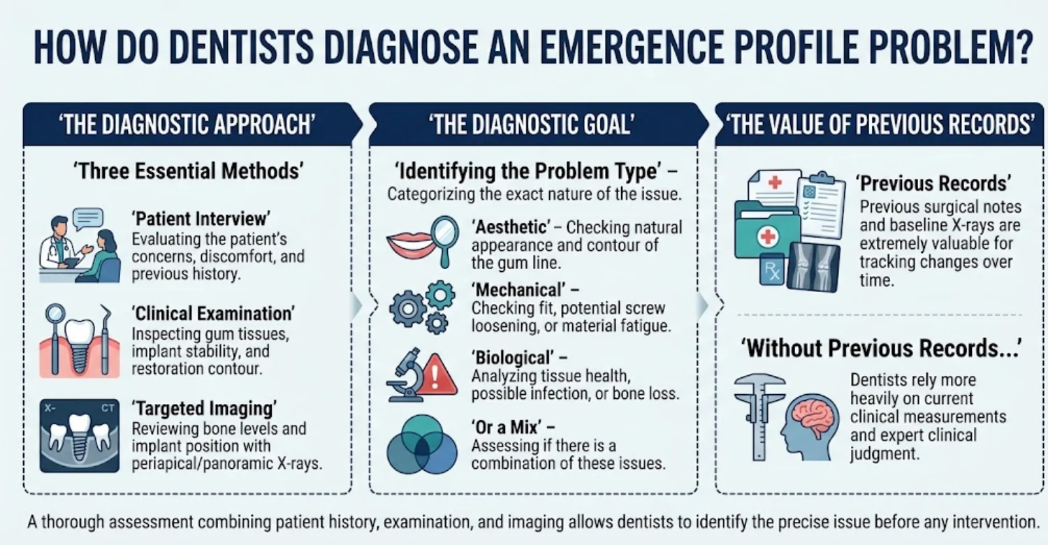
What Does a Dentist Examine During an Implant Restoration Assessment?
The dentist first asks about symptoms, timing, cleaning habits, and medical history. They then look at gum color, swelling, recession, and tissue thickness. Contact points are tested with floss to check for food traps. The bite is assessed for heavy or uneven contacts. Photographs often record the gumline for comparison with the opposite side or future visits.
How Dentists Evaluate the Implant Crown, Abutment, and Surrounding Gingiva
The crown is checked for overhangs, ledges, rough surfaces, and margin position. Dentists assess whether floss or interdental brushes can pass smoothly around it. They examine the abutment connection for looseness or gaps where visible. The gum is evaluated for bleeding, pus, blanching, and keratinized tissue width. Together, these checks show whether the Dental Implant Emergence Profile supports or stresses the tissue.
When Are Periodontal Probing and Implant Stability Assessments Appropriate?
Gentle probing is considered safe and useful around implants. It measures pocket depth and detects bleeding or pus. The 2017 World Workshop suggested probing depths of 6 mm or more, with bone loss, indicate likely peri-implantitis. Dentists compare readings with baseline values whenever possible. Stability testing checks whether the crown, abutment, or implant itself moves. Any implant mobility usually signals a serious problem requiring urgent evaluation.
How Dental X-Rays Help Assess Bone Levels and Restoration-Related Concerns
Periapical X-rays show bone levels along the implant threads. They can also reveal excess cement, open margins, or poorly seated components. Traditional success criteria accepted up to about 1.5 mm of bone remodeling in the first year. After that, ongoing loss should usually remain minimal. Comparing new images with baseline X-rays shows whether bone levels are stable or changing.
When Are CBCT Scans Necessary, and When Are They Not?
Cone-beam CT provides three-dimensional images of bone and implant position. It is helpful for assessing facial bone thickness, implant angle, or planned surgery. However, metal implants can create scatter that obscures fine detail. CBCT also involves a higher radiation dose than a single periapical X-ray. Guidelines therefore recommend CBCT only when it is likely to change treatment decisions.
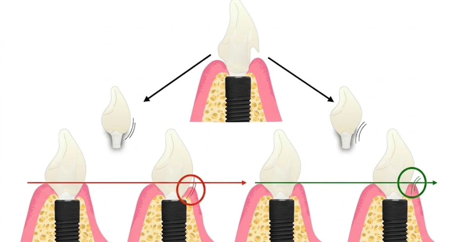
How Dentists Distinguish Mechanical, Aesthetic, and Biological Complications
Mechanical complications include loose screws, fractured porcelain, or broken components. One review reported abutment screw loosening in about 9% of single crowns over five years. Aesthetic complications involve color, shape, gum height, and symmetry. Biological complications include mucositis, peri-implantitis, and recession. Many cases combine categories, so each is assessed separately before treatment planning.
Why a Clinical Examination Is Needed Before Changing the Implant Crown
Replacing a crown without diagnosis can waste time and money. If bone loss or infection is present, a new crown will not resolve it. If the implant is malpositioned, contour changes may have limited benefit. Examination also reveals whether surrounding teeth need treatment. A proper diagnosis ensures the chosen correction addresses the real cause.
How Can a Poorly Designed Dental Implant Emergence Profile Be Corrected?
Correction ranges from simple polishing to surgery and a new restoration. The right option depends on the cause, the tissue condition, and the implant position. Many cases improve with conservative steps. Others need staged treatment over several months. A clear diagnosis always comes first.
Can a Dentist Reshape an Implant Crown to Improve Its Emergence Profile?
Yes, in selected cases a crown can be reshaped. Screw-retained crowns are easier to adjust because they can be removed without damage. The dentist or laboratory reduces bulky areas, smooths ledges, and repolishes the surface. Polishing matters because rough surfaces attract more plaque. Cemented crowns are harder to modify safely and may need replacement instead.
When Is Crown Contouring Enough, and When Is Crown Replacement Necessary?
Contouring works best for mild overbulking with healthy tissue and a well-placed implant. Replacement is usually better when margins are open, porcelain is damaged, or bulk is extensive. A new crown is also needed when shade or shape cannot be corrected by adjustment. Replacement allows a fresh digital design using the current tissue scan. It also offers an opportunity to switch from cement to screw retention.
How Abutment Modification Can Improve Restoration Contour
The abutment shapes the deeper part of the Dental Implant Emergence Profile. Replacing a stock abutment with a custom one can remove pressure points. A slimmer, more concave transmucosal section gives tissue room to thicken. Switching titanium for zirconia may reduce gray discoloration in thin tissue. Taller abutments may move restoration margins further from bone.
When Customized Provisional Crowns Can Help Reshape the Gum
A provisional crown lets the dentist retrain the gum before committing to a final design. It is especially useful in aesthetic cases with uneven margins or flat papillae. The contour is adjusted gradually over several visits. Once the tissue stabilizes, the shape is copied into the definitive crown. This phase commonly lasts about two to three months.
When Soft-Tissue Contouring or Gum Grafting May Be Considered
Grafting is considered when tissue is too thin, recession has occurred, or keratinized tissue is lacking. Connective tissue grafts, often taken from the palate, are commonly used to add thickness. Reviews report thickness gains of roughly 1 mm or more in many cases. Collagen substitutes can avoid a second surgical site in suitable patients. Minor gum contouring may refine irregular margins where tissue is abundant.
How Peri-Implant Mucositis or Peri-Implantitis Is Managed When Present
Mucositis is usually managed with professional cleaning, improved home care, and correction of plaque traps. It is often reversible within weeks. Peri-implantitis requires more intensive care. Treatment may include non-surgical debridement, surgical access, and surface decontamination. Regenerative or resective surgery may follow, depending on bone defect shape. Supportive maintenance every three to six months is then essential.
Why Treatment Depends on the Cause Rather Than Appearance Alone
Two crowns may look equally unattractive for completely different reasons. One may be overcontoured, while another sits on a poorly placed implant. Treating appearance without understanding the cause often leads to disappointing results. A cause-based plan targets the real problem first. This approach also helps control cost by avoiding unnecessary procedures.
What Relief and Medical Tips Can Help if the Gum Around an Implant Is Irritated?
Mild irritation can sometimes be eased with gentle home care. However, home measures support professional treatment rather than replace it. Persistent symptoms around a Dental Implant Emergence Profile usually need examination. The tips below are general guidance, not a personal treatment plan.
What to Do if the Implant Crown Feels Bulky or Traps Food
First, note exactly where food collects and when it happens. Remove trapped food gently with floss, an interdental brush, or a water flosser. Avoid toothpicks that may scratch or injure the gum. Do not ignore repeated trapping, because it often signals a contact or contour issue. Book an appointment so the dentist can check the crown and neighboring teeth.
How to Clean Around an Implant Crown Without Injuring the Gum
Use a soft-bristled or electric toothbrush angled gently toward the gumline. Interdental brushes in the correct size often clean implant spaces better than string floss. Implant-specific floss with a spongy section can help around bulky contours. One study found water flossers reduced bleeding at about 82% of implant sites. String floss achieved this at roughly 33%. Avoid metal instruments and abrasive whitening pastes.
Can Salt-Water Rinses or Pain Relief Help With Temporary Post-Treatment Discomfort?
Warm salt-water rinses can soothe tissue after adjustments or minor procedures. A common mix is half a teaspoon of salt in a cup of warm water. Rinse gently, without forceful swishing, several times a day. Over-the-counter pain relief may help with short-term tenderness. These measures should ease discomfort within a few days. If symptoms persist or worsen, contact your dentist.
Which Pain-Relief Medicines Should Be Discussed With a Dentist or Pharmacist?
Common options include ibuprofen and paracetamol, also called acetaminophen. Always follow the label and do not exceed the stated dose. Ibuprofen may be unsuitable for people with stomach ulcers, kidney disease, or certain asthma types. It can also interact with blood thinners and some blood-pressure medicines. Pregnant patients should seek advice before taking any medicine. A pharmacist can confirm what is safe for you.
Why You Should Not File, Adjust, or Loosen an Implant Crown at Home
Implant components are precision parts with very tight tolerances. Filing a crown can expose rough surfaces that collect more plaque. Attempting to loosen it may damage the screw, abutment, or implant connection. Home adjustments can also change your bite and overload the implant. Any change to contour or fit should be made in a dental setting.
Why Antibiotics Should Not Be Used Without Professional Assessment
Most gum irritation around implants is caused by plaque, cement, or contour problems. Antibiotics do not remove these causes. Using leftover or borrowed antibiotics can hide symptoms while the problem progresses. The World Health Organization ranks antimicrobial resistance among the top global public health threats. Your dentist will prescribe antibiotics only when they are clearly indicated.
When Persistent Irritation Means You Need a Dental Examination
Seek an examination if bleeding continues for more than about two weeks despite careful cleaning. Swelling, pus, a bad taste, or increasing pain also need assessment. A crown that feels loose or a changing bite should not be ignored. Early evaluation often allows simpler and less expensive treatment. Delaying care can allow mucositis to progress toward peri-implantitis.
Note : All images used are for editorial and illustrative purposes only and may not originate from the original news provider or associated company.
What Are the Possible Side Effects and Risks of Emergence Profile Correction?
Every corrective procedure carries some risk, even when carefully performed. Most side effects are mild and temporary. A few can affect the final aesthetic result or require further treatment. Understanding these risks helps patients make informed decisions about correcting a Dental Implant Emergence Profile.
Temporary Tenderness, Swelling, or Bleeding After Soft-Tissue Procedures
Mild tenderness is common for a few days after contouring or grafting. Swelling usually peaks around 48 to 72 hours, then gradually settles. Light bleeding or pink saliva may appear during the first day. Cold compresses on the cheek and soft foods can help. Your dentist will provide specific aftercare instructions and medication advice.
Gum Recession, Contour Changes, or Unsatisfactory Aesthetic Results
Soft tissue does not always respond exactly as planned. Some recession may continue during healing, especially in thin tissue. Papillae may fill only partly, leaving small dark triangles. Gum texture or color may differ slightly from neighboring areas. Patients should understand that improvement is likely, but perfect symmetry cannot always be guaranteed.
Risks Associated With Unnecessary Crown Adjustment or Replacement
Removing a crown repeatedly can disturb the soft-tissue seal around the abutment. Some research suggests frequent abutment disconnection may increase early bone remodeling. Adjustments can also weaken thin ceramic or expose rough surfaces. Replacement adds laboratory costs and extra appointments. For these reasons, dentists avoid crown changes unless a clear benefit is expected.
Potential Complications of Soft-Tissue Grafting or Surgical Contouring
Palatal donor sites can be sore for one to two weeks. Free gingival grafts may shrink considerably, with some studies reporting losses near 40%. Infection, graft failure, and delayed healing are uncommon but possible. Smokers and people with poorly controlled diabetes face higher complication rates. Collagen substitutes may reduce donor-site pain but are not suitable for every case.
How Proper Case Selection and Follow-Up Can Help Manage Treatment Risks
Risk management starts with choosing the right treatment for the right patient. Dentists assess medical history, smoking status, and tissue quality before surgery. Staged treatment allows healing to be checked before the next step. Follow-up visits catch early problems, such as infection or graft movement. Clear communication also helps patients follow aftercare correctly.
Ready to transform your smile?
Talk to our team and get a personal FREE treatment plan.
How Can You Prevent Dental Implant Emergence Profile Problems?
Prevention begins with careful planning and continues for the life of the implant. Surgeons, restorative dentists, technicians, and patients all play a part. Good planning reduces design errors, while good maintenance catches early changes. Together, they protect both appearance and long-term tissue health.
Why Implant Position and Soft-Tissue Assessment Matter Before Surgery
Most contour problems are easier to prevent than to correct. Pre-surgical scans show bone width, height, and the location of nerves and sinuses. Dentists also assess gum thickness and keratinized tissue width. Digital planning places the implant according to the future crown. If tissue is lacking, grafting can be planned early. This preparation creates room for a gradual, cleansable contour.
How Proper Crown Design Supports Long-Term Cleaning and Maintenance
A preventive Dental Implant Emergence Profile keeps the deeper zone slim and avoids unnecessary bulk. Margins are placed where excess cement can be removed, or screw retention is used. Contact points are designed to stop food packing. Surfaces are highly polished to resist plaque. The technician also leaves access for interdental brushes. Good design makes daily care realistic rather than frustrating.
The Role of Daily Brushing, Interdental Cleaning, and Professional Maintenance
Brush twice daily for about two minutes, focusing on the gumline around the implant. Clean between teeth once daily with interdental brushes, implant floss, or a water flosser. Professional cleanings remove hardened deposits with implant-safe instruments. A five-year study by Costa and colleagues showed the value of maintenance. Peri-implantitis affected about 44% of patients without maintenance, versus 18% with regular care.
Why Regular Implant Reviews Help Detect Changes Early
Implant problems are often painless in early stages. Bleeding and slight pocket deepening may be the only warning signs. Regular reviews let dentists compare probing depths, photographs, and X-rays with baseline records. Most patients benefit from reviews every three to six months, depending on risk. Early detection usually means simpler and cheaper treatment.
How Smoking, Gum Disease History, and Oral Hygiene Can Affect Implant Maintenance
Smoking reduces blood flow to the gums and slows healing. Meta-analyses suggest smokers have roughly twice the implant failure risk of non-smokers. A history of periodontitis also increases peri-implantitis risk. Poor plaque control remains one of the strongest risk indicators. Stopping smoking, treating gum disease, and keeping regular reviews significantly improve long-term outcomes.
How Much Does Dental Implant Emergence Profile Treatment Cost in the US, UK, and Turkey?
Dental implant emergence profile design is rarely priced as a standalone service. It is generally incorporated into implant restoration planning, abutment selection, and crown fabrication. Costs vary by country, implant system, materials, and clinical complexity. The following figures are indicative planning estimates in US dollars, not personalized quotes.
Average Dental Implant Emergence Profile Treatment Costs by Country
Country | Estimated Cost (Single Implant, Abutment, and Crown) | Average / Midpoint (USD) | Estimated Difference vs. US Midpoint |
United States | 3,000–6,000 | $4,500 | Baseline |
United Kingdom | 1,800–3,500 | $2,650 | Approximately 41% lower |
Turkey | 450–1,100 | $775 | Approximately 83% lower |
Note: Midpoints are calculated from the stated ranges, not verified national averages. These figures cover the complete single-implant restoration rather than emergence profile design alone. Actual costs depend on the treatment package and clinical requirements.
Is Dental Implant Emergence Profile Design Charged Separately?
In most clinics, emergence profile design is incorporated into the restorative workflow rather than charged as a separate service. The contour is developed through the shape of the abutment and crown, with digital planning and laboratory work contributing to the final restoration.
Additional procedures, such as custom abutment fabrication, provisional crown shaping, crown replacement, or soft-tissue grafting, may increase the overall cost.
Why Profile Design Is Usually Included in Implant Abutment and Crown Planning
Emergence profile design influences how the restoration transitions from the implant area through the gum tissue to the crown. Because this contour is created during restorative planning and fabrication, its cost is commonly included in the abutment and crown fees. Custom components and additional provisional restorations may incur extra charges.
When Crown Replacement, Gum Grafting, or Additional Procedures Can Increase the Total Cost
Corrective procedures can increase the original treatment budget. Replacing a crown or abutment may involve additional laboratory and clinical fees. Soft-tissue grafting, bone grafting, or treatment for peri-implant disease may also add costs. The amount depends on the procedure, materials, and complexity of the case.
What Is the Average Cost of a Single Dental Implant With an Abutment and Crown in the US?
In the United States, the estimated cost of a single dental implant with an abutment and crown ranges from $3,000 to $6,000. The final fee depends on the location, clinician, implant system, laboratory work, and whether additional procedures are needed.
Average US Dental Implant Restoration Costs
Cost Measure | Estimated Cost (USD) |
Lower end of the range | $3,000 |
Upper end of the range | $6,000 |
Calculated midpoint | $4,500 |
The $4,500 midpoint provides a planning reference for a standard case without major additional procedures. Imaging, temporary restorations, custom components, or grafting may increase the total. Patients should request an itemized estimate that identifies which services are included.
What Is the Average Cost of a Single Dental Implant With an Abutment and Crown in the UK?
In the United Kingdom, private dental implant treatment costs vary by clinic and region. The estimated range for a single implant, abutment, and crown is 1,800–3,500, although diagnostic imaging and additional procedures may be charged separately.
Average UK Dental Implant Restoration Costs
Cost Measure | Estimated Cost (USD) |
Lower end of the range | $1,800 |
Upper end of the range | $3,500 |
Calculated midpoint | $2,650 |
The calculated midpoint is approximately 41% lower than the US midpoint provided above. However, this is a comparison of estimates rather than a verified national average. Patients should confirm whether consultation, CBCT imaging, provisional crowns, and follow-up appointments are included.
What Is the Average Cost of a Single Dental Implant With an Abutment and Crown in Turkey?
Turkey offers a range of dental implant treatment options for local and international patients. The estimated cost of a single implant with an abutment and crown ranges from $450 to $1,100. Prices vary according to the implant brand, crown material, clinic, and treatment requirements.
Average Turkey Dental Implant Restoration Costs
Cost Measure | Estimated Cost (USD) |
Lower end of the range | $450 |
Upper end of the range | $1,100 |
Calculated midpoint | $775 |
The $775 midpoint is approximately 83% lower than the stated US midpoint and 71% lower than the UK midpoint. These are illustrative comparisons based on the supplied ranges, not guaranteed savings. International patients should also account for travel, accommodation, and arrangements for follow-up care.
Which Factors Influence the Final Implant Restoration Cost?
Several factors affect the cost of a dental implant restoration and its emergence profile. Understanding these variables helps patients compare quotations more accurately.
Implant Brand, Abutment Type, and Crown Material
The implant system, abutment design, and crown material all influence the total cost. Custom titanium or zirconia abutments may cost more than standard components because of their design and manufacturing requirements. Crown materials, including monolithic zirconia, layered zirconia, and lithium disilicate, also vary in price and aesthetic characteristics.
Customized Contouring, Temporary Restorations, and Laboratory Fees
Customized contouring may involve additional digital design, laboratory work, and clinical adjustments. Provisional crowns can be used to guide soft-tissue shaping before the definitive restoration is placed. These additional stages may increase costs, particularly when detailed aesthetic planning is required for a front-tooth implant.
Bone Grafting, Soft-Tissue Grafting, or Additional Surgery
Some patients require bone grafting or soft-tissue grafting to support the implant restoration and surrounding tissues. Other patients may need corrective treatment for gum recession or peri-implant disease. Such procedures are generally priced separately and may extend the treatment timeline.
Diagnostic Imaging, Follow-Up, Travel, Accommodation, and Aftercare
CBCT scans, X-rays, clinical reviews, and maintenance appointments may be included in the treatment package or charged separately. International patients should also consider travel and accommodation expenses, as well as access to follow-up care after returning home.
What Should Patients Check Before Accepting an Implant Treatment Quote?
A low headline price does not necessarily represent the total cost. Request a written, itemized treatment plan and confirm what is covered before accepting a quotation.
Whether the Price Includes Surgery, Implant Fixture, Abutment, and Final Crown
Check whether the quote covers implant placement, the implant fixture, the abutment, the final crown, and any required imaging. Confirm the implant system and restoration materials specified in the treatment plan.
Whether Corrective Contouring or Replacement Is Charged Separately
Ask whether crown adjustments, replacement restorations, custom abutments, and provisional crown stages are included. Some clinics may cover certain adjustments under their warranty terms, while others charge additional fees.
How Follow-Up and Complication Management Will Be Arranged
International patients should clarify how follow-up appointments will be organized and who will manage complications. Request copies of implant records, component details, and treatment documentation for future dental care. These records can help another clinician maintain and assess the restoration.
Key Takeaways: Average Dental Implant Emergence Profile Treatment Costs
Country | Estimated Treatment Range (USD) | Calculated Midpoint (USD) |
United States | 3,000–6,000 | $4,500 |
United Kingdom | 1,800–3,500 | $2,650 |
Turkey | 450–1,100 | $775 |
Dental implant emergence profile design is generally incorporated into abutment and crown planning, so a separate average price for the design alone may not be available. The figures above represent estimated costs for a complete single-implant restoration. Patients should compare itemized treatment plans, materials, aftercare, and any additional procedures before making a decision.
At Vitrin Clinic Treatment Pricing
The cost of dental implant treatment varies significantly depending on the procedure scope, implant brand, and country of treatment. The following table provides a transparent comparison of average costs across the United Kingdom, United States, and Vitrin Clinic in Istanbul from a single implant to full-arch restoration allowing patients to evaluate their options with accurate financial context before committing to a treatment plan.
Treatment type | 🇬🇧 UK | 🇺🇸 USA | 🇹🇷 Turkey |
|---|---|---|---|
Single dental implant Implant post + abutment + crown | $2,400 – $5,700 | $3,000 – $6,000 | $500 – $700 |
Implant-supported crown Crown on existing implant | $1,270 – $2,540 | $1,500 – $3,000 | $700 – $900 |
All-on-4 Per arch, fixed full-arch restoration | $10,000 – $22,000 | $18,000 – $30,000 | $3,500 – $7,500 |
All-on-6 Per arch, 6-implant full-arch restoration | $12,700 – $25,400 | $20,000 – $35,000 | $4,000 – $9,000 |
Full mouth reconstruction Both arches, upper + lower | $29,000 – $50,000+ | $36,000 – $70,000+ | $7,000 – $15,000 |
3D CT scan & consultation | Charged separately | Charged separately | Free & included |
Estimated saving | — | — | Up to 70% less |
* Prices are averages for reference only and may vary based on individual case requirements, implant brand, bone condition, and treatment complexity. A personalised quote is provided following your free consultation.






Tips for Patients
Informed patients usually achieve better long-term results with implant restorations. Asking the right questions helps you understand your options and responsibilities. Clear communication also helps your dentist identify problems early. The tips below are practical steps you can take at each stage.
Questions to Ask Before Approving Your Final Implant Crown
• Will my crown be screw-retained or cemented, and why?
• Is the abutment custom-made or stock?
• How will I clean around this crown at home?
• Will a provisional crown be used to shape my gum first?
• What warranty covers the crown, abutment, and adjustments?
• How will my Dental Implant Emergence Profile be checked over time?
What to Tell Your Dentist About Gum Discomfort, Food Trapping, or Cleaning Difficulty
Describe exactly where the problem occurs and how long it has lasted. Mention whether bleeding happens with brushing, flossing, or spontaneously. Note which foods get trapped and on which side. Explain any cleaning tools that shred, catch, or cause pain. Photographs taken at home can also help your dentist track changes.
How to Prepare for an Implant Restoration Appointment
Bring previous X-rays, implant cards, and treatment records if you have them. List your medicines, allergies, and any medical changes. Brush and clean between your teeth carefully before the visit. If you are traveling for treatment, allow extra days for adjustments. Prepare your questions in advance so nothing is forgotten.
What to Expect During Recovery After Crown Adjustment or Gum Contouring
Simple crown adjustments usually cause little or no discomfort. Gum contouring or grafting may cause tenderness for several days. Eat soft foods and avoid chewing directly on the treated area. Follow cleaning instructions carefully, as some areas may need gentle care initially. Most minor soft-tissue procedures heal substantially within one to two weeks.
Warning Signs That Require Prompt Dental Evaluation
Some symptoms should not wait for your next routine review. Contact your dentist promptly if you notice any of the following signs. Early care can prevent minor problems from becoming serious.
Persistent or Increasing Gum Swelling, Bleeding, or Pus
Bleeding that continues for more than about two weeks needs assessment. Pus always indicates infection and requires prompt care. Increasing swelling may signal worsening inflammation around the implant.
Worsening Pain, Fever, or a Persistent Unpleasant Taste
Pain should improve after treatment, not worsen. Fever may indicate a spreading infection. A constant bad taste can suggest discharge from the implant site.
A Loose Crown, Movement of the Implant, or Sudden Changes in the Bite
A loose crown often means a loose screw, which is usually fixable. Movement of the implant itself is more serious. Sudden bite changes may overload the implant and need quick review.
Facial Swelling or Difficulty Swallowing or Breathing Requiring Urgent Care
Facial swelling spreading toward the eye or neck needs same-day attention. Difficulty swallowing or breathing is a medical emergency. Call emergency services or go to the nearest emergency department immediately.
At Vitrin Clinic: Personalized Dental Implant Emergence Profile Planning
At Vitrin Clinic, implant contour is treated as part of the full treatment plan, not a final detail. Our team considers the implant, gum, crown, and your cleaning ability together. Each Dental Implant Emergence Profile is planned around your anatomy and goals. This approach supports results that look natural and remain maintainable.
Assessing Implant Position, Crown Contour, and Surrounding Soft Tissue
Every assessment begins with a careful clinical examination. Our dentists evaluate implant position, gum thickness, recession, and papilla fill. Existing crowns are checked for bulk, margins, contacts, and cleaning access. We also review bite forces and the condition of neighboring teeth. These findings help separate design issues from biological problems.
Using Digital Scans and Dental Imaging When Clinically Appropriate
Digital intraoral scans record your teeth and gum contours with high detail. X-rays help assess bone levels around existing implants. CBCT imaging is recommended only when it is likely to change treatment decisions. This keeps radiation exposure as low as reasonably achievable. Digital records also support communication with our dental laboratory.
Planning Implant Crown Shape Around Gum Health, Aesthetics, and Cleanability
Our planning balances three priorities: healthy tissue, natural appearance, and practical home care. In aesthetic cases, provisional crowns may be used to guide the gum before the final design. In back teeth, cleansable contours and secure contacts usually take priority. Each design is reviewed with you before final fabrication.
Evaluating Whether Contour Adjustment, Crown Replacement, or Soft-Tissue Treatment Is Appropriate
When a restoration needs correction, we first identify the underlying cause. Minor overcontouring may respond to careful reshaping and polishing. Larger problems may require a new abutment or crown. Thin tissue or recession may need soft-tissue treatment first. We explain why each option is or is not recommended.
How Individualized Treatment Planning Helps Set Realistic Aesthetic Expectations
Not every implant site can achieve a perfect result. Bone loss, thin tissue, or implant position may limit what is possible. We discuss these limits openly before treatment begins. Photographs and digital previews help you visualize realistic outcomes. Clear expectations support better satisfaction and informed decisions.
How Vitrin Clinic Supports Your Treatment Journey
Implant treatment often involves several stages and appointments. Our team coordinates each step so you know what comes next. Support covers assessment, planning, treatment, and aftercare. This structure is especially helpful for international patients.
Initial Consultation and Review of Existing Implant Records
Your journey starts with a consultation and review of any existing records. Previous X-rays, implant cards, and treatment notes are especially helpful. These records show which implant system and components were used. They also allow comparison of current and past bone levels. Patients can usually share photographs and records before arriving.
Coordinating Digital Diagnostics and Clinical Assessments
We schedule scans, imaging, and examinations efficiently to minimize repeat visits. Results are reviewed together to build one coordinated plan. When specialist input is needed, our team arranges it. This coordination helps prevent delays and conflicting advice.
Explaining Available Treatment Options, Expected Stages, and Cost Considerations
After assessment, we explain your options in clear, everyday language. Each plan outlines stages, expected healing times, and likely appointments. Written cost estimates show what is included and what may cost extra. You are encouraged to ask questions before making decisions.
Coordinating Appointments for International Patients
International patients often have limited time for treatment visits. Our team plans appointment sequences around healing requirements and travel dates. We advise on how many days to allow for each stage. Where staged treatment is needed, return visits are planned in advance.
Arranging Follow-Up Guidance and Ongoing Implant Maintenance Planning
Follow-up care protects your investment after treatment. We provide home-care instructions tailored to your Dental Implant Emergence Profile and cleaning tools. Patients receive records and component details to share with their local dentist. We also recommend a maintenance schedule based on individual risk.
Ready to transform your smile?
Talk to our team and get a personal FREE treatment plan.
Key Takeaways
Implant success depends on more than osseointegration. The shape where the restoration meets the gum influences health, appearance, and maintenance for years. The points below summarize the most important lessons from this guide.
Why Dental Implant Emergence Profile Design Matters for Gum Health and Natural Appearance
Contour guides how the gum heals, seals, and looks around the crown. A gradual, cleansable shape supports stable tissue and easier cleaning. With peri-implant mucositis affecting roughly 43% of implants in one meta-analysis, cleansability matters.
Which Design Factors Influence Healing, Cleaning Access, and Restoration Stability
Implant depth, position, tissue thickness, abutment height, and crown shape all interact. Research associates emergence angles above about 30 degrees with higher peri-implantitis rates. Provisional crowns help test and refine contour before the final restoration.
Why Complications Require Assessment Before Corrective Treatment
Bleeding, recession, and food trapping can share symptoms but have different causes. Excess cement, contour errors, tissue deficiency, and peri-implantitis each need different treatment. Diagnosis prevents unnecessary crown replacements and wasted costs.
How to Compare Implant Restoration Costs Across the US, UK, and Turkey
Indicative midpoints are about $4,500 in the US, $2,650 in the UK, and $775 in Turkey. Always compare what each quote includes. Factor in travel, grafting, follow-up, and the cost of any future corrections to your Dental Implant Emergence Profile.
When to Seek Professional Dental Care for Persistent Symptoms or Warning Signs
See a dentist if bleeding lasts beyond about two weeks or pus appears. A loose crown, worsening pain, or bite changes also need prompt review. Facial swelling with breathing or swallowing difficulty requires emergency care.
At Vitrin Clinic
At Vitrin Clinic, dental implant emergence profile design focuses on how the crown transitions through the surrounding gum tissue. Our team evaluates implant position, crown contour, and gum health to help address bulky restorations, food trapping, or unnatural appearance. Using individualized assessment and digital planning where appropriate, we develop a restoration plan tailored to each patient's needs. Book your FREE TREATMENT for a personalized implant assessment.
Reference
FAQs
Dr. Rifat Alsaman has more than 5 years of clinical experience in dentistry and currently serves as the Head of the Medical Team at Vitrin Clinic. He is dedicated to providing exceptional patient care, overseeing treatment planning, and ensuring the highest clinical standards across the team. His expertise, attention to detail, and commitment to continuous professional development have helped countless patients achieve healthier, more confident smiles.


