Endodontics
June 7, 2026
Direct Pulp Capping Materials: Which Ones Preserve Tooth Vitality?
 Dr. Rifat Alsaman
.webp&w=3840&q=75)
Preserving a living tooth is one of dentistry's most important clinical priorities. When the dental pulp is exposed or endangered due to deep decay or trauma, selecting the right direct pulp capping materials becomes critical. These specialized biocompatible substances are placed directly over exposed pulp tissue. They stimulate natural healing, prevent bacterial invasion, and encourage the formation of a protective dentin bridge. Understanding which materials perform best helps patients and clinicians make well-informed decisions about vital pulp therapy.
How Direct Pulp Capping Materials Help Avoid Root Canal Treatment
Direct pulp capping materials help avoid root canal treatment by protecting and preserving the living dental pulp after minor exposure. Materials such as MTA, calcium hydroxide, and bioceramics create a seal that prevents bacterial invasion. They also encourage dentin repair. By supporting pulp healing and maintaining tooth vitality, these materials allow the natural tooth structure and nerve function to remain intact.
Pulp Preservation in Modern Dentistry
Modern dentistry has shifted dramatically toward minimally invasive approaches. These approaches prioritize preserving natural tooth structure and living pulp tissue. Rather than defaulting to root canal treatment at the first sign of deep decay, clinicians now evaluate vital pulp therapy first. The strategic application of direct pulp capping materials can maintain pulp health long-term. This approach reduces treatment complexity and lowers overall patient costs. It also preserves the tooth's biological function and structural integrity for many years or potentially decades of continued service.
The Biological Goal of Vital Pulp Therapy
The primary objective of vital pulp therapy is to keep the dental pulp alive and biologically active within the tooth. When pulp tissue is healthy or only mildly inflamed, it has a remarkable capacity for self-repair through the production of tertiary dentin. Direct pulp cap materials support this process. They create a biocompatible barrier that stimulates odontoblastic activity and promotes dentin bridge development. They also shield pulp cells from ongoing microbial threats. Left unmanaged, these threats could lead to irreversible pulpitis or complete pulp necrosis.
When Dentists Consider Direct vs. Indirect Pulp Capping
Dentists choose between direct and indirect approaches based on clearly defined clinical indicators. Direct pulp capping using carefully selected direct pulp capping materials is indicated when actual pulp exposure occurs due to mechanical accident or caries removal, and the pulp appears healthy. In contrast, indirect pulp capp materials are employed when a thin residual dentin layer remains over the pulp. Direct contact with the pulp is avoided in these cases. Patient age, symptom history, and vitality testing results guide this important clinical decision.
The Historical Evolution of Pulp Capping Materials: From Early Formulas to Bioactive Cements
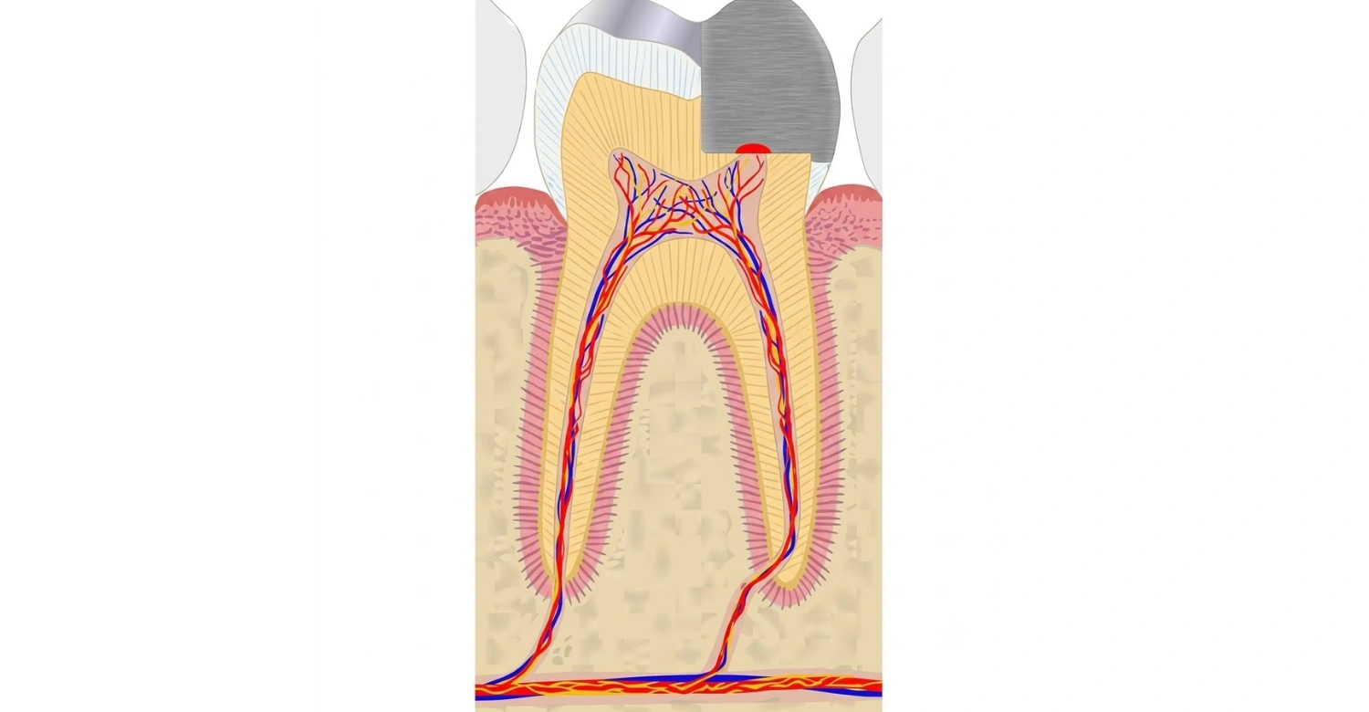
The evolution of dental pulp capping materials spans more than a century of clinical trial and refinement. Early 20th-century dentists relied on zinc oxide-eugenol pastes and simple lime compounds. Results were mixed and often unpredictable. Calcium hydroxide's introduction marked one of the first true milestones in vital pulp therapy development. It gave clinicians a material that reliably encouraged dentin bridge formation for the first time.
Direct Pulp Capping Materials: What Dentists Use Today
The range of available direct pulp capping materials has expanded significantly over the past two decades. Clinicians now have access to a spectrum of biocompatible substances, from traditional calcium hydroxide to advanced calcium silicate cements. Each has distinct biological profiles and clinical performance records. Selecting the most appropriate material depends on the degree of pulp exposure, the extent of bacterial contamination, available clinical technology, and the anticipated long-term prognosis. The goal is preserving the tooth's natural vitality and structural function throughout the patient's lifetime.
Calcium Hydroxide as a Traditional Direct Pulp Capping Material
Calcium hydroxide has historically been the most widely used direct pulp capping material in clinical dentistry worldwide. Its high alkalinity creates an antibacterial environment. It also stimulates the pulp to form a protective dentin bridge over the exposure site. Calcium hydroxide is still applied in some clinical contexts. However, it has documented limitations, including tunnel defects in the dentin bridge, material degradation over time, and inconsistent long-term success.
Calcium Silicate Cements in Contemporary Vital Pulp Therapy
Calcium silicate-based cements represent one of the most significant advances among direct pulp capping materials. These modern formulations create an excellent biological seal against bacterial microleakage. They consistently promote the formation of a thick, uniform dentin bridge. Long-term clinical studies report success rates significantly higher than those achieved with calcium hydroxide. Their outstanding biocompatibility and ability to set reliably in a moist environment make them the preferred choice in contemporary vital pulp therapy protocols worldwide.
Advanced Calcium Silicate Formulations
Newer generations of calcium silicate-based direct pulp capping materials have been developed to further improve on earlier formulations. These materials set more rapidly and offer superior mechanical strength. They also release calcium ions that actively promote dentin remineralization at the exposure site. Other advanced bioactive alternatives available in current clinical practice have also shown promising results in clinical and laboratory settings. These options collectively represent the next generation of direct pulp cap materials. They combine strong biological activity with improved clinical workability for modern practice.
Resin-Modified Materials and Their Clinical Role
Resin-modified direct pulp capping materials offer unique advantages in specific clinical scenarios. They allow immediate light-curing after placement, simplifying the procedure and reducing chair time. These materials also release calcium and fluoride ions, providing measurable biological activity alongside mechanical stability. They are not universally preferred over pure calcium silicate cements. Still, resin-modified options serve as reliable and practical alternatives where faster workflow is a clinical priority.
Indirect Pulp Cap Materials and Their Conservative Approach
This article focuses on direct pulp capping materials. Still, understanding the complementary role of indirect pulp cap materials in conservative dentistry is equally important. Indirect pulp capping is applied when residual carious dentin is intentionally left over the pulp to avoid iatrogenic exposure. The objective is to allow remineralization of the remaining dentin while protecting the pulp from bacteria. This stepwise technique has proven effective in reducing the need for direct pulp intervention in many appropriately selected clinical cases.
When Indirect Pulp Capping Is Preferable to Direct Exposure
Indirect pulp capping is the preferred strategy when removing all carious dentin would likely risk or cause pulp exposure. This is particularly true in deep lesions approaching but not breaching the pulp. In these situations, indirect pulp capping materials are placed to seal the residual decay and encourage remineralization. They also allow clinicians to monitor pulp health over a defined period. This conservative approach avoids the complications associated with direct pulp exposure. It reduces the risk of irreversible pulpitis or heavy bacterial contamination of the pulp tissue. Effective materials often sway the decision when weighing pulp capping vs root canal therapies.
Glass Ionomer Cements in Indirect Pulp Capping
Glass ionomer cements and their resin-modified counterparts are among the most widely used indirect pulp capping materials in clinical practice today. They bond chemically to dentin and release fluoride ions that inhibit residual bacterial activity. They also actively support dentin remineralization processes. Their inherent biocompatibility and tolerance for moist environments make them ideal deep cavity liners. When applied as indirect pulp cap materials, they create a protective interface between residual carious dentin and the final overlying restoration. This effectively supports long-term pulp health preservation.
Bioactive Liners Used in Indirect Pulp Capping
Bioactive substances, including calcium silicate-based cements and calcium hydroxide-based materials, are also employed as indirect pulp capping materials. This is particularly true in very deep lesions where active pulp stimulation alongside protection is clinically desirable. These substances interact with dentin proteins to activate odontoblasts. They promote the formation of tertiary dentin. Their biological signaling properties make them superior to traditional cavity liners in deep indirect pulp capping scenarios. This delivers more predictable long-term outcomes for pulp health preservation in high-risk restorative cases.
Patient Concerns About Deep Cavities and Pulp Exposure
Patients diagnosed with deep cavities often experience significant anxiety about treatment outcomes. Common concerns include pain, the risk of tooth loss, and the possibility of requiring root canal therapy. Many remain unaware that advanced direct pulp capping materials now offer highly effective conservative solutions for managing pulp exposure. Understanding these options empowers patients to have informed conversations with their dentist. This reduces fear and improves compliance. Early professional intervention using the right materials can prevent minor decay from escalating into a dental emergency requiring more aggressive treatment.
Symptoms Patients Commonly Experience
Patients with decay approaching the pulp typically report heightened sensitivity to hot, cold, or sweet stimuli. Some experience spontaneous mild discomfort, lingering aches after eating, or sharp pain during chewing. These symptoms indicate proximity of the decay to the pulp. They signal the need for prompt professional evaluation. Early identification and treatment with direct pulp capping materials significantly raises the odds of preserving tooth vitality. This holds true compared to cases where treatment is delayed until severe or unprovoked pain has already developed and progressed. Dentists use highly specialized endodontic tools to place these materials precisely.
Fear of Root Canal Treatment and Tooth Loss
Fear of root canal treatment is among the most common reasons patients delay necessary dental care. This often allows a manageable situation to deteriorate. Understanding that direct pulp capping materials can, in many appropriate cases, prevent the need for root canal therapy helps substantially reduce this anxiety. Preserving the tooth's nerve and blood supply through vital pulp therapy maintains its natural function and resilience. Encouraging patients to seek timely professional evaluation transforms fear into confidence. It ensures they receive the least invasive treatment option available.
Patient Self-Assessment: Interactive Checklists for Evaluating Reversible vs. Irreversible Symptoms
A simple pulp vitality self assessment checklist can help patients gauge whether their symptoms suggest reversible or irreversible pulp inflammation. It never replaces a professional diagnosis. Reversible symptoms typically include brief cold sensitivity that fades within seconds, mild discomfort while chewing, and no spontaneous pain when nothing is touching the tooth.
Irreversible symptoms present differently. Patients may notice pain that lingers for minutes after a hot or cold stimulus, throbbing that starts without any trigger, or pain that wakes them at night. Swelling near the gum line or a loose feeling in the tooth are also warning signs. These should prompt an urgent appointment.
Tips for Patients
Patients preparing for a possible pulp capping procedure can take a few simple steps to support a smoother experience. Keeping a short symptom diary noting when sensitivity occurs and how long it lasts gives the dentist valuable diagnostic detail.
Avoiding very hot, cold, or sugary foods in the days before an appointment can reduce discomfort. It also prevents masking important symptoms. Patients should also mention any prior dental trauma to the tooth, even years-old trauma, since it can still affect pulp health today.
Clinical Procedure: How Direct Pulp Capping Is Performed
Successfully performing direct pulp capping requires precise clinical technique, appropriate material selection, and strict adherence to infection control principles. The procedure involves diagnosing pulp status, isolating the tooth, and removing decay. Direct pulp capping materials are then applied over the exposure site, and the tooth is sealed with a durable high-quality restoration. Every step must be executed carefully. This maximizes the biological conditions necessary for pulp healing and dentin bridge formation.
Diagnosis and Case Selection
Not every tooth with pulp exposure is suitable for direct pulp capping. Clinicians must carefully evaluate for signs of irreversible pulpitis. Warning signs include spontaneous pain, uncontrolled bleeding at the exposure site, or a large bacterially contaminated exposure. Ideal candidates include patients with healthy or reversibly inflamed pulp tissue, small mechanically created exposures, and no history of unprovoked pain. Radiographic assessment combined with pulp vitality testing provides the diagnostic foundation. This determines whether this conservative treatment approach is clinically appropriate before proceeding.
Isolation and Infection Control
Rubber dam isolation is considered essential during pulp capping procedures. The presence of saliva and oral bacteria at the exposure site dramatically increases the risk of pulpal infection and treatment failure. The exposure area must be disinfected using an appropriate antimicrobial agent before material application. Achieving hemostasis at the pulp exposure is equally critical. Active uncontrolled bleeding must be managed before direct pulp capping materials are placed. Proper isolation ensures a clean, uncontaminated operative environment. This creates optimal biological conditions for successful pulp healing.
Placement of Direct Pulp Cap Materials
Once the pulp exposure is cleaned and hemostasis is achieved, the selected direct pulp capping material is carefully applied. The material is gently condensed over the exposure using a small applicator. This ensures full coverage without excessive pressure that could damage the underlying pulp tissue. It must fully contact the pulp surface and extend slightly onto the surrounding dentin walls. For some calcium silicate cements, a moist cotton pellet is temporarily placed. This supports proper setting and maintains the moisture balance needed for optimal material performance.
Final Restoration and Sealing the Tooth
After placing the capping material, a high-quality final restoration is applied to create a bacteria-tight coronal seal. Composite resin or glass ionomer is placed over the direct pulp capping material. This restores the tooth's functional anatomy and protects the underlying treatment from microbial recontamination. A well-sealed restoration is critically important. Microleakage around restoration margins can reinfect the pulp and cause treatment failure, regardless of the quality of the underlying material. Regular clinical and radiographic follow-up confirms dentin bridge formation and verifies long-term pulp vitality.
Clinical Note
Not every case of pulp exposure responds the same way to treatment, even when the correct material and technique are used. Clinicians treat each diagnosis as an individual judgment call rather than a fixed protocol.
Follow-up remains just as important as the initial procedure. A tooth that looks successfully treated at one month can still show signs of pulpal breakdown later. This is why scheduled reviews are a required part of any pulp capping treatment plan, not an optional extra.
Benefits of Using Modern Direct Pulp Cap Materials
The clinical advantages of modern direct pulp capping materials over older treatment options are thoroughly documented in dental literature. Contemporary bioactive substances stimulate natural healing responses within the pulp and minimize postoperative sensitivity. They also achieve durable seals that resist bacterial infiltration effectively over time. Beyond immediate clinical outcomes, these materials support long-term tooth preservation. They help patients avoid the expense and complexity of root canal treatment and prosthetic restorations.
Preserving Natural Tooth Vitality
Maintaining the vitality of a natural tooth provides biological and functional benefits that no prosthetic option can fully replicate. A vital tooth retains its periodontal ligament. This supports alveolar bone integrity and provides proprioceptive feedback during biting and chewing. Modern bioactive materials preserve this vitality by protecting the pulp from further damage while stimulating its inherent repair mechanisms. Long-term retention of a vital natural tooth is consistently associated with superior overall oral health outcomes. This holds true compared to root canal-treated or prosthetically replaced alternatives across long-term patient follow-up studies.
Avoiding Root Canal Treatment When Possible
Root canal treatment, while clinically effective, involves permanent removal of the tooth's living pulp. It typically necessitates crown placement to protect the subsequently weakened tooth structure. By successfully applying direct pulp capping materials at the earliest sign of pulp exposure, clinicians can often eliminate the need for this more invasive procedure. Preserving the pulp maintains the tooth's natural resilience. It significantly reduces treatment cost and clinical complexity for the patient. This objective is a primary driver behind growing global interest in vital pulp therapy protocols today.
Promoting Dentin Bridge Formation
One of the most important biological outcomes of successful pulp capping is the formation of a reparative dentin bridge across the exposure site. This mineralized tissue layer is produced by stimulated odontoblast-like cells. It creates a natural barrier between the pulp and the overlying restorative material. Calcium silicate-based direct pulp capping materials produce thicker, more uniform dentin bridges compared to calcium hydroxide, which frequently develops tunnel defects. A well-formed dentin bridge dramatically improves long-term pulp protection. It also reduces the risk of delayed treatment failure.
What We Notice Clinically
Dr. Rifat Alsaman, Head of the Medical Team at Vitrin Clinic and a cosmetic dentist, notes that patients often underestimate how much material choice affects long-term comfort. In his clinical observation, cases treated with modern calcium silicate cements show noticeably less postoperative sensitivity compared with older calcium hydroxide protocols.
Dr. Rifat Alsaman also observes that early intervention consistently produces the best outcomes he sees in practice. According to Dr. Rifat Alsaman, patients who seek care at the first sign of sensitivity are far more likely to keep their natural tooth vital without ever needing a root canal, rather than waiting until pain becomes constant.
Advanced Technology Supporting Pulp Capping Success
Advances in diagnostic technology, imaging systems, and material science have significantly improved the predictability of pulp capping outcomes. Dentists today rely on digital caries detection tools, dental operating microscopes, and advanced direct pulp capping materials. These tools help achieve consistently higher success rates in vital pulp therapy. Together, these innovations allow clinicians to detect pulp exposure earlier and manage the operative environment more precisely. They also allow the use of bioactive substances that interact with pulp tissue in ways older calcium hydroxide-based agents were never designed to achieve.
Digital Caries Detection Tools
Digital caries detection technologies allow dentists to identify decay at very early stages, often before it becomes visible on conventional radiographs. Early detection means lesions can be managed conservatively before they endanger the pulp. When caries are identified late and pulp exposure becomes unavoidable, these digital tools improve depth assessment accuracy. This supports better case selection for vital pulp therapy. It also reduces the risk of proceeding with treatment in cases where the pulp has already undergone irreversible inflammatory changes that compromise outcomes.
Magnification and Dental Microscopes
Dental operating microscopes and high-magnification loupes have fundamentally transformed the level of precision achievable during pulp capping procedures. Under magnification, clinicians can clearly identify healthy versus inflamed pulp tissue. They can also achieve more effective hemostasis and place bioactive materials with far greater clinical accuracy. Microscopic visualization confirms complete material coverage of the exposure site. It ensures no residual contamination remains at the pulp interface before the restoration is placed. This level of procedural precision significantly improves long-term success rates beyond what unaided vision can achieve.
Bioactive Material Innovations
Ongoing research in material science continues to produce direct pulp capping materials that outperform conventional options in key biological properties. Next-generation calcium silicate compounds are being developed with faster setting times and superior biocompatibility. They also offer enhanced antimicrobial activity and better aesthetic characteristics. Some innovations incorporate growth factors or bioactive peptides that directly stimulate pulp cell differentiation and accelerate mineralization. These developments suggest the future of vital pulp therapy will be defined by increasingly responsive, biologically intelligent materials.
Minimally Invasive Dentistry Philosophy
Minimally invasive dentistry aims to preserve as much natural tooth structure as possible while effectively managing dental disease. Advanced direct pulp capping materials are central to this philosophy. They enable clinicians to treat pulp exposure without removing the pulp itself. Combining early caries detection with precise technique and innovative bioactive substances reduces the biological cost of dental treatment. It supports long-term tooth survival in ways that more aggressive interventions, such as pulpectomy or root canal treatment, cannot replicate for otherwise healthy, treatable pulp exposures in suitable patients.
Improved Bonding and Sealing Techniques
Advanced adhesive dentistry has complemented progress in bioactive materials by ensuring the overlying restoration provides a bacteria-tight interface. Modern bonding agents minimize gap formation at the dentin-restoration junction, one of the most common causes of failure following pulp capping. When excellent coronal sealing is consistently achieved alongside biologically active direct pulp capping material placement, the long-term prognosis for maintaining tooth vitality improves substantially. This reinforces the importance of treating every procedural step with equal clinical precision and attention.
Dental Care at Vitrin Clinic
Vitrin Clinic is committed to evidence-based, minimally invasive dental care that prioritizes every patient's long-term oral health. As part of this commitment, Vitrin Clinic offers advanced vital pulp therapy protocols using the most effective direct pulp capping materials currently available in modern clinical dentistry. Every treatment plan is built around preserving natural tooth structure, minimizing discomfort, and delivering durable outcomes. Patients choosing Vitrin Clinic benefit from a comprehensive, research-guided approach to tooth preservation delivered by an experienced and dedicated dental team.
Clinical Philosophy Focused on Tooth Preservation
At Vitrin Clinic, tooth preservation is not merely a clinical goal. It is a foundational philosophy embedded in every diagnostic and therapeutic decision the team makes. Every deep cavity and potential pulp exposure is evaluated with the objective of maintaining vitality wherever biologically feasible. The use of advanced direct pulp capping materials is integral to this approach. It allows clinicians to address pulp exposure conservatively and effectively. This commitment significantly reduces the rate of root canal treatment referrals. It supports consistently superior long-term patient outcomes across all restorative cases.
Use of Modern Bioactive Direct Pulp Cap Materials
Vitrin Clinic applies only the most advanced generation of direct pulp capping materials in clinical practice, including the latest calcium silicate-based formulations. These materials have demonstrated superior clinical performance compared to older alternatives. Documented benefits include higher success rates, more uniform dentin bridge formation, and improved long-term pulp health outcomes. Material selection at Vitrin Clinic is guided strictly by current scientific evidence. This ensures every patient receives treatment using agents proven to maximize the probability of preserving natural tooth vitality.
Patient-Centered Treatment Planning
Vitrin Clinic believes that well-informed patients make better, more confident treatment decisions. Before proceeding with any vital pulp therapy, the clinical team explains the diagnosis thoroughly. The team discusses all available options, including both direct and indirect pulp capping approaches, and outlines expected outcomes alongside potential risks. Patients are encouraged to ask questions, share concerns, and participate actively in their care planning. This transparent, collaborative model builds lasting trust. It ensures patients feel fully supported, informed, and empowered throughout their entire dental experience at Vitrin Clinic.
Advanced Equipment for Precision Care
Vitrin Clinic continuously invests in advanced dental technology to maintain the highest standards of clinical precision in every procedure performed. Digital radiography, intraoral cameras, dental operating microscopes, and cutting-edge diagnostic devices are all available to support accurate assessment and treatment delivery. When placing direct pulp capping materials, magnification and digital imaging enable greater procedural accuracy. They reduce the risk of error and optimize overall treatment outcomes. This commitment to technological excellence ensures patients at Vitrin Clinic consistently receive care that reflects current best practices in conservative and restorative dentistry.
Follow-Up Protocol for Long-Term Success
Long-term success following vital pulp therapy depends on systematic monitoring and timely intervention if complications develop. Vitrin Clinic follows a structured follow-up protocol incorporating clinical and radiographic assessments at defined post-treatment intervals. Patients are evaluated for dentin bridge formation, pulp vitality maintenance, and restoration integrity at each visit. This monitoring approach ensures early identification of any treatment complications. It protects the patient's tooth and maximizes the return on their investment in conservative dental care. The team at Vitrin Clinic remains committed to each patient's long-term dental health.
Book a FREE Consultation Today
If you are experiencing tooth sensitivity, have been informed you have a deep cavity, or are concerned about pulp exposure, do not delay seeking professional care. Vitrin Clinic offers free consultations to assess your dental health and determine whether vital pulp therapy is appropriate for your individual situation. The clinical team will evaluate your pulp status and explain all available treatment options clearly. They will guide you toward the least invasive solution. Take the first step toward preserving your natural smile: book your free consultation with Vitrin Clinic today.
Professional Guidance: When to Choose Direct vs. Indirect Pulp Capping
Deciding between direct and indirect pulp capping requires professional clinical judgment grounded in a thorough examination of the patient's specific dental condition. Not every deep cavity requires the same treatment approach. The decision must weigh the risk of pulp exposure against the biological status of the pulp itself. Both strategies, when correctly applied using appropriate materials and technique, can produce reliable long-term outcomes. They effectively reduce the need for more aggressive interventions such as root canal treatment or tooth extraction.
Dentist Evaluation Criteria
When evaluating a patient for pulp capping, clinicians assess multiple clinical factors: the size and location of the carious lesion, radiographic depth of invasion, presence or absence of spontaneous pain, and pulp vitality test results. The biological condition of the pulp at the moment of exposure is the single most critical indicator of long-term success. Specifically, clinicians look at whether bleeding is controlled and the tissue appears clinically healthy.
Age and Pulp Vitality Considerations
A patient's age significantly influences the expected prognosis of vital pulp therapy. Younger patients with large, healthy pulp chambers and abundant vascular supply respond more favorably to pulp capping than older patients with calcified or fibrosed pulps. In younger individuals, the pulp's regenerative capacity is considerably greater. This supports more predictable healing and dentin bridge formation. For older patients, clinicians must carefully evaluate pulp vitality before committing to a conservative approach. Reduced healing capacity may increase failure risk even when the most advanced materials are applied.
Extent of Decay and Bacterial Contamination
The extent of bacterial contamination at the pulp exposure site is among the most powerful predictors of treatment outcome. Small, clean mechanical exposures in healthy pulp tissue offer the most favorable prognosis. Large exposures resulting from extensive carious lesions carry higher bacterial loads. These loads compromise the pulp's healing capacity, even when highly biocompatible materials are carefully applied. In these situations, clinicians may need to consider more invasive alternatives such as pulpotomy or root canal treatment. Thorough debridement of all infected tissue before material placement remains essential.
About Vitrin Clinic
Vitrin Clinic is an international dental clinic based in Istanbul, Turkey, welcoming patients from around the world for both restorative and cosmetic treatments. The clinic combines advanced diagnostic technology with a patient-first philosophy. This philosophy is centered on preserving natural teeth whenever biologically possible.
Every treatment plan at Vitrin Clinic, including vital pulp therapy using modern direct pulp capping materials, is guided by the clinical team led by Dr. Rifat Alsaman, Head of the Medical Team at Vitrin Clinic. Patients traveling to Istanbul for treatment receive coordinated care from consultation through follow-up. They are supported by multilingual staff at every stage of their visit.
References:
https://pocketdentistry.com/pulp-capping-materials-for-the-maintenance-of-pulp-vitality/
FAQs
Dr. Rifat Alsaman has more than 5 years of clinical experience in dentistry and currently serves as the Head of the Medical Team at Vitrin Clinic. He is dedicated to providing exceptional patient care, overseeing treatment planning, and ensuring the highest clinical standards across the team. His expertise, attention to detail, and commitment to continuous professional development have helped countless patients achieve healthier, more confident smiles.


