Dental Implant
October 11, 2026
What Is the Ideal Distance between implant and tooth ?
 Dr. Rifat Alsaman
The Ideal Distance between implant and tooth protects the root, bone, and gums. Dentists commonly use 1.5 mm as a planning reference between an implant and an adjacent natural tooth. However, this measurement is not a universal guarantee of safety. Root anatomy, implant angulation, bone levels, and restorative needs also shape placement decisions. A 2025 systematic review examined 11 studies involving 1,727 participants and more than 2,000 implants. Researchers reported possible localized complications when implants were placed very close to neighboring teeth. Overall implant survival remained high, although study limitations reduced certainty. Understanding appropriate spacing helps patients ask informed questions before surgery. Vitrin Clinic emphasizes individualized assessment rather than reliance on one measurement alone. This guide explains the reference value, its limits, the risks of close placement, and the options available.
What Does the Distance between implant and tooth Mean?
This measurement describes the space separating a dental implant from the root surface of an adjacent natural tooth. Dentists assess this relationship when planning surgery and when evaluating existing restorations. The objective is to protect neighboring structures while leaving room for healthy bones. It also allows a functional crown with a natural shape. The measurement must be interpreted carefully, because images can be misleading. An implant may look close to a tooth on an X-ray without causing damage. Conversely, an unfavorable implant angle can threaten a root despite apparently acceptable spacing. Clinical assessment therefore considers three-dimensional anatomy, bone support, tooth vitality, and the intended crown position. Dentists may use conventional radiographs or cone-beam computed tomography when clinically indicated. A number cannot replace an individual evaluation of each patient.
Understanding the Space Between a Dental Implant and a Natural Tooth Root
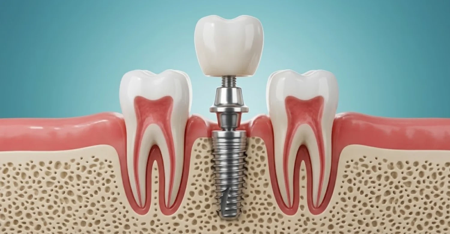
The space between an implant and a natural tooth refers to the separation between their adjacent surfaces. Clinically, dentists focus on the implant fixture and the neighboring tooth root. The visible crowns alone cannot show whether the underlying structures have adequate clearance. Natural roots vary in shape, width, curvature, and angulation. These differences influence the room available within the jawbone. An apparently wide gap between crowns may conceal closely positioned roots beneath the gums. Adequate clearance helps preserve supporting bone during and after surgery. It also reduces the risk of accidentally injuring neighboring structures while drilling. Spacing must still be evaluated alongside implant depth, direction, and surrounding anatomy. Three-dimensional imaging can add information when conventional examinations leave root relationships unclear.
How Is Implant-to-Tooth Distance Different From Implant-to-Implant Spacing?
Implant-to-tooth distance describes the separation between a dental implant and a natural tooth. Implant-to-implant spacing describes the separation between two neighboring implants. Both measurements influence planning, but they involve different anatomical considerations. Natural teeth have periodontal ligaments that connect their roots to the surrounding bone. Dental implants bond directly with bone through a process called osseointegration. These biological differences influence how tissues respond around each structure. Implant-to-implant spacing mainly affects the bone and gum tissue between two fixtures. Insufficient space there may compromise bone levels and the appearance of the gums. The distance between implant and tooth additionally requires protection of a living root and its pulp. Dentists therefore assess each relationship separately rather than applying identical assumptions everywhere.
Why Adequate Spacing Matters for Healthy Bone, Gums, and Neighboring Teeth
Adequate spacing helps protect the neighboring tooth root and preserve supporting bone. It also gives dentists room to position the implant and crown appropriately. Poor positioning can increase the risk of localized tissue damage. It may also complicate daily cleaning and long-term maintenance. Bone levels around neighboring teeth influence the gum tissue between restorations. When supporting bone is compromised, the gum contour may change over time. Patients may then notice a dark triangle or food trapping between teeth. Clinical evidence also highlights possible pulp and periapical complications when implants approach natural roots. However, these findings do not mean every closely positioned implant will cause damage. Proper planning balances anatomical safety, function, and appearance.
What Is the Ideal Distance Between a Dental Implant and a Natural Tooth?
A commonly used planning reference is approximately 1.5 mm between a dental implant and an adjacent natural tooth. This guideline helps clinicians judge available space while protecting roots and supporting tissues. However, the appropriate distance between implant and tooth depends on individual anatomy and the implant system. Root curvature, implant diameter, bone dimensions, and the planned crown all influence positioning. A measurement acceptable in one mouth may be unsuitable in another. Evidence on extremely close placement suggests possible problems with tooth vitality and surrounding bone. The 2025 systematic review found that proximity-related injuries could occur despite high implant survival. Patients should ask how their dentist assessed the neighboring roots and available bone. The objective is a safe, functional position rather than a number pursued in isolation.
Is 1.5 mm the Recommended Distance Between an Implant and an Adjacent Tooth?
The 1.5 mm figure is a commonly cited clinical reference for implant-to-tooth separation. It helps clinicians check whether enough space exists to protect adjacent structures. Nevertheless, it should not be read as an absolute safety threshold. The 2025 systematic review noted possible complications when implants sat within 1.5 mm of natural roots. Researchers also emphasized gaps in the evidence and the importance of implant direction and depth. The safest placement depends on more than the measured gap. Root position, implant diameter, bone anatomy, and surgical accuracy all affect risk. The planned crown must also fit without creating bulky or uncleansable contours. Patients should discuss these factors before surgery. Additional imaging may be advised when existing radiographs do not show the root clearly.
Why 1.5 mm Is a Common Clinical Reference Rather Than a Universal Safety Guarantee
The 1.5 mm reference offers a practical starting point for implant planning. However, anatomy varies considerably between patients. Consequently, one number cannot guarantee protection against every possible complication. An implant may still cause problems if its drill path threatens a neighboring root. Root curvature and implant depth can raise risk even when the measured gap looks acceptable. Conversely, radiographic closeness alone does not prove that meaningful injury has occurred. Studies also differ in how they define proximity and diagnose complications. The 2025 review highlighted these limitations while still identifying possible localized damage. Dentists must therefore read the distance between implant and tooth alongside clinical findings and imaging. Good planning considers the whole anatomical relationship, not only the shortest gap.
How Implant Design, Root Anatomy, and Bone Levels Influence the Required Spacing
Implant design influences how much space placement requires. Wider implants generally need more horizontal room than narrower ones. The right diameter depends on bone dimensions, restorative needs, and the expected chewing load. Natural tooth roots also differ in width, curvature, and direction. Roots that lean toward the proposed site reduce the space available for surgery. These relationships are hard to judge from the visible crowns alone. Bone levels add another layer of complexity. Reduced bone height or width may restrict positioning and alter the relationship with neighboring teeth. Digital planning and suitable imaging help evaluate these factors before surgery. Qualified clinicians must still interpret the images within the wider clinical picture.
What Is the Minimum Distance Between an Implant and an Adjacent Tooth?
Approximately 1.5 mm is a widely used planning reference, not an established minimum that guarantees safety. The clinically acceptable distance between implant and tooth depends on root position and the implant's planned path. When space is limited, dentists check whether an implant can avoid the root safely. They may consider a narrower implant, orthodontic space creation, or a different restoration. Each option needs its own assessment of suitability and long-term function. Placing an implant extremely close to a root may increase the chance of injury. The 2025 review described possible pulp, periapical, and bone complications in proximity cases. Patients should not treat one figure as permission to proceed. A qualified clinician must evaluate the complete anatomy first.
How Is Interproximal Distance Measured During Implant Planning?
Dentists evaluate available space through clinical examination and appropriate imaging. They assess the neighboring tooth, the proposed site, and the surrounding bone. The aim is to understand the three-dimensional relationship between these structures. Periapical radiographs give detailed views of individual teeth and nearby bones. Panoramic images offer a broader overview but less detail in specific areas. Cone-beam computed tomography provides three-dimensional information when clinically justified. Planning software may help assess implant diameter, angulation, and root proximity. However, measurements depend on image quality, calibration, and correct interpretation. Two-dimensional images may hide some cheek-to-tongue relationships. Patients can ask which measurements were reviewed and whether more imaging would genuinely help.
Can a Dental Implant Be Placed Less Than 1.5 mm From a Natural Tooth?
An implant may sometimes sit less than 1.5 mm from a natural tooth. However, reduced clearance demands careful evaluation because the neighboring root faces greater risk. The figure alone cannot decide whether placement is appropriate. Dentists consider root direction, implant angulation, bone anatomy, and the planned crown. They also check whether the surgical path could contact or damage the root. Alternative approaches may be wiser when anatomical space is insufficient. Research published in 2025 identified localized complications in some close-placement cases. Reported problems included changes in tooth vitality and supporting bone. These findings indicate potential risk, not inevitable damage for every patient. If an existing implant looks unusually close, an examination can decide whether investigation is needed.
Dental Implant Spacing Guidelines for Front Teeth and Back Teeth
Front and back teeth create different planning challenges. Front teeth demand close attention to gum contours, appearance, and crown shape. Small changes in the supporting bone may become visible whenever the patient smiles. Back teeth carry heavier chewing forces than front teeth. Their roots may also have complex shapes, especially in the upper molar region. Implant positioning must suit available bone, crown dimensions, and functional demands. The same general principles for the distance between implant and tooth apply in both regions. However, implant diameter, angulation, and crown design may differ by location. A measurement suitable in one area may not suit another. Digital planning can help when anatomical relationships are complex.
What Factors Affect the Safe Distance for Dental Implants?
Several anatomical and technical factors decide how closely an implant may approach a natural tooth. The distance between implant and tooth must be assessed alongside root direction, bone dimensions, implant diameter, and crown design. The 2025 systematic review included 11 studies, 1,727 participants, and over 2,000 implants. It identified potential complications involving adjacent teeth, yet overall implant survival exceeded 95%. The evidence varied in quality and included observational studies and case series. Clinical assessment should therefore focus on avoiding injury rather than meeting a number automatically. Dentists may also consider orthodontic changes, previous extractions, and surgical accuracy. Appropriate imaging helps reveal limitations before surgery begins.
Natural Tooth Root Position and Angulation
Natural tooth roots do not always follow the direction of their visible crowns. Their curvature, tilt, and width decide how much space remains for an implant. The safe distance between implant and tooth therefore depends partly on the true root position. A root angled toward the gap may narrow the available surgical corridor. Two-dimensional radiographs may not reveal every aspect of this relationship. Dentists assess neighboring roots before choosing implant diameter and trajectory. Further imaging may be appropriate when conventional views leave important questions unanswered. Careful planning reduces the risk of root contact during drilling and placement. It also helps the surgeon choose a path that keeps a safe margin from the root.
Available Jawbone Width and the Position of the Bone Crest
Jawbone width affects whether an implant can fit without compromising supporting tissues. The bone crest also influences implant depth, neighboring bone levels, and the final gum contour. Insufficient bone may limit implant diameter or require additional treatment first. However, bone grafting is not automatically needed whenever space looks restricted. Dentists evaluate bone dimensions, root anatomy, and crown requirements together. Appropriate imaging helps estimate available bone and identify anatomical limits. The resulting plan should preserve healthy structures while supporting the proposed restoration. Good bone support around both the tooth and implant helps maintain stable gums. Bone that is too thin between the two structures may slowly resorb over time.
Dental Implant Diameter, Position, and Intended Crown Shape
Implant diameter influences how much space is needed within the jaw. Wider implants generally require more horizontal clearance than narrower alternatives. The planned crown and neighboring tooth contours must also fit the available room. An implant that fits the bone may still cause restorative problems if poorly positioned. Dentists therefore plan the implant around the intended final crown. This approach helps avoid bulky contours and awkward cleaning access. Implant size must also provide enough mechanical support for the bite. A narrower implant is not automatically better. Strength, bone quality, and loading demands remain important in every case.
Narrow Tooth Gaps and Limited Space for Implant Placement
A narrow gap may restrict implant diameter and the surgical instruments that can be used. The distance between implant and tooth becomes especially important when neighboring roots lean into the gap. Dentists assess the space between crowns and the root positions beneath them. They may recommend orthodontic treatment to create room before surgery. In suitable cases, a narrow implant or alternative restoration may be considered. Placement should never proceed simply because the visible gap looks wide enough. The underlying anatomy must also allow safe placement and a cleanable crown. Each decision depends on individual bone dimensions, root anatomy, and restorative needs.
Changes in Tooth Position After Extraction or Orthodontic Treatment
Natural teeth can drift when a neighbor is missing. Orthodontic treatment also changes tooth and root positions deliberately. These movements can alter the available implant space over months or years. The planned distance between implant and tooth should therefore be checked again before surgery. Older photographs or scans may no longer reflect the current anatomy. Dentists examine present alignment, bite, and root positions. Orthodontic treatment may help restore suitable spacing when clinically indicated. After extraction, the jawbone also remodels and may shrink. Updated examinations and imaging confirm whether the proposed position remains appropriate.
Implant Placement Accuracy and Three-Dimensional Treatment Planning
Accurate placement depends on diagnosis, planning, suitable instruments, and careful execution. Spacing should be evaluated in three dimensions when the anatomy requires it. Digital planning helps clinicians examine root proximity, implant angle, and bone boundaries. Guided surgery may improve the transfer of the planned position to the mouth. Nevertheless, surgical guides do not remove every risk. Their accuracy depends on planning quality, guide fit, stability, and technique. Dentists must also confirm during surgery that the plan still suits the site. Appropriate imaging and professional judgment remain essential parts of safe implant treatment.
What Happens When a Dental Implant Is Too Close to a Natural Tooth?
An implant placed too close to a natural tooth may threaten the root, bone, or dental pulp. The distance between implant and tooth is one factor among several. Actual contact, drilling direction, and injury depth also influence outcomes. The 2025 systematic review reported that roughly 20–25% of affected teeth in contact cases needed root canal treatment. It also reported bone loss or peri-implantitis in up to 4% of implants placed under 1 mm from roots. These figures came from limited and varied research. They should not be read as personal risk estimates for any individual. Early assessment helps identify damage and decide whether monitoring or treatment fits best.
Can a Dental Implant Damage an Adjacent Tooth Root?
Yes, an implant drill or fixture can damage a neighboring root through contact or penetration. A measured gap alone cannot confirm whether damage occurred. Injury depends on implant trajectory, drilling depth, and the extent of contact. Some affected teeth stay vital, while others develop inflammation or structural damage. Dentists assess symptoms, tooth vitality, and radiographic findings. Root damage may need monitoring, root canal treatment, or extraction in severe cases. Suspected injury deserves professional assessment because symptoms may appear late. Pain is not the only warning sign worth noticing. Changes in color, tenderness, or gum swelling near the tooth also matter.
Dental Implant and Adjacent Tooth Bone Loss: What Is the Connection?
Bone separates and supports both the implant and the neighboring natural tooth. Very close placement may affect this thin bone wall. In some cases, localized bone loss affects the tooth, the implant, or both. Consequences depend on the anatomy, tissue condition, and presence of inflammation. Bone changes may appear on radiographs before obvious symptoms develop. Dentists compare current images with earlier ones whenever available. Not every close implant causes bone loss. Other conditions, such as gum disease, can produce similar findings. Diagnosis should establish the likely cause before any treatment is chosen.
How Implant Proximity Can Affect the Dental Pulp and Tooth Nerve
The dental pulp contains nerves and blood vessels that keep a tooth alive. Drilling close to a root can cause mechanical or heat-related injury. Proximity matters, but drilling direction and depth matter as well. Damage may lead to lasting sensitivity, inflammation, or death of the pulp. Some teeth stay healthy despite close implant positioning. Dentists may perform vitality tests and review radiographs to assess the neighboring tooth. Root canal treatment may be advised when irreversible pulp damage or infection is confirmed. Decisions should rest on clinical findings rather than closeness alone.
Can Incorrect Implant Spacing Cause Gum Recession or Inflammation?
Incorrect positioning can complicate gum health and change the supporting bone contour. Tight spacing may influence the bone and soft tissue between neighbors. However, the gum recession also has many other causes. Thin tissue, brushing trauma, and periodontal disease are common examples. Inflammation around an implant often signals plaque buildup or peri-implant disease. Dentists examine tissue appearance, bleeding, pocket depth, and bone levels. Treatment may include better hygiene, professional cleaning, or further intervention. A close radiographic relationship does not prove that spacing caused the inflammation.
How Close Implant Placement May Affect Crown Fit, Cleaning, and Gum Health
A poorly positioned implant can make the final crown hard to shape and clean. Limited space reduces room for a natural contour and for hygiene tools. Bulky contours may encourage plaque buildup and food trapping. Tight space can also distort contact points and the gum outline. Dentists evaluate the proposed crown before surgery to reduce these problems. Floss, interdental brushes, and water flossers may help maintain the restoration. However, cleaning tools cannot fix a fundamentally unsuitable implant position. Persistent food trapping or bleeding gums deserves professional evaluation.
Does Incorrect Spacing Always Mean the Dental Implant Will Fail?
No, incorrect spacing does not automatically mean an implant will fail. A short distance between implant and tooth may harm the neighboring tooth while the implant still integrates. The 2025 systematic review reported overall implant survival above 95% across the proximity literature. Nevertheless, localized injuries occurred in some cases. Implant survival and neighboring tooth health are separate outcomes. Dentists may monitor a stable implant while treating an affected tooth. Other cases may need corrective intervention. The right decision depends on examination, imaging, symptoms, and severity of damage.
What Causes a Dental Implant to Be Placed Too Close to a Neighboring Tooth?
Several factors can lead to inadequate clearance during implant surgery. Limited bone, unfavorable root anatomy, and inaccurate positioning are common contributors. Tooth migration can also change the available space after the original plan. Inadequate imaging may make these relationships harder to recognize. The 2025 review listed anatomy, available space, tooth drift, and surgical positioning as possible causes. These problems do not always stem from a single error. Good planning reviews current images, neighboring roots, and the final crown before surgery. When space is insufficient, clinicians should rethink the approach. Forcing an implant into an unsuitable position invites avoidable complications.
Limited Bone Space or Unfavorable Root Anatomy
Narrow jawbone and converging roots may restrict suitable implant positions. The distance between implant and tooth must account for both available bone and the adjacent root surface. A visible gap between crowns does not prove enough space exists underneath. Dentists evaluate root angulation, bone width, and the planned restoration. They may consider orthodontic space creation or another replacement when necessary. The safest approach depends on whether the anatomy can support the implant without harming neighbors. Sometimes the wisest choice is to delay surgery until the space is improved.
Incorrect Implant Angulation or Positioning
Incorrect angulation can direct the implant or drill toward a neighboring root. Clearance may become inadequate even when the original space seemed sufficient. Poor positioning can also complicate crown construction and daily cleaning. Dentists use preoperative assessment and surgical guidance to reduce avoidable errors. However, digital plans and guides do not guarantee perfect execution. If a problem is suspected afterward, clinicians assess the implant, tooth, and bone first. Corrective treatment is recommended only after that assessment.
Inadequate Preoperative Assessment of Adjacent Tooth Roots
Incomplete assessment may leave important anatomical relationships unidentified. The distance between implant and tooth should be judged alongside root direction, bone levels, and nearby structures. Depending on the case, periapical radiographs or CBCT may provide useful information. CBCT is best chosen when three-dimensional detail can change diagnosis or treatment. It is not automatically required for every implant. Reviewing dental history and existing images can also reveal past treatment or anatomical concerns. A rushed assessment saves little time and may cost far more later.
Tooth Drifting or Migration That Changes the Available Space
Natural teeth may move toward an empty space after a neighbor is lost. Orthodontic movement and bite changes can also alter root alignment. Spacing should therefore be reassessed using current records. A plan based on older images may no longer match the patient's anatomy. Dentists may recommend orthodontic correction or a different restoration when space has changed. Updated assessment reduces the risk of placing an implant against a neighboring root. Delaying implant treatment for long periods can make drift more likely.
Why Implant Placement Accuracy Matters More Than a Measurement Alone
A single figure cannot fully describe the relationship between an implant and a natural tooth. The distance between implant and tooth must be read alongside root direction, drilling depth, and implant trajectory. Published reports describe roots contacted or injured across varied spatial relationships. Accuracy therefore depends on both planning and surgical execution. Digital planning, guided techniques when appropriate, and careful judgment reduce avoidable risks. No technique removes every possible complication. Experience and attention to detail remain central to safe results.
What Symptoms Can Suggest That a Dental Implant Is Too Close to a Tooth?
Symptoms can signal injury or inflammation, but they cannot confirm the cause. Examination and suitable imaging are needed to judge implant position. Possible warning signs include persistent pain, sensitivity, tenderness, swelling, or discomfort when chewing. Bleeding gums and discharge can indicate inflammation or infection. Some patients notice nothing despite changes in neighboring tissues. Absence of symptoms therefore does not prove that an implant is ideally placed. Persistent or worsening symptoms should be assessed by a dentist. Severe swelling, fever, spreading facial swelling, or difficulty swallowing needs urgent professional attention.
Persistent Pain or Tenderness Near the Implant
Persistent discomfort may indicate inflammation, an implant complication, or a problem with a neighboring tooth. The distance between implant and tooth is considered alongside the timing and nature of the pain. Dentists assess the implant, adjacent tooth, bite, and surrounding gums. They may also review radiographs and earlier treatment records. Pain alone cannot show that the implant is too close. New, lasting, or worsening discomfort deserves prompt assessment, especially with swelling or discharge. Noting when pain started and what worsens it helps the dentist.
New Tooth Sensitivity to Hot, Cold, or Pressure
Sensitivity can arise from exposed dentin, decay, cracks, gum recession, or pulp inflammation. Root injury becomes a possibility when sensitivity begins soon after implant surgery. Dentists distinguish these causes through examination and appropriate tests. Thermal sensitivity offers clues but does not identify the cause by itself. Lasting sensitivity should be investigated rather than ignored. The affected tooth may need monitoring or treatment depending on the findings. Keep a note of when sensitivity started and what triggers it.
Pain When Biting or When the Neighboring Tooth Is Tapped
Pain on biting can come from root inflammation, a cracked tooth, bite interference, or infection. Close implant placement is one possible factor when pain starts after surgery. Dentists may check the bite and gently tap the neighboring tooth. Radiographs can reveal some root and bone changes. However, similar symptoms occur in many dental conditions. Accurate diagnosis must come before adjusting a crown or treating the natural tooth. Avoid chewing hard foods on that side until you are examined.
Swelling, Bleeding Gums, or Discharge Around the Implant
Bleeding and swelling may indicate plaque-related inflammation around the implant. Discharge can suggest infection and needs prompt assessment. Proximity may matter if nearby bone or tooth tissues are also affected. Dentists evaluate tissue condition, pocket depth, and available radiographs. Similar symptoms often occur without any root proximity problem. Avoid squeezing swollen tissue or taking leftover antibiotics. Professional assessment identifies the cause and the appropriate treatment.
Tooth Mobility or Changes in the Way the Teeth Fit Together
New looseness in a natural tooth may indicate periodontal damage, trauma, or inflammation. Bite changes can also result from crown contacts or shifting teeth. Implant position deserves review if these signs appear after surgery. Dentists examine tooth stability, supporting bone, and contact with opposing teeth. Mobility does not automatically mean the implant caused the problem. Prompt assessment shows whether periodontal, restorative, or other treatment is needed. Early action often keeps treatment simpler.
Can an Implant Be Too Close to a Tooth Without Causing Symptoms?
Yes, a closely positioned implant may cause no noticeable symptoms at first. Pain is an unreliable guide to implant position. Some root or bone changes become visible only during examination or radiographic review. Other implants stay stable without any apparent injury to neighbors. Dentists consider surgical records, current symptoms, tooth vitality, and tissue health. Routine follow-up helps catch changes that patients cannot feel. Skipping checkups removes this important safety net.
How Do Dentists Diagnose Problems With Implant-to-Tooth Spacing?
Diagnosis combines dental history, clinical examination, and appropriate imaging. The distance between implant and tooth is assessed alongside root integrity, vitality, bone support, and implant stability. Dentists also consider decay, periodontal disease, infection, and bite problems. Periapical radiographs may provide useful detail. CBCT can clarify three-dimensional relationships when clinically justified. Imaging findings must always be interpreted with symptoms and examination results. A close implant on an image does not prove injury by itself. The process aims to confirm whether a problem exists and identify its likely cause. It then guides the least invasive suitable management.
Reviewing Dental History, Previous Implant Records, and Current Symptoms
A detailed history explains when discomfort began and how symptoms have changed. Original treatment records make investigation far more effective. Dentists may review surgical notes, earlier radiographs, implant specifications, and crown details. Past root canal treatment or gum problems may also be relevant. Comparing old and new findings separates longstanding anatomy from recent change. Patients should bring all available records when seeking a second opinion. Even partial records can save time and repeat imaging.
Clinical Examination of the Implant, Adjacent Tooth, and Surrounding Gums
Clinical examination lets the dentist assess the implant crown and neighboring tooth directly. The distance between implant and tooth is weighed alongside gum condition, mobility, bite contacts, and tenderness. The clinician may look for plaque, bleeding, and abnormal pockets. Vitality tests may help assess the health of the pulp. Examination findings guide the need for imaging or referral. No single finding confirms every type of implant-related injury. The full pattern of findings matters most.
Periapical and Panoramic X-Rays for Initial Assessment
Periapical radiographs give focused images of individual teeth and nearby bone. Panoramic radiographs offer a broad view of both jaws and existing dental work. These images can show implant-to-root relationships when the structures are clearly visible. However, two-dimensional images cannot reveal every anatomical relationship. Distortion and overlapping structures may also limit interpretation. Dentists choose imaging based on the clinical question and existing records. Additional images make sense only when they are likely to add useful information.
When CBCT Imaging May Help Assess Root Position and Bone Anatomy
Cone-beam computed tomography produces three-dimensional images of teeth and jaws. It can clarify the distance between implant and tooth when conventional radiographs leave doubt. CBCT helps clinicians check for root contact and evaluate implant trajectory. However, it usually involves a higher radiation dose than standard dental X-rays. It should be used only when clinically justified. Not every patient or suspected spacing problem requires it. Dentists weigh diagnostic benefits against radiation exposure and alternative options.
How Dentists Evaluate Root Damage, Pulp Health, and Bone Changes
Dentists use clinical tests and imaging to investigate possible root injury. They examine tooth vitality, tenderness to tapping, and supporting bone levels. Findings may point to pulp inflammation, root damage, gum changes, or another condition. Some injuries need repeat testing because early results can be inconclusive. Comparing current and earlier radiographs reveals developing changes. Treatment depends on the diagnosis, extent of damage, and outlook for the tooth. Patience during this process often prevents unnecessary procedures.
Distinguishing Implant Proximity From Infection, Tooth Decay, and Bite Problems
Pain around an implant does not necessarily mean inadequate spacing. Infection, decay, cracked teeth, and bite interference can all mimic proximity problems. A poorly adjusted crown can cause chewing pain without any root contact. Gum inflammation may simply reflect plaque accumulation. Dentists use examination, vitality testing, and imaging to separate these causes. Treating the wrong condition delays relief and may create new risks. A clear diagnosis should always guide the recommended treatment.
When a Periodontist, Endodontist, or Oral Surgeon May Be Needed
Specialist referral may help when diagnosis or treatment becomes complex. An oral surgeon may assess a short distance between implant and tooth when removal is considered. An endodontist may evaluate suspected pulp injury or complex root canal needs. A periodontist may assess bone support, gum inflammation, or peri-implant disease. The right specialist depends on the specific problem. Collaboration helps coordinate care for both the implant and the natural tooth.
How Is a Dental Implant That Is Too Close to a Natural Tooth Treated?
Treatment depends on whether the implant has caused damage or simply appears close. Dentists consider symptoms, tooth vitality, bone changes, and implant stability together. Some cases need only observation and regular examinations. Others need treatment for the natural tooth, the gums, or an infection. Implant removal is considered only when findings justify its risks. The 2025 review reported localized complications despite high overall implant survival. This matters because removing a stable implant introduces new surgical risks. Dentists should explain the diagnosis, alternatives, prognosis, and limitations before any intervention.
When Monitoring May Be Appropriate for an Implant Near an Adjacent Tooth
Monitoring may suit cases where the implant is stable and injury is not progressing. The distance between implant and tooth is reviewed with vitality, symptoms, bone levels, and radiographs. Dentists may schedule regular examinations and compare new images with old ones. The follow-up interval depends on the clinical situation. Patients should report new pain, sensitivity, swelling, or mobility promptly. Monitoring is not the same as ignoring a problem. It provides a structured way to detect change and act in time.
Can a Poorly Positioned Dental Implant Be Corrected?
A poorly positioned implant can sometimes be managed successfully. Correction depends on its location and on any complications it has caused. Dentists assess implant stability, bone support, crown design, and the neighboring tooth. Modifying the crown may improve cleaning or the bite without touching the fixture. However, crown changes cannot repair root damage or an unsafe implant path. Complex cases may need specialist assessment and discussion of several options. The least invasive effective option is usually preferred.
When Implant Removal, Repositioning, or Replacement May Be Considered
Removal or replacement may be considered when an implant causes significant injury. It may also be needed when the implant cannot function safely. Spacing is only one factor in this decision. Dentists evaluate root damage, bone loss, infection, stability, and restorative limits. Removal requires further surgery and may reduce the available bone. Repositioning or replacement is not always feasible or advisable. Treating the tooth or adapting the crown may sometimes be the better choice. A specialist should explain the benefits, risks, and alternatives first.
Root Canal Treatment or Other Care for an Injured Neighboring Tooth
Root canal treatment may be advised when the neighboring pulp is irreversibly inflamed or dead. Implant-related trauma is suspected when this follows implant surgery. However, diagnosis must rule out decay, cracks, and other causes. Root canal treatment removes diseased pulp and aims to keep the remaining tooth. A crown or filling may be needed afterward, depending on the damage. Not every sensitive tooth requires root canal treatment. A dentist should confirm the diagnosis before recommending endodontic care.
Treating Gum Inflammation and Bone Loss Around the Implant
Treatment depends on whether the problem is reversible gum inflammation or advanced peri-implant disease. Spacing may matter when bone changes affect both the tooth and implant. Initial care often includes professional cleaning, plaque control, and personalized hygiene advice. More advanced conditions may require specialist treatment. Dentists monitor bone levels using clinical measurements and appropriate imaging. Antibiotics are not a universal solution for implant inflammation. Treatment should target the diagnosed cause and any contributing crown or hygiene problems.
When a Dental Bridge or Another Tooth Replacement Option May Be Considered
A bridge or another replacement may suit cases where an implant is unsuitable. Insufficient distance between implant and tooth may rule out safe implant placement altogether. A bridge uses neighboring teeth for support. A removable partial denture offers another alternative. Each option has different maintenance needs and effects on surrounding structures. Some bridges require trimming healthy supporting teeth. Dentists compare function, longevity, bone condition, cost, and patient preference before recommending one.
How Dentists Decide Between Monitoring and Further Treatment
Dentists weigh the risks of intervention against the risks of watching and waiting. Symptoms, implant stability, vitality, bone levels, and progression all inform the decision. Stable findings may support continued monitoring. Progressive injury, infection, or significant root damage usually calls for treatment. Earlier radiographs help show whether changes are developing. Patients should understand the reasoning behind each recommendation. They should also know which signs would trigger reassessment. Shared decision-making aligns clinical needs with personal preferences.
Can You Get Relief From Pain Caused by an Implant Near a Natural Tooth?
Pain relief depends on identifying the underlying cause. Medication cannot correct the distance between implant and tooth. Temporary comfort measures can help while you arrange an examination. They do not replace a proper diagnosis. A dentist may suggest suitable painkillers after reviewing your health and other medicines. Antibiotics help only in selected situations and cannot fix mechanical root damage. Persistent pain, swelling, fever, or discharge needs prompt professional assessment. Rapidly spreading facial swelling or difficulty swallowing or breathing requires emergency care.
Safe Measures for Temporary Relief While Arranging Dental Assessment
Choose soft foods and avoid chewing on the painful side for now. Brush gently to keep the area clean without irritating inflamed tissue. A cold compress on the cheek may ease swelling for short periods. Avoid pressing, probing, or trying to move the implant. These steps only buy time until you are examined. Arrange dental assessment rather than relying on home remedies. Seek urgent care if swelling spreads or you feel unwell.
When a Dentist May Recommend Appropriate Pain Relief
Pain relief may be suitable while the dentist investigates the cause. This applies when post-surgical discomfort persists or a neighboring tooth starts aching. Medication choice depends on age, medical history, allergies, pregnancy, and other medicines. A pharmacist or dentist can help choose a safe option. Follow product instructions and never exceed the recommended dose. Severe or worsening pain needs investigation even if medication brings temporary relief.
Why Pain Medication Cannot Correct Implant Position or Repair Root Damage
Pain medication eases symptoms without addressing their source. A positioning problem needs clinical evaluation, not just tablets. Painkillers cannot move an implant, repair a root, or restore lost bone. Relying on them may also delay diagnosis while damage progresses. Dentists decide whether observation, crown adjustment, root canal treatment, or surgery is required. Treatment should address the underlying condition rather than symptoms alone.
Why You Should Avoid Self-Treating With Antibiotics or Adjusting the Implant Crown
Antibiotics do not treat every cause of dental pain and can cause side effects. Taking leftover antibiotics may delay proper care. It also contributes to antimicrobial resistance. Trying to grind or loosen an implant crown can damage the restoration and surrounding tissue. Only a qualified dental professional should decide whether an adjustment is needed. Seek a diagnosis instead of attempting mechanical or medicinal self-treatment.
Warning Signs That Require Prompt or Urgent Dental Care
Persistent pain, worsening swelling, discharge, or new tooth mobility requires prompt dental assessment. These signs deserve particular attention when they begin after implant surgery. Fever, spreading facial swelling, and feeling generally unwell need urgent attention. Difficulty swallowing or breathing is an emergency requiring immediate medical care. Do not delay emergency treatment while arranging travel to another country. Early evaluation helps identify the cause and limit complications.
What Are the Possible Side Effects of Treating Incorrect Implant Spacing?
Corrective treatment carries its own risks, especially when surgery or implant removal is involved. The distance between implant and tooth guides the decision but cannot predict every complication. Possible effects include pain, swelling, bleeding, infection, or changes in supporting bone. Procedures may also affect neighboring tissues or require further restorative work. Less invasive approaches may suit cases where the implant is stable and the tooth healthy. Dentists should explain expected benefits, limitations, and risks beforehand. Follow-up visits help detect complications and track healing.
Possible Side Effects of Implant Removal or Replacement Surgery
Implant removal can affect the surrounding bone and soft tissue. Surgery becomes more delicate when a neighboring root lies very close. Potential complications include bleeding, swelling, infection, and further bone loss. The need for grafting depends on the resulting defect and future plans. Replacement may require a healing period before another implant is placed. Not every patient suits immediate replacement. A specialist should assess anatomy, healing potential, and alternatives.
Pain, Swelling, Bleeding, and Infection Risks After Corrective Procedures
Some discomfort, swelling, and minor bleeding are normal after oral surgery. Symptoms should follow the expected healing course described by your clinician. Increasing pain, persistent bleeding, fever, or worsening swelling needs professional review. Follow instructions on hygiene, diet, and any prescribed medication. Your dental team should explain which changes are expected. They should also explain which changes require urgent contact.
Potential Effects on Adjacent Teeth, Gum Tissue, and Supporting Bone
Corrective surgery may involve tissue very close to the natural tooth root. The distance between implant and tooth shapes how surgeons plan access and protect neighbors. Possible effects include temporary gum irritation, changes in gum contour, or bone damage. Actual risks depend on the implant position and the chosen procedure. Dentists assess the root and bone before selecting an approach. Careful planning and follow-up help manage these risks but cannot remove them entirely.
How Individualized Planning and Follow-Up Help Manage Treatment Risks
Individualized planning considers medical history, oral health, anatomy, and patient priorities. Likely benefits are weighed against the risks of each intervention. Dentists set out a plan for postoperative review and long-term maintenance. Patients receive guidance on hygiene, medication, and symptoms that need attention. Follow-up can detect healing problems before they grow more complex. Clear communication matters most when treatment involves several stages or specialists.
What Is the Average Cost of a Dental Implant in ?
US
UK
Turkey
Dental implant prices vary depending on the implant system, crown material, diagnostic requirements, and provider. Costs can increase when additional treatment or corrective procedures are necessary. Published 2026 estimates offer a useful starting point, but individual quotes may differ substantially.
The following table compares indicative costs for a single dental implant with a crown in the United States, the United Kingdom, and Turkey. These figures should not be treated as verified national averages or guaranteed prices. They are most useful when the same treatment components are included in each quote.
Average Dental Implant Cost Comparison in 2026
Country | Estimated Cost for One Dental Implant with Crown | Main Factors Affecting Cost |
United States | 3,000–6,000 | Provider fees, implant system, crown material, and additional procedures |
United Kingdom | $1,800–$3,500 | Clinic location, implant brand, restoration costs, and treatment complexity |
Turkey | 500–1,800 | Implant brand, crown material, diagnostic services, and treatment package |
Note: These are indicative planning estimates, not guaranteed prices. Confirm the currency, source date, and included services for each estimate before comparing clinics. Bone grafting, root canal treatment, sedation, and corrective surgery may increase the total cost.
Average Dental Implant Cost in the United States: 3,000–6,000
A single dental implant with a crown may cost approximately 3,000–6,000 in the United States. Prices vary by city, provider, implant system, and crown material. Some quotes exclude bone grafting, tooth extraction, or advanced imaging, which can increase the overall expense.
Cost Factor | What to Check |
Implant fixture | Whether the implant and its components are included |
Abutment and crown | Whether both are covered by the quoted price |
Diagnostic imaging | Whether X-rays or CBCT scans cost extra |
Additional procedures | Whether extraction, grafting, or other treatment is required |
Follow-up care | Whether postoperative appointments are included |
Before accepting a quote, confirm whether the consultation, implant fixture, abutment, crown, surgery, and follow-up visits are included. Insurance coverage, where available, may also affect your final out-of-pocket cost. A personalized treatment estimate is more useful than an advertised starting price.
Average Dental Implant Cost in the United Kingdom: $1,800–$3,500
Private dental implant treatment in the United Kingdom may cost approximately $1,800–$3,500 per tooth in some published estimates. However, the quoted amount may cover the implant alone or the implant and crown together. Location, case complexity, implant brand, and crown material all influence the final price.
Cost Factor | What to Check |
Implant surgery | Whether the surgical placement is included |
Dental crown | Whether the final restoration is included |
Implant components | Whether the abutment is charged separately |
Diagnostic tests | Whether consultation and imaging are included |
Additional treatment | Whether grafting or extraction carries separate fees |
Some practices quote the surgical implant separately from the final crown. Confirm the currency and the exact services included before comparing providers. Complex cases involving insufficient bone, gum disease, or damage to neighboring teeth may cost more than routine single-tooth replacement.
Average Dental Implant Cost in Turkey: 500–1,800
A single dental implant with a crown may cost approximately 500–1,800 in Turkey, depending on the provider and scope of treatment. Estimates vary according to implant brand, crown material, bone condition, diagnostic requirements, and whether the advertised price covers the complete restoration.
Cost Factor | What to Check |
Implant fixture | Brand, model, and warranty conditions |
Abutment and crown | Whether both are included in the package |
Diagnostic assessment | Whether consultation, X-rays, and required scans are covered |
Additional procedures | Whether extraction or bone grafting costs extra |
Follow-up and aftercare | Which appointments and services are included |
Travel expenses | Whether accommodation and transportation are separate |
Some advertised prices cover only part of the treatment. International patients should request written confirmation of the implant fixture, abutment, crown, imaging, surgery, and follow-up arrangements. Travel, accommodation, and potential return visits should be budgeted separately.
Note : All images used are for editorial and illustrative purposes only and may not originate from the original news provider or associated company.
What Is Included in the Total Cost of Dental Implant Treatment?
The total price of dental implant treatment depends on which components the provider includes. Common expenses include the implant fixture, abutment, crown, consultation, diagnostic imaging, and surgery. A short distance between an implant and a neighboring tooth may require additional assessment beyond routine placement.
Bone grafting or treatment for a neighboring tooth can increase the total cost. Some treatment packages also include temporary crowns or selected follow-up visits. An itemized estimate helps patients understand what they are paying for and compare providers more accurately.
Dental Implant Treatment Costs by Component
Treatment Component | What It Covers | Potential Effect on Total Cost |
Implant fixture | The component placed in the jawbone to replace the tooth root | Varies by brand and implant system |
Abutment | The connector between the implant and crown | May be included or charged separately |
Dental crown | The visible restoration that replaces the missing tooth | Depends on material and design |
Surgical fees | Implant placement and associated clinical work | Depends on case complexity |
Consultation | Clinical examination and treatment discussion | May be included in a package |
X-rays and CBCT scans | Diagnostic imaging when clinically indicated | Depends on the type and number of examinations |
Digital planning | Planning implant position and restoration | May be included or itemized |
Bone grafting | Rebuilding bone when additional support is needed | Depends on the extent of reconstruction |
Root canal treatment | Treating an affected neighboring tooth when indicated | Depends on the tooth's condition |
Corrective surgery | Managing a placement complication or other implant problem | Depends on the diagnosis and procedure |
Follow-up care | Review appointments and postoperative monitoring | Depends on the clinic's care policy |
Implant Fixture, Abutment, Crown, and Surgical Fees
A complete single-tooth implant restoration generally involves several components. The implant fixture replaces the root of the missing tooth, while the abutment connects the fixture to the crown. The crown restores the visible portion of the tooth and helps support chewing function. Surgical fees cover implant placement and associated clinical work.
Some quotes combine these expenses into one package, while others list them separately.
Component | Main Purpose | Question to Ask the Provider |
Implant fixture | Replaces the missing tooth root | Which brand and model are being used? |
Abutment | Connects the implant to the crown | Is the abutment included? |
Crown | Restores the visible tooth | Which material is included? |
Surgery | Places the implant | Are all surgical fees included? |
Check the materials, component brands, warranty conditions, and replacement policies before accepting a quote.
Consultation, X-Rays, CBCT Scans, and Digital Planning
Diagnostic costs depend on the examinations required for each case. Consultation fees, periapical radiographs, panoramic X-rays, and cone-beam computed tomography (CBCT) scans may be charged separately. Digital planning may also be included in the package or itemized as an additional service.
Additional imaging may be justified when neighboring roots are difficult to assess, particularly if the implant is close to an adjacent tooth. However, CBCT is not necessary for every patient and should be recommended only when the clinical situation warrants it.
Diagnostic Service | Purpose | Possible Cost Consideration |
Clinical consultation | Assesses oral health and treatment needs | May have a separate fee |
Periapical X-ray | Examines a specific tooth and surrounding structures | May be charged per image |
Panoramic X-ray | Provides a broader view of the jaws and teeth | May be included in an assessment |
CBCT scan | Provides three-dimensional information when indicated | May carry an additional fee |
Digital implant planning | Helps plan implant positioning and restoration | May be included or itemized |
Ask which tests are recommended, why they are necessary, and whether their costs are included in your estimate.
Additional Costs for Bone Grafting, Root Canal Treatment, or Corrective Surgery
Additional procedures may be necessary when the jawbone is insufficient or a neighboring tooth has been damaged. Depending on the diagnosis, treatment may involve bone grafting, root canal therapy, extraction, implant removal, or replacement. Each procedure can add fees for clinical work, materials, imaging, and appointments.
Not every patient requires these interventions, and the need for treatment cannot be determined from implant spacing alone.
Additional Procedure | When It May Be Required | Cost Consideration |
Bone grafting | When there is insufficient bone for the planned treatment | Depends on the amount and type of grafting |
Root canal treatment | When the neighboring tooth's pulp is damaged or irreversibly inflamed | Depends on the tooth and restoration needs |
Tooth extraction | When a tooth cannot reasonably be preserved | Depends on extraction complexity |
Implant removal | When removal is clinically indicated | Depends on implant stability and surrounding bone |
Implant replacement | When a new implant is appropriate after reassessment | Includes new planning and potentially additional surgery |
Specialist assessment | When the case needs additional clinical expertise | Depends on the specialist and investigations required |
Request a written estimate that separates recommended treatment from possible future procedures. This helps you understand the expected costs without assuming that every potential intervention will be necessary.
What Determines the Cost of Correcting an Implant Placed Too Close to a Tooth?
Corrective treatment costs depend on the diagnosis and the procedure required. The distance between an implant and the neighboring tooth is important, but the assessment should also consider root injury, bone loss, implant stability, symptoms, and crown design.
Some cases may be managed through monitoring and preventive care, while others require root canal treatment, implant removal, bone grafting, or replacement. Specialist fees and additional diagnostic imaging can increase the total cost.
Clinical Finding | Possible Management | Effect on Cost |
Close spacing without detected damage | Monitoring and appropriate follow-up | Usually less costly than surgery |
Suspected injury to a neighboring tooth | Further examination and imaging | Depends on diagnostic requirements |
Damage to the tooth's pulp | Root canal treatment when indicated | Adds treatment and potentially restoration costs |
Bone loss around the implant | Assessment and individualized periodontal or implant management | Depends on severity and treatment needs |
Implant position compromising restoration or adjacent structures | Corrective treatment, potentially including removal or replacement | May substantially increase costs |
Unclear diagnosis | Additional clinical assessment | Depends on the investigations required |
A dentist must assess the case before estimating the total expense. No single correction price applies to every patient, and an implant placed close to a tooth does not automatically need to be removed.
Why an Implant Price Is Not the Same as the Cost of Corrective Treatment
Routine implant placement and corrective treatment involve different clinical requirements. An original treatment package may cover surgery and a crown but exclude later treatment for root damage, infection, bone loss, or other complications.
Corrective care may require additional specialists, imaging, restorative work, or bone reconstruction. Patients should therefore distinguish the original treatment cost from the cost of managing a subsequent complication.
Comparison Factor | Routine Implant Treatment | Corrective Implant Treatment |
Main purpose | Replace a missing tooth | Address a diagnosed complication or placement problem |
Planning | Plan the initial implant position and restoration | Reassess the implant, neighboring teeth, and surrounding tissues |
Diagnostic needs | Based on routine clinical requirements | May require additional imaging or specialist examination |
Surgical complexity | Depends on anatomy and bone condition | May involve removal, replacement, or reconstruction |
Neighboring tooth care | Not always needed | May require root canal treatment or other restorative care |
Total cost | Based on the planned implant and restoration | Can be higher due to additional procedures |
A fresh clinical assessment is needed to establish the likely scope and total expense. Ask the original provider whether any warranty or treatment policy covers corrective care, and confirm exactly what that coverage includes.
How to Compare Dental Implant Quotes Across the US, UK, and Turkey
Compare equivalent treatment components rather than advertised starting prices. Individual anatomy, including the distance between an implant and an adjacent tooth, may mean that two patients who appear to need similar treatment actually require different procedures.
Check whether each estimate includes the implant fixture, abutment, crown, imaging, surgery, and follow-up. Confirm the currency, materials, payment terms, and any additional fees. For treatment abroad, calculate travel, accommodation, and possible return visits separately.
Dental Implant Quote Comparison Checklist
Comparison Point | What to Confirm |
Total price | Is the quoted amount for a complete implant and crown? |
Implant brand | Is the implant manufacturer and model identified? |
Crown material | Is the proposed restoration material specified? |
Diagnostic tests | Are the required X-rays or scans included? |
Additional treatment | Are grafting, extraction, or treatment for neighboring teeth excluded? |
Temporary restoration | Is a temporary crown provided when appropriate? |
Follow-up care | How many visits are included? |
Warranty | What does the warranty cover, and what exclusions apply? |
Currency and payment | Is the quote fixed, and are there additional fees? |
Complication management | What happens if a problem develops after treatment? |
Overseas arrangements | Are accommodation and transport included or separate? |
For international treatment, ask what happens if complications arise after you return home. An itemized written plan allows you to compare providers more fairly and understand the limits of each package.
Why Travel, Accommodation, Aftercare, and Follow-Up Should Be Considered Separately
Dental tourism involves costs beyond the clinical treatment itself. Patients may need to budget for flights, local transport, accommodation, time away from work, and return appointments. Unexpected complications can also require reassessment or further care after travelling home.
Implant treatment often involves several stages and healing periods. A short visit may not accommodate every clinical contingency, so patients should understand the proposed schedule before booking travel.
Expense or Consideration | What to Include in Your Budget |
Flights | Initial travel and potential return trips |
Accommodation | Hotel or other lodging for the required stay |
Local transport | Airport transfers and travel to appointments |
Time away from work | Lost income or leave requirements |
Follow-up appointments | Reviews required during healing and restoration |
Additional treatment | Possible extra procedures if clinically necessary |
Care after returning home | Local examinations or urgent dental treatment if needed |
Ask the clinic how follow-up is coordinated, how urgent concerns will be handled, and whether additional appointments could change the quoted price. Local dental care may still be necessary after returning home.
The lowest advertised price is not necessarily the lowest overall cost. A more meaningful comparison considers the full treatment plan, clinical suitability, included services, aftercare, and the arrangements for managing complications.
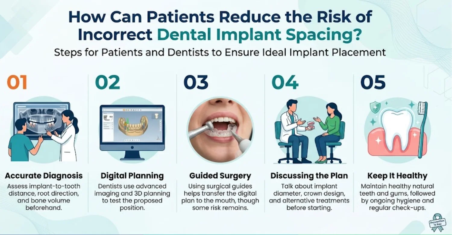
Choosing a Qualified Implant Dentist or Oral Surgeon
Choose a clinician with appropriate training and experience in implant surgery. Ask how they will evaluate the neighboring roots, bone dimensions, and final crown. Discuss their experience with complications and how they would manage them. You should receive clear explanations before consenting to treatment. The best choice is someone who can justify the plan and arrange proper follow-up. Low price alone should never be the deciding factor.
Reviewing Adjacent Tooth Roots and Bone Anatomy Before Surgery
Preoperative assessment reveals the available space and any anatomical limits. Root angulation and supporting bone are reviewed together. Dentists select imaging based on the needs of each case. Three-dimensional imaging may help when root relationships remain unclear. The results guide implant diameter, direction, and depth. Identifying limits early allows alternatives to be considered before surgery.
Using Appropriate Digital Planning and Guided Implant Placement When Indicated
Digital planning helps clinicians test implant positions before surgery. Planning software can measure the distance between implant and tooth when image quality allows. A surgical guide can help transfer the planned position into the mouth. However, accuracy depends on planning quality, guide stability, and surgical technique. Guided placement does not remove all risk or replace clinical judgment. Your dentist should explain when these tools add real benefit.
Discussing Implant Diameter, Position, and Restoration Design Before Treatment
Understand why your clinician recommends a particular implant size and location. Available space shapes both the implant and the crown that sits on it. The crown should be planned alongside the implant, not as an afterthought. Ask whether the available bone supports the chosen diameter and angle. Discuss alternatives if neighboring roots restrict placement. An informed conversation clarifies benefits, limits, and risks before surgery.
Attending Follow-Up Appointments and Maintaining Oral Hygiene
Regular follow-up monitors healing, implant stability, and neighboring tooth health. Spacing is fixed at surgery, but later examinations can catch complications early. Brush twice daily and clean around the crown with recommended tools. Attend professional maintenance visits at the intervals your dentist advises. Report persistent pain, bleeding, swelling, or bite changes. Good hygiene supports healthy tissue but cannot correct a poor implant position.
Protecting the Health of Natural Teeth Before and After Implant Placement
Healthy neighboring teeth are vital for a successful restorative plan. Dentists may treat active decay, gum disease, or infection before implant surgery. Patients should keep up oral hygiene and attend recommended examinations. After surgery, report new sensitivity or lasting discomfort promptly. Protecting natural teeth preserves function and avoids unnecessary treatment. Every natural tooth you keep healthy simplifies future dental care.
What We Notice Clinically
Clinical assessment looks beyond the apparent gap between an implant and a natural tooth. The distance between implant and tooth is read alongside root anatomy, tissue condition, bone levels, and symptoms. Some patients feel nothing despite radiographic proximity. Others develop sensitivity, localized inflammation, or changes around the neighboring root. The 2025 review identified possible biological complications while overall implant survival stayed high. These findings support careful diagnosis rather than automatic implant removal. Decisions should separate incidental proximity from evidence of real injury. The following observations are educational and do not replace an individual examination.
Why an Implant That Looks Close to a Tooth May Need Further Assessment
A radiograph can show proximity without proving damage. Root integrity, tooth vitality, and supporting bone must all be reviewed. Dentists may compare earlier images and run additional tests when needed. If the tooth stays healthy and the implant stable, monitoring may suffice. Signs of progressive damage call for further investigation. Treatment should follow the full clinical findings rather than appearance alone.
Why Patients Can Have Root Damage or Bone Changes Without Obvious Symptoms
Some dental changes develop before any pain or sensitivity appears. A neighboring tooth may feel normal during the early stages of certain problems. Bone changes may first appear during a routine examination. Dentists combine clinical tests with suitable imaging to investigate these findings. Regular follow-up catches concerns that symptoms alone would miss. Feeling fine is reassuring, but it is not proof of health.
Common Misconception: Every Implant Must Have Exactly the Same Tooth-to-Implant Distance
No single figure guarantees safe placement in every mouth. The distance between implant and tooth must reflect each patient's roots, bone, implant design, and surgical path. About 1.5 mm is a widely cited reference, but evidence does not make it a universal guarantee. The 2025 review described the figure as debated and called for individualized assessment. Clinical decisions should focus on protecting surrounding structures. Applying one number mechanically misses the point.
Distinguishing a Small Radiographic Distance From Clinically Significant Tooth Injury
A small gap on an X-ray does not automatically indicate injury. Root contact, vitality tests, symptoms, and bone findings must be considered together. Some closely positioned teeth stay healthy, while others develop complications. Dentists may repeat tests or review earlier images when results are uncertain. Progressive change tells more than a single isolated measurement. Treatment should address proven problems, not proximity alone.
Clinical Note: Dr. Rifat Alsaman
A clinical perspective attributed to Dr. Rifat Alsaman should emphasize individualized diagnosis and protection of neighboring teeth. Spacing should never be judged without assessing root anatomy, bone levels, implant direction, and tooth vitality. This proposed clinical note should be reviewed and approved by Dr. Alsaman before publication. It is educational content, not a verified quotation or report of his personal findings. This approach supports transparent, evidence-informed patient communication.
Clinical Perspective on Interpreting Implant Spacing Alongside Root Anatomy, Bone Levels, Tooth Vitality, and Symptoms
The distance between implant and tooth should be interpreted within a complete clinical examination. Root direction and bone anatomy show whether the proposed position is appropriate. Vitality testing may reveal possible pulp injury. Radiographs help assess the supporting tissues. Symptoms add information but cannot confirm or exclude damage on their own. Combining these findings strengthens diagnosis and guides sensible next steps. Individualized assessment remains essential when evidence is inconclusive.
Why Treatment Decisions Should Consider the Entire Clinical Picture Rather Than Distance Alone
A measurement alone cannot justify corrective surgery. Implant stability, tooth health, symptoms, and radiographic change must all be weighed. Removing an implant introduces surgical risks and extra costs. Monitoring may be reasonable when no progressive injury is found. Confirmed damage may justify treatment or specialist referral. Clinicians should explain the evidence, alternatives, and expected outcomes before recommending intervention.
.jpg)
.jpg)
.jpg)
.webp)
Tips for Patients
Patients can help protect their natural teeth by keeping records and reporting changes promptly. Spacing is usually decided during planning, but follow-up remains important. Ask how the neighboring root was evaluated and which symptoms need attention. Keep copies of relevant radiographs and treatment records. Maintain daily hygiene and attend recommended checkups. Never try to adjust the implant crown yourself. These habits support informed decisions and earlier detection of complications.
What to Ask Your Dentist About the Distance Between an Implant and a Natural Tooth
Ask whether the neighboring root and supporting bone were clearly assessed before surgery. Request an explanation of the distance between implant and tooth in relation to your own anatomy. Ask whether the implant is stable and the adjacent tooth healthy. Find out which findings would justify monitoring, more imaging, or corrective treatment. Request clear explanations of risks and alternatives. These questions make discussions clearer and support informed consent.
What Dental Records and Previous X-Rays to Bring to a Second Opinion
Bring previous radiographs, treatment summaries, implant details, and the surgery date if available. Comparing images over time makes evaluation far more accurate. Include records of root canal or gum treatment involving the neighboring tooth. A written list of symptoms and when they began is also useful. Original digital images are better than printed copies or photos. A second opinion can then decide whether further tests or treatment are warranted.
How to Maintain Oral Hygiene Around an Implant and Adjacent Teeth
Brush twice daily with a soft-bristled toothbrush and fluoride toothpaste. Clean between teeth with floss or correctly sized interdental brushes. Pay particular attention to the junction between the implant crown and the natural tooth. Follow your dentist's advice on tools for hard-to-reach areas. Avoid aggressive brushing that can injure the gums. Professional maintenance supports implant health and lets the dentist monitor the neighboring tooth.
What to Avoid if You Suspect Your Implant Is Too Close to a Neighboring Tooth
Do not push, twist, or try to loosen the implant or crown. Avoid placing sharp objects between the implant and the natural tooth. Do not take leftover antibiotics or exceed recommended pain-relief doses. Arrange a dental assessment if symptoms persist or worsen. Seek urgent care for spreading facial swelling or difficulty swallowing. These precautions reduce the risk of further injury while you await diagnosis.
When to Seek a Second Opinion From an Implant Specialist
A second opinion may help when findings are uncertain or recommendations conflict. It is especially valuable when removal, repositioning, or complex root treatment is proposed. The specialist will weigh the neighboring tooth's outlook against available alternatives. Bring previous images and written treatment plans where possible. Ask the specialist to explain the diagnosis, likely outcomes, and risks. Further intervention should reflect clinical need rather than anxiety about a number.
At Vitrin Clinic: Assessing Implant Position and the Health of Neighboring Teeth
Assessment should cover the implant, neighboring roots, gums, supporting bone, and intended crown. The distance between implant and tooth is one part of this broader evaluation. Vitrin Clinic can explain the available diagnostic options during consultation. Digital X-rays and CBCT may be considered when clinically appropriate. These tools clarify anatomy but do not automatically dictate treatment. The clinical team should explain findings, alternatives, and limitations before recommending anything. International patients should also understand how follow-up and urgent care would work.
Evaluating Implant Placement, Adjacent Roots, Gums, and Supporting Bone
A thorough assessment examines the implant position and the health of surrounding structures. Root integrity, gum condition, and bone levels are reviewed together. The clinician may check implant stability, tooth vitality, and the bite. Previous radiographs help reveal changes over time. The findings decide whether monitoring or further investigation is appropriate. Treatment should rest on evidence of a problem rather than proximity alone.
Using Digital X-Rays, CBCT, and 3D Scanning When Clinically Appropriate
Imaging choices depend on the diagnostic question and available records. Standard dental radiographs answer many questions about root and bone relationships. CBCT can clarify root direction and bone anatomy that flat images cannot show. Intraoral scans help evaluate crown shape and the bite. However, scans do not replace radiographic assessment of roots. Vitrin Clinic should choose imaging according to clinical need. Radiation exposure and expected diagnostic benefit must both be considered.
Developing an Individualized Plan for Monitoring or Correcting Implant-Related Problems
The treatment plan depends on actual findings and the patient's priorities. Symptoms, bone changes, vitality, and implant stability are assessed together. Monitoring may suit cases without progressive injury. Confirmed complications may need crown adjustment, root canal treatment, or specialist care. Implant removal is reserved for cases where benefits clearly outweigh risks. Vitrin Clinic can explain each option and the reasons behind every recommendation.
Considering the Condition of the Natural Tooth Before Recommending Further Treatment
The natural tooth's condition shapes both diagnosis and prognosis. Decay, cracks, gum support, and pulp health all need evaluation. A healthy neighboring tooth may need only monitoring. A tooth with confirmed pulp injury may need root canal treatment. Invasive procedures should never be recommended without a clear indication. Vitrin Clinic's discussion should show how each option protects remaining natural structures.
Internal-Link Opportunities for Further Patient Education
Relevant links can help patients understand diagnosis and treatment alternatives. This article could link to Vitrin Clinic's dental implant treatment page and contact page. Related resources might cover CBCT imaging, bone grafting, and root canal treatment. Pages on gum disease and implant aftercare would also help readers. Links should answer the reader's next question accurately. Avoid adding links purely for keyword repetition.
How Vitrin Clinic Supports Your Treatment Journey
A clear treatment journey includes assessment, explanation, coordination, and follow-up. Concerns about implant spacing may require reviewing old records or arranging new imaging. Vitrin Clinic can explain clinical findings and discuss suitable next steps. Recommendations should reflect individual anatomy, existing dental work, and overall oral health. International patients may need help coordinating appointments and understanding treatment stages. Communication should set realistic expectations without promising a particular outcome.
Initial Consultation and Review of Dental History
The first consultation establishes the patient's concerns and dental history. The clinician reviews previous procedures, medications, health conditions, and current symptoms. An examination then shows whether further tests are needed. This information supports a personalized treatment discussion. Patients should report any change in symptoms and bring available records. Questions are welcome at every stage of this first visit.
Coordinating Clinical Examinations and Relevant Diagnostic Imaging
Clinical examinations decide which tests may answer the treatment question. Focused radiographs or CBCT may be arranged when clinically justified. Vitrin Clinic can coordinate these assessments according to the clinician's advice. Imaging should be ordered only when it is likely to help. Results should be explained in plain language. Patients should know the purpose and cost of each investigation.
Explaining Treatment Options, Expected Limitations, and Potential Risks
Treatment discussions should explain the diagnosis and available alternatives. Monitoring, crown adjustment, or further intervention may each be appropriate. Clinicians should cover potential benefits, risks, limitations, and costs. Patients should understand why a particular approach is recommended. No procedure can guarantee a complication-free outcome. Informed consent needs time for questions and consideration of reasonable alternatives.
Personalized Treatment Planning for International Patients
International planning must balance clinical needs with travel constraints. Existing records may need diagnostic review before travel is booked. Vitrin Clinic can outline expected appointment stages and required documents. Patients should know whether treatment may need several visits. Healing periods and unexpected findings can affect the schedule. Travel plans should stay flexible until the treatment timeline is confirmed.
Coordinating Appointments, Treatment Stages, and Follow-Up Care
Coordinated scheduling helps patients understand the sequence of care. Implant position should be fully assessed before any corrective procedure is booked. Some cases need one examination, while others need specialist input or staged treatment. Vitrin Clinic can explain expected appointments and follow-up requirements. Healing cannot be guaranteed to follow a fixed timetable. Patients should know how to raise concerns between visits.
Supporting Aftercare and Communication With the Clinical Team
Aftercare helps patients follow hygiene instructions and recognize complications early. Follow-up may include reviewing symptoms or new radiographs. Patients should know how to contact the clinical team. They should also know when urgent local care is necessary. Instructions should cover cleaning, medications, and review appointments. International patients need a realistic plan for care after returning home. Good communication supports continuity but never replaces an examination when one is needed.
Ready to transform your smile?
Talk to our team and get a personal FREE treatment plan.
Key Takeaways
Safe implant planning protects neighboring roots, supporting bone, and the final crown. The distance between implant and tooth must always be read in its anatomical and clinical context. About 1.5 mm is a widely cited planning reference, not an absolute guarantee. Research shows close placement can cause localized problems, though many implants still function well. Persistent symptoms or suspicious findings deserve proper evaluation. Careful planning, individualized treatment, and regular follow-up protect both natural teeth and implants.
The Main Points About Safe Dental Implant Spacing
Adequate spacing protects neighboring roots, supporting bone, and gum tissue. Clearance must be assessed before surgery, not after problems appear. Root anatomy, implant diameter, angulation, and crown design all influence placement. No single measurement establishes safety for every patient. Individualized planning reveals anatomical limits and suitable alternatives.
Why Implant-to-Tooth Distance Must Be Interpreted in Its Clinical Context
A measurement alone cannot show whether a tooth has been injured. Symptoms, root anatomy, bone levels, and vitality tests complete the picture. Radiographs and clinical examinations together clarify the situation. Treatment should address confirmed problems rather than proximity alone. Monitoring may suit cases where the implant and tooth both remain healthy.
Which Symptoms Should Prompt a Dental Examination
Persistent pain, new sensitivity, swelling, discharge, or tooth mobility warrants dental assessment. These signs matter most when they develop after implant surgery. Dentists can investigate the implant, neighboring tooth, and surrounding tissues. Severe swelling, fever, or spreading facial swelling needs urgent care. Difficulty breathing or swallowing requires emergency medical attention.
How Proper Planning, Appropriate Treatment, and Follow-Up Help Protect Natural Teeth
Preoperative assessment identifies anatomical risks before implant placement. Bone support, root direction, and crown design are considered together. Suitable imaging and surgical techniques reduce avoidable injury. Follow-up helps detect complications and monitor healing. Treatment should stay individualized and evidence-informed throughout.
At Vitrin Clinic
At Vitrin Clinic, dental implant assessment considers neighboring teeth, supporting bone, gum health, and the planned crown. Clinical examination and appropriate imaging help evaluate how the implant relates to nearby roots. Recommendations depend on individual findings rather than a fixed measurement. Patients can discuss concerns, review existing records, and explore treatment alternatives. International patients receive clear information about appointments, treatment stages, and follow-up. Contact the clinic to arrange an individual assessment.
Reference
FAQs
Dr. Rifat Alsaman has more than 5 years of clinical experience in dentistry and currently serves as the Head of the Medical Team at Vitrin Clinic. He is dedicated to providing exceptional patient care, overseeing treatment planning, and ensuring the highest clinical standards across the team. His expertise, attention to detail, and commitment to continuous professional development have helped countless patients achieve healthier, more confident smiles.

.webp&w=3840&q=75)


