Dental Implant
October 15, 2026
Photogrammetry vs Digital Scanning for Full Arch Implants: Which Is More Accurate?
 Dr. Rifat Alsaman
Photogrammetry vs Digital Scanning for Full Arch Implants is one of the most debated topics in modern implant dentistry. Both methods record where your implants sit before the laboratory builds your bridge. The difference between them can affect how well that bridge fits on the day of delivery. According to the World Health Organization, about 23% of adults aged 60 and over have lost all their teeth. Many of them will consider a fixed full arch bridge supported by four to six implants. This guide explains the accuracy evidence, the clinical workflow, the costs, and the questions worth asking before treatment. This article on Photogrammetry vs Digital Scanning for Full Arch Implants is written for patients, not engineers.
Quick Facts | Details |
Methods compared | Intraoral photogrammetry (PG) and intraoral scanning (Intraoral Scanner) |
What they record | Three dimensional implant positions for a full arch bridge |
Current evidence | Recent meta analyses favor photogrammetry, mostly from lab studies |
Radiation exposure | None; both methods use light, not X rays |
Cost per arch | About 3,250–8,450 in Turkey vs. 18,000–30,000 in the US |
.jpg)
.jpg)
.jpg)
.jpg)
.jpg)
What Is Photogrammetry vs Digital Scanning for Full Arch Implants?
Photogrammetry vs Digital Scanning for Full Arch Implants describes a choice between two digital ways of recording implant positions. Each method produces a digital file that guides the design of your final bridge. Neither method is a treatment by itself. Both are measurement tools that sit between implant surgery and the finished prosthesis. Understanding them helps patients follow the conversation during their consultation.
What Is Intraoral Photogrammetry in Implant Dentistry?
Intraoral photogrammetry uses a special camera to photograph coded markers screwed onto each implant. Software calculates the exact position and angle of every implant from overlapping images. The method borrows principles from engineering, surveying, and aerospace measurement. Systems such as PIC Dental, iCam4D, and MicronMapper are used in implant dentistry today. Photogrammetry captures implant positions only, so it does not record gums or teeth. The camera records markers from a short distance and does not touch the gums. A separate scan is therefore still needed for soft tissue and bite information.
What Is Digital Intraoral Scanning (Intraoral Scanner)?
Digital intraoral scanning uses a handheld wand that captures thousands of images as it moves around the mouth. Software stitches these images together into one continuous three dimensional model. For implants, small scan bodies are attached so the scanner can locate each implant. An intraoral scanner records teeth, gums, scan bodies, and bites in one session. It has largely replaced putty impressions for single implants and short bridges. Its accuracy over a whole arch, however, depends heavily on technique and conditions. Intraoral scanners are now standard equipment in many practices worldwide. Their adoption grew rapidly over the last decade as prices fell.
Intraoral Photogrammetry vs Digital Implant Scanning: What Is the Difference?
The key difference is how each method builds its measurement. An intraoral scanner stitches many small images in sequence, and small errors can accumulate across the arch. Photogrammetry measures all markers simultaneously from several angles, which limits that cumulative effect. Intraoral scanning captures soft tissue, while photogrammetry captures only implant positions. In practice, Photogrammetry vs Digital Scanning for Full Arch Implants is a comparison between a specialist tool and a versatile one.
Feature | Photogrammetry | Intraoral Scanning |
Records implant positions | Yes | Yes |
Records gums and teeth | No | Yes |
Error build up across arch | Limited | Possible |
Equipment availability | Less common | Widely available |
How Do Digital Impressions for Full Arch Dental Implants Work?
A digital impression begins with attaching scan bodies or photogrammetry markers to each implant or abutment. The device then records their positions and converts them into a digital file. Software matches each scan body to a library file of known geometry. This tells the laboratory where every implant connection sits in space. The file is combined with gum, bite, and facial information. Technicians then design the bridge using CAD/CAM software before milling or printing it. Digital files avoid the distortion that can occur when impression materials shrink or stone models expand. They can also be stored for future repairs.
Are Photogrammetry and Intraoral Scanning Alternatives or Complementary Technologies?
They are often complementary rather than competing. Photogrammetry supplies highly reliable implant coordinates, while intraoral scanning supplies the gums, teeth, and bite. Many clinics merge both datasets into a single design file. This hybrid approach uses each tool for what it does best. Within Photogrammetry vs Digital Scanning for Full Arch Implants, the real question is often how to combine them. Your dentist chooses the workflow based on your case, not on the technology alone. Hybrid workflows are increasingly described in recent clinical reports.
Ready to transform your smile?
Talk to our team and get a personal FREE treatment plan.
Full Arch Implant Impression Accuracy: How Do the Two Methods Compare?
Accuracy is the central issue in Photogrammetry vs Digital Scanning for Full Arch Implants. A full arch bridge connects several implants with one rigid structure. Even tiny recording errors can prevent that structure from seating passively. This section explains how accuracy is measured and what the latest research reports.
What Does Accuracy Mean in Full Arch Implant Dentistry?
In dentistry, accuracy follows the international ISO 5725 definition. It combines two ideas, trueness and precision. A recording is accurate when it is both close to reality and consistently repeatable. For full arch implants, accuracy is usually measured in micrometers and degrees. One micrometer equals one thousandth of a millimeter. Errors are tiny, yet they matter because the bridge does not flex like natural teeth.
Trueness vs. Precision in Digital Implant Impressions
Trueness describes how close a scan is to the real implant positions. Precision describes how similar repeated scans are to each other. A device can be precise but not true, repeating the same error every time. Researchers usually compare scans with a reference model measured by an industrial coordinate machine. For context, a human hair is about 70 micrometers thick. Lower deviation values mean better performance. Both measures matter when choosing between Photogrammetry vs Digital Scanning for Full Arch Implants.
Linear, Angular, and Three Dimensional Deviations
Linear deviation measures the distance error between implants, usually in micrometers. Angular deviation measures how much the recorded tilt of an implant differs from reality, in degrees. Three dimensional deviation combines errors along all spatial axes into one value. Angular errors are especially important for long full arch bridges. Even a small angular error can grow into a larger gap at the far end of a bridge. A 2025 meta analysis noted that deviations above 1 degree may compromise passive fit.
Photogrammetry Accuracy in Full Arch Implant Dentistry
Photogrammetry was designed specifically for measuring multiple fixed points over long distances. In a 2026 meta analysis, photogrammetry showed three dimensional trueness deviations of about 78–88 micrometers. In the same studies, intraoral scanners showed deviations of 148–536 micrometers. Photogrammetry angular deviations were about 0.72–0.81 degrees. Precision results were also strong, with some studies reporting values near 5–14 micrometers. These numbers explain why photogrammetry attracts attention for complex full arch cases. For Photogrammetry vs Digital Scanning for Full Arch Implants, these figures represent the clearest measurable gap.
Intraoral Scanner Accuracy for Full Arch Implants
Intraoral scanners perform very well for short spans and single implants. Over a full arch, stitching errors can accumulate from one side to the other. Reported angular deviations for scanners ranged from about 1.1 to 1.7 degrees in pooled studies. Newer scanners, splinting aids, and calibrated techniques have narrowed this gap. Some recent studies show scanner results that approach clinically acceptable levels. Performance still varies widely between devices, software versions, and operators. In one pooled comparison, scanner precision measured 39 micrometers versus 14 micrometers for photogrammetry.
What Does Current Scientific Evidence Say About Photogrammetry vs. Intraoral Scanner?
Several systematic reviews published between 2025 and 2026 address Photogrammetry vs Digital Scanning for Full Arch Implants. Most conclude that photogrammetry delivers better trueness and precision. However, most included studies were performed in laboratories rather than patients. The evidence is promising, yet clinical trials remain limited. The following subsections summarize the main findings.
Evidence from the 2026 Systematic Review and Meta Analysis
Jain and colleagues published a systematic review in the 2026 volume of the European Journal of Dentistry. They included 13 studies, of which 9 were laboratory studies and 4 were clinical. Eight studies entered quantitative meta analysis. Photogrammetry showed significantly better three dimensional trueness, angular trueness, and precision. Heterogeneity was high, with I² values above 90% for most outcomes. The authors concluded that stereophotogrammetry scanners are more accurate than intraoral scanners. This review is the newest pooled summary of Photogrammetry vs Digital Scanning for Full Arch Implants.
Findings from the 2025 Systematic Review and Meta Analysis
Pozzi and colleagues published a 2025 review in Clinical Implant Dentistry and Related Research. They analyzed 13 studies, including 10 laboratory and 3 clinical studies. All three meta analyses favored photogrammetry for surface trueness, surface precision, and angular trueness. They also reported that intraoral scans often exceeded the 1 degree angular threshold. A separate 2025 review in the Journal of Prosthetic Dentistry included 14 studies. It also found significantly better trueness and precision with photogrammetry. Three independent reviews reaching similar conclusions strengthens confidence in the overall direction of evidence.
Limitations of Laboratory Studies and Available Clinical Evidence
Laboratory studies use stone or resin models without saliva, tongue movement, or bleeding. Real mouths are far less predictable. Some clinical studies included very small samples, sometimes only one patient. Many studies tested a single brand of each device. Risk of bias was rated high in many laboratory studies. Consequently, the evidence supports photogrammetry, but conclusions should be applied with clinical judgment. Readers should view Photogrammetry vs Digital Scanning for Full Arch Implants research as strong but still maturing.
Does Greater Scanning Accuracy Guarantee a Better Fitting Implant Bridge?
No, greater scanning accuracy does not guarantee a better fitting bridge. The scan is only one step in a long chain. Design, milling, material shrinkage, and screw tightening all add their own small errors. A perfect scan can still produce a misfitting bridge if later steps fail. For this reason, Pozzi and colleagues recommend a rigid prototype try before the final bridge. Accuracy improves the odds, but verification confirms the result. In practice, accuracy and verification work best together.
What Factors Affect Full Arch Digital Impression Accuracy?
Many variables influence digital impression accuracy besides the device itself. Some relate to the implants, others to the operator or the patient. Recognizing these factors explains why results differ between clinics. It also shows why Photogrammetry vs Digital Scanning for Full Arch Implants cannot be judged by device alone.
Implant Number, Distribution, Angulation, and Interimplant Distance
More implants mean more positions to record and more chances for error. Wide distances between implants make stitching harder for intraoral scanners. Tilted posterior implants, common in All on 4, increase angular recording challenges. Studies show scanner deviations tend to rise as interimplant distance increases. Photogrammetry is less affected by distance because it measures all markers together. Your dentist considers these factors when planning the impression. In an All on 4 case, the two posterior implants can sit more than 40 millimeters apart.
Implant Scan Body Accuracy and Precision
Scan bodies are the reference points every digital method depends on. If a scan body is worn, loose, or poorly matched, the recorded position will be wrong. Even the best scanner cannot correct a faulty scan body. This makes scan body handling as important as the device itself. In Photogrammetry vs Digital Scanning for Full Arch Implants, both methods rely on these components.
Scan Body Design, Seating, Compatibility, and Calibration
Scan body shape, material, and surface finish affect how easily software recognizes them. Each scan body must fully seat on its implant or multiunit abutment. Incomplete seating by a fraction of a millimeter creates a measurable error. Scan bodies must match the correct library file in the design software. Some manufacturers recommend limiting reuse because repeated sterilization can cause wear. Photogrammetry markers also require correct calibration before each session.
How Scan Body Errors Can Affect the Final Restoration
A scan body error moves the virtual implant away from its real position. The laboratory then designs the bridge around incorrect coordinates. When delivered, the bridge may rock, resist seating, or create tension on screws. Errors may be invisible on screen yet obvious in the mouth. That is why scan body checks are a routine step in careful workflows. Some clinics photograph the seated scan bodies before recording as an extra check.
How Operator Experience and Scanning Strategy Influence Accuracy
Intraoral scanning is highly technique sensitive. The path the wand follows, its speed, and its angle all influence stitching quality. Experienced operators often achieve noticeably lower deviations than beginners. Manufacturers recommend specific scanning patterns for edentulous arches. Photogrammetry depends less on hand movement, though marker positioning still requires training. Skill and protocol often matter as much as the brand of equipment. Laboratory studies have reported meaningful accuracy differences between novice and experienced operators using the same scanner.
How Saliva, Soft Tissue Movement, and Limited Visibility Affect Scanning
Saliva reflects light and can confuse optical scanners. The tongue, cheeks, and lips may move into the scanning field. Edentulous gums are smooth and lack the landmarks software uses for stitching. Limited mouth opening makes reaching the back implants difficult. These conditions are absent in laboratory studies, which is a key limitation of current evidence. Good isolation and patient positioning reduce these problems. Edentulous arches are widely considered the most difficult scanning situation in dentistry.
How Scanner Technology, Software, and Data Processing Influence Results
Each scanner uses different optics, frame rates, and stitching algorithms. Software updates can change accuracy, sometimes significantly. Data processing steps, such as scan body alignment and file export, introduce further small deviations. Studies testing older scanner generations may not reflect current performance. Photogrammetry systems also differ in camera design and marker coding. Results from one device should not be generalized to all devices. This variability makes Photogrammetry vs Digital Scanning for Full Arch Implants comparisons device specific.
What Causes Inaccurate Full Arch Implant Impressions?
Inaccurate impressions usually result from several small problems rather than one large error. Common causes include:
Scan body issues: incomplete seating, wear, or the wrong library file.
Stitching errors: accumulated deviations across long edentulous spans.
Patient factors: saliva, movement, or limited mouth opening.
Operator factors: inconsistent scanning path or insufficient training.
Processing errors: incorrect alignment or data merging.
Identifying the cause allows the team to correct it before fabrication. Most of these errors are preventable with consistent protocols.
Passive Fit Accuracy in Full Arch Implant Prostheses
Passive fit is the practical goal behind every impression method. It decides whether a bridge seats without stress on implants and screws. Photogrammetry vs Digital Scanning for Full Arch Implants matters mainly because of its influence on passive fit. This section explains what passive fit means and why it is verified carefully.
What Is Passive Fit in an Implant Supported Full Arch Bridge?
Passive fit means the bridge seats on all implants at once without forcing or bending. When screws are tightened, no strain should be transferred to the implants or bone. Implants are fused to bone and cannot move like natural teeth. Natural teeth have a ligament that allows about 25–100 micrometers of movement. Implants move only about 3–5 micrometers. This lack of flexibility makes accurate fit essential. Put simply, the implant connection behaves almost like a rigid joint in bone.
Why Is Passive Fit Important for Implant Prosthesis Stability?
A passive bridge distributes chewing forces evenly across all implants. A misfitting bridge may create constant stress even when you are not chewing. Over time, stress can contribute to screw loosening or component fracture. Some researchers link significant misfit to biological complications, though evidence remains debated. Early research by Jemt suggested misfits below about 150 micrometers were clinically tolerable. Modern protocols aim for considerably lower values. Patients rarely feel small misfits, which is why clinical checks are so important.
How Can an Inaccurate Impression Affect the Final Implant Bridge?
An inaccurate impression places one or more virtual implants in the wrong position. The laboratory builds the bridge to those wrong positions. When delivered, the bridge may seat on some implants but not others. Tightening screws can then pull the bridge down and create hidden stress. The dentist may need to section and rejoin the frame or remake it entirely. Each remake can add time, cost, and extra visits. This is why accurate records are considered an investment rather than an extra.
What Are the Possible Signs of Implant Prosthesis Misfit?
Some misfit signs are noticed by the dentist during seating. Others appear later and are noticed by the patient. Recognizing them early allows timely assessment and correction.
Difficulty Seating the Bridge or Repeated Fit Adjustments
A bridge that will not seat fully during try in suggests a positional error. The dentist may see gaps between the bridge and one implant. Repeated adjustments over several visits can also indicate an underlying fit problem. These findings warrant a systematic check of the impression and design. Some dentists describe this as the bridge rocking when pressed on one side.
Unexpected Bite Changes, Loosened Screws, or Bridge Movement
A bite that suddenly feels different may suggest the bridge has shifted. Screws that loosen repeatedly are a recognized warning sign of misfit or overload. Any rocking or movement of a fixed bridge needs prompt evaluation. Patients should report these signs instead of waiting for the next routine visit. Early reporting often allows simpler fixes, such as retightening or minor adjustment.
Persistent Discomfort or Pain That Requires Professional Assessment
Mild soreness after delivery is common and usually settles within days. Persistent pressure, aching, or pain around implants is different. It may relate to misfit, bite overload, infection, or other causes. Only a clinical examination can identify the reason. Patients should not assume pain is normal or attempt home adjustments. Keeping a short symptom diary can help your dentist identify patterns.
Can Photogrammetry Help Improve Passive Fit?
Photogrammetry can improve the reliability of implant position records, which supports passive fit. Better coordinates give the laboratory a more trustworthy starting point. Several clinical reports describe passive frameworks produced from photogrammetry data. However, large randomized clinical trials confirming improved long term outcomes are still lacking. Photogrammetry reduces one source of error, but it does not remove the others. Within Photogrammetry vs Digital Scanning for Full Arch Implants, this is the main clinical argument for photogrammetry.
Why Should Passive Fit Be Verified Before Final Prosthesis Delivery?
Verification confirms that the digital chain produced a real fit. Common methods include a printed or milled prototype, the one screw test, and visual inspection. Radiographs may be used where gaps cannot be seen directly. Catching errors at the prototype stage costs far less than remaking a finished zirconia bridge. Remaking a final zirconia bridge can add several weeks to treatment. Verification turns a promising scan into a confirmed result.
for All on 4 Dental Implants
Photogrammetry
Intraoral Scanner
All on 4 and similar concepts are common reasons patients research Photogrammetry vs Digital Scanning for Full Arch Implants. These designs use few implants, often tilted, to support a full arch. Long-term studies of All on 4 report implant survival above 94% at ten years. Accurate records help protect those results.
Which Impression Method Is Better for All on 4 Restorations?
There is no single best method for every All on 4 case. Photogrammetry handles tilted posterior implants and long spans with consistently low deviations in studies. Intraoral scanning can work well when used with validated protocols and verification steps. Some clinics add splinting aids to improve scanner accuracy. The best method depends on equipment, experience, and the patient's anatomy. A prototype try-in remains valuable whichever method is used. Pozzi and colleagues found scanner angular errors often exceeded 1 degree, which matters with tilted implants.
Is Photogrammetry Useful for All on 6 and Other Full Arch Implant Designs?
Yes, photogrammetry is useful for All on 6 and other multi implant designs. More implants increase the number of positions that must align perfectly. Photogrammetry measures all markers together, so additional implants do not add stitching steps. It also suits zygomatic implants and cases with wide implant spreads. Its value grows as cases become more complex. Patients with six or more implants often benefit most from simultaneous measurement.
When Can an Intraoral Scanner Be Appropriate for Full Arch Implant Restorations?
An intraoral scanner can be appropriate when implants are well distributed and accessible. It works well for recording provisional bridges, soft tissue, and bite. Experienced operators using calibrated techniques can achieve clinically acceptable results. Scanners are also suitable for follow up records and repairs. Verification with a prototype helps confirm scanner accuracy before final fabrication. These cases show that Photogrammetry vs Digital Scanning for Full Arch Implants is not an all or nothing choice.
Can Dentists Combine Photogrammetry and Intraoral Scanning?
Yes, combining both is a common contemporary workflow. Photogrammetry records implant positions, while the intraoral scanner records gums, teeth, and bite. Software merges these files using shared reference points on the scan bodies. This hybrid approach captures the strengths of both methods. Merged files also give the laboratory a richer picture of the case. Many experts consider it the most complete solution in Photogrammetry vs Digital Scanning for Full Arch Implants.
What Clinical Factors Should Guide the Choice of Impression Method?
The choice should depend on clinical facts rather than marketing claims. Relevant factors include:
Number, spread, and angulation of implants.
Implant or abutment level connection.
Mouth opening and gag reflex.
Soft tissue condition and saliva control.
Available equipment and team experience.
Planned bridge material and design.
Your dentist weighs these together to select the most reliable workflow.
Full Arch Implant Digital Workflow: From Implant Placement to the Final Bridge
A full arch digital workflow links surgery, records, design, and delivery. Photogrammetry vs Digital Scanning for Full Arch Implants affects one early stage, yet every step shapes the outcome. The steps below show the typical sequence used in modern clinics.
Step 1 : Clinical Examination and Implant Assessment
Treatment starts with a full examination of gums, bone, bite, and facial features. CBCT imaging shows bone volume and implant positions in three dimensions. The dentist checks that implants are stable and tissues are healthy before recording. Any inflammation is treated first. Medical clearance may be requested for some patients with systemic conditions. This step confirms that the patient is ready for definitive records.
Step 2 : Implant Position Recording with Photogrammetry or Intraoral Scanner
Scan bodies or photogrammetry markers are attached to implants or multiunit abutments. The dentist records their positions using the selected method. Photogrammetry capture is often completed within a few minutes. Intraoral scanning of a full arch may take longer depending on conditions. The goal is a clean, complete record of every implant. This is the stage where Photogrammetry vs Digital Scanning for Full Arch Implants becomes a practical decision.
Step 3 : Scan Body Verification and Digital Data Processing
The team checks the digital file for missing areas, distortion, or misaligned scan bodies. Each scan body is matched to its correct library file. Software then calculates implant connection positions. Some clinics compare two recordings to check consistency. Errors found here are corrected before any design begins. This quality check takes minutes but can prevent weeks of rework.
Step 4 : Combining Implant Position Data with Soft Tissue and Bite Records
Implant positions are merged with gum scans, bite records, and often facial photographs. Existing provisional bridges may be scanned to transfer tooth position and vertical height. This combination ensures the bridge fits the implants, the gums, and the smile. Accurate merging is essential because errors can enter during alignment. Facial references help align the smile with the eyes and lips.
Step 5 : CAD/CAM Design and Laboratory Fabrication
Technicians design the bridge in CAD software using the merged data. They plan tooth shape, bite, cleaning access, and screw channel positions. The design is then milled or printed in the chosen material. Common final materials include zirconia, titanium frameworks, and high performance polymers. Precise milling preserves the accuracy captured during the impression. Design files are typically reviewed with the dentist before fabrication.
Step 6 : Prototype Try In and Passive Fit Verification
A prototype bridge, often printed or milled in resin, is tried in the mouth. The dentist checks seating, passive fit, bite, speech, and appearance. The one screw test helps reveal hidden gaps. The patient can also give feedback on aesthetics. Changes are made at this stage before the final bridge is produced. Many clinicians consider this the most valuable safety check in the entire workflow.
Step 7 : Final Prosthesis Delivery and Bite Adjustment
The final bridge is seated and screws are tightened to the manufacturer's recommended torque. The dentist checks fit, bite contacts, and cleaning access. Small bite adjustments are made where needed. Screw channels are sealed with composite or protective material. Patients receive hygiene instructions and a follow up schedule. The first review visit usually takes place within a few weeks.
Where Can Errors Enter the Full Arch Digital Workflow?
Errors can enter at almost every stage. Recording may suffer from scan body or stitching issues. Data merging may misalign files. Design software may introduce tolerances that do not suit the case. Milling and sintering can shrink or distort materials slightly. Screw tightening can also reveal hidden tension. Careful checks at each stage keep these small errors from adding up. A checklist at each stage helps teams catch problems early.
Ready to transform your smile?
Talk to our team and get a personal FREE treatment plan.
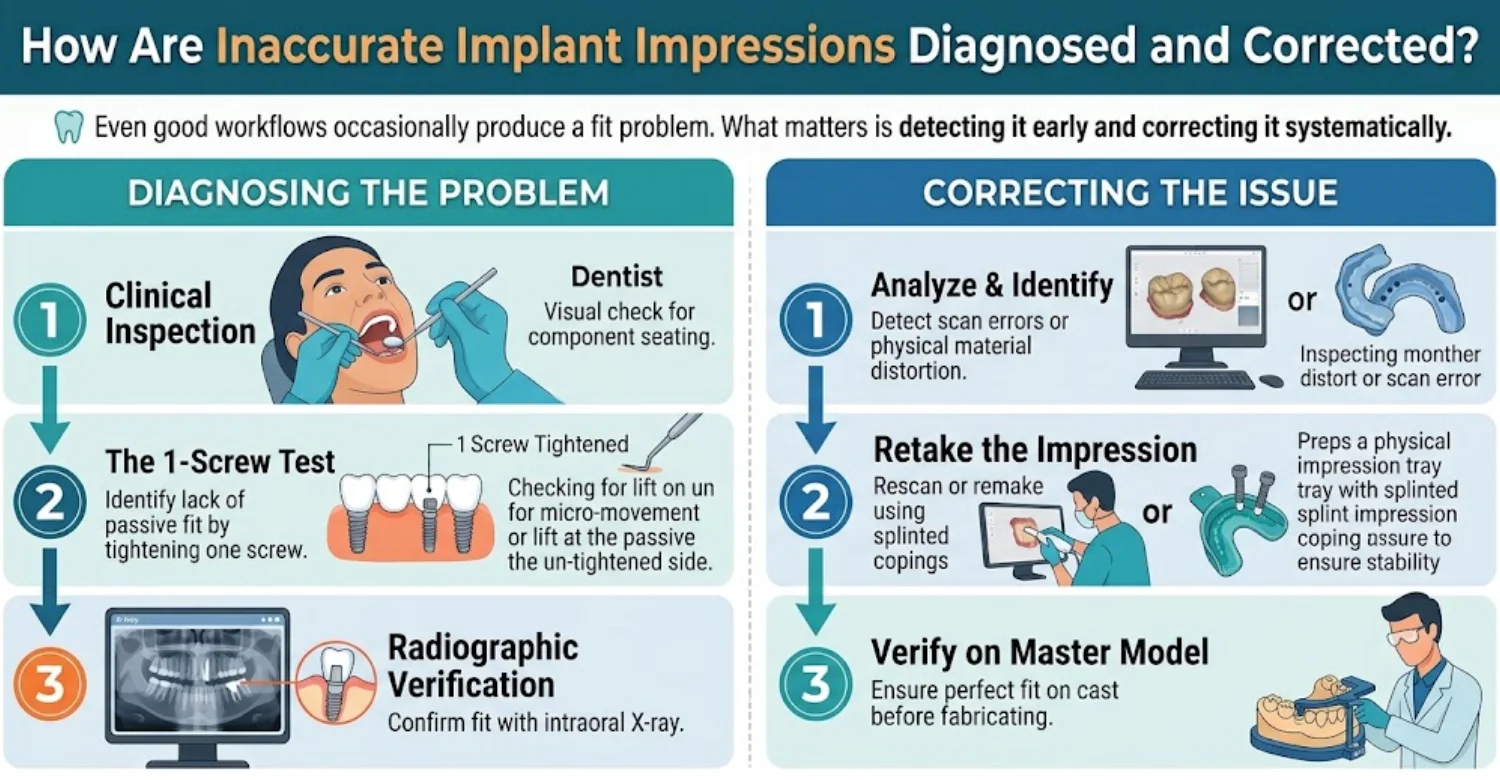
How Does a Dentist Evaluate the Fit of a Full Arch Implant Bridge?
The dentist first inspects the bridge visually and with magnification. Each implant connection is examined for gaps or uneven contact. The one screw test involves tightening a single screw and checking whether other sites lift. Patients may be asked about pressure or tension during seating. These simple checks often reveal problems quickly. Magnification loupes help reveal gaps invisible to the naked eye.
What Is the Role of Clinical Fit Checks and Verification Jigs?
A verification jig is a rigid device connecting all implants to confirm their recorded positions. It may be printed, milled, or made from splinted components. If the jig seats passively, the recorded positions are likely accurate. If it cracks or rocks, the impression may need repeating. Some clinics use printed prototypes instead of traditional jigs. Both approaches aim to confirm accuracy before final fabrication.
When Are Radiographs Appropriate During Implant Prosthesis Evaluation?
Radiographs help where implant connections lie below the gum and cannot be seen. Small periapical images can show gaps between the bridge and implant. Images should be taken only when clinically justified, following radiation safety principles. They are also used to assess bone levels around implants during follow up. Your dentist decides whether imaging adds useful information. Digital radiographs generally use lower doses than older film systems.
When Should an Implant Impression Be Repeated?
An impression should be repeated when verification reveals a clear positional error. Other reasons include missing scan data, distorted files, or a jig that does not seat. Repeating the record is usually faster than adjusting a misfitting final bridge. In some cases, switching to photogrammetry or a hybrid method improves the next record. The decision depends on the size and source of the error. Hybrid recording is one practical outcome of Photogrammetry vs Digital Scanning for Full Arch Implants research.
How Are Digital Design, Fabrication, or Seating Errors Corrected?
Design errors can be corrected in software and the bridge redesigned. Fabrication errors may require remilling or reprinting the affected part. A metal framework can sometimes be sectioned and rejoined to restore passive fit. Seating errors may relate to soft tissue trapped beneath the bridge. Each correction targets the specific step where the error entered. Clear records of each step make troubleshooting faster.
When Should a Patient Seek Professional Dental Care?
Some symptoms should never be ignored after implant treatment. Prompt care can prevent minor issues from becoming serious complications. Contact your dentist if you notice any of the following signs.
Persistent Pain, Swelling, Bleeding, or Discharge Around Implants
Pain or swelling that lasts beyond the normal healing period needs examination. Bleeding gums around implants may signal inflammation. Discharge or a bad taste may suggest infection. A widely cited meta analysis estimated peri implantitis at about 22% of implant patients. Early treatment is generally simpler and more effective. Smokers and patients with diabetes may face higher risk and need closer monitoring.
A Loose or Moving Implant Supported Bridge
A fixed bridge should not move when you chew or touch it. Movement may come from loose screws, component fracture, or implant problems. Continuing to chew on a loose bridge can worsen damage. Book an appointment promptly and avoid hard foods until assessed. Screw loosening is among the most commonly reported mechanical complications with screw retained bridges.
Sudden Bite Changes or a Fractured Prosthesis
A sudden change in how your teeth meet may indicate shifting or fracture. Chipped or cracked bridge material should be assessed before it spreads. Keep any broken pieces and bring them to the appointment. Prompt repair helps protect the implants and the remaining bridge.
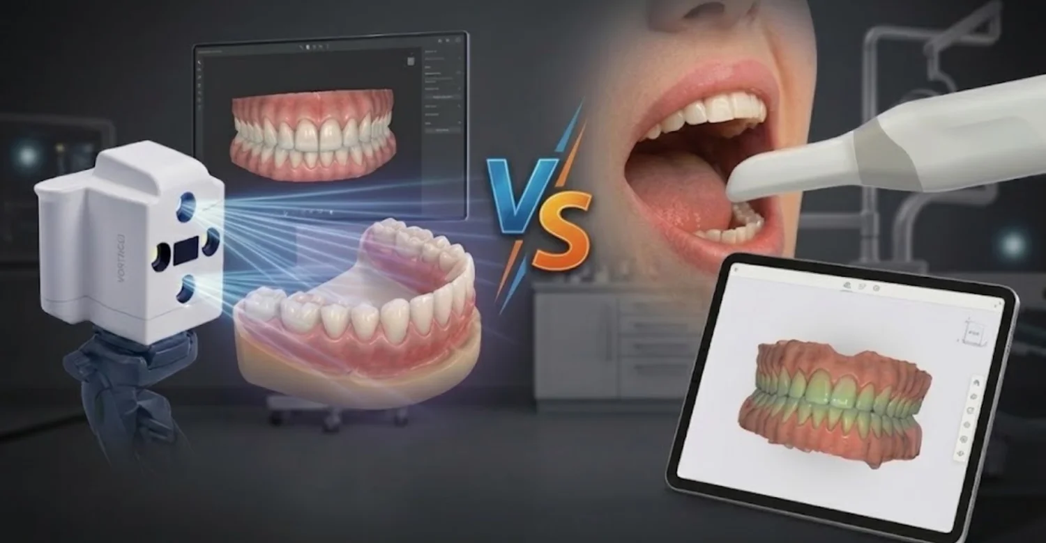
Note : All images used are for editorial and illustrative purposes only and may not originate from the original news provider or associated company.
Can More Accurate Digital Impressions Provide Relief from Repeated Fitting Problems?
Many patients searching Photogrammetry vs Digital Scanning for Full Arch Implants have experienced fitting frustrations. Better records can reduce some causes of repeated visits. They cannot solve every problem, however. This section sets realistic expectations.
Can Better Implant Position Records Reduce Repeated Impressions and Adjustments?
Yes, more reliable records can reduce the need for repeated impressions. When implant positions are captured accurately the first time, prototypes tend to seat more predictably. Fewer remakes mean fewer appointments and less chair time. Studies showing lower photogrammetry deviations support this expectation. Clinical confirmation from large trials is still developing. This benefit is one practical reason patients ask about Photogrammetry vs Digital Scanning for Full Arch Implants.
Can Digital Workflows Help Reduce Unnecessary Additional Appointments?
Digital workflows allow files to be shared instantly with the laboratory. Designs can be reviewed remotely before fabrication. Prototypes can be printed quickly for try in. These efficiencies often shorten the overall treatment timeline. For international patients, fewer trips can be a meaningful advantage. Planning still requires realistic time for healing and verification.
Why Can Scanning Accuracy Not Guarantee Pain Relief or a Perfect Fit?
Pain after implant treatment can have many causes unrelated to scanning. These include bite overload, gum inflammation, infection, nerve sensitivity, or jaw muscle tension. A perfect scan does not address these factors. Fabrication and seating also influence the final fit. Honest clinicians present scanning accuracy as one helpful factor, not a guarantee.
What Should Patients Do if an Existing Implant Bridge Feels Uncomfortable?
Contact your treating dentist and describe your symptoms clearly. Note when discomfort starts, what triggers it, and whether the bridge moves. Avoid adjusting or removing the bridge yourself. If your original clinic is unavailable, seek an implant experienced dentist. Bring treatment records, implant details, and X rays if possible. A structured assessment identifies the cause before any correction.
Side Effects, Discomfort, and Limitations of Digital Implant Scanning
Digital impressions are generally comfortable and well tolerated. Still, patients may experience minor, temporary discomfort. Both methods also have technical limits worth understanding. This section covers both aspects honestly.
What Discomfort Can Patients Experience During Intraoral Scanning?
Most patients find intraoral scanning more comfortable than traditional putty impressions. Some may feel jaw tiredness from keeping the mouth open. The scanner tip can press lightly on cheeks or lips. Patients with a strong gag reflex may find posterior scanning challenging. Short breaks during the scan help most people manage comfortably. A full arch scan typically takes several minutes per jaw.
Can Scan Bodies or Soft Tissue Retraction Cause Temporary Discomfort?
Attaching scan bodies may cause mild pressure, especially soon after surgery. Healing gums can feel tender when components are placed or removed. Cheek retractors may cause brief stretching of the lips. These sensations usually disappear shortly after the appointment. Local anesthetic is rarely needed but can be used if tissues are sensitive.
Can Photogrammetry Trigger Gagging or Jaw Fatigue?
Photogrammetry is usually quick, and the camera stays outside the mouth. This reduces the chance of gagging compared with a scanner tip reaching the back of the mouth. Patients must still keep the mouth open while markers are attached and photographed. Some jaw fatigue may occur during longer sessions. Overall, many patients find photogrammetry easy to tolerate.
Are Digital Impressions Associated with Radiation Exposure?
No, neither photogrammetry nor intraoral scanning uses ionizing radiation. Both rely on visible or near visible light and optical cameras. Radiation exposure in implant treatment comes from X rays and CBCT scans instead. Those are prescribed separately when clinically justified. Patients can undergo digital impressions without radiation concerns. By contrast, CBCT imaging delivers a measurable, though generally low, radiation dose.
What Are the Technical Limitations of Photogrammetry and Intraoral Scanner?
Each method has limitations:
Limitation | Photogrammetry | Intraoral Scanning |
Soft tissue capture | Not captured | Captured |
Long span error | Low | Can accumulate |
Equipment cost | Higher, specialized | Lower, widely available |
Technique sensitivity | Moderate | High |
Clinical evidence | Mostly laboratory | Broader overall |
Understanding these limits helps explain why many clinics combine both methods.
How Can Dental Teams Minimize Discomfort and Avoid Repeated Scans?
Teams can minimize discomfort with clear explanations, short breaks, and comfortable positioning. Good saliva control and isolation improve scan quality the first time. Checking scan bodies before recording prevents avoidable errors. Reviewing the file before the patient leaves catches missing data early. These habits reduce repeat visits and improve the overall experience. Short appointments also help patients with dental anxiety.
Average Cost of Photogrammetry vs Digital Scanning for Full-Arch Implants
Cost is an important consideration when choosing full-arch implant treatment. Digital impressions are rarely priced separately, so the total usually reflects the entire treatment. Prices vary by country, clinic, restoration materials, and case complexity. The figures below are illustrative planning ranges, not guaranteed quotes.
Average Full-Arch Implant Treatment Costs by Country
Does Photogrammetry Cost More Than Intraoral Scanning?
Photogrammetry systems are specialized and may cost more to purchase than intraoral scanners. Clinics may account for this investment in their overall treatment fees. However, digital impressions are commonly included in full-arch treatment packages rather than charged separately.
More accurate records may help reduce remakes and adjustments, although outcomes depend on the clinical workflow and case complexity. Patients should compare complete treatment packages rather than individual technology charges.
Average Full-Arch Implant Treatment Cost in the United States
The United States has relatively high full-arch implant treatment fees. Costs reflect clinician fees, laboratory charges, restoration materials, and regional operating expenses.
Cost Category | Estimated Cost |
Lower planning estimate per arch | $18,000 |
Upper planning estimate per arch | $30,000 |
Planning midpoint per arch | $24,000 |
Estimated midpoint for two arches | $48,000 |
Lower-cost options may involve acrylic hybrid bridges, while higher-cost treatments may include zirconia restorations, extractions, or bone grafting. Some complex cases may exceed these estimates. Patients should request an itemized quote covering surgery, temporary and final restorations, and follow-up care.
Average Full-Arch Implant Treatment Cost in the United Kingdom
Full-arch implant treatment in the United Kingdom is primarily provided through private dental practices. Prices vary by region, with some clinics in London charging more than practices elsewhere.
Cost Category | Estimated Cost |
Lower planning estimate per arch | $10,000 |
Upper planning estimate per arch | $18,000 |
Planning midpoint per arch | $14,000 |
Estimated midpoint for two arches | $28,000 |
The final price depends on implant selection, bridge material, clinical complexity, and the number of additional procedures required. Patients should confirm whether temporary restorations, follow-up appointments, adjustments, and maintenance are included.
Average Full-Arch Implant Treatment Cost in Turkey
Turkey is a popular destination for international patients seeking full-arch implant treatment. Differences in operating costs can make treatment less expensive than in the United States and United Kingdom. However, the final price depends on the clinic, materials, surgical requirements, and restoration design.
Cost Category | Estimated Cost |
Lower planning estimate per arch | $3,250 |
Upper planning estimate per arch | $8,450 |
Planning midpoint per arch | $5,850 |
Estimated midpoint for two arches | $11,700 |
Using these planning midpoints, the estimated cost in Turkey is approximately 76% lower than in the United States and 58% lower than in the United Kingdom per arch. These comparisons exclude travel, accommodation, and other costs that may not be included in treatment packages.
What Is Usually Included in a Full-Arch Implant Treatment Quote?
The components included in a quote differ between clinics. Reviewing the following items can help patients compare treatment packages accurately.
Treatment Component | What to Confirm |
Implant surgery | Number of implants, surgical fees, and anesthesia |
Abutments | Whether multi-unit abutments and related components are included |
Temporary prosthesis | Whether a temporary fixed bridge is included |
Final restoration | Material, design, and laboratory fees |
Digital impressions | Whether photogrammetry or intraoral scanning is included |
Prototype try-in | Whether a trial restoration and fit verification are included |
Additional procedures | Separate costs for extractions, bone grafting, or sinus lifts |
Follow-up care | Reviews, X-rays, adjustments, and maintenance |
Remakes and repairs | Policies and potential additional charges |
What Factors Can Increase the Total Cost of Full-Arch Implant Treatment?
Several clinical and restorative factors can increase the final treatment cost.
Cost Factor | Why It May Increase the Price |
Bone grafting or sinus lifts | Additional surgical procedures and materials may be required |
Zygomatic implants | Complex surgery and specialized planning may increase costs |
Premium implant components | Component selection can affect the overall treatment fee |
Zirconia final bridges | Laboratory work and material costs may be higher |
More implants per arch | Additional components and surgical work may be needed |
Sedation or general anesthesia | Separate professional and facility fees may apply |
Complex bite correction | Additional planning, adjustments, or restorative work may be necessary |
Prototypes, remakes, and maintenance | Extra laboratory work and follow-up procedures may add costs |
Before committing to treatment, request a written, itemized quote that identifies included services, exclusions, follow-up arrangements, and potential additional charges.
How Should Patients Compare Full Arch Implant Quotes Across Countries?
Compare quotes on equal terms rather than headline prices. Check implant brands, bridge materials, number of implants, and included visits. Ask about warranty terms and aftercare arrangements. For treatment abroad, add flights, accommodation, and time away from work. Consider how follow up will be handled after you return home. A cheaper quote with missing items may cost more overall.
Why Should Photogrammetry and Intraoral Scanner Prices Not Be Compared Without Checking What Is Included?
Impression technology is only one part of a full arch package. A clinic advertising photogrammetry may still exclude prototypes or the final bridge material you want. Another clinic using intraoral scanning may include extensive verification steps. Comparing technology alone can be misleading. In Photogrammetry vs Digital Scanning for Full Arch Implants, value depends on the complete workflow and its safeguards.
At Vitrin Clinic Treatment Pricing
The cost of dental implant treatment varies significantly depending on the procedure scope, implant brand, and country of treatment. The following table provides a transparent comparison of average costs across the United Kingdom, United States, and Vitrin Clinic in Istanbul from a single implant to full-arch restoration allowing patients to evaluate their options with accurate financial context before committing to a treatment plan.
Treatment type | 🇬🇧 UK | 🇺🇸 USA | 🇹🇷 Turkey |
|---|---|---|---|
Single dental implant Implant post + abutment + crown | $2,400 – $5,700 | $3,000 – $6,000 | $500 – $700 |
Implant-supported crown Crown on existing implant | $1,270 – $2,540 | $1,500 – $3,000 | $700 – $900 |
All-on-4 Per arch, fixed full-arch restoration | $10,000 – $22,000 | $18,000 – $30,000 | $3,500 – $7,500 |
All-on-6 Per arch, 6-implant full-arch restoration | $12,700 – $25,400 | $20,000 – $35,000 | $4,000 – $9,000 |
Full mouth reconstruction Both arches, upper + lower | $29,000 – $50,000+ | $36,000 – $70,000+ | $7,000 – $15,000 |
3D CT scan & consultation | Charged separately | Charged separately | Free & included |
Estimated saving | — | — | Up to 70% less |
* Prices are averages for reference only and may vary based on individual case requirements, implant brand, bone condition, and treatment complexity. A personalised quote is provided following your free consultation.






What We Notice Clinically
Clinical experience adds context that laboratory studies cannot provide. Patients often arrive with strong opinions about technology. Our observations focus on practical outcomes rather than device claims. These insights reflect common patterns in full arch care. Questions about Photogrammetry vs Digital Scanning for Full Arch Implants are now common during our consultations.
Why Full Arch Implant Cases Require Careful Implant Position Recording
A full arch bridge links implants that may sit four or five centimeters apart. Small angular errors at one end can produce larger gaps at the other. Tilted implants and multiunit abutments add further complexity. Careful recording protects the bridge, the screws, and the implants. We treat this step as a foundation rather than a formality. An angular error of 1 degree over 40 millimeters can shift a far point by roughly 0.7 millimeters.
Common Misconceptions About Digital Scanning Accuracy
Many patients believe a digital scan is automatically perfect. Others assume any new scanner guarantees an ideal fit. Some think photogrammetry makes verification unnecessary. In reality, Photogrammetry vs Digital Scanning for Full Arch Implants is about reducing errors, not eliminating them. Every method still depends on correct scan bodies, skilled operators, and careful checks. Clear explanations help patients set realistic expectations.
Why a More Advanced Scanner Cannot Replace Clinical Verification
Technology captures data, but verification confirms reality in the mouth. Even accurate records can be affected by later fabrication or seating steps. A prototype try in reveals problems that no screen can show. Patient feedback on bite, speech, and appearance also matters. We consider verification essential regardless of the recording device.
Clinical Note : Dr. Rifat Alsaman
Dr. Rifat Alsaman reviews full arch implant cases with a focus on prosthetic accuracy. His approach emphasizes matching the workflow to the patient rather than to a device. The following points summarize his clinical perspective.
The Importance of Case Selection, Scan Verification, and Passive Fit Assessment
Dr. Alsaman stresses that good outcomes start with proper case selection. Healthy tissues and stable implants are prerequisites for reliable records. He recommends checking every scan file before the patient leaves. Passive fit is confirmed with a prototype before the final bridge. This sequence reduces surprises at delivery.
Why the Best Impression Method Depends on the Clinical Situation
According to Dr. Alsaman, no single method suits every patient. Photogrammetry offers strong advantages in long spans and tilted implants. Intraoral scanning remains valuable for soft tissue, bite, and simpler distributions. Combining both often gives the most complete record. The final choice should follow the clinical findings, not marketing claims.
Tips for Patients
Being prepared helps patients make confident treatment decisions. The following tips cover consultation preparation, key questions, and aftercare. They apply whether you are treated at home or abroad.
What Should Patients Prepare Before a Full Arch Implant Consultation?
Gather recent X rays, CBCT scans, and previous dental records if available. List current medications and medical conditions, including diabetes and smoking habits. Note your main goals, such as chewing comfort, appearance, or speech. Write down your questions before the appointment. Bring details of any existing implants, including brand and placement date. Photos of your current smile can also help with aesthetic planning.
What Questions Should Patients Ask About Photogrammetry and Digital Scanning?
Useful questions include:
Which method will record my implant positions, and why?
Do you combine photogrammetry with intraoral scanning?
How do you verify scan body seating?
Will I have a prototype try before the final bridge?
What happens if the fit is not passive?
Clear answers suggest a structured approach to Photogrammetry vs Digital Scanning for Full Arch Implants.
How Can Patients Compare Treatment Plans and Itemized Quotes?
Ask each clinic for a written, itemized plan. Compare implant brands, number of implants, abutments, and bridge materials. Check whether prototypes, X rays, and follow up visits are included. Ask about warranty conditions and maintenance requirements. Compare total costs, including travel if relevant. The lowest headline price is not always the best value.
What Should Patients Know About Temporary and Final Implant Bridges?
Temporary bridges are usually made of acrylic and placed soon after surgery. They allow function while implants integrate with bone, typically over three to six months. Temporary bridges are less strong and may need gentle care. Final bridges are often zirconia or reinforced materials designed for long term use. Final records are usually taken after healing, when tissues are stable.
How Can Patients Maintain Implant Health After Final Restoration?
Clean around your bridge daily using a soft brush, water flosser, or interdental brushes. Professional maintenance visits are typically recommended every three to six months. Your dentist may remove the bridge periodically for deeper cleaning. Avoid smoking, which increases implant complication risk. Wearing a night guard may help if you grind your teeth. Research consistently links regular maintenance with lower rates of peri implant disease.
What Should Patients Avoid If an Implant Bridge Feels Loose or Painful?
Avoid chewing hard or sticky foods on the affected side. Do not try to tighten, remove, or glue the bridge yourself. Avoid delaying care, because small problems can become complex. Do not rely on painkillers to mask ongoing symptoms. Contact your dentist promptly and describe what you notice.
Full Arch Implant Planning and Digital Diagnostics at Vitrin Clinic
At Vitrin Clinic, full arch treatment begins with careful diagnosis and planning. We use digital tools to support clinical judgment, not replace it. Our goal is a stable, comfortable, and well fitting bridge. The following steps describe our planning approach.
Clinical Assessment Before Full Arch Implant Restoration
Every patient receives a detailed examination of teeth, gums, bone, and bite. We review medical history, medications, and lifestyle factors. Facial proportions and smile lines are assessed for aesthetic planning. Existing implants or bridges are evaluated for stability and fit. Photographs and digital smile records help us communicate the planned result. This assessment forms the basis of an individualized plan.
Digital Implant Records and Appropriate Diagnostic Imaging
We use CBCT imaging when clinically indicated to evaluate bone and anatomy. Digital records capture implant positions, soft tissues, and bite relationships. Imaging is prescribed only when it adds diagnostic value. All records are reviewed before fabrication begins. This combination supports precise and predictable planning. Records are stored securely for future reference and maintenance.
Personalized Selection of the Impression and Restoration Workflow
We select the recording method according to each patient's clinical situation. Implant number, angulation, and distribution guide this decision. Soft tissue condition and patient comfort are also considered. Our team explains the chosen approach and why it suits your case. The restoration workflow is then planned around reliable records. Where helpful, we combine more than one recording method.
Implant Supported Bridge Design and Fit Verification
Bridge design considers function, aesthetics, cleaning access, and long term durability. We use prototype try ins to check fit, bite, and appearance before final fabrication. Passive fit is verified carefully at each relevant stage. Adjustments are made before the final material is produced. This step-by-step verification protects your investment.
Individualized Treatment Planning Based on Oral Health and Prosthetic Needs
No two full arch cases are identical. We tailor implant number, bridge material, and timeline to your oral health. Patients with bone loss may need grafting or alternative implant designs. We discuss benefits, limitations, and realistic expectations openly. Your plan reflects your needs, not a standard template.
How Vitrin Clinic Supports Your Treatment Journey
Full arch treatment involves several stages over months. We aim to make each stage clear and well coordinated. Support continues from first contact to long term maintenance. Here is how we guide patients through the process.
Initial Consultation and Review of Existing Dental Records
Your journey starts with a consultation, either in person or remotely. You can share X rays, CBCT scans, and photographs in advance. Our team reviews these records and identifies any additional information needed. We discuss your goals, concerns, and medical background. This review allows a preliminary assessment before your visit. Remote reviews can save international patients an extra trip.
Personalized Treatment Planning and Discussion of Available Options
After assessment, we present a written treatment plan with clear options. We explain implant designs, bridge materials, and impression methods relevant to your case. Each option includes expected stages, timelines, and costs. You have time to ask questions and consider your choice. We never pressure patients toward a particular option.
Coordinating Implant Surgery, Digital Records, and Prosthetic Fabrication
Our team coordinates surgery, digital records, and laboratory work in a planned sequence. Clear communication between clinicians and technicians reduces delays. Digital files are shared securely with the laboratory. Prototype and final stages are scheduled around healing requirements. Coordination keeps your treatment efficient and organized. You receive a clear schedule before treatment starts.
Treatment Coordination for International Patients
We support international patients with appointment planning and travel guidance. Treatment visits are scheduled to make the most of each trip. A dedicated coordinator answers questions before, during, and after your stay. We explain healing periods and when return visits may be needed. Communication continues after you travel home. Many international patients complete full arch treatment across two main visits.
Follow Up, Prosthesis Maintenance, and Aftercare Planning
After delivery, we provide clear aftercare instructions and a maintenance schedule. Follow up visits check fit, bite, gum health, and screw stability. International patients receive guidance on local maintenance options. We remain available if concerns arise between visits. Long term care helps protect your implants and bridge.
Ready to transform your smile?
Talk to our team and get a personal FREE treatment plan.
Key Takeaways
This guide covered accuracy, workflow, costs, and patient considerations. The points below summarize the most important lessons. Use them as a checklist when discussing treatment with your dentist.
The Main Differences Between Photogrammetry and Intraoral Scanning
Photogrammetry measures implant positions simultaneously and resists cumulative error across long spans. Intraoral scanning records teeth, gums, and bite but may accumulate stitching errors. Recent meta analyses from 2025 and 2026 favor photogrammetry for trueness and precision. Most evidence comes from laboratory studies. Many clinics combine both methods for complete records.
Why Implant Position Accuracy and Passive Fit Matter
Implants cannot move like natural teeth, so the bridge must fit without strain. Misfit can contribute to loose screws, component fractures, and repeated adjustments. Accurate records support passive fit but do not guarantee it. Verification with a prototype remains essential before final delivery. Ask your clinic how it verifies fit before delivery.
How Clinical Assessment Helps Determine the Most Appropriate Method
Implant number, angulation, spacing, and tissue condition guide the choice of method. Operator experience and available equipment also matter. A thorough assessment matches the workflow to your specific case. Photogrammetry vs Digital Scanning for Full Arch Implants is best decided clinically, case by case.
What Patients Should Consider Before Choosing a Full Arch Implant Treatment Plan
Compare itemized quotes rather than headline prices. Ask which recording and verification methods the clinic uses. Confirm implant brands, bridge materials, and aftercare arrangements. Consider travel and follow up logistics for treatment abroad. Choose a team that explains its process clearly.
When to Seek Professional Evaluation for Persistent Discomfort or a Loose Implant Bridge
Seek prompt evaluation for persistent pain, swelling, bleeding, or discharge around implants. A moving or loose bridge needs urgent assessment. Sudden bite changes or fractured material also require attention. Early care usually means simpler treatment and better protection of your implants. Keep your treating clinic's contact details easily accessible.
At Vitrin Clinic
At Vitrin Clinic, we combine careful clinical assessment with modern digital records for full arch dental implant treatment. We select the impression method that suits your implants, anatomy, and goals. Every bridge is planned with fit verification and long term maintenance in mind. Our team explains each step and provides transparent, itemized treatment plans. If you are researching Photogrammetry vs Digital Scanning for Full Arch Implants, we can review your case. Share your X rays or scans with us for a personalized assessment. Contact Vitrin Clinic today to book your consultation and discuss your options.
Sources
FAQs
Dr. Rifat Alsaman has more than 5 years of clinical experience in dentistry and currently serves as the Head of the Medical Team at Vitrin Clinic. He is dedicated to providing exceptional patient care, overseeing treatment planning, and ensuring the highest clinical standards across the team. His expertise, attention to detail, and commitment to continuous professional development have helped countless patients achieve healthier, more confident smiles.


