General Dentistry
August 31, 2026
Cementum: Definition, Function, Composition, and Its Role in Dental Health?

Cementum is the mineralized tissue that covers the root surface of every tooth. It sits beneath the gum line, where it usually stays hidden from view. Patients often ask about it when they notice gum recession, root sensitivity, or signs of periodontal disease, since these issues can expose the root. Unlike enamel, which protects the visible crown, this layer supports the tooth from below. Understanding its role helps patients recognize when exposure or damage needs professional attention rather than home remedies. This guide explains it from a clinical perspective, covering its structure, function, and care.
Cementum: What Is It and Why Is It Important?
It is the thin, calcified layer that surrounds the anatomical root of a tooth. It is not visible during a normal smile, since it lies below the gumline. Still, it plays a central role in keeping teeth anchored in the jaw. This tissue differs from the hard enamel covering the crown; it is softer and works together with the periodontal ligament. In simple terms, the short answer comes down to this: a protective, living layer that supports the tooth from below. Patients rarely think about it. Dentists, however, consider this layer clinically important for long-term periodontal health.
Where Is Cementum Located?
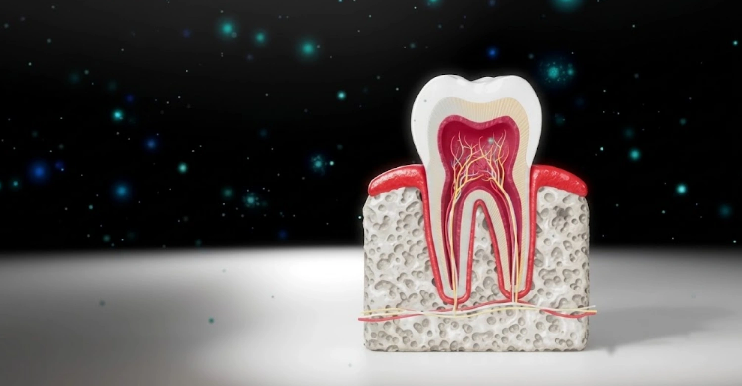
This tissue is located around the root of the tooth, beneath the gum line and adjacent to the periodontal ligament and surrounding bone. It connects with dentin on one side. It meets enamel at a specific point near the neck of the tooth, known as the enamel junction. Dentists use this junction as an important anatomical landmark during clinical examinations. Its condition can indicate gum recession, wear, or structural changes. These signs may need further evaluation and monitoring over time.
Cementum vs. Enamel and Dentin
Enamel, dentin, and this layer are the three mineralized tissues that make up a tooth, each with a different role. Enamel covers the crown and is the hardest substance in the body. Dentin lies beneath it and extends into the root. This layer covers the root itself and is softer than both enamel and dentin, containing less mineral content. Because it lacks enamel's hardness, exposed root surfaces are far more vulnerable to wear, sensitivity, and decay once the protective gum tissue recedes.
Cementum Composition and Structure
Composition-wise, this layer is made mostly of mineral content, primarily calcium phosphate, combined with an organic matrix of collagen fibres and water. This mixture gives it enough rigidity to anchor periodontal fibres while remaining slightly flexible compared with enamel. Rather than a passive coating, it functions as a living, biologically active tissue that responds to mechanical forces on the tooth. Its structure allows continuous, gradual deposition throughout life. This supports ongoing attachment as the periodontal ligament adapts to normal function.
Cellular vs. Acellular Cementum
There are two recognized types. Acellular tissue forms first and covers most of the root surface, particularly near the crown, without embedded cells. Cellular tissue develops later and contains cementocytes within its structure, produced by cementoblasts. It is typically found in the apical third of the root and in furcation areas of multi-rooted teeth. This cellular form is especially important there because it can continue adapting to functional changes. It can also repair minor damage over time.
Acellular Cementum
This acellular type covers most of the root surface, extending from the enamel junction near the crown toward the root apex. It forms slowly and does not contain cementocytes, giving it a denser, more stable structure. Its main role is anchoring the periodontal ligament. Sharpey's fibres embed directly into this layer to hold the tooth firmly within its socket. Because it develops early and remains relatively static, this layer is considered the primary attachment tissue for long-term periodontal stability and tooth support.
Cellular Cementum
This cellular type is found mainly toward the root apex and in furcation regions between multiple roots. It contains cementocytes housed in small spaces called lacunae, similar to bone tissue. Because of this cellular activity, it can continue to form and adapt throughout a person's life. It responds to occlusal forces and minor root surface changes. This adaptability makes it useful for ongoing attachment as periodontal ligament fibres adjust, although it is generally considered less mineralized than the acellular layer covering the rest of the root.
Tips for Patients
Use a soft-bristled toothbrush.
Avoid aggressive horizontal brushing.
Use fluoride toothpaste as recommended by a dental professional.
Maintain daily flossing or interdental cleaning.
Limit frequent exposure to acidic foods and drinks.
Do not ignore persistent tooth sensitivity.
Attend regular dental and periodontal examinations.
Follow professional recommendations after periodontal or restorative treatment.
When Should You See a Dentist?
Persistent sensitivity.
Gum recession.
Bleeding or swollen gums.
Visible root surfaces.
Pain while brushing.
Signs of root decay.
How to Protect Cementum and Exposed Root Surfaces
Use a soft-bristled toothbrush and gentle strokes rather than aggressive horizontal scrubbing, which can wear down exposed root surfaces. Fluoride toothpaste, recommended by a dental professional, helps strengthen vulnerable areas. Daily flossing or interdental cleaning keeps plaque from accumulating near the gumline. Limiting frequent exposure to acidic foods and drinks reduces surface erosion. Persistent sensitivity should never be ignored. Regular dental and periodontal check-ups support long-term protection. So does following professional advice after any periodontal or restorative treatment.
When Should You See a Dentist?
A dental visit is recommended for persistent sensitivity that does not improve, visible gum recession, or bleeding and swollen gums. Visible root surfaces, pain while brushing, and signs of root decay are also reasons to seek assessment. Tooth mobility or noticeable changes in bite should never be dismissed, since these can indicate underlying periodontal involvement. Early evaluation allows the cause to be identified accurately. This generally leads to simpler, more conservative treatment than waiting until symptoms progress further.
Cementum Function Teeth: What Does Cementum Do?
The main role it plays for teeth is providing an attachment surface for periodontal ligament fibres, which connect the tooth to surrounding bone. This attachment contributes directly to tooth anchorage and stability within the jaw. It also protects the underlying root dentin from external exposure. Ongoing deposition allows the root to adapt gradually over time. This helps maintain a stable relationship between the tooth and its supporting structures, which is essential for long-term periodontal health and normal chewing function.
Cementum and Periodontal Ligament
The relationship between this tissue and the periodontal ligament is central to how teeth stay anchored. Collagen bundles known as Sharpey's fibres run from the ligament directly into the root surface layer, creating a strong mechanical attachment. This connection holds the tooth suspended within its socket rather than fused rigidly to bone. It allows slight, healthy movement during chewing. A well-functioning periodontal ligament depends on this attachment remaining intact. This is why any disruption to either tissue can affect overall tooth stability.
What Happens When Cementum Becomes Exposed?
Exposure of this root surface tissue commonly results from gum recession or periodontal disease, both of which uncover areas normally protected by gum tissue. Aggressive brushing over time can also contribute, gradually wearing away both gum and root surface. Root exposure sometimes follows periodontal treatment as tissue levels adjust. Once uncovered, the area often becomes more sensitive, since it lacks enamel's protective hardness. Recognizing these causes helps patients understand why sensitivity develops and why professional evaluation is worthwhile.
Common Symptoms of Exposed Cementum
Common symptoms include noticeable sensitivity to cold drinks or foods, discomfort while brushing, and reactions to acidic or sweet items. Patients may also see visible root surfaces near the gumline, which can appear yellowish compared with the whiter enamel above. Because the exposed area lacks enamel's protection, there is an increased risk of root-surface caries developing in that location. These symptoms often prompt a dental visit. Identifying them early allows for simpler, more conservative management options.
Does Exposed Cementum Mean the Tooth Is Damaged?
Exposure alone does not automatically mean a tooth is structurally failing. In many cases, it reflects gum-level changes rather than deep damage to the tooth itself. However, professional assessment remains important, since exposure can sometimes accompany underlying periodontal disease or wear that needs treatment. Dentists distinguish between normal, gradual exposure associated with ageing or minor recession and pathological exposure linked to active disease. This distinction determines whether monitoring or active treatment is the appropriate next step.
Common Dental Problems Affecting Cementum
Several conditions can affect this root surface tissue, including root caries, abrasion from aggressive brushing, and general sensitivity. Gingival recession is a frequent cause of exposure, often linked with periodontal disease and root surface damage. Less commonly, excessive formation known as hypercementosis can occur, thickening the layer beyond normal levels. Each of these problems has different causes and treatment approaches. This is why an accurate diagnosis matters more than treating surface symptoms like sensitivity alone.
Cementum and Gum Recession
Gingival recession exposes root surfaces that are normally covered by healthy gum tissue. Contributing factors include aggressive brushing technique, periodontal disease, thin gum tissue, and in some cases orthodontic movement or grinding habits. Mild recession may simply need monitoring and improved hygiene technique. Progressive or symptomatic recession, though, often requires professional treatment. A dentist evaluates the extent of recession, underlying cause, and periodontal health before recommending whether preventive care, periodontal therapy, or surgical correction is appropriate for a given case.
Cementum and Root Caries
Exposed root surfaces are more susceptible to decay than enamel-covered crowns, since this tissue is softer and less mineralized. Plaque accumulation near an exposed root can lead to root caries relatively quickly if left unmanaged. Preventive measures include fluoride use, improved cleaning around the gumline, and reducing sugar or acid exposure. When decay has already started, fluoride treatment alone may not be sufficient. Restorative treatment is generally recommended to remove decayed tissue and protect the remaining root structure.
How Do Dentists Assess Cementum and Root-Surface Problems?
Assessment typically starts with a clinical examination and periodontal probing to measure gum attachment levels around each tooth. Dentists evaluate the extent of gingival recession, assess reported sensitivity, and check for plaque or calculus buildup near exposed areas. The periodontal ligament and surrounding supporting tissues are also reviewed for signs of inflammation or mobility. Dental radiographs are used when clinically indicated. They help confirm findings that cannot be assessed through visual examination or probing alone.
When Is Dental Imaging Necessary?
X-rays help evaluate the root and surrounding supporting structures when clinical findings suggest deeper involvement, such as bone loss, root caries extending below the gumline, or unclear symptoms. Imaging is not used routinely for every patient. Instead, it is selected according to individual clinical need. A dentist decides whether radiographs are necessary based on examination findings, reported symptoms, and periodontal probing results. This ensures imaging adds useful diagnostic information rather than being performed as a standard, unnecessary step.
Clinical Note
This tissue is biologically important because it provides the attachment surface for periodontal ligament fibres that anchor each tooth. Exposure is often a consequence of another condition, such as gingival recession or periodontal disease, rather than a standalone disease itself. Treatment should target the underlying cause. Patients with persistent sensitivity, visible root exposure, bleeding gums, or changes in tooth stability should receive a professional dental examination. As Dr. Rifat Alsaman notes, diagnosis should always precede any cosmetic or restorative intervention.
Treatment Options for Cementum and Exposed Root Surfaces
Treatment depends entirely on the underlying cause identified during examination. Options range from conservative approaches like fluoride application and desensitizing products, to restorative solutions such as bonding or fillings for decayed areas. When gum recession is significant, periodontal therapy or gum grafting may be considered. Rather than treating sensitivity in isolation, dentists aim to address why the root surface became exposed in the first place. Resolving the underlying issue offers more predictable, lasting results for patients.
Fluoride Treatment
Professional fluoride applications can help reduce sensitivity and strengthen vulnerable exposed root surfaces by supporting mineral content in the affected area. This treatment is often recommended for patients with mild to moderate sensitivity or early signs of root surface softening. It is typically applied during a dental visit and may be repeated periodically depending on individual risk factors. Fluoride treatment works best as part of a broader preventive plan that includes improved hygiene and dietary adjustments, rather than as a standalone fix.
Desensitizing Treatments
Managing root sensitivity can involve both professional and at-home approaches. In the clinic, dentists may apply desensitizing agents directly to the exposed area to block nerve signals within the dentin. At home, patients can use desensitizing toothpaste formulated for sensitive teeth as part of their daily routine. These options are often combined, with professional treatment providing faster relief and home care maintaining results over time. Persistent sensitivity despite these measures usually indicates a need for further evaluation of the underlying cause.
Dental Bonding for Exposed Root Surfaces
Tooth-colored composite material may be used to cover exposed root surfaces, particularly where sensitivity or aesthetic concerns are present. Bonding offers both a protective barrier and an improved appearance, blending with surrounding tooth structure. However, it has limitations: bonded areas may wear over time and require maintenance or replacement. It is generally considered a conservative option compared with more involved periodontal surgery. It suits smaller areas of exposure rather than extensive recession affecting multiple teeth.
Periodontal Treatment
When exposure relates to gum disease, treatment focuses on managing the underlying periodontal condition. This usually involves professional cleaning to remove plaque and calculus, along with periodontal therapy tailored to the severity of inflammation and attachment loss. Treating periodontal inflammation is essential, since ongoing disease can worsen recession and root exposure over time. Once inflammation is controlled, the risk of further root surface exposure typically decreases. Any remaining sensitivity or aesthetic concerns can then be addressed separately.
Gum Grafting and Root Coverage
Periodontal plastic surgery, such as gum grafting, may be considered for more significant recession where root coverage is desired. The goal is generally to improve coverage and reduce sensitivity rather than promise complete restoration of original gum levels. Outcomes are influenced by factors including the extent of recession, tissue thickness, and overall periodontal health. A dentist evaluates whether a patient is a suitable candidate. They also discuss realistic expectations before recommending this more involved surgical treatment option.
Restorative Treatment for Root Caries
A restoration becomes necessary when decay has affected the root surface and simple preventive measures are no longer sufficient. Dentists determine appropriateness based on the extent of decay, its location, and how much sound tooth structure remains. Restorative treatment typically involves removing decayed material and placing a suitable filling material to protect the area. Because root surfaces differ from enamel, material choice and technique are adapted accordingly. This ensures a durable, well-sealed result for the patient.
What Can Patients Expect Before and After Treatment?
Before any treatment, patients undergo a clinical examination, diagnosis of the underlying cause, and assessment of periodontal health, sensitivity, and root exposure. This informs an individualized treatment plan suited to their specific situation. After treatment, patients can expect reduced sensitivity when the cause has been properly managed, along with improved protection of exposed root surfaces. Aesthetic improvement is possible depending on the treatment chosen. Ongoing preventive care remains important, and outcomes vary based on gum health, recession severity, and hygiene.
Before Treatment
Before treatment begins, a thorough clinical examination is carried out to diagnose the exact cause of symptoms. This includes assessing periodontal health, evaluating the degree of sensitivity, and reviewing the extent of root exposure. Based on these findings, an individualized treatment plan is developed rather than applying a generic approach. This preparatory stage ensures that any recommended treatment directly addresses the underlying issue. It may involve preventive care, periodontal therapy, restorative work, or a combination of these approaches.
After Treatment
After appropriate treatment, patients generally experience reduced sensitivity once the underlying cause has been properly managed. Exposed root surfaces receive better protection, lowering the risk of further wear or decay. Some treatments, particularly restorative or periodontal options, may also offer aesthetic improvement. However, ongoing preventive care remains essential to maintain results long-term. Outcomes vary according to individual factors such as gum health, severity of recession, oral hygiene habits, and which treatment approach was selected for that patient.
What We Notice Clinically
Dr. Rifat Alsaman emphasizes protecting exposed root surfaces early, favoring conservative management of sensitivity whenever possible before considering more involved procedures. He notes that patients should avoid self-diagnosing root-surface problems, since symptoms like sensitivity can stem from several different causes that require professional differentiation. Regular periodontal examinations are highlighted as essential, allowing early detection of recession or attachment loss before it progresses. This approach prioritizes preserving natural tooth structure whenever a conservative option remains appropriate for the patient.
How Vitrin Clinic Approaches Patient-Centred Dental Care
Vitrin Clinic prioritizes comprehensive dental assessment before recommending any treatment, ensuring decisions are based on accurate diagnosis rather than symptoms alone. Modern diagnostic and digital dental technologies are used where clinically appropriate to support this process. Treatment planning can combine periodontal, restorative, and cosmetic considerations depending on individual needs, with Dr. Rifat Alsaman contributing to the clinic's professional perspective. Individualized treatment plans are favored over one-size-fits-all procedures. Patients are encouraged to receive a proper examination and discuss their concerns directly with the team.
Why Professional Assessment Matters
Concerns involving this root surface tissue can stem from several different underlying causes, from recession to decay to periodontal disease. Early assessment helps prevent progression of these root-surface problems before they become more complex to manage. A dentist can determine whether preventive, periodontal, restorative, or cosmetic treatment is the appropriate path forward for a given patient. Without proper assessment, treatment risks addressing only surface symptoms. It may fail to resolve the actual issue affecting long-term periodontal health and tooth stability.
Final Takeaway
It is essential to tooth support, providing the attachment surface that periodontal ligament fibres rely on for stability. Understanding the difference between this tissue, enamel, and dentin helps patients recognize why exposed root surfaces behave differently from the visible crown. Exposed root structure may indicate an underlying periodontal or gingival issue rather than being a problem on its own. Persistent sensitivity, gum recession, root exposure, or root decay all warrant professional assessment. Vitrin Clinic offers individualized consultations to evaluate these concerns and plan appropriate treatment.
References:
FAQs

Dr. Rifat Alsaman has more than 5 years of clinical experience in dentistry and currently serves as the Head of the Medical Team at Vitrin Clinic. He is dedicated to providing exceptional patient care, overseeing treatment planning, and ensuring the highest clinical standards across the team. His expertise, attention to detail, and commitment to continuous professional development have helped countless patients achieve healthier, more confident smiles.
.webp&w=3840&q=75)


