General Dentistry
August 31, 2026
What Is Tooth Enamel and What Does It Do?

Tooth enamel is the highly mineralized outer layer that protects every visible tooth surface. It helps teeth resist chewing forces, temperature changes, acids, and bacterial attacks. Understanding its structure can explain why enamel preservation matters throughout life. Enamel contains no living cells, so significant damage cannot naturally regenerate like skin. Early mineral loss can sometimes be repaired through saliva, fluoride, and improved oral care. However, advanced enamel loss requires professional evaluation and treatment. At Vitrin Clinic, enamel preservation is considered before cosmetic or restorative treatment. A careful assessment helps identify whether damage results from erosion, abrasion, attrition, or decay. This distinction allows dentists to select conservative treatment whenever possible. The following guide explains enamel structure, functions, damage, prevention, and modern treatment options.
What Is Tooth Enamel?
Enamel is the hard outer covering surrounding the crown of every tooth. It protects the underlying dentin and deeper dental tissues from daily environmental challenges. Enamel is primarily composed of mineral crystals arranged into an organized microscopic structure. It is harder than bone and is considered the hardest material in the human body. Despite its strength, enamel can gradually lose minerals under repeated acidic conditions. Bacterial acids can also contribute to mineral loss during tooth decay. Unlike many body tissues, mature enamel contains no cells capable of repairing major structural damage. Saliva can support mineral replacement during early demineralization. Fluoride can further improve resistance against future acid attacks. According to NIDCR, early tooth decay can sometimes be stopped or reversed before cavitation develops.
Where Is Tooth Enamel Located?
Enamel covers the crown of each tooth, which is the portion visible above the gumline. It forms a protective shell around the underlying dentin. The enamel layer is thickest over areas receiving strong chewing forces. Its thickness varies between different teeth and different regions of each tooth. Incisal edges and chewing surfaces experience particularly significant mechanical stress. Enamel becomes thinner toward the cervical region near the gums. It also meets cementum around areas where the tooth transitions toward the root. This arrangement helps protect the tooth while allowing attachment to surrounding periodontal tissues. When gums recede, previously protected root surfaces may become exposed. Those surfaces lack the same thick enamel covering found on the crown. Therefore, exposed roots can become more vulnerable to sensitivity and wear.
What Is the Enamel Layer Made Of?
The enamel layer is mainly composed of calcium and phosphate arranged into mineral crystals. These crystals form a highly organized material called hydroxyapatite. Water and small amounts of organic material are also present within the structure. Mineral content gives enamel its exceptional hardness and resistance to mechanical forces. However, mineral content alone does not explain its remarkable fracture resistance. Researchers increasingly recognize that enamel's microscopic organization also influences strength. NIDCR reported that enamel can appear normal in thickness yet still become structurally weaker. The organization of mineral rods helps distribute forces through the tooth. This structure allows enamel to tolerate repeated chewing cycles. Acid exposure can disturb the mineral balance and weaken susceptible areas. Saliva continually supplies minerals that support surface repair during early mineral loss.
Why Is Tooth Enamel Important?
Enamel protects the more sensitive structures located beneath the tooth surface. It shields dentin from temperature changes, mechanical forces, and chemical challenges. Without adequate enamel, everyday eating can become uncomfortable or painful. Enamel also contributes significantly to the natural appearance of teeth. Its translucency allows underlying dentin characteristics to influence perceived tooth color. When enamel becomes thinner, teeth can appear more yellow because dentin becomes more visible. Enamel also provides the foundation for many conservative cosmetic dental treatments. Preserving healthy enamel can support predictable bonding and restoration outcomes. Its condition can influence decisions involving veneers, bonding, crowns, and whitening procedures. Early professional evaluation can identify changes before substantial structural loss occurs. For these reasons, enamel preservation remains central to preventive and restorative dentistry.
What Is the Structure of Tooth Enamel?
The structure consists of tightly packed mineral crystals organized into microscopic enamel rods. These rods extend from the dentinoenamel junction toward the external surface. Their orientation changes across different regions of the tooth. This arrangement helps distribute chewing forces rather than concentrating stress at one location. The structure also creates differences in hardness between enamel regions. Surface enamel can differ from deeper enamel in mineral composition and organization. The dentinoenamel junction provides an important transition between hard enamel and resilient dentin. This interface helps limit crack propagation through the tooth. Modern research shows that structural organization influences enamel durability. Therefore, enamel strength depends on architecture as well as mineral concentration.
How Is Tooth Enamel Structured?
Enamel consists of millions of microscopic rods or prisms packed closely together. Each rod contains highly organized mineral crystals. The rods follow complex paths from the dentin toward the external surface. Their changing orientation helps manage repeated chewing forces. Between rods, specialized regions contribute to the overall structural organization. The outer surface can also contain a more mineralized protective zone. Enamel is not a completely uniform material throughout its thickness. Mineral density, crystal orientation, and organic components can vary microscopically. These differences influence how enamel responds to acids and mechanical stress. Research continues to investigate why some enamel structures resist cracking better than others. NIDCR's recent findings emphasize that internal organization can influence enamel fragility.
What Minerals Make Up Tooth Enamel?
The dominant mineral is calcium phosphate arranged primarily as hydroxyapatite crystals. Calcium and phosphate provide much of enamel's hardness. Fluoride can become incorporated into mineral structures during remineralization processes. Fluoride-containing mineral phases are generally more resistant to acid dissolution. Magnesium, sodium, carbonate, and other trace elements can also occur within enamel. Their concentrations vary according to location, age, and developmental conditions. Water and organic components occupy relatively small proportions of the mature tissue. Mineral composition therefore influences both mechanical performance and chemical resistance. Dietary acids can dissolve mineral components when exposure becomes frequent or intense. Saliva helps restore mineral ions during periods between acidic challenges. Fluoride can further support this mineral balance through topical exposure.
What Gives Tooth Enamel Its Strength?
Enamel strength comes from both mineral content and microscopic structural organization. Highly packed hydroxyapatite crystals provide exceptional hardness. Enamel rods distribute forces through carefully organized crystal arrangements. The dentinoenamel junction also helps reduce the risk of catastrophic cracking. Dentin underneath provides a more flexible foundation than enamel. This combination creates a functional relationship between a hard shell and resilient core. Recent NIDCR research highlights the importance of enamel's internal architecture. Some enamel can have normal mineral levels yet demonstrate increased fragility. This finding shows why thickness alone cannot determine tooth durability. Chewing habits, acid exposure, grinding, and previous damage can also influence performance. Professional assessment considers these factors when evaluating weakened or worn teeth.
Enamel Rods and Crystals
Enamel rods are microscopic structural units extending through the enamel layer. Their mineral crystals are arranged in highly organized patterns. Adjacent rods can change orientation to improve resistance against mechanical stress. This organization contributes to enamel's ability to withstand repeated chewing. Rod architecture also influences how cracks travel through the tissue. When structural organization becomes disrupted, enamel may become more vulnerable. NIDCR researchers recently studied how rod arrangement affects fracture resistance. Their findings challenge the idea that mineral density alone determines enamel strength. This insight may eventually influence future preventive and restorative strategies. For patients, the key message is simple. Healthy enamel depends on both its chemistry and microscopic architecture.
Mineral Density and Enamel Strength
Mineral density strongly influences enamel hardness and resistance to chemical dissolution. Highly mineralized regions generally provide greater resistance to mechanical wear. However, density does not completely determine fracture resistance. Crystal orientation and rod organization also influence how forces move through enamel. Acid exposure can reduce mineral density near the tooth surface. Repeated episodes may create progressively softer areas. Saliva can help restore minerals during early stages of demineralization. Fluoride can strengthen the mineralized surface and increase acid resistance. NIDCR research suggests that two enamel samples with similar mineral density can behave differently. This highlights the importance of structural integrity during clinical assessment.
Clinical Note: Why Enamel Structure Matters
Enamel structure matters because damage rarely occurs from one factor alone. Dentists often see combinations of erosion, abrasion, attrition, and bacterial acid damage. A smooth worn surface may indicate chemical softening followed by mechanical forces. Cracks may reflect repeated stress rather than simple mineral loss. NIDCR's recent research also highlights the importance of internal enamel architecture. Clinicians therefore assess texture, thickness, cracks, wear patterns, and exposed dentin. Bite relationships can provide additional information about mechanical loading. Dietary history can reveal frequent exposure to acidic beverages or foods. The goal is identifying the underlying cause before selecting treatment. At Vitrin Clinic, this assessment can help guide conservative enamel-preserving treatment decisions.
What Is the Function of Tooth Enamel?
The primary function is protecting the tooth from mechanical and chemical challenges. Enamel absorbs and distributes forces generated during biting and chewing. It also protects dentin from temperature changes and acidic environments. This protective layer reduces direct exposure of deeper dental tissues. Enamel contributes to the appearance, shape, and surface texture of natural teeth. It also plays an important role in preventing progression of early mineral loss. Saliva and fluoride can support its mineral balance after mild acid exposure. However, enamel cannot regenerate after extensive structural destruction. Preventive care therefore remains more effective than replacing lost tissue later. Understanding these functions helps patients appreciate why conservative dentistry prioritizes preservation.
How Does Enamel Protect the Tooth?
Enamel forms the first physical barrier against mechanical and chemical challenges. It withstands pressure created during biting, chewing, and food processing. Its mineralized structure also reduces penetration toward the dentin. Acid attacks can temporarily soften the surface and increase vulnerability. Saliva helps buffer acids and supplies minerals during recovery periods. Fluoride increases resistance against acid-related mineral loss. Together, these protective mechanisms reduce the likelihood of progressive tooth destruction. However, repeated challenges can eventually overwhelm natural protective processes. The ADA identifies frequent acidic exposure as an important risk factor for erosive tooth wear.
Does Enamel Prevent Tooth Decay?
Enamel provides important protection against tooth decay, but it cannot prevent every cavity. Decay develops through interactions among bacteria, dietary carbohydrates, saliva, fluoride, and tooth structure. Bacteria metabolize sugars and produce acids that remove minerals from the tooth. Repeated acid exposure can shift the balance toward progressive mineral loss. Early lesions may appear as white spots without obvious cavities. NIDCR states that early decay can sometimes be stopped or reversed. Once a cavity forms, lost tooth structure cannot simply regenerate. Preventive strategies therefore aim to maintain mineral balance before cavitation develops.
How Does Enamel Protect Dentin?
Enamel covers dentin across the crown and reduces its exposure to external stimuli. Dentin contains microscopic tubules connected toward the dental pulp. When exposed, these tubules can transmit temperature and pressure changes. This can produce the sharp discomfort commonly associated with dentin sensitivity. Maintaining the enamel layer therefore reduces direct stimulation of dentinal structures. Acid erosion can thin the protective layer gradually. Abrasion and grinding can further accelerate tissue loss. Once dentin becomes exposed, sensitivity may become more noticeable. Professional treatment focuses on controlling the cause and protecting exposed surfaces.
How Does Enamel Affect Tooth Appearance?
Enamel influences tooth color, brightness, translucency, and surface texture. Healthy enamel is naturally translucent rather than completely opaque. Its optical properties allow underlying dentin to influence overall tooth color. When enamel becomes thinner, yellowish dentin can become increasingly visible. Acid erosion can also create smoother, flatter, or more translucent surfaces. Staining may become more noticeable when structural changes alter light reflection. Cosmetic dentistry therefore begins with understanding the condition of natural enamel. Whitening cannot rebuild lost enamel or restore structural thickness. Conservative cosmetic planning should consider whether discoloration reflects staining, dentin visibility, or enamel loss.
How Strong Is Tooth Enamel?
Enamel is exceptionally hard and highly mineralized. NIDCR identifies it as the hardest material in the human body. Its hardness allows teeth to withstand substantial repeated chewing forces. However, hardness does not mean immunity from cracking, erosion, or wear. Acid exposure can soften the surface and make mechanical damage easier. Grinding can generate repeated forces capable of producing wear and cracks. Trauma can also fracture enamel despite its hardness. Recent research demonstrates that internal structure influences fracture resistance. Therefore, strong enamel still requires protection from preventable chemical and mechanical challenges.
Is Tooth Enamel the Hardest Substance in the Human Body?
Yes, enamel is widely recognized as the hardest material in the human body. Its exceptional hardness comes from its very high mineral content. Hydroxyapatite crystals form the primary structural component. However, hardness does not make enamel completely resistant to damage. Enamel can fracture under sudden trauma or repeated excessive forces. It can also lose minerals during frequent acid exposure. NIDCR describes enamel as the body's hardest material while emphasizing its inability to heal itself. This distinction is clinically important. A hard material can still be vulnerable to specific chemical and mechanical stresses.
What Makes Enamels So Strong?
The exceptional strength comes from densely packed mineral crystals and organized microscopic architecture. Enamel rods create a complex structural network throughout the tissue. Crystal orientation helps distribute forces during chewing. The dentinoenamel junction also provides support beneath the hard outer layer. Saliva and fluoride help maintain mineral integrity after minor acid challenges. However, repeated exposure can gradually shift the balance toward mineral loss. Recent NIDCR research suggests that rod architecture also influences fracture resistance. This explains why thickness and mineral density alone cannot predict performance.
Can Strong Enamels Still Crack or Wear Away?
Yes, strong enamel can crack, chip, flatten, or gradually wear. Excessive biting forces can create microscopic cracks that enlarge over time. Bruxism can produce repeated loading during sleep or waking hours. Acid erosion can soften surfaces before mechanical forces remove weakened minerals. Hard objects can also cause localized chips or fractures. Enamel loss is generally permanent once substantial tissue has disappeared. Early detection can prevent minor defects from becoming larger restorative problems. Dentists may recommend behavioral changes, protective appliances, fluoride, or restorative treatment. The correct approach depends on the cause and severity.
What Can Damage Tooth Enamel?
Several chemical, biological, and mechanical factors can damage enamel. Frequent acidic foods and beverages can cause erosive mineral loss. Bacterial acids generated from dietary sugars contribute to tooth decay. Grinding can cause mechanical wear and stress-related fractures. Aggressive brushing can contribute to abrasion under certain circumstances. Reduced saliva can weaken natural acid-buffering and mineral-replenishing protection. These factors often interact rather than occurring independently. For example, acid-softened surfaces may become more vulnerable to mechanical abrasion. Identifying the dominant cause is essential before treatment begins. ADA guidance emphasizes reducing frequent acid exposure and maintaining appropriate oral hygiene.
How Do Acidic Foods and Drinks Affect Enamel?
Acids can dissolve minerals from the tooth surface through chemical erosion. Frequent exposure gives saliva less time to restore mineral balance. Carbonated drinks, sports drinks, fruit juices, and acidic foods can contribute. The ADA identifies frequent acidic beverage consumption as a major erosion risk. One study found that 93% of 379 tested beverages had pH values below 4.0. Lower pH generally indicates greater potential for acid-related mineral dissolution. However, pH is not the only factor determining erosive potential. Buffering capacity, calcium content, exposure frequency, and contact time also matter.
Can Sugar and Plaque Damage Enamel?
Sugar itself does not directly dissolve enamel in the same way as dietary acids. Plaque bacteria metabolize sugars and produce acids that lower local pH. Repeated acid production can remove minerals from susceptible tooth surfaces. Frequent sugary snacks can therefore increase the duration of acid challenges. NIDCR explains that bacteria exposed to sugars and starches generate acids. These acids attack enamel and cause mineral loss. Early mineral loss can appear as a white spot. Fluoride and saliva can support repair during early stages. Reducing sugar frequency can help shift the balance toward remineralization.
Does Teeth Grinding Wear Down Enamel?
Teeth grinding can produce significant mechanical wear over time. Bruxism repeatedly loads opposing tooth surfaces during clenching or grinding. Common signs include flattened cusps, polished surfaces, cracks, and sensitivity. Grinding can also place excessive stress on fillings, crowns, and other restorations. Acid exposure may increase vulnerability by temporarily softening the surface. Dentists may recommend a custom night guard when clinically appropriate. Stress management and treatment of contributing factors may also be considered. The correct approach depends on whether grinding occurs during sleep or waking hours. Early assessment can help prevent progressive structural damage.
Can Aggressive Brushing Damage the Enamel Surface?
Normal brushing with appropriate technique is unlikely to remove healthy enamel significantly. However, excessive pressure and abrasive products can contribute to tooth wear. The ADA recommends soft-bristled toothbrushes and fluoride toothpaste. Aggressive brushing can be particularly problematic when surfaces have already been softened by acids. Brushing immediately after acidic exposure may increase mechanical wear on softened surfaces. Rinsing with water first can provide a gentler response. Patients should use controlled movements rather than forceful scrubbing.
How Does Dry Mouth Affect Enamel Protection?
Saliva provides several protective functions for teeth. It helps neutralize acids, clear food particles, and supply minerals. Reduced salivary flow therefore decreases important natural protection. Dry mouth can increase the risk of tooth decay and mineral loss. Certain medications, dehydration, medical conditions, and mouth breathing can reduce salivary comfort. Drinking water can support hydration and improve oral comfort. Sugar-free chewing gum can stimulate saliva in suitable patients. Persistent dry mouth should receive professional assessment because its underlying cause may require treatment.
What Are the Signs of Damaged Tooth Enamel?
Enamel damage can produce several visible and functional changes. Sensitivity may develop when underlying dentin becomes increasingly exposed. Teeth can appear yellow because thinner enamel reveals more dentin. Surfaces may become rough, smooth, flattened, or uneven. Chips and cracks can indicate mechanical stress or structural weakness. Cavities may develop when mineral loss progresses beyond early stages. Some early damage causes no symptoms at all. This makes routine dental examinations important even when teeth feel normal. ADA guidance notes that early erosive changes can sometimes be difficult to detect clinically.
Tooth Sensitivity
Sensitivity can occur when protective enamel becomes thinner and dentin becomes exposed. Cold drinks, hot foods, sweets, and air may trigger short sharp sensations. Not every sensitivity problem results from enamel loss. Gum recession, cracks, decay, and defective restorations can produce similar symptoms. Therefore, sensitivity should not automatically be attributed to weakened enamel. Dentists evaluate the location, duration, intensity, and triggering factors. Treatment depends on the underlying cause. Fluoride products, desensitizing toothpaste, bonding, or restorative treatment may sometimes help. Persistent or worsening sensitivity deserves professional evaluation.
Yellow or Darker-Looking Teeth
Teeth can appear darker when enamel becomes thinner. The underlying dentin naturally has a more yellow appearance. Enamel erosion can therefore change perceived tooth color without external staining. Surface staining from foods, drinks, or tobacco can create another type of discoloration. These causes require different treatment approaches. Whitening can address certain surface and internal discoloration patterns. However, whitening cannot replace lost enamel. Excessive whitening may also increase temporary sensitivity in some patients. A dental examination can distinguish staining from structural color changes.
Rough or Uneven Enamel Surface
A rough surface can indicate erosion, abrasion, developmental defects, or other structural changes. Patients may notice unusual texture when their tongue touches affected teeth. Erosion may create smooth depressions, flattened areas, or cupping. Abrasion can produce localized grooves near the gumline. Attrition often creates matching wear facets on opposing teeth. These patterns provide valuable diagnostic clues. Dentists evaluate location, shape, symmetry, and surrounding tissues. Identifying the pattern helps determine which behavior or condition should be addressed first.
Chipped or Cracked Teeth
Chips can occur when enamel experiences sudden excessive forces. Cracks may develop from repeated loading, trauma, grinding, or weakened tooth structure. Small enamel fractures may cause no symptoms initially. Larger cracks can produce pain during biting or sensitivity to temperature. Some fractures extend into dentin or the dental pulp. Prompt evaluation can prevent further structural deterioration. Treatment may range from monitoring to bonding, crowns, or other restorative procedures. The appropriate treatment depends on fracture depth, location, symptoms, and remaining tooth structure.
Increased Risk of Cavities
Damaged or demineralized surfaces can become more vulnerable to caries development. Bacterial acids remove minerals during repeated low-pH episodes. If mineral loss exceeds remineralization, the lesion can progress. NIDCR explains that early decay may be stopped or reversed before cavitation. Once a cavity forms, restorative treatment becomes necessary. Fluoride, saliva, and reduced sugar frequency can support prevention. Regular dental examinations can identify early changes before major structural damage occurs.
What Is Enamel Demineralization?
Demineralization occurs when acids remove minerals from the tooth surface. It represents a normal part of the ongoing mineral balance in the mouth. Saliva and fluoride can support remineralization after mild mineral loss. Problems arise when mineral loss occurs more frequently than mineral replacement. Dietary acids and bacterial acids can both contribute. Frequent sugar consumption can increase bacterial acid production. Frequent acidic drinks can directly expose tooth surfaces to erosive acids. The balance between demineralization and remineralization determines whether damage progresses.
What Causes Enamel Demineralization?
Common causes include bacterial acids, dietary acids, and reduced protective saliva. Sugar and starch exposure can feed acid-producing plaque bacteria. Carbonated drinks, fruit juices, and sports drinks can provide direct acidic challenges. Stomach acid can contribute when reflux or recurrent vomiting occurs. Dry mouth can reduce natural buffering and mineral replacement. Poor oral hygiene can allow plaque to remain against tooth surfaces longer. Frequent exposure matters because recovery requires time between challenges. The ADA emphasizes exposure frequency as an important factor in erosive tooth wear.
What Is Enamel Remineralization?
Remineralization is the natural replacement of minerals lost from early tooth surfaces. Saliva supplies calcium, phosphate, and other components needed for mineral recovery. Fluoride can enhance this process and improve resistance to future acid attacks. Early caries lesions can therefore sometimes become arrested or reversed. NIDCR specifically notes that saliva and fluoride can help repair early mineral loss. Remineralization cannot rebuild a large cavity or replace a missing enamel fragment. The process works best when mineral loss remains limited and the surface is intact.
Can Fluoride Help Strengthen Enamel?
Yes, fluoride can strengthen the tooth surface and support remineralization. It can become incorporated into mineral structures that resist acid dissolution more effectively. Fluoride also helps reduce bacterial acid production under appropriate conditions. Fluoride toothpaste is a widely used preventive measure. Professional fluoride treatments may benefit patients with elevated caries risk. NIDCR states that fluoride strengthens the hard outer tooth surface. ADA guidance also recognizes fluoride as effective for preventing and controlling dental caries.
Can Lost Enamels Be Naturally Replaced?
Minor mineral loss can sometimes be repaired through remineralization. However, physically lost enamel cannot grow back naturally. Once substantial tissue has been removed through erosion, abrasion, or fracture, the body cannot recreate it. Early lesions are therefore different from advanced structural defects. Fluoride and saliva can support early mineral recovery when the surface remains intact. Restorative dentistry becomes necessary when significant tissue has disappeared. Dentists may use bonding, veneers, or crowns depending on severity. Preventing further loss remains essential after any restorative treatment.
Can Tooth Enamel Grow Back?
Mature enamel cannot regenerate after significant structural loss. It contains no living cells capable of producing new enamel tissue. This differs from some body tissues that can repair themselves through cellular activity. Early mineral loss is different because minerals can return to an intact surface. Saliva and fluoride can support this process. Once enamel chips away or erodes permanently, replacement requires dental materials. This distinction is one of the most important concepts in preventive dentistry. Protecting natural enamel is therefore preferable to replacing it whenever possible.
Can Damaged Enamels Regenerate?
Extensive enamel damage cannot regenerate naturally. Mature enamel lacks the living cells needed to rebuild missing tissue. Early demineralization may improve when the mineral balance becomes favorable. Fluoride and saliva play important roles during this early repair process. However, a visible chip or deep erosion defect cannot simply grow back. Dentists can restore lost structure using materials designed for different clinical situations. The treatment choice depends on remaining tooth structure, function, aesthetics, and disease activity. Preventing continued damage remains essential after restoration.
Can You Remineralize Tooth Enamel?
You can support remineralization when mineral loss remains early and the surface remains intact. Fluoride toothpaste is one of the most established preventive tools. Saliva supplies minerals and buffers acids naturally. Reducing frequent sugar and acidic drink exposure creates better recovery conditions. Good plaque control also reduces bacterial acid production. NIDCR explains that early decay may stop or reverse with minerals from saliva and fluoride. Remineralization cannot rebuild a missing piece of enamel. Professional assessment can determine whether a lesion remains suitable for preventive management.
How Can Dentists Treat Enamel Loss?
Treatment depends on the amount and cause of structural loss. Early mineral changes may require prevention, fluoride, and monitoring. Small defects may be treated with dental bonding. Larger cosmetic defects can sometimes involve veneers. Extensive structural loss may require crowns or other restorations. Dentists should also identify the cause before restoring damaged surfaces. Otherwise, grinding, acid exposure, or poor hygiene may continue damaging the restoration. Treatment planning should therefore combine restoration with prevention. At Vitrin Clinic, preserving remaining natural tooth structure can guide restorative decisions.
Fluoride Treatments
Professional fluoride treatments can provide concentrated topical fluoride exposure. They may be recommended for patients with increased caries risk. Fluoride supports remineralization and improves resistance to acid-related mineral loss. Treatment type and frequency depend on individual risk factors. Professional applications do not rebuild physically missing enamel. Their main role is preventive and protective. Patients should continue daily fluoride toothpaste use unless their dentist recommends otherwise. NIDCR and ADA recognize fluoride as an effective strategy against tooth decay.
Dental Bonding
Dental bonding can restore small chips, worn edges, and selected areas of enamel loss. Tooth-colored resin is carefully shaped to reproduce natural contours. Bonding is often considered conservative because it can preserve more natural tooth structure. It can also improve appearance when discoloration or minor shape changes are present. However, bonding does not regenerate natural enamel. Its longevity depends on bite forces, habits, location, and material conditions. Patients who grind their teeth may require additional protective measures. Professional evaluation determines whether bonding is appropriate.
Veneers
Veneers are thin restorations designed to improve selected front-tooth cosmetic concerns. They can address certain changes involving shape, color, and surface structure. Enamel preservation is important because bonding to enamel can support predictable veneer retention. The amount of preparation depends on the clinical design and existing tooth anatomy. Not every patient needs extensive enamel reduction. Modern conservative planning aims to preserve healthy tooth structure whenever possible. Vitrin Clinic can evaluate enamel condition before recommending cosmetic treatment. Veneers should not be used simply to hide active disease or untreated structural problems.
Dental Crowns
Crowns cover a larger portion of the tooth when substantial structural support is needed. They may be considered for severely worn, fractured, or weakened teeth. A crown can protect remaining tooth structure while restoring shape and function. However, crown preparation generally requires more tooth alteration than conservative bonding. Therefore, crowns should be selected according to clinical need. Dentists first assess cracks, decay, remaining structure, occlusion, and symptoms. Addressing the underlying cause remains essential before definitive restoration.
How Can You Protect Tooth Enamel?
Protection begins with reducing repeated chemical and mechanical challenges. Brush twice daily with fluoride toothpaste and use appropriate technique. Clean between teeth daily to control plaque accumulation. Limit frequent acidic drinks and sugary snacks. Drink water regularly to support saliva and oral clearance. Avoid using teeth to open packages or bite hard objects. Discuss grinding symptoms with a dentist when appropriate. Routine dental examinations can identify early structural changes. These habits help preserve natural tooth structure for longer. ADA guidance recommends reducing acid exposure and using soft-bristled toothbrushes.
Brush Your Teeth Gently and Correctly
Gentle brushing removes plaque without unnecessary mechanical pressure. Use a soft-bristled toothbrush and fluoride toothpaste. Hold the brush comfortably and avoid aggressive horizontal scrubbing. Excessive pressure can contribute to abrasion, especially around exposed root surfaces. Brushing should cover outer, inner, and chewing surfaces systematically. Replace toothbrushes when bristles become significantly worn. Electric toothbrushes can help control brushing pressure when used correctly. The goal is effective plaque removal rather than forceful cleaning. ADA guidance supports soft-bristle brushing and fluoride toothpaste for daily care.
Use Fluoride Toothpaste
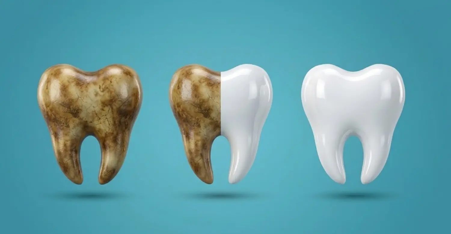
Fluoride toothpaste helps strengthen tooth surfaces and prevent decay. It supports remineralization of early mineral loss. The ADA notes that cavity-protection toothpastes carrying its Seal contain fluoride. Brushing twice daily is a common evidence-based recommendation. After brushing, avoid immediately washing away all toothpaste residue when appropriate. Children require age-appropriate fluoride guidance from dental professionals. High-risk adults may sometimes need prescription-strength fluoride products. These should be used according to professional recommendations.
Limit Frequent Acid Exposure
Frequency of acid exposure can be as important as the specific acidic food. Repeated sipping keeps teeth exposed for longer periods. Try consuming acidic drinks during meals instead of throughout the day. Water can help rinse and dilute acids afterward. Avoid holding or swishing acidic beverages around the mouth. Using a straw may reduce contact with front teeth for some drinks. The ADA recommends reducing dietary acid exposure to lower erosion risk.
Maintain Good Oral Hygiene
Good hygiene reduces plaque accumulation and bacterial acid production. Brush twice daily with fluoride toothpaste. Clean between teeth using floss or interdental cleaners. Regular dental visits allow early detection of mineral loss and structural changes. Patients with high caries risk may require personalized preventive schedules. Diet and hygiene should be considered together rather than separately. ADA guidance emphasizes reducing sugar frequency and maintaining effective oral hygiene.
Stay Hydrated and Support Healthy Saliva
Saliva naturally protects teeth by buffering acids and supplying minerals. Dehydration can reduce salivary comfort and potentially increase oral vulnerability. Drinking water regularly supports hydration and helps clear food residues. Sugar-free chewing gum can stimulate salivary flow for suitable patients. Persistent dry mouth should receive professional evaluation. Dentists can identify contributing medications, habits, or underlying conditions. Additional saliva substitutes may sometimes provide symptomatic relief. Protecting saliva is an important part of maintaining the oral environment.
Tips for Patients
Avoid frequent acidic drinks between meals whenever possible. Choose water when you need regular hydration throughout the day. Brush gently using fluoride toothpaste and a soft-bristled toothbrush. Do not brush immediately after a significant acidic exposure. Rinse with water first and allow the mouth to recover. Reduce frequent sugary snacks to limit bacterial acid production. Ask a dentist about sensitivity, grinding, cracks, or visible wear. Never assume severe tooth discoloration results from surface staining alone. Regular examinations can identify early structural changes before treatment becomes more complex. At Vitrin Clinic, personalized assessment can help connect symptoms with their underlying cause.
Tooth Enamel vs. Dentin: What Is the Difference?
Enamel and dentin are both mineralized dental tissues, but they have different structures. Enamel forms the outer crown surface. Dentin lies underneath enamel and surrounds the dental pulp. Enamel is harder and more highly mineralized. Dentin contains microscopic tubules and has greater resilience. Because dentin transmits stimuli through its tubules, exposure can cause sensitivity. Enamel protects dentin from direct environmental challenges. When enamel wears away, dentin can become increasingly exposed. This changes both tooth appearance and sensory response. Understanding this relationship helps explain why preserving enamel is clinically important.
What Is the Difference Between Enamel and Dentin?
Enamel is the hardest tissue and covers the crown. Dentin lies beneath enamel and forms most of the tooth's internal structure. Enamel contains a very high proportion of minerals. Dentin contains more organic material and water than enamel. Dentin is therefore less hard but more resilient. The two tissues work together during chewing and function. Enamel resists external forces while dentin provides supportive flexibility underneath. Their interface helps distribute stress throughout the tooth. Damage to one tissue can eventually affect the other.
Why Is Dentin More Sensitive Than Enamel?
Dentin contains microscopic tubules connected toward the dental pulp. Fluid movement within these tubules can stimulate nerves indirectly. Enamel lacks these open tubules and therefore does not respond similarly. When enamel becomes thin, exposed dentin can react to cold, heat, sweets, or touch. Sensitivity therefore often increases after substantial surface loss. However, sensitivity has many possible causes beyond enamel wear. A dentist should evaluate persistent or localized symptoms. Correct diagnosis prevents unnecessary treatment and identifies cracks, decay, or gum recession.
What Happens When Enamel Wears Away?
As enamel becomes thinner, dentin becomes increasingly exposed. Teeth may appear more yellow because dentin becomes more visible. Sensitivity can increase, particularly with cold or sweet foods. Tooth surfaces may become flatter, smoother, or more irregular. Mechanical forces can accelerate wear after acid exposure softens surfaces. Cavities may also develop when mineral loss progresses. Severe structural loss can compromise tooth shape and function. Treatment aims to stop the damaging process and restore appropriate structure. Early intervention generally allows more conservative management.
How Does Tooth Enamel Affect Cosmetic Dentistry?
Enamel condition strongly influences cosmetic dental planning. Healthy enamel can provide excellent support for adhesive restorative procedures. Excessive preparation can unnecessarily remove valuable natural tissue. Dentists therefore evaluate thickness, surface integrity, discoloration, cracks, and wear patterns. Cosmetic treatment should not mask untreated disease or active erosion. Patients with significant wear may require functional stabilization before cosmetic procedures. The final treatment should address both appearance and long-term structural stability. Conservative dentistry aims to improve aesthetics while preserving healthy natural tissue.
Why Is Preserving Natural Enamels Important?
Natural enamel is biologically integrated with the tooth's underlying structures. Once physically removed, it cannot naturally grow back. Preserving it can maintain stronger natural protection against environmental challenges. It may also support adhesive procedures and future treatment flexibility. Excessive preparation can reduce remaining tooth structure unnecessarily. Conservative dentistry therefore emphasizes minimal intervention when clinically appropriate. Patients should ask whether treatment can achieve the desired result while preserving healthy tissue. A thorough assessment helps determine the safest approach.
How Does Enamel Affect Dental Veneers?
Enamel condition influences veneer planning, bonding, preparation, and long-term stability. Healthy enamel can provide a favorable surface for adhesive bonding. Significant enamel loss may change the available bonding substrate. The dentist may then need to modify the restoration design. Veneers should not be used to treat uncontrolled grinding or active erosion alone. The underlying cause must be addressed first. Conservative veneer planning aims to preserve healthy enamel whenever possible. At Vitrin Clinic, enamel assessment can form part of cosmetic treatment planning.
How Does Enamel Affect Dental Bonding?
Bonding can work effectively when sufficient healthy tooth structure remains. Resin material is attached to prepared tooth surfaces using adhesive techniques. Preserving enamel can improve the available bonding substrate in appropriate cases. Bonding can restore small chips, worn edges, and selected cosmetic imperfections. However, it cannot recreate the biological properties of natural enamel. Its durability depends on location, bite forces, habits, and maintenance. Patients with grinding may need protective measures. Dentists assess enamel conditions before recommending bonding.
When Is Enamel Loss Severe Enough to Require Restoration?
Restoration may become appropriate when structural loss affects function, appearance, sensitivity, or tooth integrity. Small stable changes may require monitoring rather than immediate restoration. Larger defects can expose dentin and increase sensitivity. Significant wear may alter bite relationships or reduce chewing efficiency. Cracks can create additional structural risks. Restorative options depend on remaining tissue and the cause of damage. Treatment should not proceed without controlling active erosion, decay, or excessive loading. A dentist can determine the least invasive appropriate option.
What We Notice Clinically
Clinical assessment often reveals patterns that patients cannot identify themselves. Wear location can provide clues about its cause. Smooth widespread surfaces may suggest chemical erosion. Matching facets may suggest tooth-to-tooth attrition. Localized grooves can indicate mechanical abrasion. Cracks may indicate excessive loading or weakened structure. Dentists also examine discoloration, sensitivity, exposed dentin, and restoration conditions. Combining these observations improves diagnostic accuracy. Treatment should target both the visible damage and its underlying cause.
Clinical Note: Common Patterns of Enamel Wear
Different wear patterns often point toward different contributing factors. Acid erosion commonly creates smooth, shallow, flattened, or cupped surfaces. Attrition often creates matching facets where opposing teeth contact. Abrasion may produce localized grooves or defects near accessible surfaces. Bruxism can produce widespread flattening combined with cracks or muscle symptoms. Several mechanisms can occur simultaneously in the same patient. This makes history-taking especially important. Dietary habits, brushing technique, reflux symptoms, and grinding should all be considered. A pattern-based approach can reduce unnecessary restorative treatment.
How Dentists Identify the Cause of Enamel Damage
Dentists combine clinical examination with patient history. They evaluate location, shape, texture, color, sensitivity, and severity. Dietary questions can identify frequent acidic drink consumption. Brushing habits can reveal possible mechanical abrasion. Bite evaluation can identify excessive contacts and grinding patterns. Medical history may reveal reflux or medication-related dry mouth. Photographs and dental records can help monitor progression over time. In selected cases, radiographs or other diagnostic tools may be appropriate. The objective is identifying why damage occurred before restoring it.
Dr. Rifat Alsaman’s Opinion on Enamel Preservation
Dr. Rifat Alsaman emphasizes the value of protecting healthy tooth structure whenever treatment allows. Enamel loss should first be evaluated rather than immediately covered cosmetically. Identifying the cause can prevent repeated damage after restoration. Conservative treatment may be preferable when structural loss remains limited. Patients should understand that restorations replace missing structure but do not recreate natural enamel. A long-term plan should therefore include prevention and maintenance. This approach can support both oral health and cosmetic outcomes.
How Does Vitrin Clinic Approach Tooth Enamel Preservation?
Vitrin Clinic approaches enamel-related concerns through individualized clinical assessment. The first objective is understanding the condition and cause of structural change. Treatment recommendations can then be matched to the patient's functional and cosmetic needs. Preventive options may be considered before restorative procedures. When restoration is necessary, preserving remaining natural structure remains an important consideration. Cosmetic treatment should also account for bite forces and long-term maintenance. This approach helps avoid unnecessary intervention. Each patient's enamel condition can differ significantly, requiring personalized planning.
Comprehensive Enamel Assessment
Assessment begins with examining tooth surfaces and identifying visible structural changes. Dentists may evaluate wear, cracks, discoloration, sensitivity, and exposed dentin. Bite relationships can reveal possible mechanical loading. Dietary and oral hygiene histories can identify chemical or bacterial risk factors. Previous restorations can also influence current structural stability. Photographic documentation may help monitor changes over time. The purpose is not simply finding damaged areas. The goal is understanding the overall condition of the patient's teeth. This information supports more precise treatment planning.
Identifying the Cause of Enamel Damage
Identifying the cause is essential because restoration alone may not stop progression. Acid erosion requires reducing repeated acid exposure. Grinding may require occlusal management or protective appliances. Plaque-related mineral loss requires improved hygiene and caries prevention. Dry mouth may require hydration strategies and professional management. Multiple causes may coexist in the same patient. Dentists therefore consider lifestyle, diet, medical history, and oral function together. Treating the cause helps protect both natural teeth and future restorations.
Personalized Treatment Planning
Treatment planning should match the severity and cause of structural loss. Early changes may benefit from preventive measures and monitoring. Minor defects may be suitable for conservative bonding. Larger structural problems may require veneers or crowns. Functional problems may require bite stabilization before cosmetic treatment. The objective is achieving appropriate function while preserving natural tissue. Vitrin Clinic can develop treatment recommendations according to individual clinical findings. Patients should receive clear explanations before making irreversible treatment decisions.
Why Choose Vitrin Clinic for Enamel-Related Dental Care?
Enamel-related treatment requires both preventive and restorative knowledge. Vitrin Clinic combines clinical assessment with individualized treatment planning. The focus remains on understanding the cause before selecting a procedure. Patients can discuss cosmetic concerns alongside functional and preventive considerations. This integrated approach can be especially useful when enamel wear affects appearance. Treatment recommendations should always reflect individual anatomy and clinical findings. Patients should understand the benefits, limitations, and maintenance requirements of proposed restorations.
Experienced Dental Team
An experienced dental team can recognize subtle differences between erosion, abrasion, attrition, and decay. These distinctions influence treatment decisions significantly. Clinical experience also helps dentists evaluate restorative needs conservatively. Vitrin Clinic's dental team can assess both functional and cosmetic concerns. The objective is not simply correcting visible defects. Long-term stability and preservation of remaining tooth structure also matter. Individual treatment plans should reflect each patient's clinical situation.
Modern Dental Technology
Modern dental technology can support accurate diagnosis and treatment planning. Digital photographs can document surface changes and cosmetic concerns. Digital scans can assist with analyzing tooth shape and restoration design. Diagnostic imaging can help evaluate deeper structures when clinically indicated. Technology does not replace clinical judgment. Instead, it provides additional information for better decision-making. Vitrin Clinic can integrate appropriate digital tools into individualized dental workflows.
Personalized Patient Care
Every patient has different enamel thickness, wear patterns, bite forces, and dietary habits. Treatment therefore should not follow a single standardized cosmetic formula. Personalized care begins with listening to the patient's concerns. Dentists then combine those concerns with clinical findings and preventive needs. Treatment options can be discussed according to severity and long-term goals. Follow-up care helps monitor results and prevent recurrence. This patient-centered approach supports informed decision-making.
Focus on Preserving Natural Tooth Structure
Preserving natural tooth structure is a core principle of conservative dentistry. Removing healthy tissue should have a clear clinical justification. Small defects may sometimes be treated without extensive preparation. Preventive measures can also delay or avoid restorative intervention. When restorations become necessary, dentists should consider how much healthy structure remains. At Vitrin Clinic, treatment planning can prioritize preservation alongside aesthetics and function. This approach supports long-term dental health.
Conclusion About Tooth Enamel
Tooth enamel provides the first major defense against daily dental challenges. Its mineralized structure allows teeth to withstand substantial chewing and environmental stress. However, hardness does not make enamel indestructible. Acid exposure, bacterial acids, grinding, abrasion, trauma, and dry mouth can contribute to damage. Early mineral loss can sometimes improve through saliva and fluoride. Significant structural loss cannot naturally regenerate. This makes prevention and early diagnosis particularly valuable. Patients should maintain fluoride exposure, control plaque, limit frequent acids, and protect saliva. Persistent sensitivity, visible wear, cracks, or discoloration should receive professional evaluation. At Vitrin Clinic, enamel preservation can be considered during preventive, restorative, and cosmetic treatment planning. Protecting natural structure today can support healthier teeth for years.
Reference
FAQs

Dr. Rifat Alsaman has more than 5 years of clinical experience in dentistry and currently serves as the Head of the Medical Team at Vitrin Clinic. He is dedicated to providing exceptional patient care, overseeing treatment planning, and ensuring the highest clinical standards across the team. His expertise, attention to detail, and commitment to continuous professional development have helped countless patients achieve healthier, more confident smiles.
.webp&w=3840&q=75)


