General Dentistry
August 31, 2026
What Are the Different Parts of a Tooth?

Parts of a Tooth , Understanding the structures of a tooth helps explain how teeth function, resist damage, and respond to dental treatment. Each tooth contains several specialized structures that work together during chewing, speaking, and biting. The visible crown is protected mainly by enamel, while dentin forms much of the internal tooth structure. Beneath the dentin, dental pulp contains nerves, blood vessels, and connective tissue. The root anchors the tooth inside the jaw and connects with surrounding supporting tissues. These structures include cementum, periodontal ligaments, alveolar bone, and gingival tissue. Understanding tooth anatomy can also explain symptoms such as sensitivity, pain, fractures, and tooth decay. Dentists use this knowledge when planning fillings, crowns, veneers, root canal treatment, and other procedures. At Vitrin Clinic, understanding individual tooth anatomy helps dentists develop treatment plans that preserve healthy structures whenever possible.
What Are the Parts of a Tooth?
The tooth structures include the crown, root, enamel, dentin, pulp, cementum, and supporting tissues. Each structure has a specific role in maintaining tooth strength, function, sensation, and stability. The crown is the visible portion above the gumline in most healthy teeth. The root extends into the jawbone and provides anchorage for the tooth. Enamel forms the hard outer covering of the crown, protecting deeper tissues from daily forces. Dentin lies beneath enamel and surrounds the dental pulp. The pulp contains blood vessels, nerves, and connective tissues that help maintain tooth vitality. Cementum covers the root surface and provides attachment for periodontal ligament fibers. These structures operate together rather than functioning as isolated components.
What Are the Main Parts of a Tooth?
The main components of a tooth can be divided into the crown and root. The crown is generally the portion visible above the gumline. The root lies inside the jawbone and provides stability during chewing. The crown contains enamel, dentin, and pulp tissue. The root contains dentin, pulp tissue, and cementum surrounding its external surface. The periodontal ligament connects cementum with the surrounding alveolar bone. Gingival tissue surrounds the tooth near the cervical region and helps protect underlying structures. According to dental anatomy principles, each structure contributes differently to mechanical protection or biological function. Understanding these divisions makes dental diagrams easier to interpret.
What Is Inside a Tooth?
The internal anatomical structures of a tooth include dentin and dental pulp, surrounded by harder external tissues. Dentin forms the largest portion of many adult teeth and surrounds the pulp chamber. The pulp contains nerves, blood vessels, connective tissue, and specialized cells. These tissues communicate through tiny channels and openings extending toward the root. The pulp provides sensory functions and supports the biological health of the tooth. Dentin contains microscopic tubules that can transmit stimuli toward the pulp. This explains why exposed dentin may produce sensitivity to cold, heat, sweetness, or touch. Dental decay can eventually progress through enamel and dentin toward the pulp. Early diagnosis can help prevent deeper structural damage.
The Two Main Sections of a Tooth
The two primary tissues that make up a tooth are the crown and root. These sections have different anatomical positions and perform different mechanical functions. The crown interacts directly with food and opposing teeth during chewing. The root remains within the supporting tissues and helps stabilize the tooth. The boundary between these regions is called the cervical area. The crown is mainly protected externally by enamel. The root is covered externally by cementum rather than enamel. The internal structures of both regions contain dentin and extensions of the pulp. The proportions of crown and root vary among different tooth types.
Crown
The crown is one of the most important Parts of a Tooth because it performs much of the visible chewing function. The anatomical crown refers to the portion covered by enamel. The clinical crown describes the portion visible inside the mouth. The crown contains enamel overlying dentin and underlying pulp tissue. Its shape differs between incisors, canines, premolars, and molars. Cusps, ridges, grooves, and fossae help posterior teeth process food efficiently. Enamel provides strong resistance against mechanical wear and chemical challenges. The crown can become damaged through decay, erosion, abrasion, trauma, or excessive biting forces. Restoration choices depend on the amount and condition of remaining tooth structure.
Root
The root is one of the essential structures of a tooth because it anchors the tooth within the jaw. The root usually contains dentin surrounding the root canal and pulp tissue. Cementum covers the external root surface and provides attachment for periodontal ligament fibers. The periodontal ligament connects the tooth to alveolar bone while allowing slight physiological movement. Root anatomy varies according to tooth type and can include multiple roots or canals. Molars commonly have more complex root anatomy than many anterior teeth. Root fractures and infections can compromise tooth stability and function. Dentists evaluate root shape carefully before procedures such as root canal treatment.
What Are the Main Layers of a Tooth?
The main tooth structures include several layers with different compositions and functions. Enamel forms the highly mineralized external covering of the crown. Dentin lies beneath enamel and forms much of the internal tooth structure. Dental pulp occupies the central chamber and contains living connective tissue. Cementum covers the root and supports attachment to the periodontal ligament. These tissues differ significantly in mineral content, elasticity, cellular activity, and biological function. Enamel is extremely mineralized and contains very little organic material. Dentin is less mineralized and contains microscopic tubules that communicate with the pulp. Pulp tissue is soft and vascular, making it biologically different from surrounding hard tissues. Cementum resembles bone in several characteristics but has important anatomical differences.
Enamel
Enamel is among the most important components of a tooth because it protects the crown from mechanical and chemical challenges. It is the hardest tissue in the human body and contains a very high proportion of mineral content. Enamel is primarily composed of hydroxyapatite crystals arranged into highly organized structures. Despite its hardness, enamel can be damaged by acids, trauma, grinding, and bacterial decay. Enamel does not contain blood vessels or nerves, so it cannot regenerate like living soft tissue. However, saliva can support remineralization of early mineral loss. Fluoride can also enhance resistance against acid-related mineral dissolution. Regular brushing with fluoride toothpaste helps protect enamel from decay. Cavities can develop when bacterial acids repeatedly challenge susceptible enamel surfaces.
Dentin
Dentin is one of the major anatomical structures of a tooth and lies beneath enamel and cementum. It forms much of the tooth's internal hard structure and surrounds the dental pulp. Dentin contains microscopic tubules that extend toward the pulp. These tubules help explain why exposed dentin can become sensitive. Dentin is less mineralized than enamel, making it more resilient but also more vulnerable to decay once exposed. Primary, secondary, and tertiary dentin can develop under different biological conditions. Dentin provides structural support for enamel and helps distribute forces through the tooth. When enamel becomes severely worn, dentin may become exposed to the oral environment. Dentists consider remaining dentin thickness when evaluating restorations and tooth prognosis.
Dental Pulp
Dental pulp is one of the living tissues that make up a tooth and occupies the central internal space. The pulp contains nerves, blood vessels, connective tissue, and specialized cells. It extends from the pulp chamber toward the root canals. Pulp tissue contributes to nutrition, sensation, defense, and continued dentin formation in developing teeth. Pulp inflammation can occur after deep decay, trauma, cracks, or repeated dental procedures. When inflammation becomes severe or irreversible, patients may experience spontaneous or persistent dental pain. Pulp testing helps dentists determine whether the tissue responds normally to stimulation. Infected or irreversibly inflamed pulp may require root canal treatment. Preserving healthy pulp tissue is generally preferred whenever clinically possible.
Cementum
Cementum is one of the Parts of a Tooth that covers the external root surface. It provides an attachment surface for periodontal ligament fibers that connect the tooth with alveolar bone. Cementum is less mineralized than enamel and is biologically closer to bone than enamel. It continues forming slowly throughout life under normal conditions. Exposure can occur when gum tissue recedes or periodontal attachment is lost. Exposed cementum is softer than enamel and may be more susceptible to wear and root caries. Proper brushing technique can help reduce mechanical damage to exposed root surfaces. Periodontal treatment may be necessary when root exposure results from gum disease. Regular dental examinations help identify cementum and root-related problems early.
Clinical Note
The structures of a tooth respond differently when exposed to decay, trauma, erosion, or periodontal disease. Enamel damage can sometimes be managed conservatively when mineral loss remains early and non-cavitated. Dentin involvement usually requires closer clinical assessment because progression toward pulp tissue can occur. Pulp inflammation may cause sensitivity or pain and requires appropriate diagnostic testing. Root exposure can increase sensitivity and susceptibility to root surface decay. Clinical examination, radiographs, and vitality testing help determine which structures are affected. The exact treatment depends on disease severity, remaining tooth structure, and periodontal support. Evidence-based dental care focuses on removing disease while preserving healthy tissue whenever possible.
Parts of a Tooth Diagram: A Complete Guide
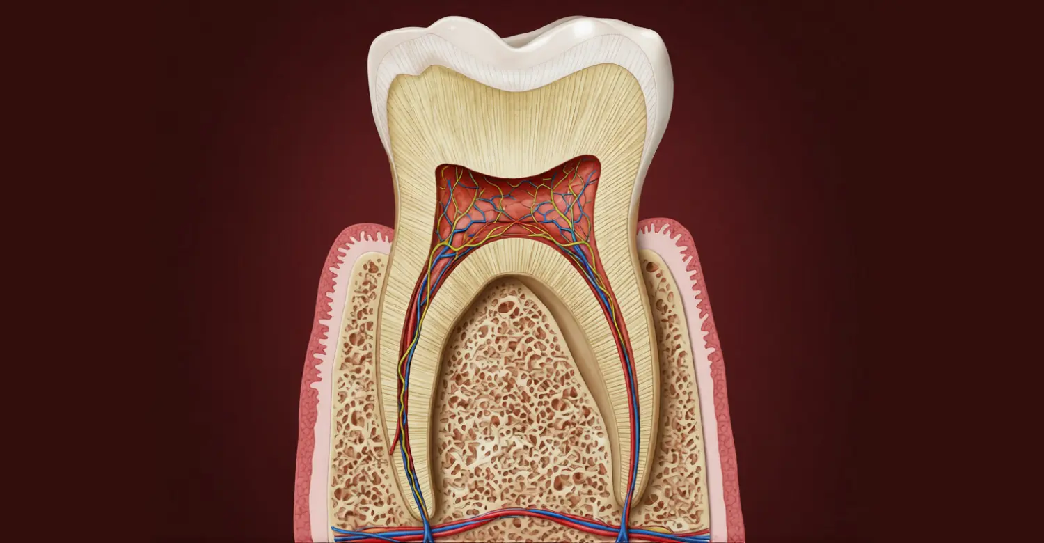
A tooth structures diagram visually identifies the crown, root, enamel, dentin, pulp, cementum, and supporting structures. Diagrams usually show a tooth in cross-section so internal anatomy becomes easier to understand. The crown appears above the gumline, while the root extends into the jawbone. Enamel usually appears as the outermost crown layer. Dentin occupies the larger region beneath enamel and surrounds the pulp chamber. The pulp is typically shown as the central soft-tissue space extending into root canals. Cementum appears as a thin covering around the root surface. The periodontal ligament connects the root with surrounding alveolar bone. Using diagrams can make dental terminology easier for patients and students to understand.
How to Read a Parts of a Tooth Diagram
A component of a tooth diagram should be read from the outside toward the center. Begin by identifying the crown and root because these provide the basic anatomical orientation. Next, locate enamel covering the crown and cementum covering the root. Dentin should appear beneath these external mineralized tissues. The central pulp chamber continues into root canals within the roots. The periodontal ligament surrounds the root and separates cementum from alveolar bone. The gingiva is usually represented around the cervical area near the tooth surface. Each labeled structure represents a different biological or mechanical function. Dentists use similar anatomical concepts when interpreting clinical images and planning treatment.
Crown, Neck, and Root
The crown, neck, and root represent important regions within the anatomical structures of a tooth. The crown is the upper portion and contains the enamel-covered anatomical crown. The neck, or cervical region, lies near the junction between crown and root. The root extends below the gumline and provides attachment within the jaw. The cementoenamel junction marks the transition between enamel and cementum. The position of the clinical crown can change when gums recede or become swollen. Therefore, the visible portion of a tooth does not always equal its anatomical crown. Understanding these regions helps dentists describe recession, periodontal attachment, restorations, and tooth fractures accurately.
Layers of a Tooth From Outside to Inside
The Parts of a Tooth can be understood by following its layers from outside toward the center. The crown begins with enamel as its external protective layer. Dentin lies underneath enamel and surrounds the pulp. The pulp occupies the central chamber and contains living tissues. On the root, cementum covers dentin externally instead of enamel. The root canal contains pulp tissue extending toward the tooth apex. The surrounding periodontal ligament connects cementum with alveolar bone. These layers provide protection, structural strength, sensation, and anchorage. The arrangement also explains why damage can become more serious as it moves toward the pulp.
How the Different Tooth Parts Work Together
The tissues that make up a tooth function as an integrated biological and mechanical system. Enamel protects deeper structures from chewing forces and environmental challenges. Dentin supports enamel and forms a protective barrier around the pulp. The pulp provides nerves, blood vessels, and cells needed for tooth vitality. Cementum provides attachment for periodontal ligament fibers around the root. The periodontal ligament transfers chewing forces between the tooth and supporting bone. Alveolar bone provides the socket surrounding the root. Gingival tissues create a protective soft-tissue barrier around the tooth. Healthy interaction among these structures allows teeth to remain functional and stable. Evidence-based preventive dentistry aims to preserve this entire system rather than treating isolated symptoms.
What Is the Crown of a Tooth?
The crown is one of the most visible structures of a tooth and performs much of the tooth's chewing function. The anatomical crown is covered by enamel, which provides strong external protection. The clinical crown refers to the portion visible in the mouth at a particular time. Crown shape differs significantly between incisors, canines, premolars, and molars. The crown contains enamel, dentin, and pulp tissue beneath its external surface. Caries, fractures, erosion, and excessive wear can alter crown structure. Restorations may replace lost crown tissue while attempting to preserve healthy remaining structure. Dentists assess crown anatomy carefully before placing fillings, crowns, veneers, or other restorations.
What Is the Anatomical Crown?
The anatomical crown is one of the key tooth structures and refers specifically to enamel-covered tooth structure. It extends from the incisal or occlusal surface toward the cementoenamel junction. The anatomical crown remains defined by its enamel covering regardless of gum position. Gum recession can expose root surfaces while leaving the anatomical crown unchanged. The anatomical crown differs from the clinical crown because the latter depends on what is visible in the mouth. Understanding this distinction is important when discussing periodontal recession and restorative treatment. Dental professionals use anatomical landmarks to describe tooth surfaces and treatment areas accurately.
What Is the Clinical Crown?
The clinical crown describes the portion of the components of a tooth that is visible inside the mouth. The clinical crown can include anatomical crown structure and exposed root structure in some situations. Gum recession can make the clinical crown appear longer without changing the anatomical crown. Gingival enlargement can make the visible crown appear shorter. The clinical crown is therefore influenced by the position of surrounding soft tissues. This distinction matters during periodontal evaluation, cosmetic treatment, and restorative planning. Dentists assess tooth exposure before determining whether changes involve tooth structure or gum position.
What Parts Make Up the Crown?
The crown contains several important anatomical structures of a tooth, including enamel, dentin, and dental pulp. Enamel forms the outer protective shell over the anatomical crown. Dentin lies underneath and provides structural support for enamel. The pulp occupies the central internal space and contains living tissue. The relationship among these tissues affects sensitivity, decay progression, and restorative decisions. Crown damage can remain superficial or extend progressively toward deeper structures. The severity depends on the location and depth of the affected tissue.
Enamel
Enamel is the outermost crown component among the major Parts of a Tooth. It protects dentin from chewing forces, temperature changes, acids, and bacterial activity. Enamel contains highly organized mineral crystals that provide exceptional hardness. However, acids can dissolve minerals from enamel during repeated episodes of demineralization. Saliva and fluoride can support remineralization of early mineral changes before cavitation develops. Good oral hygiene reduces the bacterial acids responsible for many carious lesions.
Dentin
Dentin forms the substantial internal framework beneath enamel within the crown. Dentin is one of the most important tissues that make up a tooth because it supports enamel and protects the pulp. It contains microscopic tubules extending toward the pulp. Dentin can transmit external stimuli through these tubules, contributing to sensitivity when exposed. Dentin also provides flexibility that helps support the more brittle enamel layer. Decay can progress through dentin faster than through healthy enamel in certain circumstances. Early restorative treatment can help prevent deeper structural involvement.
Pulp
The dental pulp is the living center of the crown and one of the most biologically important structures of a tooth. It contains nerves, blood vessels, connective tissue, and cells involved in dentin formation and defense. The pulp occupies the pulp chamber and continues into the root canals. Inflammation can develop after deep decay, trauma, cracks, or repeated procedures. Severe inflammation may cause prolonged sensitivity or spontaneous pain. Dentists use clinical examination and pulp testing to evaluate its condition.
What Is the Root of a Tooth?
The root is one of the essential tooth structures because it anchors the tooth within its socket. The root extends below the gumline and is surrounded by periodontal tissues. Most roots contain dentin, pulp tissue, cementum, and periodontal ligament attachments. Root shape and number vary depending on the type of tooth. Incisors often have simpler root anatomy, while molars may have multiple roots. Complex root anatomy can influence root canal treatment and surgical procedures. The root transfers chewing forces toward surrounding alveolar bone. Proper periodontal support is essential for maintaining root stability.
What Does the Tooth Root Do?
The tooth root is among the most important components of a tooth for stability and force distribution. It anchors the tooth through the periodontal ligament and surrounding alveolar bone. Roots help transfer functional forces from the crown into supporting tissues. Cementum provides the surface where periodontal ligament fibers attach. The root also contains canals carrying nerves and blood vessels toward the pulp. The shape and length of roots vary according to tooth type. Root damage, periodontal bone loss, or fractures can compromise tooth stability. A complete dental assessment considers both root structure and surrounding support.
What Is Inside the Tooth Root?
The internal anatomical structures of a tooth root include dentin, root canals, pulp tissue, and associated neurovascular structures. Dentin forms the main hard tissue surrounding the root canal. The root canal contains pulp tissue, including nerves and blood vessels. The canal system may have branches, curves, and anatomical variations. The root terminates near an opening called the apical foramen. This opening allows nerves and blood vessels to communicate with surrounding tissues. Root canal anatomy can be complex, especially in molars and teeth with multiple roots. Digital imaging helps dentists evaluate these structures before endodontic procedures.
Root Canal
The root canal is one of the internal Parts of a Tooth and contains pulp tissue. The canal extends through the root toward the apical opening. Its shape varies considerably between individual teeth and tooth types. Root canals can curve, divide, merge, or contain accessory anatomy. This complexity is important during root canal treatment because untreated canal spaces can allow persistent infection. Root canal procedures involve cleaning, shaping, disinfecting, and sealing the canal system. Dentists use radiographs and specialized instruments to identify and treat canal anatomy accurately.
Nerves and Blood Vessels
Nerves and blood vessels form essential living components within the internal tissues that make up a tooth. They enter the tooth through openings near the root apex. Blood vessels provide nutrients and help maintain pulp tissue. Nerves provide sensory information and contribute to pain responses when inflammation occurs. Pulp inflammation can increase pressure within the confined pulp chamber. This process may contribute to significant dental pain when inflammation becomes severe. Pulp testing helps determine whether nerve responses are within expected clinical ranges.
How Is the Root Attached to the Jawbone?
The root connects with the jaw through several supporting structures of a tooth and surrounding structures. Cementum covers the root and provides attachment for periodontal ligament fibers. The periodontal ligament connects these fibers with alveolar bone. The alveolar bone forms the tooth socket and surrounds the root. Gingival tissues provide additional protection around the cervical region. This attachment system allows slight physiological tooth movement during normal chewing. It also distributes functional forces and reduces direct stress transmission to bone. Damage to periodontal support can lead to increased tooth mobility.
Cementum
Cementum covers the external root surface and represents one of the important tooth structures. It provides attachment for periodontal ligament fibers and protects underlying root dentin. Cementum is continuously deposited throughout life under normal biological conditions. It is softer and less mineralized than enamel. Root exposure can increase the risk of cementum wear and root caries. Gentle oral hygiene and periodontal care help protect exposed root surfaces.
Periodontal Ligament
The periodontal ligament is one of the key supporting structures associated with the components of a tooth. It consists of specialized connective tissue fibers between cementum and alveolar bone. The ligament helps suspend the tooth within its socket. It also contains sensory receptors that contribute to awareness of biting forces. The periodontal ligament can adapt to functional forces through continuous tissue remodeling. Periodontal inflammation can damage this attachment system and contribute to tooth mobility.
Alveolar Bone
Alveolar bone forms the socket surrounding the root and supports the tooth within the jaw. It is one of the essential supporting structures associated with the Parts of a Tooth. Bone levels depend on periodontal health, functional forces, age, and other biological factors. Periodontal disease can cause destruction of supporting bone around affected teeth. Radiographs help dentists evaluate bone height and identify patterns of bone loss. Preserving periodontal health is therefore essential for long-term tooth stability.
What We Notice Clinically
The anatomical structures of a tooth often show different clinical signs depending on the affected structure. Enamel problems may appear as discoloration, surface breakdown, or cavities. Dentin exposure commonly produces sensitivity to cold, sweet foods, or touch. Pulp inflammation can cause spontaneous pain or prolonged sensitivity. Root exposure may increase sensitivity and root caries risk. Periodontal attachment loss can produce mobility or changes in gum position. Clinical examination helps identify whether symptoms originate from the tooth or surrounding tissues. Radiographs may reveal hidden decay, bone loss, root pathology, or other abnormalities.
Dr. Rifat Alsaman's Clinical Opinion
According to Dr. Rifat Alsaman, understanding the tissues that make up a tooth is essential before selecting restorative treatment. "A tooth should be evaluated as a complete biological structure," he explains. Treatment should consider remaining enamel, dentin, pulp condition, root anatomy, and periodontal support. Preserving healthy tooth tissue should remain a priority whenever clinically possible. He also emphasizes that symptoms should not be interpreted without proper clinical diagnosis. Different structures can produce similar symptoms, making professional assessment important. At Vitrin Clinic, treatment planning considers tooth anatomy alongside the patient's functional and aesthetic needs.
What Is Tooth Enamel and Why Is It Important?
Tooth enamel is one of the strongest structures of a tooth and protects the underlying dentin and pulp. It forms the external surface of the anatomical crown. Enamel is highly mineralized and designed to withstand repeated chewing forces. Despite its hardness, enamel can experience demineralization from bacterial acids and dietary acids. Enamel cannot regenerate after significant structural loss because it contains no living cells. Early mineral changes can sometimes be reversed through remineralization. Saliva, fluoride, and good oral hygiene support enamel protection. Regular dental examinations can identify early changes before substantial tissue loss occurs.
What Is Tooth Enamel?
Tooth enamel is the highly mineralized outer layer covering the anatomical crown. It is one of the most protective tooth structures and shields deeper tissues from environmental challenges. Enamel consists primarily of hydroxyapatite minerals arranged into organized microscopic structures. It contains very little organic material compared with dentin and pulp. Enamel thickness varies across different areas of individual teeth. Molars generally require strong enamel protection because they experience substantial chewing forces. Enamel can lose minerals when repeatedly exposed to acidic conditions.
Why Is Enamel the Hardest Part of a Tooth?
Enamel is considered the hardest biological tissue in the human body because of its exceptionally high mineral content. Its tightly organized crystals provide strong resistance to compression and surface wear. Enamel is among the most mineralized components of a tooth, allowing it to tolerate significant chewing forces. The mineral structure also explains why enamel can be brittle without adequate dentin support. This combination of hardness and brittleness makes underlying dentin important for overall tooth strength. Enamel remains vulnerable to acid dissolution despite its hardness.
Can Damaged Enamel Repair Itself?
Enamel cannot biologically regenerate once substantial tissue has been permanently lost. However, early mineral loss can undergo remineralization under favorable oral conditions. Saliva supplies minerals that can help restore mineral balance on early lesions. Fluoride can increase enamel resistance to future acid attacks. Dental professionals may recommend fluoride treatments, dietary changes, and improved plaque control depending on risk. Enamel damage involving actual cavitation cannot be naturally rebuilt by the body. The appropriate treatment depends on lesion depth, location, symptoms, and structural loss.
How Can You Protect Tooth Enamel?
Protecting enamel helps preserve one of the most important parts of a tooth. Brush twice daily with fluoride toothpaste and use a soft-bristled toothbrush. Avoid frequent exposure to acidic drinks and foods throughout the day. Limit sugary snacks because oral bacteria can convert sugars into enamel-damaging acids. Do not brush immediately after strong acidic exposure because enamel may temporarily soften. Waiting before brushing can reduce mechanical abrasion of softened surfaces. Regular dental examinations help identify early enamel changes. Fluoride exposure remains one of the strongest evidence-based approaches for preventing dental caries.
What Is Dentin and How Does It Protect the Tooth?
Dentin is one of the major anatomical structures of a tooth and forms the structural foundation beneath enamel. It surrounds the pulp and extends through both crown and root. Dentin contains microscopic tubules that connect toward the pulp. These tubules contribute to sensitivity when dentin becomes exposed. Dentin is less mineralized than enamel and has greater structural resilience beneath the harder outer layer. It also provides support that helps prevent enamel from fracturing under functional forces. Dentin can develop additional protective layers after certain forms of irritation. Protecting dentin is important because exposure can increase sensitivity and decay progression.
What Is Dentin?
Dentin is a mineralized tissue located beneath enamel and cementum. It forms a major portion of the tissues that make up a tooth and surrounds the dental pulp. Dentin contains hydroxyapatite minerals within an organic collagen-rich matrix. Its microscopic tubules extend from the pulp toward the outer dentin surface. Dentin is harder than bone but softer than enamel. Its relative flexibility helps support the brittle enamel layer during chewing. Dentin can also form in response to biological stimulation throughout life.
Where Is Dentin Located?
Dentin lies beneath enamel in the crown and beneath cementum in the root. It surrounds the dental pulp throughout much of the tooth. Dentin is therefore present across most internal regions of the structures of a tooth. Its thickness varies between tooth regions and individual teeth. Dentin forms the main structural mass between external protective tissues and the pulp. The relationship between dentin and pulp is particularly important when decay becomes deep. Dentists assess remaining dentin carefully during restorative treatment.
Why Does Exposed Dentin Cause Sensitivity?
Exposed dentin can cause sensitivity because its microscopic tubules communicate with the underlying pulp. These tubules can transmit fluid movement and stimuli toward nerve-containing pulp tissue. This mechanism helps explain sensitivity to cold, air, touch, and sweet foods. Dentin exposure may result from gum recession, enamel loss, tooth wear, or periodontal problems. The severity of symptoms varies among individuals and depends on exposure and pulp condition. Desensitizing toothpaste, fluoride treatments, and treatment of the underlying cause can reduce symptoms. Persistent sensitivity should receive professional evaluation.
How Does Dentin Protect the Pulp?
Dentin protects the pulp by forming a substantial mineralized barrier around the internal soft tissue. It is one of the tooth structures that separates the pulp from external environmental challenges. Dentin reduces direct transmission of mechanical and thermal forces toward the pulp. Its tubular structure also participates in defensive responses when irritation occurs. Dentin can produce additional mineralized tissue under certain biological conditions. This response may reduce the space available to irritants as the tooth ages.
What Is Dental Pulp and What Does It Do?
Dental pulp is the living internal tissue located inside the tooth. It represents one of the most biologically active components of a tooth. The pulp contains connective tissue, nerves, blood vessels, immune-related cells, and odontoblasts. Odontoblasts contribute to dentin formation and repair responses. The pulp provides nutrients and sensory functions within the tooth. It also participates in defensive responses against bacterial invasion and injury. When inflammation becomes severe, pulp tissue may become irreversibly damaged. Root canal treatment may then be necessary to remove infected or irreversibly inflamed tissue.
What Is Inside the Dental Pulp?
Dental pulp contains several specialized tissues and cell types that maintain tooth vitality. These include blood vessels, nerve fibers, connective tissue, immune cells, and odontoblasts. The pulp occupies the pulp chamber and root canal system among the internal Parts of a Tooth. Odontoblasts are located near the dentin-pulp interface and produce dentin throughout life. The pulp also contains cells involved in immune defense and tissue maintenance. The confined space of the pulp chamber makes severe inflammation clinically significant.
What Are the Nerves and Blood Vessels in the Pulp?
Pulp nerves provide sensory information and contribute strongly to the experience of dental pain. Blood vessels supply oxygen and nutrients while supporting metabolic activity. These structures enter the tooth through openings near the root apex. They are essential living components among the internal anatomical structures of a tooth. Inflammation can alter blood flow and tissue pressure within the pulp chamber. Pulp testing evaluates nerve response but does not directly measure every aspect of pulp health. Clinical findings must therefore be interpreted alongside symptoms and imaging.
What Happens When the Dental Pulp Becomes Infected?
Pulp infection can occur when bacteria reach the internal tissue through deep decay, cracks, or traumatic damage. Infected pulp represents serious involvement of the living tissues that make up a tooth. Inflammation may initially cause sensitivity and pain. Progressive infection can eventually lead to tissue death and infection around the root apex. Some patients experience severe symptoms, while others may have minimal discomfort despite significant disease. Dental examination and imaging are needed to determine the extent of infection. Treatment may involve root canal therapy or extraction depending on tooth restorability.
When Is Root Canal Treatment Needed?
Root canal treatment may be recommended when pulp tissue becomes irreversibly inflamed or infected. The procedure addresses disease affecting the internal structures of a tooth rather than simply treating surface symptoms. Common causes include deep decay, significant trauma, cracks, or repeated procedures. A dentist evaluates symptoms, pulp testing, clinical findings, and radiographs before recommending treatment. The objective is to remove diseased pulp, disinfect the canal system, and seal it. Root canal treatment can allow a suitable tooth to remain functional when adequate supporting structure remains.
What Is Cementum of a Tooth?
Cementum is a mineralized tissue covering the root surface. It is one of the tooth structures that connects the tooth with periodontal supporting tissues. Cementum provides an attachment surface for periodontal ligament fibers. Unlike enamel, cementum can continue forming throughout life. Cementum is softer and less mineralized than enamel, making exposed areas more vulnerable to wear. Root exposure may occur because of gum recession or periodontal attachment loss. Exposed cementum can become sensitive and may develop root surface caries. Regular periodontal care helps protect cementum and surrounding root structures.
Where Is Cementum Located?
Cementum covers the external surface of the tooth root. It lies over root dentin and extends toward the cementoenamel junction. Cementum is therefore located within the root-related Parts of a Tooth rather than covering the crown. It provides the attachment surface for periodontal ligament fibers. Cementum thickness can vary according to location, tooth age, and biological conditions. Gum recession can expose cementum to the oral environment.
What Does Cementum Do?
Cementum attaches periodontal ligament fibers to the tooth root and helps anchor the tooth. Cementum is among the components of a tooth that contribute directly to periodontal attachment. It also covers and protects the underlying root dentin. Cementum can continue forming throughout life, particularly in response to functional or biological demands. Its attachment function allows chewing forces to transfer between tooth and supporting bone.
Cementum vs Enamel
Cementum and enamel are mineralized tissues, but they differ significantly in location and function. Enamel covers the crown, while cementum covers the root. They are both among the protective anatomical structures of a tooth, but their biological characteristics differ. Enamel is much harder and more highly mineralized than cementum. Cementum provides periodontal ligament attachment, while enamel primarily protects the crown. Enamel cannot regenerate after significant loss, whereas cementum can continue depositing throughout life.
What Happens When Cementum Is Exposed?
Exposed cementum can become sensitive because it provides less protection than enamel. Root exposure often occurs because of gum recession or periodontal attachment loss. Cementum is one of the tissues that make up a tooth that requires protection when roots become exposed. Exposed root surfaces can also be more vulnerable to abrasion and root caries. Gentle brushing, fluoride toothpaste, and professional periodontal care can help protect these surfaces. Persistent sensitivity should be evaluated to identify the underlying cause.
What Structures Support the Tooth?
Several supporting structures work with the structures of a tooth to keep teeth stable. These include the periodontal ligament, alveolar bone, gingiva, blood vessels, and nerves. The periodontal ligament connects cementum with the surrounding bone. Alveolar bone forms the socket that surrounds the tooth root. Gingival tissues provide a protective barrier around the cervical tooth region. Blood vessels supply living periodontal tissues and surrounding structures. Nerves provide sensation and help detect pressure, temperature, and pain. These structures can be affected by periodontal disease, trauma, inflammation, and other conditions.
Periodontal Ligament
The periodontal ligament is a specialized connective tissue surrounding the tooth root. It connects cementum with alveolar bone and supports the tooth structures within the jaw. The ligament contains collagen fibers arranged to resist functional forces. It also contains blood vessels, nerves, and specialized cells. The periodontal ligament allows slight physiological movement during normal chewing. This movement helps distribute forces and provides sensory information about biting pressure. Periodontal disease can damage these attachment fibers and reduce tooth support.
Alveolar Bone
Alveolar bone forms the bony socket surrounding each tooth root. It provides structural support for the Parts of a Tooth and interacts closely with the periodontal ligament. Bone levels change in response to functional forces and periodontal inflammation. Advanced periodontal disease can destroy alveolar bone around affected teeth. Radiographic evaluation can help dentists assess the amount and pattern of bone support. Preserving bone requires controlling periodontal inflammation and maintaining effective oral hygiene.
Gingiva and Gums
Gingiva, commonly called gums, surrounds the teeth and covers portions of the underlying supporting tissues. Healthy gums provide a protective barrier around the components of a tooth near the cervical region. Healthy gingiva is generally firm and does not bleed during routine gentle brushing. Persistent bleeding can indicate gingival inflammation and should not be ignored. Gum recession can expose cementum and root dentin, increasing sensitivity and root caries risk. Regular professional examinations help monitor gingival health and periodontal attachment.
Blood Vessels and Nerves
Blood vessels and nerves supply and monitor the tooth and its surrounding supporting structures. Blood vessels provide nutrients to living tissues associated with the anatomical structures of a tooth. Nerves transmit sensory information about pressure, pain, and other stimuli. The dental pulp contains important neurovascular structures that enter through the root apex. Periodontal tissues also contain nerves that detect functional forces and inflammation. Injury or inflammation can alter normal sensory responses and produce pain.
Clinical Note
The tissues that make up a tooth cannot be evaluated separately from their supporting periodontal structures. A tooth may appear structurally intact while losing significant support from surrounding bone or periodontal tissues. Periodontal probing, radiographs, and clinical examination help evaluate these supporting structures. Early periodontal inflammation can often be managed before advanced attachment loss develops. Patients should report persistent bleeding, swelling, mobility, or gum recession to a dentist. Regular maintenance is important for protecting both teeth and their supporting tissues.
How Do the Different Parts of a Tooth Work Together?
The structures of a tooth function as a coordinated system that provides strength, sensation, protection, and stability. Enamel protects dentin from the oral environment. Dentin supports enamel and surrounds the pulp. The pulp maintains biological vitality and provides sensory responses. Cementum attaches periodontal ligament fibers to the root. The periodontal ligament connects the tooth with alveolar bone. Alveolar bone supports the tooth within its socket. Gingiva provides an external protective barrier around the cervical region. The health of one structure can influence the condition of neighboring structures.
How Enamel Protects Dentin
Enamel forms a highly mineralized barrier over the crown and protects underlying dentin. It is one of the tooth structures most exposed to chewing forces and dietary acids. Enamel limits direct exposure of dentin to the oral environment. When enamel is lost through decay, erosion, or trauma, dentin becomes more vulnerable. Exposed dentin can produce sensitivity and may allow decay to progress more rapidly. Protecting enamel therefore helps preserve deeper tooth structures.
How Dentin Protects the Pulp
Dentin surrounds the pulp and creates a substantial mineralized barrier against external influences. It is one of the Parts of a Tooth that provides structural and biological protection. Dentin reduces direct transmission of thermal and mechanical stimuli toward the pulp. Dentin can also respond biologically by producing additional mineralized tissue under certain conditions. Deep decay can eventually breach this protective barrier and expose the pulp. Once bacterial contamination reaches pulp tissue, inflammation and infection may develop.
How the Pulp Keeps the Tooth Vital
The pulp contains blood vessels that supply nutrients and oxygen to its living tissues. It also contains nerves and cells involved in defense and dentin formation. The pulp is therefore among the most biologically active components of a tooth. Healthy pulp supports continued dentin formation throughout life. It can respond to certain injuries by activating defensive processes. Severe inflammation or infection can compromise these functions and threaten tooth vitality.
How Cementum Anchors the Root
Cementum covers the root and provides attachment for periodontal ligament fibers. It is one of the anatomical structures of a tooth directly involved in tooth anchorage. The periodontal ligament connects these fibers with alveolar bone. This attachment system allows teeth to withstand functional forces during chewing. Cementum can continue forming throughout life and contributes to long-term attachment. Root exposure can compromise comfort and increase vulnerability to root surface damage.
Dr. Rifat Alsaman's Clinical Opinion
Dr. Rifat Alsaman emphasizes that successful dental treatment requires understanding how the tissues that make up a tooth interact. He explains that treating one structure without considering neighboring tissues can compromise long-term results. Tooth restorations should account for remaining enamel, dentin, pulp status, root anatomy, and periodontal support. Functional forces should also be considered when selecting restorative materials and treatment approaches. At Vitrin Clinic, comprehensive assessment helps dentists develop treatment plans based on individual anatomy. Preserving natural tooth structure remains an important principle whenever clinically appropriate.
What Happens When Different Parts of a Tooth Are Damaged?
Damage can affect different structures of a tooth, and symptoms depend on the structure involved. Enamel damage can lead to sensitivity, discoloration, or cavities. Dentin exposure can produce sharp sensitivity to temperature and touch. Pulp damage may cause prolonged pain, inflammation, or infection. Root damage can include fractures or structural weakening. Cementum exposure can increase sensitivity and root caries risk. Periodontal damage can reduce the bone and ligament support surrounding a tooth. Early diagnosis can prevent minor structural problems from progressing into more complex disease.
Enamel Damage and Tooth Decay
Enamel damage may begin as mineral loss caused by acids produced by oral bacteria. Caries are among the most common conditions affecting the Parts of a Tooth. Early lesions may appear as white or chalky areas before cavitation develops. If decay progresses, bacteria can penetrate deeper into dentin and approach the pulp. Risk depends on factors such as diet, fluoride exposure, plaque accumulation, saliva, and individual susceptibility. Preventive treatment can stop or slow early disease before extensive structural loss occurs.
Dentin Damage and Sensitivity
Dentin damage can expose microscopic tubules connected toward the dental pulp. Exposed dentin is among the common causes of sensitivity involving the tooth structures. Patients may experience short, sharp discomfort from cold drinks, sweet foods, air, or touch. Causes include enamel wear, gum recession, abrasion, erosion, and dental decay. Treatment depends on identifying and controlling the underlying cause. Desensitizing products and fluoride may help certain patients. Persistent or severe symptoms require professional evaluation.
Pulp Damage and Infection
Pulp damage occurs when inflammation or infection affects the living tissue inside the tooth. Deep decay, trauma, cracks, and repeated procedures can compromise this part of the components of a tooth. Inflammation may initially cause temperature sensitivity or pain. Severe inflammation can become irreversible and eventually lead to pulp necrosis. Bacteria can then spread through the root canal toward surrounding tissues. Root canal treatment may preserve a restorable tooth when appropriate.
Root Damage and Fractures
Root fractures can compromise the structural integrity of the tooth and its supporting tissues. Fractures are serious problems involving the deeper anatomical structures of a tooth. Symptoms can include pain during chewing, sensitivity, swelling, or increased mobility. However, some fractures produce limited symptoms and remain difficult to diagnose. Dentists may use clinical examination, radiographs, and additional imaging when necessary. Treatment depends on fracture location, severity, periodontal involvement, and tooth restorability.
Cementum Damage and Root Exposure
Cementum damage commonly occurs when root surfaces become exposed through gingival recession. Exposed cementum represents vulnerability within the root-related tissues that make up a tooth. Because cementum is softer than enamel, exposed surfaces can wear more easily. Improper brushing technique can contribute to mechanical abrasion on exposed roots. Root caries can also develop when exposed surfaces remain exposed to bacterial acids. Gentle brushing and fluoride use can help protect vulnerable root surfaces.
When Should You See a Dentist?
You should seek dental care when symptoms persist, worsen, or interfere with normal activities. Problems affecting the structures of a tooth can sometimes progress without severe symptoms. Warning signs include persistent pain, swelling, bleeding, sensitivity, broken teeth, or changes in tooth mobility. Dental trauma requires prompt assessment, particularly when a tooth becomes displaced or fractured. Early treatment often provides more conservative options than delayed treatment. Regular checkups can also detect problems before symptoms become obvious.
How Do Dentists Examine the Parts of a Tooth?
Dentists evaluate the Parts of a Tooth using clinical examination, imaging, and diagnostic tests. The examination begins with visual inspection of teeth, gums, and surrounding tissues. Dentists assess tooth surfaces for decay, fractures, wear, discoloration, and restorations. Probing can evaluate periodontal tissues and attachment levels. Radiographs reveal structures that cannot be seen directly inside the mouth. Digital scans and three-dimensional imaging may provide additional anatomical information when indicated. Pulp testing helps evaluate the sensory response of dental tissues. Combining these methods produces a more complete diagnostic picture.
Clinical Dental Examination
A clinical examination allows dentists to directly evaluate visible tooth structures and surrounding tissues. Dentists inspect enamel surfaces for decay, cracks, erosion, and discoloration. They also evaluate restorations, gum tissues, tooth mobility, and bite relationships. Periodontal probing measures the depth of the spaces around teeth. Palpation and percussion can help identify certain inflammatory conditions. A thorough examination connects symptoms with observable clinical findings. Diagnostic conclusions should consider the complete oral environment rather than isolated symptoms.
Dental X-Rays
Dental X-rays allow dentists to visualize hidden components of a tooth and supporting structures. Radiographs can reveal decay, root anatomy, bone levels, infections, and other abnormalities. They provide information that cannot be obtained through visual examination alone. Different radiographic views answer different clinical questions. Radiation exposure from modern dental imaging is generally kept as low as reasonably achievable. Dentists recommend radiographs according to individual diagnostic needs rather than using identical imaging for every patient.
Bitewing X-Rays
Bitewing X-rays provide detailed views of the crowns of upper and lower posterior teeth. They are particularly useful for detecting decay between teeth and evaluating bone levels. Bitewings help assess crown-related anatomical structures of a tooth that may not be visible clinically. Interproximal decay can remain hidden until it becomes relatively advanced. These radiographs can therefore support earlier diagnosis in appropriate patients. Dentists determine the need and frequency based on individual risk.
Periapical X-Rays
Periapical X-rays show an entire tooth from crown through root and surrounding bone. They provide important information about deeper tissues that make up a tooth and supporting structures. Periapical images can help identify root anatomy, infections, fractures, and changes near the tooth apex. They are commonly used during root canal diagnosis and treatment planning. These images complement clinical examination and other diagnostic methods.
Digital Scans and 3D Imaging
Digital scanning can create detailed representations of tooth surfaces and dental arches. Three-dimensional imaging can provide additional information about complex structures of a tooth and surrounding anatomy. Cone beam computed tomography may be recommended for selected cases requiring three-dimensional assessment. It can help evaluate complex root anatomy, impacted teeth, bone structures, and certain treatment-planning questions. Three-dimensional imaging should be used when the additional information is clinically justified.
Pulp and Nerve Testing
Pulp testing evaluates how dental pulp responds to specific sensory stimuli. These tests help dentists assess the condition of internal Parts of a Tooth. Common methods include cold testing, electric pulp testing, and other clinical assessments. Results should be compared with neighboring and controlled teeth when appropriate. A positive response does not automatically mean the pulp is healthy. Likewise, a lack of response requires interpretation alongside clinical findings and imaging.
What We Notice Clinically
The tooth structures can produce different clinical signs depending on disease location. Dentists compare symptoms with examination findings to identify the likely source. For example, brief cold sensitivity differs diagnostically from spontaneous prolonged pain. Periodontal findings may indicate that symptoms originate from supporting tissues rather than the tooth itself. Imaging can reveal hidden pathology that clinical examination cannot detect alone. Combining diagnostic information reduces the risk of treating the wrong structure.
How Do Tooth Parts Affect Dental Treatments?
Knowledge of the components of a tooth directly influences restorative, cosmetic, and surgical treatment decisions. Fillings require sufficient remaining tooth structure to support the restoration. Crowns may be recommended when significant structural loss compromises tooth strength. Veneers primarily affect the visible facial surfaces of anterior teeth. Root canal treatment addresses infected or irreversibly inflamed pulp tissue. Dental implants replace missing teeth rather than restoring the original natural tooth structure. Each treatment requires careful assessment of remaining anatomy, function, and surrounding tissues.
Tooth Anatomy and Dental Fillings
Dental fillings restore tooth structure lost through decay or minor damage. The dentist must consider enamel, dentin, and pulp when treating affected anatomical structures of a tooth. Small cavities may require limited removal of diseased tissue. Larger lesions may involve deeper dentin and increase the risk of pulpal irritation. Restoration materials are selected according to cavity location, size, functional demands, and patient factors. Preserving healthy enamel and dentin is generally preferred during conservative restorative treatment.
Tooth Anatomy and Dental Crowns
Dental crowns cover and protect teeth with significant structural compromise. Crown planning depends heavily on the remaining tissues that make up a tooth. Dentists evaluate remaining enamel, dentin, pulp condition, tooth shape, and periodontal support. A crown can restore chewing function, appearance, and structural protection when appropriately indicated. The preparation must balance adequate material space with preservation of healthy tooth tissue. Proper occlusion and periodontal considerations also influence long-term restorative performance.
Tooth Anatomy and Veneers
Dental veneers primarily cover selected visible surfaces of teeth for cosmetic or restorative purposes. Veneer planning depends on the condition of enamel and other Parts of a Tooth. Adequate enamel can support predictable bonding for many veneer applications. The amount of preparation depends on the clinical situation and chosen material. Veneers are not appropriate for every tooth or every type of damage. Dentists assess bite, enamel quality, tooth position, gum health, and aesthetic goals before treatment.
Tooth Anatomy and Root Canal Treatment
Root canal treatment addresses disease within the pulp and root canal system. It focuses on internal structures of a tooth that have become infected or irreversibly inflamed. The dentist removes diseased pulp tissue, cleans the canal system, and seals it. Afterward, the tooth may require additional restoration depending on remaining structure and functional demands. Proper restoration helps protect the treated tooth from fracture and reinfection.
Tooth Anatomy and Dental Implants
Dental implants replace missing teeth rather than repairing the original natural tooth structures. An implant is placed into the jawbone and supports a prosthetic crown. Success depends on adequate bone, healthy soft tissues, appropriate planning, and proper maintenance. Digital imaging can help evaluate available bone and anatomical structures. The final restoration must also consider bite, aesthetics, cleansability, and long-term function. Dental implants are therefore planned as complete restorative systems rather than isolated components.
Tips for Patients: How to Protect Every Part of Your Teeth
Protecting the components of a tooth requires consistent prevention rather than focusing only on visible surfaces. Daily oral hygiene helps control bacterial plaque and reduce the risk of dental decay. Fluoride toothpaste strengthens enamel and supports prevention of caries. A balanced diet can reduce repeated acid challenges to tooth surfaces. Gum health also matters because periodontal tissues support the tooth roots. Protective equipment can reduce dental trauma during contact sports. Regular examinations help identify early changes before they become extensive problems.
Protect Your Enamel
Protecting enamel helps preserve the strongest external layer of the anatomical structures of a tooth. Brush twice daily using fluoride toothpaste and a soft-bristled toothbrush. Avoid frequent sugary snacks and acidic drinks throughout the day. Drinking water can help neutralize acids and support saliva function. Do not use teeth as tools to open packages or break hard objects. If you grind your teeth, discuss appropriate protection with a dentist. Regular fluoride exposure can help reduce the risk of dental caries.
Prevent Dentin Exposure
Preventing dentin exposure protects one of the major internal tissues that make up a tooth. Use gentle brushing pressure to avoid unnecessary abrasion of enamel and root surfaces. Address gum recession and periodontal problems with professional care. Limit repeated exposure to acidic foods and beverages because acids can contribute to tooth surface loss. Treat dental decay before it progresses deeper into dentin. Desensitizing toothpaste may help manage sensitivity for some patients.
Keep Your Gums Healthy
Healthy gums protect the supporting structures surrounding the Parts of a Tooth. Brush along the gumline and clean between teeth regularly. Floss or use an appropriate interdental cleaning device according to professional advice. Gum bleeding can indicate inflammation and should not be considered normal. Persistent swelling, recession, or bleeding warrants dental evaluation. Periodontal maintenance is especially important for patients with previous gum disease.
Protect Your Teeth From Trauma
Trauma can damage several structures of a tooth, including enamel, dentin, pulp, and roots. Wear a properly fitted mouthguard during activities with significant impact risk. Avoid chewing extremely hard objects such as ice or non-food items. Do not use teeth to open bottles or packages. If a dental injury occurs, seek professional assessment promptly. Some traumatic injuries may require treatment even when visible damage appears limited.
Do Not Ignore Tooth Pain or Sensitivity
Persistent pain can indicate disease involving deeper tooth structures. Brief sensitivity may have a relatively simple cause, but prolonged or spontaneous pain requires evaluation. Swelling, fever, facial swelling, or severe pain can indicate significant infection. Do not rely on pain disappearance as proof that a problem has been resolved. Pulp tissue can lose vitality while symptoms temporarily change. Early professional diagnosis can help determine appropriate treatment.
Maintain Regular Dental Checkups
Regular dental examinations help monitor the components of a tooth and surrounding tissues. Dentists can identify early decay, gum inflammation, enamel wear, and other changes. Regular preventive care also provides opportunities for personalized oral hygiene guidance. Professional cleaning can remove deposits that cannot be effectively removed through routine brushing alone. The appropriate examination interval depends on individual disease risk and clinical history.
Parts of Baby Teeth vs Permanent Teeth
The anatomical structures of a tooth are broadly similar in primary and permanent teeth, but important differences exist. Baby teeth generally have thinner enamel and dentin than permanent teeth. Their pulp chambers are relatively large compared with overall crown dimensions. Primary teeth also have roots that undergo natural resorption before replacement by permanent teeth. These anatomical differences can affect the speed and progression of dental decay. Caring for primary teeth remains important because they support chewing, speech, and space maintenance. They also help guide the development and eruption of permanent teeth.
How Baby Teeth Differ From Permanent Teeth
Baby teeth are generally smaller and have thinner mineralized tissues than permanent teeth. Their anatomical tissues that make up a tooth include enamel, dentin, pulp, cementum, and supporting periodontal tissues. Primary teeth have relatively large pulp chambers and thinner dentin. These characteristics can allow decay to approach the pulp more quickly. Primary tooth roots eventually resorb as permanent successors develop beneath them. Proper care helps preserve primary teeth until their natural exfoliation.
Primary Tooth Structure
Primary teeth contain the same major structural tissues found in permanent teeth. These include enamel, dentin, pulp, cementum, and supporting structures among the Parts of a Tooth. However, enamel and dentin are generally thinner. Primary pulp chambers are proportionally larger and contain relatively prominent pulp horns. Primary roots are designed to undergo physiological resorption before exfoliation. These differences influence restorative procedures and treatment decisions in children.
Permanent Tooth Structure
Permanent teeth contain enamel, dentin, pulp, cementum, periodontal ligament, and supporting bone. The major structures of a tooth develop through complex biological processes during tooth formation. Permanent teeth generally have thicker mineralized tissues than primary teeth. Their roots provide long-term anchorage and remain within the jaw for many years with healthy support. Individual permanent teeth differ substantially in crown shape, root number, and canal anatomy. Proper preventive care helps preserve these structures throughout adulthood.
Why Baby Teeth Need Proper Care
Healthy primary teeth support chewing, speech development, and normal oral function. They also help maintain appropriate space for permanent teeth and guide eruption. Protecting the tooth structures in baby teeth can reduce pain and infection risk. Decay in primary teeth can progress rapidly because their hard tissues are relatively thin. Early dental visits allow clinicians to identify decay risk and provide preventive guidance. Children benefit from age-appropriate brushing, fluoride exposure, dietary guidance, and professional monitoring.
How Vitrin Clinic Evaluates Tooth Structure
At Vitrin Clinic, evaluation of the components of a tooth begins with a comprehensive clinical assessment. The dental team considers tooth structure, gum health, bite, aesthetics, symptoms, and treatment goals. Digital diagnostic tools can support evaluation when clinically appropriate. The objective is to understand the condition of each tooth before recommending treatment. Preserving healthy natural structure remains an important consideration in treatment planning. Treatment recommendations are individualized according to clinical findings rather than based solely on cosmetic preferences.
Comprehensive Tooth and Oral Examination
A comprehensive examination evaluates the visible and supporting anatomical structures of a tooth. Dentists inspect enamel, dentin exposure, restorations, gum tissues, and tooth position. Periodontal assessment evaluates gingival health, attachment, and supporting bone when indicated. The bite is also assessed because excessive forces can influence tooth wear and fractures. Symptoms are documented and correlated with clinical findings. This approach helps create a complete picture before treatment begins.
Digital Diagnostics and Treatment Planning
Digital diagnostics can provide detailed information about tooth surfaces, roots, bone, and surrounding structures. These technologies support evaluation of complex Parts of a Tooth when conventional examination is insufficient. Digital photographs, intraoral scans, radiographs, and three-dimensional imaging may be used when indicated. The selected technology depends on the clinical question and treatment requirements. Digital planning can also improve communication between patients and dental professionals.
Preserving Healthy Tooth Structure
Preserving natural tooth tissue is an important principle when treating the tissues that make up a tooth. Unnecessary removal of healthy enamel or dentin can reduce remaining structural support. Conservative techniques may be considered when clinically appropriate. Treatment selection depends on decay severity, fractures, function, aesthetics, and long-term prognosis. At Vitrin Clinic, treatment planning considers both immediate results and preservation of remaining healthy structure.
Personalized Treatment Based on Tooth Anatomy
Every tooth has individual anatomical characteristics that can affect treatment planning. The condition of the structures of a tooth determines which procedures may be appropriate. Root shape, pulp condition, enamel thickness, tooth position, and periodontal support can vary considerably. Personalized assessment helps dentists choose suitable restorative or surgical approaches. This approach can be particularly important for complex cases involving multiple treatments.
Why Choose Vitrin Clinic for Dental Care?
Choosing Vitrin Clinic for dental care involves comprehensive assessment, modern technology, and individualized planning. The clinic evaluates the tooth structures alongside the surrounding oral structures. Treatment recommendations are developed according to clinical findings, functional requirements, and patient goals. Aesthetic procedures are planned with attention to tooth structure and overall smile harmony. The dental team can evaluate restorative, cosmetic, and implant-related treatment needs within a coordinated approach. Long-term oral health remains an important consideration alongside immediate aesthetic outcomes.
Experienced Dental Team
An experienced dental team can recognize differences between individual components of a tooth and their clinical conditions. Proper diagnosis requires understanding tooth anatomy, periodontal structures, occlusion, and treatment limitations. Different specialists may contribute to complex restorative or implant cases when needed. Clinical experience supports appropriate treatment selection and communication with patients. Comprehensive assessment remains essential regardless of the procedure being considered.
Modern Dental Technology
Modern dental technology can improve visualization and planning of complex anatomical structures of a tooth. Digital radiography provides detailed information with efficient image acquisition. Intraoral scanning can create digital representations of tooth surfaces and dental arches. Three-dimensional imaging may assist selected implant, surgical, or endodontic cases. Digital technologies complement clinical expertise rather than replacing professional examination.
Personalized Treatment Plans
A personalized treatment plan considers the condition of each patient's Parts of a Tooth. The same procedure may not be suitable for every tooth or patient. Dentists consider structural integrity, gum health, bite, aesthetics, symptoms, and long-term prognosis. Personalized planning can help avoid unnecessary procedures and focus treatment on actual clinical needs. Patients should receive clear explanations of proposed treatments, alternatives, benefits, and limitations.
Focus on Long-Term Oral Health
Long-term oral health requires protecting teeth, gums, bone, and other supporting structures. The health of the tissues that make up a tooth depends partly on the surrounding oral environment. Preventive care reduces the risk of decay, periodontal disease, and avoidable structural damage. Restorative treatment should ideally be combined with appropriate maintenance and hygiene. Dental care is most effective when patients understand how to protect treatment results at home.
Key Takeaways About the Parts of a Tooth
Understanding the structures of a tooth makes dental symptoms, diagrams, and treatments easier to understand. The crown provides the primary visible chewing surface. The root anchors the tooth through cementum, periodontal ligament, and alveolar bone. Enamel protects the crown, while dentin supports enamel and surrounds the pulp. The pulp contains nerves and blood vessels that maintain tooth vitality. Cementum covers the root and provides attachment for periodontal ligament fibers. Each structure has a different role, but all work together as one functional system.
Understanding Your Tooth Structure
Understanding the tooth structures can help patients recognize why different dental problems produce different symptoms. Enamel protects the external crown. Dentin forms the main internal hard structure and can become sensitive when exposed. Pulp contains living tissues that can become inflamed or infected. Cementum covers the root and connects with periodontal attachment fibers. Supporting tissues stabilize the tooth inside the jaw. Learning this anatomy can make dental consultations easier and more informative.
Why Every Tooth Part Matters
Every structure contributes to the strength, function, sensation, or stability of the tooth. The components of a tooth cannot always be treated as independent structures. Damage to enamel can eventually affect dentin. Dentin damage can threaten pulp health. Pulp infection can spread toward surrounding root tissues. Periodontal disease can compromise the supporting bone and ligament. Preserving each structure whenever possible supports long-term oral function.
When to Seek Professional Dental Care
Seek professional care when symptoms involve pain, sensitivity, swelling, bleeding, mobility, or visible tooth damage. Problems affecting the anatomical structures of a tooth can worsen even when symptoms initially seem mild. Dental trauma should also be assessed promptly because internal damage may not be immediately visible. Regular examinations help detect decay, periodontal disease, fractures, and other problems early. Early diagnosis can sometimes allow simpler and more conservative treatment.
Reference
FAQs

Dr. Rifat Alsaman has more than 5 years of clinical experience in dentistry and currently serves as the Head of the Medical Team at Vitrin Clinic. He is dedicated to providing exceptional patient care, overseeing treatment planning, and ensuring the highest clinical standards across the team. His expertise, attention to detail, and commitment to continuous professional development have helped countless patients achieve healthier, more confident smiles.
.webp&w=3840&q=75)


