General Dentistry
August 31, 2026
What Is Dentin and How Does It Protect Your Tooth?

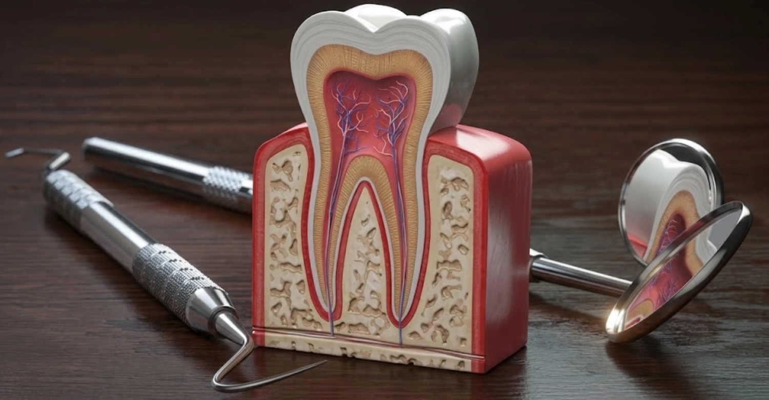
The tissue is the strong mineralized tissue beneath enamel and cementum, forming most of the tooth structure. It supports the enamel and surrounds the dental pulp, helping the tooth withstand everyday chewing forces. Unlike enamel, it contains microscopic channels called dentinal tubules, which connect toward the pulp. These tubules help explain why exposed tooth surfaces can become sensitive to temperature, touch, or sweet foods.
What Is Dentin?
The tissue is the mineralized tissue located underneath enamel in the crown and cementum around the root. It forms the largest portion of the tooth and provides structural support during normal function. This tissue is produced by specialized cells called odontoblasts, which remain associated with the pulp throughout tooth development. Its composition includes hydroxyapatite minerals, collagen, water, and other organic components.
What Is Dentin Made Of?
The tissue contains mineral crystals, organic matrix, and water arranged within a highly organized biological structure. Approximately 70% of its weight is mineralized material, mainly calcium phosphate crystals related to hydroxyapatite. The organic portion contains predominantly type I collagen, which provides flexibility and structural support. Water accounts for approximately 10% of its weight and contributes to its hydrated nature. This tissue also contains non-collagenous proteins involved in mineralization and tissue development.
Where Is Dentin Located in a Tooth?
The tissue lies directly beneath the enamel in the visible crown and beneath cementum along the root. It surrounds the dental pulp, creating a protective mineralized layer around the tooth's innermost tissues. In the crown, enamel provides the primary external protective covering over this tissue. Around the root, cementum covers the underlying surface where the tooth connects with periodontal tissues. This arrangement creates different levels of protection across the tooth.
What Does Dentin Do?
The tissue provides structural support, protects the pulp, and helps distribute forces throughout the tooth. Its combination of mineral and collagen gives teeth resistance against repeated chewing stresses. The tissue also acts as a barrier between the oral environment and the sensitive dental pulp. Its tubules create a pathway through which external stimuli can influence pulpal nerves. This explains why exposed surfaces may respond quickly to cold drinks or tactile stimulation.
How This Tissue Supports Enamel
The tissue provides a resilient foundation beneath the highly mineralized enamel layer. Enamel is extremely hard but comparatively brittle, making support from the underlying tissue important during chewing. This tissue has greater elasticity because its organic matrix contains collagen and water. This difference helps distribute mechanical stresses across the tooth rather than concentrating them entirely within enamel. The junction between enamel and the underlying tissue also contributes to resistance against crack propagation.
How This Layer Protects the Dental Pulp
The tissue surrounds the dental pulp and creates a mineralized barrier between the pulp and oral environment. The pulp contains blood vessels, connective tissue, odontoblasts, and sensory nerves. This tissue reduces direct exposure of these delicate structures to external mechanical and chemical challenges. Its thickness can also influence how quickly external stimuli affect pulpal tissues. The dentinal tubules provide communication pathways, but their organization also contributes to defensive mechanisms.
Dentin vs Enamel: What Is the Difference?
The tissue and enamel are mineralized dental tissues, but they differ significantly in composition, hardness, and biological behavior. Enamel is the hardest tissue in the human body and contains approximately 96% mineral by weight. This tissue contains substantially more organic material and water, making it less hard but more resilient. Enamel covers the crown, while this tissue extends throughout the crown and root beneath protective outer layers.
Is Dentin Harder or Softer Than Enamel?
The tissue is softer than enamel but harder than bone and cementum. Enamel contains a much higher proportion of mineral, which gives it exceptional hardness and wear resistance. This tissue contains more organic matrix and water, making it less rigid but more resilient. This difference is important because teeth need both hardness and resistance to fracture. Enamel withstands substantial forces during chewing, while the underlying tissue helps absorb and distribute those stresses.
Composition and Structure Compared With Enamel
Enamel and this tissue differ in mineral content, organic composition, water content, and microscopic organization. Enamel is approximately 96% mineral by weight, with relatively small amounts of organic material and water. The tissue contains approximately 70% mineral, 20% organic matrix, and 10% water. Its organic matrix consists predominantly of type I collagen. Enamel contains densely packed hydroxyapatite crystals arranged in highly organized structural units.
How Dentin and Enamel Respond to Damage
Enamel and this tissue respond differently because they have different cellular structures and biological capabilities. Enamel cannot regenerate substantially after mature tissue is lost because it lacks living cells. Early mineral loss can sometimes be reversed through remineralization before cavitation develops. The tissue can continue forming through odontoblast activity, including secondary and tertiary forms under specific conditions. However, this does not mean that severely destroyed this tissue simply grows back.
Why Enamel Cannot Regrow but This Tissue Can Form
Mature enamel cannot naturally regenerate because ameloblast cells disappear after enamel development is completed. The tissue has a different biological relationship because odontoblasts remain associated with the pulp. These cells can continue producing secondary tissue slowly throughout life. When irritation occurs, odontoblasts or related cells can also contribute to tertiary tissue formation. This response can create additional mineralized tissue between the irritation source and pulp.
What Are Dentinal Tubules?
Dentinal tubules are microscopic channels extending through this tissue toward the dental pulp. They contain fluid and contribute significantly to the tissue's permeability and sensitivity characteristics. Tubule density and diameter can vary depending on their location within the tooth. Tubules generally become more numerous and wider closer to the pulp. When protective enamel or cementum is lost, these channels can become exposed to the oral environment.
What Are Dentinal Tubules?
Dentinal tubules are tiny channels distributed throughout this mineralized tissue. They extend from the pulp toward the outer dentinal surface and contain dentinal fluid. Their density and diameter vary across different regions of the tooth. Tubules are generally wider and more numerous closer to the pulp. Their microscopic structure creates pathways for fluid movement and contributes to the tissue's permeability. Under healthy conditions, protective enamel or cementum prevents direct exposure of most tubules.
Why Do Dentinal Tubules Cause Tooth Sensitivity?
Dentinal tubules can contribute to sensitivity because they provide pathways through which external stimuli influence the pulp. Cold, heat, touch, airflow, and osmotic changes can alter fluid movement inside exposed tubules. Rapid fluid movement can stimulate sensory mechanisms associated with the pulp. The hydrodynamic theory remains the most widely accepted explanation for this process. Sensitivity is therefore more likely when tubules are exposed and remain relatively open.
How Fluid Movement Inside Dentinal Tubules Triggers Pain
External stimuli can create pressure changes that move fluid inside exposed dentinal tubules. Rapid fluid movement can stimulate sensory nerve mechanisms associated with the dental pulp. Experimental research has demonstrated that pressure changes applied to exposed tooth surfaces can produce sharp pain. Faster pressure changes produced stronger pain responses than slower changes in that study. This finding supports the hydrodynamic explanation for sensitivity.
The Connection Between Dentinal Tubules and This Hypersensitivity
This hypersensitivity commonly involves exposed dentinal surfaces with tubules that can transmit stimulus-related fluid movement. The condition usually produces short, sharp pain following a recognizable external trigger. Cold drinks, sweet foods, touch, and airflow are common examples. The hydrodynamic theory explains how fluid movement may activate sensory receptors associated with the pulp. Studies have reported considerable variation in prevalence because diagnostic methods and populations differ.
Clinical Note
Clinically, sensitivity should not automatically be attributed to this exposure without examination. Similar symptoms can occur with tooth decay, cracked teeth, defective restorations, or pulpal inflammation. The timing, duration, location, and trigger of pain can provide important diagnostic clues. Brief pain following cold stimulation often differs from spontaneous or lingering pulpal pain. Dentists may use air, cold, tactile stimulation, visual inspection, and radiographs when appropriate.
What Is Dentin Hypersensitivity?
This hypersensitivity is a common dental condition involving brief, sharp pain from exposed dentinal surfaces. The discomfort typically occurs after thermal, tactile, chemical, or osmotic stimulation. Cold beverages are among the most frequently reported triggers. The pain usually stops shortly after the stimulus is removed when the condition is uncomplicated. A systematic review of 77 studies found prevalence estimates ranging from 1.3% to 92.1%.
What Causes This Hypersensitivity?
This hypersensitivity develops when protective enamel or cementum becomes lost and underlying dentinal tubules become exposed. Common causes include gum recession, enamel erosion, tooth abrasion, and periodontal treatment. Acidic foods and beverages can gradually remove mineral from exposed tooth surfaces. Aggressive brushing can also contribute to cervical wear when excessive pressure is repeatedly applied. Gum recession exposes root surfaces because cementum provides thinner protection than enamel.
What Does This Feel Like?
This hypersensitivity usually feels like sudden, sharp, short-lasting pain following a specific external stimulus. Patients commonly describe discomfort after drinking something cold or eating sweet foods. Cold air can also trigger pain when exposed root surfaces are present. Brushing may cause a brief shock-like sensation around affected areas. The discomfort typically disappears after the triggering stimulus is removed. This short duration can help distinguish sensitivity from some forms of pulpal inflammation.
Why Are Cold Foods and Drinks Common Triggers?
Cold foods and drinks can trigger sensitivity because temperature changes influence fluid movement within exposed dentinal tubules. Rapid fluid movement can stimulate sensory mechanisms connected with the dental pulp. The hydrodynamic theory provides the primary explanation for this response. Ice cream, chilled water, and cold beverages commonly produce noticeable symptoms in sensitive teeth. Cold stimulation can also help dentists identify areas requiring further examination.
Can Brushing Make Dentin Sensitivity Worse?
Brushing can worsen sensitivity when excessive force, abrasive products, or incorrect technique contribute to cervical tooth wear. Repeated mechanical pressure can remove surface tissue and increase exposure of underlying dentinal tubules. A hard-bristled toothbrush may increase mechanical abrasion when combined with aggressive brushing. The tissue can become increasingly sensitive when protective structures are gradually lost. Acid exposure can make surfaces softer before mechanical brushing creates additional wear.
This Hypersensitivity vs Tooth Decay
This hypersensitivity and tooth decay can both produce sensitivity, but their underlying mechanisms differ. Hypersensitivity generally involves exposed dentinal surfaces responding to external stimuli. Tooth decay involves progressive demineralization and destruction caused by acids produced by bacterial metabolism. This decay can advance more rapidly after the protective enamel barrier has been breached. Sensitivity from decay may become increasingly frequent as the lesion progresses.
This Hypersensitivity vs Pulpitis
This hypersensitivity usually causes brief, stimulus-dependent pain from exposed surfaces. Pulpitis involves inflammation of the dental pulp and can produce more persistent or spontaneous symptoms. Reversible pulp inflammation may cause short responses to colds that resolve quickly. Irreversible inflammation can produce prolonged pain that continues after the stimulus disappears. This hypersensitivity generally does not cause spontaneous pain when uncomplicated. Pain that wakes someone during sleep requires careful professional assessment.
What We Notice Clinically
Clinically, sensitivity patterns often provide valuable information about the condition affecting a tooth. Brief cold sensitivity commonly differs from prolonged or spontaneous pain. The number of affected teeth can also provide useful diagnostic clues. Multiple sensitive teeth may suggest generalized recession, erosion, abrasion, or periodontal factors. A single sensitive tooth may require closer examination for decay, cracks, or restoration problems. This exposure is frequently visible around areas affected by gum recession.
Dr. Rifat Alsaman's Opinion
Dr. Rifat Alsaman emphasizes that sensitivity should be investigated before treatment is selected. A sensitive tooth does not always have the same underlying cause. This exposure may result from recession, erosion, abrasion, decay, or combinations of these factors. The patient's symptoms should therefore be evaluated alongside tooth structure and gum condition. Short stimulus-related pain can suggest sensitivity, while prolonged pain may require deeper assessment.
What Causes Exposed Dentin?
This exposure occurs when protective enamel, cementum, or surrounding gum tissue no longer covers the underlying surface. Several processes can create this exposure, including gum recession and enamel erosion. Tooth abrasion can remove cervical tissue through repeated mechanical friction. Cracks or structural damage may also reveal underlying surfaces. Acidic dietary habits can contribute to enamel erosion and eventually expose deeper tissues.
Gum Recession and Exposed Dentin
Gum recession occurs when the gingival margin moves toward the root and exposes previously covered tooth surfaces. Root surfaces are naturally protected by cementum rather than the thicker enamel found on crowns. Cementum can wear relatively easily when exposed to aggressive brushing or acidic conditions. Recession can therefore reveal the underlying tissue and increase the likelihood of sensitivity. Periodontal disease, traumatic brushing, and anatomical factors can contribute to recession.
Enamel Erosion and Exposed Dentin
Enamel erosion involves chemical loss of tooth mineral caused by acids that are not produced by bacteria. Frequent acidic drinks, foods, gastric acid, and certain habits can contribute to erosion. As enamel becomes thinner, the underlying tissue may eventually become visible. This exposure can increase sensitivity because its tubular structure responds to external stimuli. Acidic environments can also soften the tooth surface and increase susceptibility to mechanical wear.
Tooth Abrasion and Aggressive Brushing
Tooth abrasion occurs when repeated mechanical forces remove tooth tissue through friction. Aggressive brushing is a common contributing factor, especially around the cervical region. Excessive pressure, abrasive toothpaste, and hard-bristled brushes can increase mechanical wear. This exposure may develop when protective enamel or cementum becomes progressively removed. Patients may notice sensitivity during brushing or when consuming cold foods. The affected areas can sometimes appear as shallow grooves near the gumline.
Cracked or Damaged Enamel
Cracked or damaged enamel can reduce protection over the underlying dentinal layer. Cracks may develop from biting forces, trauma, grinding, or repeated temperature-related stresses. Some cracks remain superficial, while others extend deeper into the tooth. This exposure can occur when structural damage removes or separates protective enamel. Patients may experience sensitivity when biting, releasing a bite, or consuming cold foods.
Can Teeth Whitening Expose or Irritate This Layer?
Teeth whitening does not normally remove healthy enamel or intentionally expose this tissue when professionally performed. Whitening agents can temporarily increase sensitivity by allowing peroxide molecules to move through enamel and the underlying tissue. This process can influence the pulp and produce temporary sensory responses. Existing enamel erosion, recession, or exposed surfaces can increase sensitivity during whitening. This exposure may therefore make some patients more likely to experience discomfort.
How This Exposure Changes Tooth Sensitivity
This exposure can increase sensitivity because protective outer tissues no longer fully isolate its dentinal tubules. External stimuli can then influence fluid movement through the tubular network. Cold, touch, air, and sweet substances may produce short, sharp discomfort. This hypersensitivity is particularly associated with exposed surfaces containing open or patent tubules. Research has demonstrated differences in tubule number and diameter between sensitive and nonsensitive areas.
What Is Dentin Decay?
This decay occurs when dental caries progresses beyond enamel and reaches the underlying mineralized tissue. Once bacteria-driven acids penetrate enamel, the lesion can advance through the softer dentinal structure. The tissue contains less mineral and more organic material than enamel, affecting its response to acid attack. The lesion can spread laterally and toward the pulp as destruction progresses. This decay may produce sensitivity to cold, sweets, or chewing pressure.
How Does Tooth Decay Reach This Layer?
Tooth decay begins when bacterial acids repeatedly dissolve mineral from susceptible tooth surfaces. Initial lesions commonly develop within enamel before progressing deeper toward the dentinoenamel junction. Once the lesion reaches this junction, its spread pattern can change. The tissue contains more organic material and is less mineralized than enamel. Bacterial acids and enzymes can therefore facilitate faster structural breakdown after penetration. This decay may extend toward the pulp through the tubular network.
Why Does This Decay Progress Faster Than Enamel Decay?
The tissue is less mineralized than enamel and contains a substantial organic matrix. This composition makes it more susceptible to structural breakdown once caries reaches the dentinoenamel junction. Enamel contains approximately 96% mineral by weight, while this tissue contains approximately 70%. The difference means this tissue generally offers less resistance to acid-mediated demineralization. Its tubular structure can also provide pathways through which microorganisms and acids interact with deeper tissue.
What Are the Signs of Dentin Decay?
Signs of this decay can include sensitivity, discoloration, food trapping, rough surfaces, or visible cavities. Some lesions produce no symptoms during their early stages. As decay progresses, patients may experience increasing sensitivity to cold or sweet foods. Chewing discomfort can occur when structural damage becomes deeper or affects the tooth's integrity. Dark, brown, or yellow areas can sometimes indicate various changes, but discoloration alone does not confirm decay.\
Can This Decay Be Reversed?
Very early tooth decay may be arrested or remineralized when mineral loss has not produced a permanent cavity. Fluoride, saliva, plaque control, and dietary changes can support remineralization of early lesions. Once a cavity forms, the missing tooth structure cannot naturally return to its original shape. This decay therefore requires different management depending on lesion stage and activity. Noncavitated lesions may sometimes receive preventive treatment and monitoring.
When Does This Decay Require a Filling?
A filling may become necessary when tooth decay creates a cavitated lesion that cannot be cleaned effectively. The decision depends on lesion activity, depth, accessibility, and remaining tooth structure. Early noncavitated lesions may sometimes be managed without drilling through preventive approaches. Once this decay creates a significant cavity, restorative treatment can restore form and function. The dentist first evaluates the lesion clinically and may use radiographs when appropriate.
Clinical Note
This decay should be assessed according to lesion activity rather than color alone. Some dark areas may represent arrested lesions rather than active disease. Conversely, early active decay may have limited visible discoloration. Radiographs can be valuable when decay develops between teeth or beneath existing restorations. Clinical examination also evaluates texture, cavitation, plaque accumulation, and lesion accessibility. This exposure can sometimes resemble decay when viewed without appropriate diagnostic context.
What Is Tertiary Dentin?
This tissue is mineralized material formed locally in response to irritation or injury. It develops beneath areas affected by processes such as caries, abrasion, erosion, or restorative procedures. Unlike primary tissue, which forms mainly during tooth development, tertiary tissue develops after an external stimulus. Its formation can help increase the barrier between the irritation source and the dental pulp. The tissue responses vary according to the intensity and duration of the stimulus.
Why Does the Tooth Form This Tissue?
The tooth forms tertiary tissue as a defensive response to localized irritation. Stimuli can include bacterial caries, mechanical wear, erosion, trauma, or dental procedures. The response attempts to increase the mineralized barrier between the irritant and the pulp. This formation can therefore reduce the risk of direct pulpal exposure. The quality and structure of tertiary tissue depend on the severity and speed of the original stimulus.
Primary vs Secondary vs Tertiary Layers
Primary tissue forms during normal tooth development and creates most of the tooth's initial dentinal structure. Secondary tissue develops slowly after root formation and continues throughout life. Tertiary tissue forms locally in response to specific irritation or injury. The tissue therefore includes tissues formed during development, normal aging, and defensive responses. Primary tissue generally has a relatively regular tubular organization.
How This Tissue Protects the Pulp
Tertiary tissue increases the physical barrier separating an irritant from the dental pulp. This additional mineralized tissue can reduce the effective distance between the pulp and external damage. This formation may therefore help protect pulpal cells from bacterial products and mechanical irritation. The response depends on the type, intensity, and duration of the stimulus. A mild stimulus may produce organized reactionary tissue from surviving odontoblasts.
Reactionary vs Reparative Tissue Formation
Reactionary and reparative tissue represent different cellular responses to injury. Reactionary tissue is produced by surviving original odontoblasts after relatively mild irritation. Reparative tissue develops when the original odontoblasts are destroyed and replacement odontoblast-like cells become involved. The tissue produced through these pathways can differ in organization and structure. Reactionary tissue may retain characteristics closer to normal secondary tissue. Reparative tissue can be more irregular because it develops through a different cellular pathway.
Dr. Rifat Alsaman's Opinion
Dr. Rifat Alsaman explains that this defensive tissue demonstrates the tooth's ability to respond to irritation. However, this biological response should not be interpreted as permission to leave decay untreated. This formation may reduce the risk of direct pulpal exposure, but it cannot eliminate bacterial disease. Early intervention gives the tooth a better opportunity to preserve healthy pulp tissue.
Can Dentin Repair Itself?
The tissue has limited biological repair capacity, but it cannot fully regenerate large areas of lost tooth structure. Odontoblasts continuously produce secondary tissue throughout life, gradually reducing the size of the pulp chamber. Injury can stimulate tertiary tissue formation beneath affected areas. This response may create an additional protective barrier around the pulp. This repair therefore differs from regeneration because the newly formed tissue does not necessarily recreate the original architecture.
Can Dentin Regrow?
The tissue cannot completely regrow in the same way that some living tissues regenerate. However, teeth can continue producing secondary tissue throughout life. Injury can also stimulate tertiary tissue formation as a defensive response. These processes create additional mineralized tissue but do not recreate large areas of destroyed anatomy. This tissue therefore has greater biological activity than mature enamel, but its regenerative capacity remains limited.
How Does Secondary Formation Work?
Secondary tissue forms gradually after the tooth completes its primary development. Odontoblasts along the pulp boundary continue depositing small amounts of mineralized tissue throughout life. This process slowly reduces the internal size of the pulp chamber. This formation is generally slower than the rapid tissue production that occurs during tooth development. The pattern can also vary between different teeth and individuals.
Can Damaged Dentin Heal Naturally?
Mildly affected tissue can participate in defensive biological responses, but severely damaged areas cannot fully heal themselves. Secondary and tertiary tissue formation can create additional mineralized barriers around the pulp. Early carious lesions may also become arrested when the oral environment improves. The tissue damage becomes more difficult to manage naturally once substantial structural loss or cavitation occurs. Bacterial infection can continue inside a cavity if the underlying cause remains untreated.
Repair vs Enamel Remineralization
This repair and enamel remineralization are related but biologically different processes. Enamel remineralization involves mineral deposition into early areas of mineral loss. Saliva and fluoride can support this process before a permanent cavity forms. The tissue can also undergo mineral changes, but its biological response includes activity from odontoblast-related cells. Tertiary tissue formation creates new mineralized material beneath an area of irritation. This repair therefore involves both mineral balance and cellular defensive mechanisms.
What Is the Best Dentin Hypersensitivity Treatment?
The best treatment depends on why sensitivity developed and how severe the symptoms are. Common approaches include desensitizing toothpaste, fluoride applications, tubule-sealing materials, bonding, and periodontal treatment. Patients with gum recession may require treatment directed toward exposed root surfaces. Severe structural loss can sometimes require restorative coverage. This hypersensitivity should first be distinguished from decay, cracked teeth, and pulpal disease. A systematic review found considerable variation in treatment evidence and study outcomes.
Desensitizing Toothpaste
Desensitizing toothpaste is commonly used to reduce discomfort associated with exposed dentinal surfaces. Different formulations use ingredients that either reduce nerve responsiveness or decrease fluid movement through dentinal tubules. Potassium-based formulations can influence nerve excitability, while other ingredients promote tubule occlusion. This sensitivity may improve gradually with consistent use rather than after one application. Patients should follow product instructions and avoid excessive brushing pressure.
Fluoride Treatments
Fluoride treatments can help protect exposed tooth surfaces by supporting mineral stability and reducing demineralization. Professional fluoride applications may be considered when sensitivity accompanies exposed root surfaces or elevated caries risk. These surfaces can benefit from mineral deposition that reduces permeability under appropriate conditions. Fluoride also plays an important role in preventing progression of early carious lesions. The effectiveness of treatment depends on application method, frequency, oral hygiene, and individual risk.
Dentin Sealants and Bonding Agents
These sealants and bonding agents can reduce sensitivity by covering exposed surfaces and limiting fluid movement through dentinal tubules. These materials create a physical barrier between external stimuli and the underlying tooth structure. This bonding may be particularly useful when localized exposure causes persistent sensitivity. Treatment selection depends on surface condition, moisture control, location, and remaining tooth structure. Professional application can provide more targeted coverage than home-based products.
Treatment for Gum Recession
Treatment for gum recession begins by identifying why the gum margin has moved apically. Contributing factors can include periodontal inflammation, traumatic brushing, anatomical characteristics, and local mechanical forces. Exposed root surfaces can cause sensitivity because cementum provides limited protection over underlying dentinal tissue. This exposure may improve when contributing trauma is controlled and the surface receives appropriate care. Some recession cases can be monitored when symptoms are minimal and tissue remains stable.
Restorative Treatment for Severe Exposure
Severe exposure may require restorative treatment when substantial tooth structure has been lost or sensitivity remains uncontrolled. Options can include composite bonding, inlays, onlays, or crowns depending on the location and extent of damage. This restoration aims to protect vulnerable surfaces while restoring tooth shape and function. Treatment should preserve as much healthy structure as possible. The appropriate restoration depends on remaining enamel, tooth strength, bite forces, aesthetics, and lesion size.
When Does This Hypersensitivity Need Professional Treatment?
Professional treatment is appropriate when sensitivity persists, worsens, or interferes with normal eating and drinking. Pain without an obvious trigger also deserves dental assessment. Prolonged pain after cold or heat may suggest pulpal inflammation rather than simple hypersensitivity. This exposure accompanied by visible cavities requires evaluation for possible decay. Cracks, defective restorations, and gum disease can also produce similar symptoms. A dentist can identify these conditions through clinical testing and appropriate imaging.
Tips for Patients
Patients can reduce sensitivity by using gentle brushing techniques and avoiding excessive pressure around the gumline. A soft-bristled toothbrush can help limit mechanical abrasion of exposed surfaces. This protection also benefits from reducing frequent exposure to highly acidic foods and drinks. Patients should avoid brushing immediately after consuming acidic products. Desensitizing toothpaste may provide additional symptom relief when used consistently. Fluoride toothpaste supports mineral protection and helps reduce caries risk.
How to Seal Exposed Dentin
This exposure can sometimes be sealed to reduce sensitivity and protect vulnerable tooth surfaces. Dentists may use fluoride products, desensitizing agents, resin bonding materials, or restorative composites. The choice depends on the degree of exposure and the reason the surface became exposed. This sealing aims to reduce permeability by limiting access to dentinal tubules. The procedure may provide rapid improvement when sensitivity is localized and structurally uncomplicated.
Can Exposed Dentin Be Sealed?
Yes, this exposure can often be sealed when clinical conditions are suitable. Dentists may use fluoride, resin-based desensitizers, bonding agents, or composite restorations. This sealing works by reducing fluid movement through exposed dentinal tubules. The selected material depends on surface location, moisture conditions, defect size, and underlying cause. Localized exposure caused by abrasion may respond well to restorative bonding. Generalized sensitivity may require broader preventive and desensitizing measures.
How Dentists Seal Dentinal Tubules
Dentists can seal dentinal tubules using materials that reduce their diameter or physically block their openings. Fluoride compounds can encourage mineral deposition under appropriate conditions. Resin-based materials can create a more direct physical barrier over exposed surfaces. Some desensitizing products contain calcium-based compounds designed to reduce permeability. This tubule sealing aims to reduce fluid movement that contributes to sensitivity. Treatment choice depends on exposure severity, tooth location, and clinical diagnosis.
Dental Bonding for Exposed Tooth Surfaces
Dental bonding uses tooth-colored resin to cover exposed areas and restore lost surface structure. The procedure can be useful for localized cervical defects caused by abrasion or erosion. This bonding may reduce sensitivity by covering exposed tubules and creating a protective barrier. It can also improve the appearance of visible defects near the gumline. Treatment usually involves cleaning the surface, preparing it, applying bonding materials, and placing composite resin.
Fluoride and Desensitizing Treatments
Fluoride and desensitizing treatments can help reduce symptoms from exposed dentinal surfaces. Fluoride supports mineral stability and can contribute to reduced permeability under suitable conditions. Desensitizing agents can work by modifying nerve response or blocking pathways through dentinal tubules. This sensitivity often requires repeated management rather than one permanent intervention. Patients should follow professional recommendations regarding frequency and product selection.
How Dentists Choose the Right Treatment for Exposed Surfaces
Dentists consider the cause, severity, location, and extent of exposure before selecting treatment. They also assess gum health, enamel condition, bite forces, caries risk, and patient symptoms. This exposure caused by recession may require a different approach than exposure caused by abrasion. A small cervical defect may respond to bonding, while generalized sensitivity may require preventive management. Active decay requires caries treatment rather than simple surface sealing.
What We Notice Clinically
Clinicians often see exposed cervical surfaces in patients with recession, abrasion, or erosion. The appearance can vary from shallow grooves to visible yellow root surfaces. This sensitivity may occur even when structural loss appears relatively minor. Symptoms often become more noticeable during cold drinks, brushing, or air exposure. Multiple affected teeth can suggest generalized behavioral or dietary factors. A single affected tooth may require investigation for decay, cracks, or restoration defects.
Dr. Rifat Alsaman's Opinion
Dr. Rifat Alsaman recommends identifying the reason for exposure before sealing a sensitive surface. Simply covering the tissue may provide temporary relief if the underlying cause continues. Aggressive brushing, acid exposure, recession, and untreated decay can all contribute to recurring sensitivity. A careful clinical examination helps distinguish these conditions. Conservative treatment should focus on preserving healthy tissue whenever possible.
Can Dentin Become Stronger?
The tissue can become more resistant to demineralization through mineral-related processes, although it does not transform into enamel. Fluoride can support mineral stability and reduce susceptibility to acid-related mineral loss. Saliva provides calcium, phosphate, and buffering functions that support the oral environment. This tissue can also undergo natural changes with age, including sclerosis of some dentinal tubules. These changes can reduce permeability in certain areas.
How Fluoride Affects Dentin
Fluoride supports mineral stability and can reduce the rate of demineralization under acidic conditions. It can interact with mineral surfaces and promote formation of fluoride-containing mineral phases. The tissue contains less mineral than enamel, making protection particularly relevant when surfaces become exposed. Fluoride may also contribute to reduced permeability when mineral deposition occurs within superficial areas. This protection through fluoride works best alongside effective plaque control and dietary management.
The Role of Saliva in Protecting Dentin
Saliva plays several important roles in protecting mineralized tooth tissues. It helps neutralize acids, clear food debris, and supply calcium and phosphate ions. These functions support mineral balance after everyday acidic challenges. The tissue can benefit from this environment when exposed surfaces remain accessible to saliva. Saliva also contributes to the formation of protective acquired films over tooth surfaces. Reduced salivary flow can increase the risk of mineral loss, caries, and sensitivity.
Can Diet Help Protect Dentin?
Diet can influence tooth mineral stability by changing how frequently teeth encounter acids and fermentable carbohydrates. Frequent acidic drinks can increase the number of daily demineralization episodes. Sugary snacks can also support bacterial acid production when consumed repeatedly. The tissue becomes more vulnerable when protective enamel is already thin or absent. Choosing water between meals can reduce repeated acid exposure.
Habits That Can Weaken This Layer
Several habits can increase the risk of surface wear and sensitivity when protective tissues are already compromised. Aggressive brushing can cause mechanical abrasion around exposed cervical surfaces. Frequent acidic drinks can soften mineralized tissue and increase susceptibility to wear. Holding acidic beverages in the mouth prolongs contact with tooth surfaces. Grinding or clenching can increase mechanical stress on vulnerable teeth. This exposure can become more significant when several damaging habits occur together.
How Can You Protect Your Dentin?
Protecting tooth structure requires controlling the main causes of mineral loss, abrasion, recession, and decay. Gentle brushing helps reduce unnecessary mechanical wear around exposed surfaces. Fluoride toothpaste supports mineral protection and reduces caries risk. The tissue can remain better protected when acidic foods and drinks are consumed less frequently. Maintaining healthy gums also reduces the likelihood of root exposure through recession. Regular dental examinations can identify early enamel loss, cavities, and cervical defects.
Brush Without Damaging Enamel or Dentin
Effective brushing does not require heavy pressure or aggressive scrubbing. A soft-bristled toothbrush can provide effective plaque removal while reducing unnecessary mechanical trauma. This exposure can worsen when excessive pressure is repeatedly applied near the gumline. Patients should hold the toothbrush gently and use controlled movements. Brushing for approximately two minutes twice daily is commonly recommended for general oral hygiene. Fluoride toothpaste should be used regularly unless a dentist recommends otherwise.
Protect Teeth From Acid Erosion
Acid erosion can gradually remove mineral from tooth surfaces when exposure is frequent or prolonged. Common sources include acidic beverages, citrus products, gastric acid, and certain dietary habits. The tissue may become exposed after substantial enamel thinning occurs. Reducing the frequency of acid exposure is therefore an important preventive strategy. Water can help rinse acids and support oral clearance after acidic foods. Patients should avoid swishing acidic drinks around the mouth.
Treat Gum Recession Early
Early management of gum recession can help reduce ongoing root exposure and sensitivity. The first step is identifying factors contributing to the recession. This exposure can occur when the gum moves away from the crown and leaves root surfaces uncovered. Aggressive brushing can worsen recession in susceptible areas. Periodontal inflammation can also contribute to attachment and tissue loss. Dentists evaluate gum thickness, recession depth, inflammation, plaque, and tooth position.
Prevent Tooth Decay From Reaching This Layer
Preventing enamel decay is one of the most effective ways to protect underlying dentinal tissue. Daily fluoride toothpaste helps reduce demineralization and supports remineralization of early lesions. The tissue becomes vulnerable when bacterial acids penetrate the enamel barrier. Regular brushing and interdental cleaning help control plaque accumulation. Limiting frequent sugary snacks reduces repeated acid production by oral bacteria. Regular dental examinations can identify early lesions before they become extensive cavities.
Daily Habits for Long-Term Protection
Long-term protection depends on consistent habits that reduce mechanical, chemical, and bacterial challenges. Brush twice daily with fluoride toothpaste and use gentle pressure. Clean between teeth regularly to reduce plaque accumulation in difficult areas. Limit frequent sugary snacks and acidic drinks throughout the day. Choose water regularly, especially between meals and after acidic foods. This protection also requires avoiding habits that place excessive mechanical stress on teeth.
When Should You See a Dentist About Dentin Problems?
Dental evaluation is recommended when sensitivity persists, worsens, or begins without an obvious trigger. Changes in tooth color, shape, texture, or surface can also indicate underlying structural problems. This exposure may produce sensitivity, but similar symptoms can arise from decay or pulpal inflammation. Persistent pain deserves greater attention than brief stimulus-related sensitivity. Pain during chewing may suggest cracks, decay, or problems with an existing restoration.
Persistent Tooth Sensitivity
Persistent sensitivity should not automatically be considered normal or harmless. This hypersensitivity often produces brief pain, but prolonged symptoms may indicate another dental condition. Decay, cracks, defective fillings, recession, and pulpal inflammation can all cause sensitivity. The duration of pain provides useful information during clinical assessment. Sensitivity that continues after cold stimulation may require evaluation of the pulp. Pain occurring spontaneously also deserves professional attention.
Visible Yellow or Dark Areas on the Tooth
Yellow areas can become visible when enamel becomes thinner and underlying dentinal color shows through. The tissue naturally has a more yellowish appearance than translucent enamel. Dark areas can also result from staining, arrested decay, or active carious lesions. Color alone cannot determine whether a tooth has decay. Dentists assess surface texture, hardness, location, plaque accumulation, and lesion activity. Radiographs may provide additional information when a lesion is hidden from direct view.
Pain When Eating or Drinking
Pain while eating or drinking can result from sensitivity, decay, cracks, or pulpal inflammation. Cold foods commonly trigger brief sensitivity when dentinal tubules are exposed. Sweet foods can also produce discomfort when this tissue is exposed or decay is present. Pain during chewing may suggest a structural problem rather than uncomplicated sensitivity. The tissue involvement can occur as caries progresses through enamel. The duration and location of pain provide important diagnostic information.
Signs of Exposed Dentin
Signs of this exposure can include yellowish cervical surfaces, visible recession, sensitivity, or shallow wear grooves. The affected area may feel different when touched with a toothbrush or dental instrument. This exposure can occur after enamel erosion, abrasion, recession, or structural damage. Some exposed areas remain painless despite visible changes. Others produce sharp sensitivity to cold air, drinks, or touch. The location of exposure can help identify its likely cause.
Symptoms That May Indicate Deeper Tooth Damage
Certain symptoms suggest that sensitivity may involve deeper tooth structures rather than simple surface exposure. Lingering pain after cold or heat can indicate pulpal inflammation. Spontaneous pain without stimulation can also require urgent dental assessment. Pain that wakes a patient during sleep should not be ignored. Swelling, tenderness, or fever can indicate infection requiring prompt treatment. This decay may eventually approach the pulp and produce stronger symptoms.
Clinical Note
Clinical diagnosis should combine symptoms, examination findings, and imaging when indicated. No single symptom can reliably identify the exact source of tooth sensitivity. This exposure is common, but decay and pulpal disease can present similarly. Dentists evaluate the trigger, duration, intensity, location, and frequency of pain. They may also compare the affected tooth with neighboring teeth. Visual examination can identify recession, erosion, cavities, and structural defects.
How Is Dentin Evaluated by a Dentist?
Dentists evaluate this tissue through visual examination, clinical testing, radiographs, and assessment of surrounding structures. The goal is to determine whether exposure, sensitivity, decay, or structural damage is present. The tissue can appear yellowish when enamel becomes thin or root surfaces become exposed. Dentists also inspect surface texture, wear patterns, cracks, cavities, and restoration margins. Sensitivity testing can help identify teeth responding abnormally to cold or tactile stimuli.
Visual Examination
Visual examination allows dentists to assess tooth color, surface texture, enamel thickness, recession, and visible structural defects. The tissue may appear yellow or darker when protective enamel becomes thinner. Clinicians look for erosion patterns, cervical grooves, cavities, cracks, and defective restorations. Good lighting and magnification can improve detection of subtle surface changes. Visual examination also includes assessment of surrounding gum tissues.
Sensitivity Testing
Sensitivity testing helps dentists determine how individual teeth respond to controlled external stimuli. Cold testing is commonly used when evaluating pulpal and dentinal responses. Air and tactile stimulation can also reproduce symptoms associated with exposed surfaces. This hypersensitivity typically causes a brief response that stops when stimulation ends. Abnormal lingering responses may indicate pulpal inflammation rather than simple sensitivity. Dentists often compare the suspected tooth with nearby teeth to interpret the response.
Dental X-Rays
Dental X-rays help identify tooth decay, bone changes, and structural problems that may not be visible clinically. Bitewing radiographs are particularly useful for detecting decay between posterior teeth. Periapical images can provide information about roots and surrounding bone. This decay may appear beneath apparently intact enamel when the lesion extends internally. Radiographs can also help assess deep cavities approaching the pulp.
Checking for Gum Recession and Enamel Loss
Dentists assess gum position and enamel condition because both influence exposure of underlying tooth surfaces. Gum recession can uncover root areas that were previously protected by periodontal tissues. Enamel loss can expose this tissue on the crown when erosion or abrasion becomes advanced. This exposure can therefore originate from different anatomical regions. Clinicians measure or visually assess recession and inspect surfaces for wear patterns.
How Dentists Determine the Cause of This Exposure
Dentists determine the cause by examining exposure patterns and considering mechanical, chemical, periodontal, and bacterial factors. Localized cervical grooves may suggest abrasion from brushing or other mechanical forces. Smooth, shallow, broad surface changes can suggest erosive processes. Gum recession can expose root surfaces without substantial tooth wear. This decay usually produces changes associated with bacterial caries activity. Cracks may create localized symptoms without widespread surface loss.
Dentin Treatment at Vitrin Clinic
Vitrin Clinic approaches tissue-related concerns through comprehensive examination and individualized treatment planning. The first step is determining whether sensitivity results from exposure, erosion, recession, decay, or another condition. This treatment can range from preventive advice to professional desensitization or restorative procedures. The appropriate option depends on tooth structure, symptoms, gum health, and underlying causes. Clinicians may evaluate multiple teeth when sensitivity appears generalized.
How Vitrin Clinic Evaluates These Problems
Vitrin Clinic begins evaluation by reviewing symptoms, triggers, duration, and affected areas. The dental team then examines tooth surfaces, gums, restorations, and visible structural changes. This exposure can be assessed alongside possible signs of erosion, abrasion, recession, or decay. Sensitivity testing may help identify the tooth responsible for symptoms. Dental X-rays can be considered when hidden decay or deeper disease is suspected.
Personalized Treatment for This Hypersensitivity
Treatment for hypersensitivity should reflect the cause, severity, and distribution of symptoms. This hypersensitivity can sometimes improve with desensitizing toothpaste and improved brushing technique. Professional fluoride or tubule-sealing treatments may be considered when symptoms persist. Localized defects can sometimes benefit from dental bonding. Gum recession may require periodontal management when root exposure is significant. Patients with suspected decay need caries treatment rather than sensitivity treatment alone.
Restorative Options for Exposed or Damaged Tooth Structure
Restorative treatment can protect exposed or structurally compromised surfaces when preventive measures are insufficient. Composite bonding may cover localized cervical defects and reduce sensitivity. Larger structural defects may require indirect restorations such as inlays, onlays, or crowns. This restoration should preserve healthy tissue whenever possible. The choice depends on defect size, tooth location, remaining structure, bite forces, and aesthetic requirements. Active decay must be controlled before definitive restoration is placed.
Why Accurate Diagnosis Comes Before Treatment
Accurate diagnosis prevents dentists from treating symptoms without addressing their underlying cause. This sensitivity can result from several different conditions requiring different management strategies. A patient with recession may need periodontal care, while another patient may require treatment for decay. Cracked teeth may require structural protection rather than desensitizing toothpaste. Pulpal inflammation can require endodontic treatment when symptoms indicate deeper disease.
Dr. Rifat Alsaman's Opinion
Dr. Rifat Alsaman emphasizes that preserving natural tooth structure begins with correct diagnosis. These problems can have multiple causes, including recession, erosion, abrasion, caries, and structural damage. Treating sensitivity alone may provide incomplete care when the underlying factor continues. A careful examination helps determine whether preventive, periodontal, restorative, or endodontic treatment is needed. Conservative approaches are preferred when they can safely control the problem.
Why Choose Vitrin Clinic for Dentin and Tooth Sensitivity Care?
Vitrin Clinic provides an integrated approach to dental sensitivity and exposed tooth surfaces. The process begins with comprehensive assessment before treatment recommendations are made. These concerns may involve enamel, gums, decay, bite forces, or previous dental procedures. Evaluating these factors helps create a more accurate treatment plan. Patients can receive preventive guidance, sensitivity management, periodontal care, or restorative treatment according to their needs.
Comprehensive Dental Assessment
A comprehensive dental assessment examines more than the tooth causing sensitivity. Dentists evaluate surrounding teeth, gums, restorations, bite relationships, and potential sources of exposure. These problems can involve several contributing factors at the same time. A patient may have both gum recession and enamel erosion, for example. Identifying these relationships can improve treatment planning and prevention. Clinical testing helps determine whether symptoms originate from sensitivity or deeper disease.
Personalized Treatment Planning
Personalized treatment planning recognizes that sensitivity does not affect every patient for the same reason. This exposure may require different treatment depending on its location and severity. Some patients benefit from preventive changes and desensitizing products. Others may require professional sealing, bonding, periodontal treatment, or restorative procedures. The treatment plan should also consider the patient's aesthetic and functional expectations. Vitrin Clinic develops recommendations based on examination findings and individual dental needs.
Modern Restorative and Preventive Dentistry
Modern dentistry combines preventive care with conservative restorative techniques to preserve natural tooth structure. The tissue-related treatment may involve fluoride, desensitizing agents, bonding, or indirect restorations. Digital diagnostic technologies can support accurate treatment planning when appropriate. Preventive dentistry remains important because restoration alone does not stop erosion, recession, or caries activity. Patients receive better long-term protection when treatment is combined with behavior and hygiene guidance.
Patient-Focused Long-Term Oral Health
Long-term oral health requires more than treating symptoms after they appear. Patients need practical strategies for controlling the causes of sensitivity and tooth damage. This protection includes gentle brushing, fluoride use, dietary awareness, and regular dental examinations. Patients with recession or erosion may require additional monitoring. Those with elevated caries risk may benefit from individualized preventive programs.
Key Takeaways About Dentin
The tissue is a mineralized tissue that forms most of the tooth and supports the overlying enamel. It contains approximately 70% mineral, 20% organic material, and 10% water by weight. Its microscopic tubules contribute to tooth sensitivity when protective surfaces become exposed. Gum recession, erosion, abrasion, decay, and structural damage can expose underlying surfaces. This hypersensitivity usually causes brief sharp pain after specific external stimuli.
What You Should Remember About Dentin
The tissue sits beneath enamel and cementum and forms the majority of the tooth's internal structure. It combines mineral, collagen, water, and microscopic tubules to provide strength and resilience. Exposed surfaces can become sensitive because external stimuli influence fluid movement through dentinal tubules. This hypersensitivity usually causes brief pain, while persistent symptoms may indicate another condition. Gum recession, erosion, abrasion, and decay are common reasons for exposure.
Reference
FAQs

Dr. Rifat Alsaman has more than 5 years of clinical experience in dentistry and currently serves as the Head of the Medical Team at Vitrin Clinic. He is dedicated to providing exceptional patient care, overseeing treatment planning, and ensuring the highest clinical standards across the team. His expertise, attention to detail, and commitment to continuous professional development have helped countless patients achieve healthier, more confident smiles.
.webp&w=3840&q=75)


