General Dentistry
October 4, 2026
Frenum Mouth: What Is the Frenum in the Mouth and When Does It Need Treatment?
 Dr. Rifat Alsaman
A Frenum Mouth concern usually refers to one of several small tissue folds inside the oral cavity. These folds connect movable tissues with nearby gums, bone, or the floor of the mouth. Common examples include the labial, lingual, and buccal frenula. Their appearance can differ significantly between healthy people, and most variations are harmless. Problems arise when an attachment creates functional restriction, trauma, gum recession, or hygiene difficulties. Diagnosis should consider anatomy, movement, symptoms, periodontal health, and dental function together. Surgery is therefore not automatically justified by appearance alone. Cleveland Clinic describes oral frenums as supportive structures for the lips and tongue. Clinical research also shows substantial anatomical variation between individuals. This guide explains what is normal, what needs attention, and how treatment decisions are made.
What Is a Frenum in the Mouth?
A frenum is a fold of connective tissue that joins two nearby anatomical structures. Inside the mouth, these folds help stabilize the lips, cheeks, and tongue during movement. The most noticeable examples are the upper and lower labial frenula. Another important structure lies beneath the tongue and connects it with the floor of the mouth. Smaller buccal frenula also occur along the vestibule near the cheeks. A normal Frenum Mouth structure may appear thin, thick, short, long, broad, or narrow. Those differences do not automatically indicate disease. Clinicians instead consider whether the attachment produces functional or periodontal consequences. Cleveland Clinic notes that most people have several noticeable oral frenums.
What Is the Difference Between a Frenum and a Frenulum?
Frenum and frenulum are anatomical terms describing the same general structure. Frenulum is the formal anatomical word, while frenum appears more often in dental and patient-facing language. For example, clinicians may say labial frenulum or labial frenum interchangeably. Both terms describe the tissue connecting the lip with the gingiva. The same naming applies to the lingual frenulum beneath the tongue. Searching either spelling therefore leads patients toward similar clinical information. Terminology does not determine whether the structure is normal or restrictive. The important factor remains the anatomy and its effect on oral function.
What Does a Normal Oral Frenum Look Like?
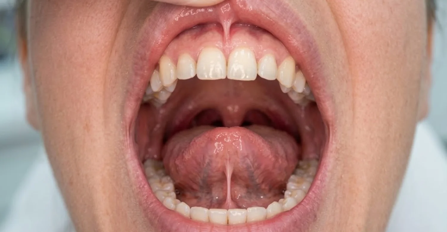
A normal oral frenum usually appears as a small, soft mucosal fold. Its color often resembles the surrounding gum or oral lining. Some frenula look nearly transparent when the tissue is gently stretched. Others appear thicker because connective tissue or muscle fibers are more prominent. The upper central labial fold is often the easiest to see in a mirror. The lingual fold becomes visible when the tongue is raised toward the palate. A visible fold should not automatically be interpreted as excessive tissue. Professional assessment becomes relevant when pain, recession, spacing, or restricted movement develops. Cleveland Clinic also emphasizes variation in frenum size, shape, and attachment.
What Does the Mouth Frenum Do?
Oral frenula helps stabilize movable soft tissues during everyday movements. They guide the position of the lips, cheeks, and tongue. This support contributes to coordinated speaking, chewing, swallowing, and oral hygiene. The structures do not perform these functions alone. Instead, they provide anatomical support while the surrounding muscles produce movement. A healthy Frenum Mouth attachment allows adequate movement without excessive tissue tension. A restrictive attachment, however, can sometimes interfere with those movements. The clinical question is therefore functional rather than purely visual. Research on oral vestibular frenula also links abnormal attachments with possible gingival and lip effects.
How Oral Frenums Support Lip and Tongue Movement
Frenula provides stabilizing connections while still allowing controlled movement. The labial structures connect the lips with tissues around the dental arches. The lingual structure connects the tongue with the floor of the mouth. During movement, these folds change shape and tension dynamically. Healthy tissue permits the required range without painful pulling. Excessive tension may become noticeable during smiling, brushing, speech, or tongue elevation. However, limited movement can have causes beyond the frenum itself. Muscle coordination, pain, dental anatomy, and neurological factors can also influence movement. Assessment should therefore consider the entire functional pattern rather than one visible fold.
Why Frenum Shape and Attachment Vary Between People
Frenum anatomy naturally varies in thickness, attachment level, width, and elasticity. This variation reflects differences in soft tissue anatomy and facial growth. The lingual structure has particularly complex relationships with floor-of-mouth tissues. Cadaveric research found considerable variation in its fascial thickness and attachments. Therefore, no single visual shape defines a healthy Frenum Mouth structure. A thicker fold can still function normally, while a thin fold can occasionally restrict movement. Age and dental development can also change how attachments appear clinically. Findings should always be interpreted within the patient's developmental stage.
Learn about the anatomy and health of the dental pulp
How Many Frenums Are in the Mouth?
There is no single universally useful number for every noticeable oral frenum. Most people have several labial, buccal, and lingual folds. The most prominent structures are usually the easiest for patients to identify. Smaller vestibular folds may be less obvious during everyday inspection. Cleveland Clinic highlights the upper and lower labial frenula and the lingual frenulum. Anatomical literature also describes multiple buccal frenula around the oral vestibule. Counting them depends partly on how individual folds are defined. That is why a fixed number can be misleading. Determining whether an attachment causes symptoms matters more than counting structures.
Labial Frenums of the Upper Lip
The upper labial frenum lies between the upper lip and the front gum tissues. It is usually visible above the central incisors when the lip is lifted. Its attachment may extend toward the alveolar mucosa or closer to the gingival margin. Anatomical studies describe this structure as a collagenous mucosal fold. Its appearance often changes during childhood and dental development. A prominent upper Frenum Mouth attachment does not automatically mean treatment is necessary. Clinicians consider spacing, gum tension, hygiene, and lip movement. The relationship with the upper front teeth also matters during orthodontic planning.
Labial Frenums of the Lower Lip
The lower labial frenum connects the lower lip with tissues around the lower dental arch. Patients may notice it when pulling the lower lip downward. Its position and thickness vary naturally between individuals. Most lower labial frenula do not create significant problems. Occasionally, excessive tension can pull against the gingival margin. That tension may complicate hygiene or contribute to localized periodontal concerns. Some people also experience irritation from repeated lip movement. Clinical evaluation should distinguish normal anatomy from harmful mechanical tension.
The Lingual Frenum Under the Tongue
The lingual frenum is the fold beneath the tongue. It connects the underside of the tongue with the floor of the mouth. Its structure is more complex than a simple rigid cord of tissue. Cadaveric research describes it as a dynamic fold formed by floor-of-mouth fascia. Some people have excellent tongue movement despite a prominent fold. Others have restricted movement because of an unusually tight attachment. Tongue function therefore matters more than visual appearance alone. Children with feeding or speech concerns may need a broader functional assessment.
Buccal Frenums Along the Cheeks
Buccal frenula are soft tissue folds located along the oral vestibule near the premolars. They connect cheek tissues with gingival or alveolar mucosal areas. Several may be present on each side of the mouth. Anatomical research found important structural differences between buccal and labial frenula. Most buccal frenula remain unnoticed throughout life. They may become relevant when dentures, periodontal tissues, or oral movements are affected. Persistent traction can sometimes make brushing uncomfortable. A clinician can assess whether this Frenum Mouth fold is truly responsible for symptoms.
Why There Is No Single Fixed Number of Noticeable Oral Frenums
The mouth contains several anatomical folds that can be described as frenula. Some are prominent, while others are extremely subtle. Clinical terminology can also differ between anatomical references and everyday dental practice. Cleveland Clinic focuses on the most noticeable structures for patients. Anatomical research recognizes additional labial and buccal folds. Asking for an exact number therefore rarely helps clinical decision-making. What matters is identifying the structure causing concern and assessing its function.

Labial Frenum vs Lingual Frenum
The labial frenum connects a lip with nearby gingival tissues. The lingual frenum connects the underside of the tongue with the floor of the mouth. They therefore influence different movements and oral functions. Labial concerns may involve gum traction, lip movement, or tooth spacing. Lingual concerns more often involve tongue mobility and functional restriction. Both structures can become irritated after trauma. Neither automatically requires surgery simply because it looks prominent. Cleveland Clinic distinguishes these two principal categories clearly.
Buccal Frenums and Their Role in Oral Tissue Movement
Buccal frenula sit where cheek movement interacts with vestibular tissues. They help organize soft tissue movement around the dental arch. Their anatomy includes connective tissue and relationships with nearby facial muscles. Problems are relatively uncommon compared with lingual concerns. A restrictive attachment may matter near dentures or thin periodontal tissues. Localized pulling can also make oral hygiene uncomfortable. Treatment should only be considered when objective clinical findings justify intervention.
What Is a High or Low Frenum Attachment?
A high or low attachment describes where the tissue inserts. The term is mainly used when discussing labial or lingual anatomy. A higher labial attachment can extend closer toward the gingival margin. A restrictive lingual attachment may connect the tongue and floor unusually closely. Attachment height alone does not determine disease. Clinical significance depends on tissue tension, movement, symptoms, and surrounding structures. Modern Frenum Mouth assessment increasingly emphasizes function alongside anatomy. This approach reduces overdiagnosis based on visual appearance alone.
Labial Frenum Attachment and the Front Teeth
The upper labial attachment can sometimes extend between the central incisors. Some attachment patterns are associated with a gap called a maxillary midline diastema. However, not every gap is caused by the frenum. Other factors include tooth size, missing teeth, oral habits, and eruption patterns. A systematic review found no consensus supporting routine early frenectomy for every diastema. It recommends considering dental development and orthodontic treatment first. This is particularly important in growing children. Surgery may become appropriate in selected cases after proper diagnosis.
Lingual Frenum Attachment and Tongue Mobility
Lingual attachment matters when it limits useful tongue movement. Clinicians assess tongue elevation, protrusion, lateral movement, and functional coordination. Appearance alone cannot reliably establish restrictions. Recent reviews found substantial variation among available diagnostic tools. Some tools emphasize anatomy, while others include movement measurements. Many visible attachments function adequately without intervention. Treatment should follow a documented functional problem rather than a visual label.
See the risks and timeline of smoking after tooth extraction
What Problems Can Affect a Mouth Frenum?
Most oral frenula remain healthy without any treatment. Problems can develop after trauma, inflammation, excessive tension, or developmental variation. Possible concerns include soreness, tearing, gum recession, tooth spacing, and restricted movement. Some unusual tissue folds may actually represent unrelated lesions, so accurate examination is important. A new mass should not automatically be labeled a Frenum Mouth issue. Clinical photographs can help monitor changes over time. Persistent symptoms may require further examination or imaging. The underlying cause should be established before any surgical decision.
Sore or Inflamed Labial Frenum
A labial frenum can become sore after repeated friction or accidental trauma. Hard foods, sharp teeth, appliances, and vigorous brushing can all contribute. Inflammation may produce tenderness, redness, or mild swelling. Most minor injuries improve as the tissue heals. Cleveland Clinic notes that irritation or injury can cause temporary pain and swelling. Persistent symptoms should be examined rather than repeatedly treated at home. Pain near the frenum does not prove the frenum caused it. The adjacent teeth and mucosa should also be examined.
Short or Tight Lingual Frenum
A short or restrictive lingual attachment can limit tongue movement. This condition is commonly called ankyloglossia or tongue-tie. Its clinical significance varies greatly between individuals. A restrictive appearance does not necessarily create functional problems. Current evidence supports evaluating both anatomy and tongue function. In children, feeding concerns may require a multidisciplinary assessment. Speech concerns also have many possible causes besides restricted tissue. Surgery should therefore not be recommended solely from a visible Frenum Mouth attachment.
High Labial Frenum Attachment and Tooth Spacing
A prominent upper attachment sometimes occurs alongside a front-tooth gap. The relationship is not always causal. A systematic review found associations with certain papillary attachment patterns. However, evidence does not support routine surgery for every childhood diastema. Permanent tooth eruption can naturally change spacing over time. Orthodontic closure may be recommended before surgery in suitable cases. This sequence can also reduce the likelihood of relapse. Treatment should consider age, eruption stage, tooth position, and tissue attachment.
Gum Recession Related to Frenum Tension
A restrictive frenum can exert traction near the gingival margin. Repeated tension may contribute to localized soft tissue problems in selected cases. Anatomical literature recognizes this possibility for labial and buccal structures. However, the gum recession has many more common causes. Brushing trauma, periodontal inflammation, tooth position, and thin tissue can all contribute. The frenum should be considered within the complete periodontal picture. Periodontal management may be needed before any frenular procedure.
Traumatic Framing: Friction, Braces, Bites, or Accidental Injury
Repeated mechanical contact can irritate frenular tissue. Braces, aligners, biting injuries, and sharp surfaces can all produce friction. The tissue may become red, tender, or slightly swollen. Small tears can also develop after sudden trauma. Removing the source of friction often supports uncomplicated healing. Orthodontic appliances should not be discontinued without dental advice. Instead, the clinician can adjust the irritating component. Persistent lesions require examination to exclude infection or unrelated pathology. Never cut a damaged Frenum Mouth fold yourself.
Tissue Tags and Other Lesions That Can Resemble a Frenum
Not every tissue fold inside the mouth is a frenum. Small tags can result from normal anatomy, trauma, scar tissue, or other lesions. A new growth should therefore be assessed carefully. Visual self-diagnosis is unreliable for unusual oral tissue. The dentist may document its size, color, surface, location, and duration. Persistent bleeding or rapid enlargement deserves prompt evaluation. A biopsy may sometimes be necessary for a definitive diagnosis. Pediatric oral surgery guidance recognizes biopsy as an important diagnostic option.
When a Tissue Tag Is Simply Normal Anatomy
Normal oral anatomy contains many folds, ridges, papillae, and mucosal variations. Some become more visible when the lips or tongue are stretched. A fold that remains stable for years may simply reflect individual anatomy. Normal structures are usually painless, unchanged, and similar in surface to nearby mucosa. They should not bleed repeatedly without a clear mechanical explanation. A dentist can confirm whether the appearance matches normal anatomy. Normal variation does not require removal unless functional or restorative needs arise.
When a New or Changing Tissue Tag Needs Examination
New oral tissue deserves attention when it changes or remains symptomatic. Warning features include persistent ulceration, unusual color, bleeding, rapid enlargement, and significant pain. A lesion that interferes with eating also needs assessment. The same applies when it persists after removing a suspected source of trauma. The examination can distinguish a Frenum Mouth fold from another soft tissue condition. A biopsy may be considered when the diagnosis remains uncertain. Early evaluation avoids self-treatment and delayed diagnosis.
Discover all about endosteal implants
Why Is My Mouth Frenum ?
Sore
Swollen
Irritated
Soreness near a frenum usually has a mechanical or inflammatory explanation. Common triggers include accidental biting, rough foods, appliances, and repetitive friction. Inflammation can also affect nearby gingival or oral tissues. A visible frenum does not automatically explain the pain. The exact location and timing of symptoms provide useful clues. Pain that worsens during stretching suggests mechanical irritation. Pain while eating may reflect a broader mucosal injury. Persistent swelling should be assessed by a dental professional. Cleveland Clinic notes that minor frenum injuries often resolve on their own.
Common Causes of a Sore Labial Frenum
The upper and lower labial folds can become irritated by repeated movement. Sharp food edges can create small abrasions. Brushing too aggressively may also traumatize the nearby mucosa. Orthodontic wires can rub against the lip or vestibule. A stretched fold may feel tender after prolonged lip movement. Most minor irritation improves once the mechanical source is removed. Persistent symptoms can indicate a deeper periodontal or mucosal problem. Treatment should target the underlying cause rather than the soreness alone.
Common Causes of Lingual Frenulum Irritation
The tissue beneath the tongue is exposed to constant movement. Sharp food, biting, dehydration, and accidental trauma can cause temporary irritation. Tongue jewelry introduces additional mechanical forces. Poor oral hygiene can also contribute to nearby inflammation. A salivary duct problem may create discomfort in the same region. That distinction matters because the duct openings lie beside the frenulum. Persistent pain should not automatically be labeled a Frenum Mouth or tongue-tie issue. Clinical examination identifies whether the tissue, duct, gland, or another structure is involved.
Mouth Injury, Friction, and Orthodontic Appliances
Orthodontic appliances can rub repeatedly against oral soft tissues. Brackets, wires, and attachments sometimes contact the lips or cheeks. The resulting irritation may appear near a frenum. Orthodontic wax may temporarily reduce friction when recommended by the orthodontist. However, recurring injury may require an appliance adjustment. A sudden injury can produce a small tear and some bleeding. Most minor trauma improves with basic wound protection. Patients should never cut or reshape oral tissue themselves.
How Long Can Minor Frenum Irritation Last?
Minor irritation usually improves as the damaged mucosa heals. The duration depends on injury severity and continued mechanical irritation. Repeated rubbing can prolong symptoms and delay healing. Improvement should generally follow removal of the provoking factor. A lesion that persists or worsens deserves clinical examination. Increasing swelling can suggest infection or more significant tissue injury. Cleveland Clinic notes that uncomplicated frenum irritation usually resolves without major treatment.
Frenum Pain Relief and Practical Home Care
For minor irritation, the first step is protecting the tissue from further trauma. Choose softer foods when chewing causes discomfort. Use gentle oral hygiene around the affected area. Rinsing with clean water can help remove food debris. Avoid repeatedly stretching the sore tissue to inspect it. Over-the-counter pain medicine may help when medically appropriate. Follow the package directions and consider existing health conditions. Do not place harsh chemicals directly on oral wounds. Seek dental advice if a Frenum Mouth problem persists, worsens, or recurs.
Safe Ways to Reduce Irritation
Avoid hard, sharp, or highly abrasive foods while the tissue heals. Use a soft toothbrush with light pressure. Maintain routine oral hygiene without scrubbing the injured area. Cold foods may temporarily soothe minor discomfort for some people. If an appliance causes friction, contact the treating clinician. Do not apply unapproved topical substances. Improvement should be monitored rather than assumed.
What to Avoid While the Tissue Is Healing
Avoid smoking because it irritates oral tissues and impairs healing. Avoid alcohol-containing mouth products if they increase burning. Do not bite, pull, or repeatedly stretch the injured fold. Never attempt self-piercing or self-cutting. Do not scrape away tissue that appears swollen. Avoid foods that repeatedly traumatize the area. Persistent bleeding or worsening pain should trigger professional evaluation.
When Pain Relief Is No Longer Enough
Pain relief should not replace diagnosis when symptoms persist. Repeated pain may indicate ongoing trauma or another oral condition. Swelling can sometimes signal infection or a deeper tissue problem. Persistent ulceration needs professional assessment. Difficulty swallowing or breathing requires urgent care, as does uncontrolled bleeding. A dentist can examine nearby teeth, gums, ducts, and mucosa. Pain location alone is not enough to identify the cause of a Frenum Mouth complaint.
Find out why no exercise after dental implant
Can a Mouth Frenum ?
Tear
Get Cut
Split
Yes, frenular tissue can tear after accidental trauma. Common events include falls, bites, sports injuries, and objects striking the mouth. The tissue may bleed because oral tissues contain many small blood vessels. Pain and swelling can follow immediately. Most small uncomplicated tears heal without surgery. The wound should be protected from additional trauma. Persistent bleeding requires professional attention. Large or deep wounds may need examination for associated injuries. Never deliberately cut the tissue at home to improve movement.
What Can Cause an Accidental Frenum Tear?
Falls are a common cause of injuries involving the lips and mouth. Children may tear tissue when toys or objects strike the oral cavity. Contact sports can create sudden stretching forces. Accidental biting may damage a labial or lingual fold. Orthodontic appliances and piercing jewelry are other potential sources of laceration. The severity depends on the direction and force of the injury. Nearby teeth may also be damaged during the same event.
What Symptoms Can Follow a Frenum Injury?
A torn fold can cause sudden pain and localized bleeding. Swelling may appear during the first hours after injury. The area can feel tender during eating or speaking. A small flap of tissue may remain visible. Some patients notice discomfort when stretching the lip or tongue. Most minor injuries become less painful as healing progresses. Persistent bleeding or increasing swelling is not typical and needs attention. Cleveland Clinic lists tearing, irritation, and swelling among recognized Frenum Mouth problems.
What Should You Do After a Mouth Frenum Injury?
Rinse gently with clean water after the injury. Apply gentle pressure with clean gauze if bleeding occurs. Avoid repeatedly pulling the lip or tongue to look at the wound. Choose softer foods if chewing causes pain. Watch for increasing swelling or discharge. Check whether a tooth was chipped or displaced. Seek urgent assessment when bleeding is difficult to control. Large wounds also need professional evaluation.
Can a Torn Frenum Heal Without Surgery?
Many small tears heal without surgical repair. The oral mucosa has a substantial capacity for healing. Conservative care is often appropriate for superficial uncomplicated injuries. The wound should remain clean and protected from continued trauma. Surgery becomes more relevant when tissue damage is extensive or bleeding persists. Associated tooth or gum injuries may require separate treatment. A clinician can confirm whether the Frenum Mouth tissue is closing normally.
When a Cut or Split Frenum Needs Professional Evaluation
Professional evaluation is appropriate when symptoms are severe or persistent. A large tear can involve deeper tissues beyond the visible fold. Significant swelling can interfere with eating or speaking. Signs of infection require prompt assessment. Associated dental trauma should not be overlooked. Children with substantial facial injuries may need broader medical evaluation. Do not judge a wound simply by comparing it with online photographs.
Persistent Bleeding, Increasing Swelling, or Severe Pain
Persistent bleeding requires prompt professional attention. Increasing swelling may indicate ongoing bleeding, infection, or significant tissue damage. Severe pain also suggests the injury may be more than superficial. Apply steady gentle pressure while arranging care. Avoid repeatedly removing the gauze to inspect the wound. Emergency services may be necessary if the airway is threatened. These warning signs should never be managed through home experimentation.
Signs of Infection or Significant Tissue Injury
Possible infection signs include increasing pain, swelling, discharge, and tenderness. Fever and an unpleasant odor can be additional warning signs. Significant injury may cause persistent bleeding or expose deeper structures. Difficulty swallowing or breathing is an emergency. Do not delay evaluation because the original injury seemed minor. A professional can decide whether antibiotics or other treatments are needed. Antibiotics should never be self-prescribed for a Frenum Mouth wound.
Why Is Saliva Coming From the Area Near the Lingual Frenum?
Saliva naturally enters the floor of the mouth close to the tongue. The major submandibular ducts open on either side of the lingual frenulum. These openings sit on small raised papillae called sublingual caruncles. Saliva can therefore appear to come directly from the frenulum. Tongue movement can visibly change the tissue around these openings. Brief fluid jets during eating or yawning are usually normal salivary flow. Persistent swelling, pain, or reduced flow may indicate another problem. A clinician can determine whether the duct, gland, or frenulum is involved.
Where the Salivary Duct Openings Are Located
The submandibular ducts open on both sides of the lingual frenulum. The openings sit on small mucosal elevations known as sublingual caruncles. This arrangement explains why saliva may seem to emerge from the fold itself. The sublingual glands also lie nearby beneath the floor-of-mouth tissues. Several structures therefore share this small anatomical region. Pain here does not automatically indicate a Frenum Mouth problem.
Why Saliva May Appear to “Leak” or “Squirt” Under the Tongue
Saliva is continuously produced and delivered through several ducts. The submandibular ducts release fluid right beside the lingual fold. Tongue movement and eating make this flow easier to notice. Patients may describe it as leaking or squirting, which is not necessarily abnormal. Pain, swelling, a foul taste, or reduced flow changes the clinical picture. These symptoms may suggest obstruction, inflammation, or another salivary condition. Professional evaluation is appropriate when the change persists or causes discomfort.
How the Lingual Frenum Differs From Nearby Salivary Structures
The lingual frenum is a tissue fold beneath the tongue. The submandibular duct is a separate tube that carries saliva. Its opening lies very close to the frenulum, and the sublingual glands sit nearby. This crowded anatomy makes self-identification difficult with a mirror. A dental professional can examine the floor of the mouth directly. Accurate identification prevents unnecessary procedures on the wrong structure.
When Salivary Changes Near the Floor of the Mouth Need Evaluation
Seek evaluation for persistent pain beneath the tongue. Also seek care for swelling that increases during meals. Reduced salivary flow, a firm swelling, or a recurrent lump deserves examination. Blood-stained or unusual discharge should not be ignored. Fever or spreading swelling may indicate infection. A dentist or oral specialist can examine the ducts and glands. Imaging may be appropriate depending on the findings.
Learn how to detect a tooth infection on x-ray
What Is Frenum Mouth Piercing and Is It Safe?
A Frenum Mouth piercing places jewelry through a fold of oral soft tissue. One popular example is the upper lip smiley piercing. Although marketed as body art, it carries real oral health risks. The American Dental Association advises against cosmetic intraoral and perioral piercing. Reported complications include bleeding, swelling, infection, tooth damage, and gingival recession. Jewelry can also become embedded in surrounding tissues. Repeated contact can damage teeth and restorations. These risks matter because the mouth contains many microorganisms. Professional dental advice should come before any oral piercing decision.
What Is a Smiley or Frenum Piercing?
A smiley piercing is usually placed through the upper labial frenulum. The jewelry becomes visible when the person smiles broadly. This location places metal directly against gums and teeth. Some people choose it for aesthetic or body-art reasons. However, normal lip movement creates repeated mechanical contact. The ADA discourages cosmetic oral piercing because of these risks. Potential complications include gum recession, tooth damage, infection, and soft tissue trauma.
Where Oral Frenum Piercings Are Typically Placed
The upper labial fold is the most common site for frenum piercings. A lower lip version, sometimes called a frowny, also exists. Other oral piercings involve the lips, cheeks, or tongue. Frenular tissue is relatively thin in many people. That thinness does not make piercing risk-free. The jewelry stays close to teeth and gingiva during every movement. Repeated rubbing can create chronic injury.
Common Side Effects of Oral Piercing
Common early effects include pain, swelling, and bleeding. Infection can develop during healing or later. Long-term contact may damage teeth or periodontal tissues. Jewelry also collects plaque and bacteria, which complicates oral hygiene. The ADA reports multiple documented complications of oral piercing. These include tooth fracture, wear, gingival recession, lacerations, scarring, and embedded jewelry. Risk depends on location, jewelry type, habits, hygiene, and individual anatomy. Complications can appear immediately or months after a Frenum Mouth piercing is placed.
Bleeding, Swelling, Pain, and Infection
Piercing creates an intentional wound inside the mouth. Pain and swelling are therefore common early effects. Bleeding can occur during placement or after accidental trauma. The oral cavity contains many microorganisms, which raises infection concerns. Increasing swelling and unusual discharge require prompt assessment. Severe swelling can create serious complications in some cases. Good hygiene reduces but does not eliminate these risks.
Tooth Chipping, Wear, and Gingival Recession
Jewelry can repeatedly strike or rub against nearby teeth. Over time, this may cause chipping, wear, or periodontal trauma. A 2023 review cited by the ADA reported recession in up to 50% of lip-piercing patients. The same review reported recession in up to 44% of tongue-piercing patients. Tooth damage was reported in 26% of people with tongue piercings. These figures describe study findings, not guaranteed individual outcomes.
Scarring, Jewelry Embedding, and Other Complications
Jewelry can become embedded within swollen oral tissue. Removal may then require a minor surgical procedure. Scarring can also change the appearance or movement of the tissue. Some patients develop persistent irritation around the jewelry. Jewelry may also interfere with dental examinations or imaging. Rare complications include aspiration or significant airway swelling. These consequences explain why the ADA discourages any cosmetic Frenum Mouth piercing.
Why Self-Piercing or Cutting the Frenum Is Unsafe
Self-piercing creates serious infection and uncontrolled bleeding risks. Home cutting can damage nearby nerves, vessels, gums, or salivary ducts. Sterility cannot be reliably maintained in an improvised setting. Incorrect placement may leave permanent scarring. It can also worsen functional problems instead of solving them. Anyone concerned about restrictive tissue should seek professional assessment. Frenular surgery requires structured diagnosis, controlled technique, and planned aftercare.
Why Tongue or Oral Tissue Splitting Is a Different and Higher-Risk Procedure
Tongue splitting deliberately divides the tongue into two sections. It is fundamentally different from treating a restrictive frenum. The American Dental Association describes it as invasive and dangerous. Potential complications include severe bleeding, infection, nerve damage, and significant pain. Swelling may also threaten the airway. It is performed for body modification rather than medical benefit. No cosmetic goal justifies self-performed tongue cutting.
Is a Short or Tight Frenum Always a Problem?
A short or prominent attachment is not automatically pathological. Some people have restricted-looking anatomy yet completely normal movement. Others experience genuine limitations that affect eating, speech, hygiene, or comfort. Modern evidence emphasizes combining anatomical and functional findings. This distinction is especially important for infants and children. The AAPD states that not all children with ankyloglossia require surgery. A specific functional concern should therefore be established first. Appearance alone should not determine a Frenum Mouth treatment plan.
Normal Anatomical Variation vs Functional Restriction
Normal variation can make a frenum look thicker or closer to the tongue tip. Functional restriction means the tissue actually limits useful movement. Telling the two apart requires more than visual inspection. Clinicians observe tongue elevation, protrusion, lateral movement, and coordination. They also consider effects on speech, feeding, swallowing, and hygiene. Recent reviews found considerable inconsistency among diagnostic systems. This supports individualized assessment instead of one visual threshold.
How Dentists Assess Tongue Mobility and Lip Function
The clinician observes active tongue and lip movement. The patient may be asked to raise, extend, and move the tongue sideways. Functional tasks can reveal restrictions that resting anatomy hides. Tissue tension during movement is inspected closely. Symptoms are documented rather than assumed from appearance. Reviews support tools that combine structure and function. Still, any single score must be read within the broader clinical context.
Why Appearance Alone Does Not Determine the Need for Surgery
A prominent frenum can be entirely functional. Conversely, a subtle attachment may still create meaningful restriction. Visual appearance therefore cannot provide the whole diagnosis. The clinician should identify the actual problem requiring treatment. For example, gum traction may matter more than tissue thickness. The AAPD supports individualized decisions and acknowledges uncertainty about some indications. This reduces unnecessary Frenum Mouth procedures and unrealistic expectations.
Children, Tongue-Tie, Breastfeeding, and Functional Assessment
Infant tongue-tie assessment requires particular care. Feeding difficulties have many possible causes. The AAPD states that causes other than ankyloglossia are more common for breastfeeding problems. Recent systematic evidence supports evaluating both structure and feeding function. A 2026 review found very-low certainty evidence for current diagnostic criteria. Surgery should therefore follow a comprehensive assessment. Parents deserve balanced information about expected benefits and limitations. A team approach involving lactation and speech professionals may help.
.jpg)
.jpg)
.jpg)
.jpg)
.jpg)
How Is a Mouth Frenum Problem Diagnosed?
Diagnosis begins with a detailed oral examination. The clinician first identifies which structure is causing the concern. Attachment, tension, movement, and surrounding tissues are then evaluated. Symptoms are compared with objective findings. Teeth, gums, bite, and hygiene are also considered. A lesion that only resembles a frenum may need a different diagnostic pathway. Imaging is not routinely needed for every Frenum Mouth concern. Treatment planning should follow the confirmed diagnosis.
What Happens During a Dental Examination?
The dentist first reviews the main symptom and its duration. The mouth is then examined under good lighting. The affected fold is inspected at rest and during movement. The clinician may gently stretch the lip or lift the tongue. Nearby teeth and gums are evaluated as well. Previous trauma or orthodontic treatment may influence the findings. The goal is to identify the real anatomical or functional problem.
What the Clinician Looks For
The examination focuses on attachment, tension, mobility, and surrounding tissue health. The clinician also checks for inflammation, trauma, and unusual tissue changes. Dental spacing and periodontal findings may influence labial decisions. Tongue movement is central when ankyloglossia is suspected. Symptoms should correlate with the observed anatomy. A visible fold without symptoms may need no treatment at all.
Frenum Attachment and Tissue Tension
Attachment level describes where the fold connects with surrounding tissues. Tension describes how strongly movement pulls on those tissues. A high labial attachment can pull near the gingival margin. A restrictive lingual attachment may limit tongue elevation. Neither finding automatically indicates surgery. The clinician considers whether the tension creates a measurable problem.
Lip and Tongue Movement
Movement testing reveals restrictions that resting anatomy cannot show. The upper lip may be lifted or stretched during the examination. The tongue may be extended and raised toward the palate. Lateral movement is also observed. The clinician records discomfort, compensatory movement, or visible tissue blanching. Literature supports combining these functional findings with structural assessment.
Teeth, Gums, Bite, and Oral Hygiene
Surrounding dental structures can influence treatment decisions. A labial attachment may be assessed alongside a midline diastema. Gum recession may require periodontal evaluation. Plaque retention can make a minor attachment clinically relevant. Bite relationships may also shape orthodontic planning. Considering these findings prevents isolated treatment of a single fold.
Symptoms, Trauma, and Functional Limitations
The clinician asks when symptoms began and what triggers them. Trauma can explain sudden pain without any need for surgery. Orthodontic appliances can explain recurring irritation. Functional limitation may suggest a restrictive attachment. Persistent lesions without an obvious cause need closer evaluation. Clinical history is therefore as important as visual inspection.
When Additional Dental Imaging May Be Useful
Most superficial frenular concerns do not require advanced imaging. Imaging helps when deeper dental or bony involvement is suspected. It may also support orthodontic or restorative planning. A simple dental radiograph may answer a narrow question. CBCT is reserved for specific clinical indications. Imaging should never be ordered simply because a frenum looks prominent.
Differential Diagnosis for an Unusual Tissue Fold or Tag
Differential diagnosis means considering alternative explanations for an appearance. A new fold may represent trauma, scar tissue, inflammation, or another lesion. A salivary structure may be mistaken for abnormal tissue near the tongue. The clinician weighs duration, surface, color, symptoms, and growth. Persistent lesions may need a biopsy for a definitive answer. This process reduces the risk of treating the wrong structure.
Learn all about how teeth whitening works
When Is Frenum Mouth Surgery Needed?
Frenum Mouth surgery is considered when tissue creates a clearly defined clinical problem. Examples include functional restriction, persistent periodontal traction, or selected denture concerns. Some orthodontic situations may also justify treatment. Conservative care is often appropriate for minor irritation or asymptomatic anatomy. The procedure should target the specific anatomical problem identified. Not every prominent fold requires removal. Evidence for some indications remains uncertain. The AAPD emphasizes individualized decision-making for pediatric patients.
Situations Where Conservative Care May Be Enough
Observation is appropriate when anatomy causes no functional problem. Minor trauma can usually be managed without surgery. Orthodontic friction may improve after an appliance adjustment. Temporary irritation usually settles once the source is removed. A stable, painless frenum generally needs no intervention. The plan should be reviewed if symptoms change.
Functional Reasons for Frenum Surgery
Surgery may be considered when a restrictive attachment causes meaningful functional limitation. Tongue movement is often the key factor. Lip movement may also be affected by some labial attachments. The clinician should define the exact limitation before recommending surgery. Each procedure should have a clear functional objective. Expected outcomes should be discussed beforehand with the patient.
Periodontal and Gum-Related Considerations
A frenum can sometimes exert traction near the gingival margin. This may complicate hygiene or add localized tissue stress. However, the gum recession has many possible causes. Inflammation, traumatic brushing, and tooth position may contribute more. A periodontal examination identifies the dominant risk factor. Frenum treatment should not replace care for underlying gum disease. Surgery may help when attachment tension remains clinically relevant.
Orthodontic Considerations for a Labial Frenum
Labial attachments can appear alongside a maxillary midline diastema. They are not always the primary cause, however. A systematic review found no consensus supporting routine early frenectomy. Orthodontic closure is often considered before surgical intervention. Timing matters because an eruption can change the appearance naturally. Coordinating orthodontic and surgical planning improves treatment logic for a Frenum Mouth concern.
Tongue-Tie and the Importance of Individual Assessment
Tongue-tie should be evaluated through both anatomy and function. No single appearance reliably predicts treatment needs. Recent reviews found inconsistent diagnostic tools and thresholds. Surgery may be considered when restriction causes documented problems. Infant cases require special attention to feeding function. The AAPD does not recommend surgery for every affected infant.
What Frenum Surgery Can and Cannot Be Expected to Correct
Surgery can release or remove restrictive tissue. It cannot automatically correct every speech, feeding, orthodontic, or periodontal problem. Other anatomical or functional issues may remain afterward. Orthodontic alignment may still require braces or aligners. Gum recession may need separate periodontal management. Feeding and speech concerns can require additional professional support. Patients should receive realistic expectations before treatment begins.
What Is the Difference Between ?
Frenectomy
Frenotomy
Frenuloplasty
These terms describe different surgical approaches to frenular tissue. A frenectomy generally removes the frenum or a substantial portion of it. A frenotomy releases or divides restrictive tissue without full removal. A frenuloplasty reshapes and closes the tissue after release. Terminology can vary slightly between clinicians. The key issue is selecting an approach that matches the problem. The choice considers tissue location, function, healing, and recurrence risk.
Frenectomy: Removing or Excising the Frenum
Frenectomy involves excising the relevant frenular tissue. It is commonly discussed for labial attachments. The clinician removes tissue that creates undesirable traction or obstruction. The depth of removal depends on the attachment and surgical goal. Indications may include periodontal, orthodontic, functional, or prosthetic concerns. Frenectomy should never be performed simply because a fold looks large.
Frenotomy: Releasing or Dividing the Restrictive Tissue
Frenotomy focuses on releasing a restrictive attachment. It is often considered for selected lingual restrictions in infants. The goal is improved movement rather than complete tissue removal. The extent of release depends on anatomy and function. Decisions in infants require full feeding and anatomical assessment. The AAPD emphasizes that not every ankyloglossia case needs surgery.
Frenuloplasty: Reshaping the Tissue for Functional Correction
Frenuloplasty releases the tissue and then reshapes the resulting area. The technique can reduce tension while supporting functional healing. Closure patterns depend on the procedure and clinician preference. It is useful when simple division would leave an unfavorable defect. Patients receive postoperative instructions specific to the technique performed.
Why the Procedure Should Match the Anatomical Problem
Different anatomical problems require different treatment strategies. A labial attachment may need a different approach from tongue restriction. A traumatic tear does not automatically require elective frenectomy. A normal visible fold does not need surgery at all. Correct diagnosis must come before technique selection. This principle reduces unnecessary procedures and clarifies the intended outcome.
Read a complete guide on how to care for your dentures
What Happens During Frenum Mouth Surgery?
Frenum Mouth surgery begins with a confirmed clinical assessment. The clinician explains the indication and the expected outcome. Local anesthesia is used for most office procedures. The technique may involve a scalpel, electrosurgery, or a laser. The choice depends on anatomy, clinician expertise, equipment, and treatment goals. Some procedures require sutures, while others do not. Postoperative instructions cover hygiene, diet, pain, and wound care. Follow-up confirms that healing is progressing well.
Examination and Treatment Planning Before Surgery
Before surgery, the clinician confirms the anatomical diagnosis. The patient's symptoms and functional concerns are reviewed. Dental and periodontal findings are documented. Orthodontic timing may matter for labial cases. Tongue function should be assessed before lingual procedures. Patients should hear about alternatives, including observation where reasonable. This preparation supports informed consent and realistic expectations.
Local Anesthesia and Patient Comfort
Local anesthesia numbs the treatment area during the procedure. Patients may still feel pressure, movement, or vibration. The amount of anesthetic depends on the procedure and the patient. Anxiety should be shared with the dental team beforehand. Mild soreness can appear after the numbness wears off. Postoperative instructions explain appropriate pain management.
Conventional Surgical Techniques
Conventional methods use a scalpel or similar surgical instrument. The clinician releases or removes the targeted tissue. Bleeding is controlled during the procedure. Sutures may be placed depending on the wound design. These traditional approaches remain well-established treatment options. Their success depends heavily on case selection and surgical technique.
Laser Frenum Surgery
Laser techniques can be used for selected frenectomy procedures. They may offer useful control of soft tissue and bleeding. A 2026 meta-analysis evaluated six randomized trials involving 324 patients. Laser treatment reduced day-one pain compared with conventional scalpel surgery. It also reduced intraoperative bleeding and operative time. However, the authors rated evidence certainty as low to very low. These results should therefore be interpreted cautiously for any Frenum Mouth case.
How Clinicians Decide Between Laser and Conventional Techniques
Technique selection depends on anatomy and the clinical objective. Laser equipment may offer helpful soft tissue control. Conventional instruments remain effective and widely used. Clinician training and experience also influence the choice. A laser is not automatically better for every patient. The 2026 meta-analysis favored diode lasers in selected labial cases. However, small study sizes and heterogeneity remain important limitations.
Pain, Bleeding, Swelling, and Operative Considerations
The 2026 meta-analysis found lower pain on days one and seven with diode lasers. Day-three pain did not differ significantly between techniques. Intraoperative bleeding was significantly lower with laser treatment. Operative time was shorter by roughly seven minutes. Swelling did not differ significantly. Technique choice should reflect the individual case rather than marketing claims.
Sutures and Wound Management
Some procedures require sutures to bring tissue edges together. Others heal without conventional suturing. The choice depends on surgical design and tissue characteristics. The wound should be kept clean without aggressive manipulation. Eating may feel uncomfortable during the first days. Sutures should only be removed according to professional instructions.
What Are the Side Effects After Frenum Surgery?
Most procedures cause some temporary local discomfort. Possible effects include tenderness, swelling, minor bleeding, and eating difficulty. Severity varies with the procedure and individual healing. Less common complications include infection, delayed healing, scarring, or persistent symptoms. Patients should leave the clinic knowing the warning signs. Symptoms should be interpreted according to the specific Frenum Mouth procedure performed.
Common Short-Term Effects
Mild soreness is common after any soft tissue surgery. Swelling may develop around the treated area. Minor bleeding or blood-tinged saliva can occur initially. Eating may feel uncomfortable for a short period. Speech can feel different after lingual procedures. These effects should gradually improve during healing.
Pain and Tenderness
Tenderness usually begins as the anesthesia wears off. Pain levels depend on tissue location and surgical technique. Over-the-counter analgesics may be recommended when medically appropriate. Follow dosage instructions and clinician guidance carefully. Severe, worsening, or unchanging pain after several days deserves review.
Swelling and Minor Bleeding
Mild swelling is a normal response to tissue surgery. Minor bleeding can occur shortly after the procedure. Gentle pressure with gauze usually controls it. Persistent or heavy bleeding is not expected. Rapidly increasing swelling requires urgent evaluation. Breathing or swallowing difficulty is an emergency.
Temporary Eating or Speaking Discomfort
Eating can feel uncomfortable while the wound remains tender. Soft foods are easier during early healing. Lingual procedures can temporarily change how tongue movement feels. Speech sensations may differ for a short period. Most patients adapt as the tissue heals. Persistent functional changes should be discussed at follow-up.
Less Common Complications
Less common problems include infection, delayed healing, and excessive scarring. Restriction can persist if the underlying problem was not fully addressed. Some symptoms may also come from an unrelated condition. This reinforces the importance of accurate diagnosis before any Frenum Mouth procedure. Follow-up helps identify healing problems early.
Infection and Delayed Healing
Infection may cause increasing pain, swelling, discharge, or fever. Delayed healing can occur when the tissue remains traumatized. Smoking also slows oral tissue healing. Protect the wound from avoidable irritation. Contact the treating clinic if healing seems abnormal. Antibiotics should be based on professional assessment only.
Scarring or Persistent Functional Symptoms
Scar tissue can sometimes remain after soft tissue surgery. Most scars become less noticeable as healing matures. Persistent restriction may require further assessment. The original diagnosis should be reconsidered if symptoms continue. Additional surgery is not automatically the right solution. A multidisciplinary approach may help in complex cases.
When Post-Surgery Symptoms Require Urgent Attention
Uncontrolled bleeding requires urgent assessment. Rapidly increasing swelling also needs immediate attention. Difficulty breathing or swallowing is an emergency. High fever or spreading infection signs need prompt care. Do not wait for a routine follow-up when severe symptoms appear. The right emergency pathway depends on local healthcare services.
How Long Does It Take to Recover From Frenum Surgery?
Recovery varies with the procedure and individual healing. Minor soft tissue procedures often allow a quick return to routine activities. Initial tenderness usually settles before complete tissue remodeling finishes. Laser and conventional methods can differ in their immediate postoperative profiles. The 2026 meta-analysis found shorter operative times with diode lasers. That finding does not guarantee faster complete healing for every patient. Follow-up remains important for judging functional recovery after treatment.
What Recovery Usually Involves
Early recovery focuses on protecting the surgical area. Patients may notice tenderness and mild swelling. Food choices may need temporary adjustment. Oral hygiene should continue carefully. Specific instructions depend on the technique used. The clinician decides when normal activities can fully resume.
Eating, Brushing, and Oral Hygiene During Healing
Soft foods reduce discomfort during early healing. Keep up oral hygiene while avoiding direct trauma to the wound. A soft toothbrush helps around sensitive areas. Do not abandon brushing, because plaque will accumulate. Follow the clinician's instructions for rinses and wound cleaning. Avoid pulling the wound open to inspect it.
Follow-Up Appointments and Wound Monitoring
Follow-up allows the clinician to examine healing directly. The tissue is checked for infection or delayed closure. Functional improvement can also be evaluated. Tongue movement may be reassessed after lingual surgery. Periodontal tension can be reviewed after labial procedures. Patients can also report any persistent symptoms.
Returning to Normal Activities
Many patients resume routine activities gradually after uncomplicated procedures. Timing depends on discomfort and procedure type. Contact sports may need extra caution while the tissue is vulnerable. Follow your individualized postoperative restrictions. Returning too quickly may cause avoidable trauma.
When the Final Result Should Be Assessed
Early appearance does not represent the final tissue result. Swelling temporarily changes the shape of the area. Scar maturation continues after initial healing. Functional outcomes should be judged after sufficient recovery. Orthodontic or periodontal results may need longer observation. The clinician sets the appropriate assessment timing.
Note : All images used are for editorial and illustrative purposes only and may not originate from the original news provider or associated company.
Can You Stretch a Mouth Frenum Safely?
Forceful stretching is not recommended without professional guidance. A frenum is living tissue connected to nearby structures. Aggressive pulling can cause tears, bleeding, and further inflammation. Self-cutting is particularly dangerous. Clinician-guided mobility exercises may form part of some treatment plans. Their purpose depends on the underlying functional problem. After surgery, exercises may support healthy movement. Always follow the specific instructions given by your treating clinician.
Why Forceful Stretching or Cutting Is Not Recommended
Stretching tissue beyond its tolerance can cause injury. Repeated trauma creates inflammation and delays healing. Cutting produces an uncontrolled wound with infection and bleeding risks. It can also damage nearby structures. A restrictive attachment needs diagnosis before any intervention. Professional treatment is safer and more predictable.
Clinician-Guided Mobility or Oral-Motor Exercises
Exercises may help when they address a documented functional goal. The exact movements depend on age and clinical condition. Some patients benefit from coordinated oral-motor therapy. Others do not need exercises at all. The clinician should explain the purpose and expected benefit. Exercises should never cause significant pain or repeated injury.
When Postoperative Exercises May Be Part of a Treatment Plan
Some postoperative plans include gentle movement exercises. The goal is usually maintaining mobility while the tissue heals. Instructions differ according to the surgical technique. Patients should not invent aggressive stretching routines. The clinician determines suitable timing and intensity. Stop any exercise that causes bleeding or worsening pain.
How Much Does Frenum Mouth Surgery Cost?
Frenum Mouth surgery pricing varies widely between countries and clinics. The final price depends on anatomy, technique, provider, setting, anesthesia, and complexity. Published figures should be treated as planning references only. They are not universal treatment quotes. Current 2026 listings show substantial variation within each market. A personalized clinical assessment is needed before any accurate quote.
Average Frenum Surgery Cost in the United States
A current 2026 cost reference places many in-office cases at 250–800. Hospital procedures under general anesthesia usually cost more. Laser tongue releases may also cost more than simple labial procedures. Provider experience, location, and complexity all affect the fee. Insurance coverage varies by indication and policy. Request an itemized estimate before treatment.
Average Frenum Surgery Cost in the United Kingdom
Current private UK listings show substantial price differences. Several London clinics list frenectomy at around $350. Specialist oral surgery listings can approach $450 or more. Some specialist providers list considerably higher prices for specific procedures. These differences reflect provider type, technique, and complexity. Use published prices as planning references only.
Average Frenum Surgery Cost in Turkey
Current Istanbul listings show considerable variation. One 2026 medical travel source estimates roughly 216–1,081. Specific Turkish clinic listings include frenectomy prices around $287 and $337. Higher package estimates also appear online. This shows why source quality and treatment scope matter. Confirm exactly what each quotation includes. A clinical examination is still required to determine the actual cost.
US vs UK vs Turkey Frenum Surgery Cost Comparison Table
Country | Published planning range | Planning midpoint* | Main cost factors |
United States | 250–800 for many office-based cases | ~$525 | Procedure type, provider, location, anesthesia, complexity |
United Kingdom | 350–995+ in published private examples | ~$673 | Specialist involvement, technique, complexity, clinic |
Turkey | 216–1,081 in current Istanbul estimates | ~$649 | Clinic, surgeon, technique, diagnostics, complexity |
*Midpoints are simple planning calculations, not average national prices. Published prices change over time and may exclude additional services.
What May or May Not Be Included in the Quoted Surgery Cost
A quoted fee may cover only the procedure itself. Other providers bundle consultation, anesthesia, and follow-up. This makes similar-looking prices hard to compare. Always request an itemized quotation before accepting treatment.
Consultation and Dental Examination
Some clinics include the examination in the surgical fee. Others charge for consultation separately. The consultation determines whether surgery is actually indicated. It may also reveal whether imaging or specialist referral is needed. A separate examination fee can therefore be clinically justified.
Local Anesthesia or Sedation
Local anesthesia is often included in straightforward procedures. Sedation usually adds a separate charge. Intravenous sedation may involve specialist fees and monitoring. Ask whether anesthesia is part of the quoted amount.
Laser or Conventional Surgical Technique
Some clinics price procedures differently according to the technology used. Laser procedures carry different equipment and operating costs. Technique should still be chosen clinically, not just financially. The 2026 evidence suggests potential benefits for diode lasers in selected cases.
Follow-Up and Postoperative Care
Follow-up visits may be included or billed separately. Suture removal can also carry a separate charge. International patients should clarify how postoperative communication works. A complete quotation should explain all of these details clearly.
Ready to transform your smile?
Talk to our team and get a personal FREE treatment plan.
What We Notice Clinically
Patients often become concerned after seeing normal oral anatomy online. A visible fold can look abnormal without causing any problem. This happens especially with labial and lingual structures. Clinical decisions should focus on symptoms and function. Research demonstrates substantial natural variation in frenular anatomy. That variation makes appearance-only diagnosis unreliable. At Vitrin Clinic, an individualized dental assessment helps separate normal anatomy from a true Frenum Mouth problem.
Why Patients Often Mistake Normal Frenum Anatomy for a Dental Problem
A mirror shows only the surface of oral tissues. It cannot reveal deeper attachments or functional relationships. Many healthy frenula look prominent when the lip or tongue is stretched. Online images can make normal variation appear unusual. Patients may also compare themselves with idealized illustrations. A dentist can assess movement, tension, gums, teeth, and symptoms together.
The Important Difference Between a Visible Frenum and a Functionally Restrictive Frenum
Visibility does not equal restriction. A prominent fold may allow excellent movement and cause no symptoms. A less noticeable fold can still create functional limitations. This difference is central to evidence-based assessment. Recent reviews emphasize combined structural and functional evaluation. Treatment should begin only with a documented functional concern.
Why a “Thick,” “Long,” or “Tight” Frenum Does Not Automatically Mean Surgery Is Needed
Words such as thick or tight describe appearance without proving disease. Clinical impact depends on movement and surrounding tissue health. Some large-looking frenulas are completely symptom-free. Surgery should always have a defined purpose. Unnecessary tissue removal exposes patients to pain, scarring, and cost. Conservative observation is often more appropriate.
Why a Sore Tissue Fold Can Have Causes Other Than the Frenum Itself
Pain near a frenum can result from trauma or appliance friction. Nearby gums may also be inflamed. Under the tongue, salivary ducts sit close to the visible fold. A salivary obstruction can therefore mimic frenular discomfort. A small mucosal lesion can also develop nearby. Diagnosis should follow examination rather than location alone.
Clinical Note
Clinical assessment should focus on anatomy, function, symptoms, and treatment goals. The appearance of a fold should never be the sole reason for surgery. Individualized evaluation shows whether treatment is truly necessary. This approach aligns with current evidence on functional assessment and careful technique selection.
Dr. Rifat Alsaman, Head of the Medical Team at Vitrin Clinic and Cosmetic Dentist, on Individualized Frenum Assessment
At Vitrin Clinic, the guiding principle is individualized assessment. The clinician first establishes why the tissue is causing concern. The evaluation considers teeth, gums, oral tissues, bite, and movement. Treatment then addresses the identified problem rather than appearance alone. This approach supports informed decisions and realistic expectations for every patient.
Tips for Patients
Most concerns become easier to understand after a professional examination. Avoid comparing your anatomy with isolated online photographs. Minor irritation often improves once the source of friction is removed. Persistent symptoms always deserve assessment. Anyone considering surgery should understand its intended benefit and alternatives. Anyone considering oral piercing should understand the documented dental risks.
What to Do if Your Frenum Is Mildly Sore or Irritated
Protect the area from further mechanical irritation. Brush gently and avoid aggressive stretching. Choosing softer foods while chewing is uncomfortable. Monitor the area for steady improvement. Never cut or pierce the tissue yourself. Persistent soreness should be reviewed by a dentist.
What to Avoid After a Frenum Injury
Avoid repeatedly touching or pulling the injured area. Do not use sharp objects to inspect the wound. Avoid foods that scrape the injury. Do not apply strong chemicals or unapproved medications. Never ignore persistent bleeding. Seek professional evaluation if symptoms worsen.
What to Ask Your Dentist Before Frenum Surgery
Ask what exact problem the surgery is intended to fix. Ask whether observation remains a reasonable option. Ask which technique is recommended and why. Ask about expected healing and possible complications. Ask whether orthodontic or periodontal care will also be needed. Ask what follow-up is included in the fee.
Ask What Functional Problem the Procedure Is Intended to Address
A clearly defined treatment goal improves informed consent. Examples include movement restriction, tissue traction, or selected orthodontic concerns. Ask how the clinician measured or observed the problem. A visual description alone may not be sufficient.
Ask Which Technique Is Recommended and Why
Ask whether frenectomy, frenotomy, or frenuloplasty fits your case. Ask why laser or conventional surgery is preferred. Evidence suggests diode lasers may improve some short-term outcomes in labial cases. The right technique still depends on your individual anatomy.
Ask About Healing, Side Effects, and Follow-Up
Understand the expected soreness, swelling, diet changes, and hygiene steps. Ask which symptoms should prompt urgent contact. Confirm when the clinic wants to review healing. International patients should clarify remote follow-up arrangements.
Medical Tips for Patients Considering Oral Piercing
Discuss oral piercing risks with a dental professional first. Consider the potential for tooth damage and gum recession. Jewelry can harbor plaque and cause ongoing tissue trauma. The ADA advises against cosmetic oral piercing. Avoid self-piercing and self-cutting completely. Seek assessment if existing jewelry causes bleeding or swelling.
When Not to Delay Professional Assessment
Do not delay care for uncontrolled bleeding or rapid swelling. Persistent ulcers, lumps, or changing tissue need examination. Severe unexplained pain also deserves assessment. Swallowing or breathing difficulty requires urgent medical care. These symptoms should never be dismissed as normal anatomy.
How Vitrin Clinic Evaluates Frenum Concerns
Vitrin Clinic approaches frenular concerns through individualized dental assessment. The objective is to identify the true source of the patient's concern. The examination can include labial, lingual, or buccal tissues. Teeth, gums, bite, and functional movement are also reviewed. Additional diagnostics are used only when clinically appropriate. The resulting plan matches the diagnosis and the treatment goal. Frenum care may also form part of a broader dental plan.
Individualized Clinical Assessment of Labial, Lingual, or Buccal Frenum Concerns
The examination starts with the specific concern the patient reports. The relevant fold is inspected at rest and during movement. Attachment level and tissue tension are carefully considered. The clinician also reviews surrounding gum and dental conditions. This avoids treating anatomy that has no clinical significance.
Evaluating Teeth, Gums, Oral Tissues, Bite, and Functional Movement
A complete dental assessment looks beyond the visible fold. Tooth spacing and periodontal tissues are examined when relevant. Tongue and lip movement can be tested directly. The bite may change how symptoms are interpreted. Oral hygiene and tissue condition also affect treatment decisions.
Digital X-Rays, CBCT, or 3D Scanning When Clinically Appropriate
Imaging is selected according to the clinical question. Routine frenular assessment does not automatically require CBCT. Digital X-rays may help with related dental findings. CBCT provides three-dimensional detail when deeper structures need evaluation. 3D scanning may support broader digital planning when indicated. The aim is useful information without unnecessary imaging.
Personalized Treatment Planning for Frenum-Related Dental Concerns
Recommendations should reflect the patient's anatomy and goals. Some patients need observation rather than surgery. Others may benefit from frenectomy, frenotomy, or another approach. The plan can also include periodontal or orthodontic treatment. The intended benefit is explained before any treatment begins.
Frenum Treatment When It Is Part of a Wider Orthodontic, Periodontal, or Smile Plan
Frenum treatment sometimes interacts with other dental procedures. Orthodontic spacing may require coordinated timing. Periodontal concerns can influence surgical planning. Restorative work may also make tissue position relevant. A coordinated approach prevents isolated treatment decisions.
Internal Link Opportunity: Vitrin Clinic Treatments Page
Add a link to the Vitrin Clinic treatments page within this section. The page gives readers an overview of available dental services. Descriptive anchor text, such as dental treatments at Vitrin Clinic, matches the destination accurately. Place the link where broader orthodontic, periodontal, or smile planning is discussed. Readers can then explore related care after understanding their frenum concern. Avoid repeating the same exact anchor elsewhere on the page.
How Vitrin Clinic Supports Your Treatment Journey
For international patients, treatment involves more than the clinical procedure. Clear communication helps patients understand each appointment. Assessment findings are explained before treatment begins. Careful coordination can also reduce unnecessary visits. Vitrin Clinic organizes the patient's dental journey around individualized planning. The goal is complete clarity before any treatment proceeds.
Consultation and Review of the Patient’s Main Concern
The process begins by understanding the patient's main dental concern. The clinical team then decides which examination is appropriate. Patients receive an explanation of the relevant findings. This creates a clear foundation for treatment planning.
Coordinating Assessment and Treatment Appointments
International patients often need careful appointment coordination. Assessment and treatment can sometimes be scheduled efficiently together. The exact schedule depends on the clinical plan. Patients should receive clear expectations before they travel.
Explaining the Recommended Treatment Plan Before Proceeding
Patients should understand why treatment is recommended. The clinician explains alternatives where appropriate. Expected benefits, limitations, risks, and recovery are also discussed. This supports informed decision-making before any procedure.
Supporting International Patients With Treatment Coordination
International dental care involves transport, accommodation, scheduling, and follow-up communication. Vitrin Clinic can coordinate these practical aspects around the dental plan. Patients still receive individualized advice from qualified professionals. Travel arrangements never replace appropriate clinical evaluation.
Post-Treatment Follow-Up and Communication
After treatment, patients may need guidance on healing and daily care. Follow-up helps identify unexpected symptoms early. International patients may need communication after returning home. Clear written instructions support continuity of care.
Internal Link Opportunity: Relevant Existing Vitrin Articles on Orthodontics, Gum Health, or Oral Surgery
Link to relevant Vitrin articles on orthodontic treatment, gum health, and oral surgery. These links expand topical coverage and strengthen semantic relationships. Anchor text should accurately describe each destination article. Avoid repeating exact-match anchors across the website.
Evidence for frenum mouth
Current evidence supports a careful, individualized approach. Anatomical studies demonstrate substantial natural variability. Pediatric guidance highlights uncertainty around some surgical indications. Recent diagnostic reviews show inconsistency among assessment tools. A 2026 meta-analysis found potential advantages for diode laser labial frenectomy. However, evidence certainty was low to very low. These findings support balanced counseling rather than universal recommendations.
Cleveland Clinic for Oral Frenum Anatomy, Types, Function, and Common Conditions
Cleveland Clinic describes oral frenums as connective tissue structures supporting the lips and tongue. It identifies labial and lingual frenula as the major oral types. It also describes common issues such as irritation, injury, and abnormal attachment. This makes it a useful patient-facing anatomy reference.
American Academy of Pediatric Dentistry for Frenulum Management and Individualized Surgical Decision-Making
The AAPD emphasizes individualized management of pediatric frenulum concerns. It notes that not all infants with ankyloglossia require surgery. It also calls for stronger evidence on long-term outcomes. This supports cautious decision-making in children.
PubMed and Peer-Reviewed Literature for Lingual Frenum Anatomy and Frenectomy Evidence
Peer-reviewed studies show that lingual frenulum structure is dynamic and variable. Research also examines diagnostic approaches and surgical techniques. Recent reviews still identify inconsistent definitions and measurement tools. No single visual measurement should therefore dominate clinical decisions.
American Dental Association for Oral Piercing Risks and Patient Safety
The ADA advises against cosmetic intraoral and perioral piercing. Reported complications include infection, bleeding, tooth damage, and gingival recession. The ADA also warns strongly against tongue splitting. These positions are highly relevant to cosmetic frenular modification.
Current 2026 Research on Laser vs Conventional Labial Frenectomy
A 2026 meta-analysis included six randomized controlled trials with 324 patients. Diode lasers reduced day-one pain, intraoperative bleeding, and operative time. Swelling did not differ significantly between techniques. Evidence certainty ranged from low to very low. The findings suggest potential benefits without proving universal superiority.
Key Takeaways
Understanding oral frenula requires more than looking at their shape. Most variations are completely normal. Treatment becomes relevant when a functional, periodontal, traumatic, or restorative problem is documented. Current evidence supports individualized clinical assessment.
What the Frenum in the Mouth Is and What It Does
Oral frenula are tissue folds connecting movable structures with nearby tissues. They support lip, cheek, and tongue positioning. Labial, lingual, and buccal frenula play different anatomical roles. Their appearance varies normally between people.
Which Changes Are Usually Minor and Which Need Assessment
Minor irritation often follows trauma or friction. Stable, painless anatomy usually needs no intervention. Persistent swelling, bleeding, changing lesions, or functional limits require examination.
When Relief and Observation May Be Reasonable
Observation is reasonable when the tissue is stable and functional. Minor trauma often heals without surgery. Removing the source of irritation may be enough.
When Frenum Surgery May Be Considered
Surgery may be considered when restriction produces a clear clinical problem. The indication should be documented before treatment. The procedure should match the anatomical issue.
The Main Side Effects and Recovery Considerations
Temporary pain, swelling, tenderness, and eating discomfort can occur. Healing varies with procedure type and individual factors. Follow-up confirms appropriate recovery.
Warning Signs That Should Prompt Dental or Medical Evaluation
Persistent bleeding, increasing swelling, severe pain, infection signs, or changing lesions need assessment. Breathing difficulty requires urgent medical attention.
Why Professional Examination Is More Reliable Than Self-Diagnosis
Oral anatomy contains many nearby structures that look similar. A professional examination evaluates anatomy, function, gum health, and symptoms together. This is far more reliable than comparing isolated photographs online.
Reference
FAQs
Dr. Rifat Alsaman has more than 5 years of clinical experience in dentistry and currently serves as the Head of the Medical Team at Vitrin Clinic. He is dedicated to providing exceptional patient care, overseeing treatment planning, and ensuring the highest clinical standards across the team. His expertise, attention to detail, and commitment to continuous professional development have helped countless patients achieve healthier, more confident smiles.


