
The gingival sulcus is a narrow, shallow space between the tooth surface and the surrounding gum tissue. It forms part of the normal anatomical relationship between the tooth and gingiva. In healthy conditions, this space is usually about 1–3 mm deep when measured with a periodontal probe. The area contains gingival crevicular fluid, which supports local immune defense and tissue protection. Its inner surface includes specialized epithelial tissues, while connective tissue provides structural support underneath. Plaque can accumulate along this area and trigger gingival inflammation when oral hygiene is inadequate. Bleeding, swelling, or increasing probing measurements may indicate changes requiring dental evaluation. Understanding this anatomy helps explain why careful gumline cleaning is essential. At Vitrin Clinic, dentists assess this area alongside bleeding, attachment levels, recession, and bone support. This comprehensive approach helps distinguish healthy anatomy from early gingival inflammation or periodontal disease.
What Is the Gingival Sulcus?
The gingival sulcus is the small crevice between the tooth and surrounding marginal gingiva. It forms part of the dentogingival interface and contains gingival crevicular fluid. The space is lined by specialized epithelial tissues that help protect periodontal structures. Its depth can be assessed clinically using a periodontal probe. Healthy measurements are commonly around 1–3 mm. The sulcus is important because it represents an interface between dental biofilm and periodontal defense mechanisms. When plaque accumulates, inflammatory changes can develop around this area. Persistent inflammation may eventually contribute to periodontal tissue destruction. Therefore, understanding this small anatomical space can help patients recognize why gumline hygiene matters.
Gingival Sulcus Definition
The term describes the shallow crevice located between the tooth surface and marginal gingival tissue. The sulcus extends from the gingival margin toward the junctional epithelium. Its walls contain specialized epithelial tissues that interact with the oral environment. Gingival crevicular fluid can also be found within this space. In healthy conditions, the space remains relatively shallow and clinically stable. Plaque accumulation can trigger inflammatory responses within surrounding tissues. The resulting swelling may change clinical probing measurements. Therefore, a deeper measurement does not automatically mean permanent tissue destruction. Dentists interpret measurements alongside bleeding, attachment levels, recession, and radiographic findings.
Where Is the Gingival Sulcus Located?
The sulcus surrounds the cervical portion of the tooth where the gum contacts the tooth surface. It is positioned between the free gingival margin and the junctional epithelium. The sulcular epithelium forms much of its inner soft-tissue lining. The junctional epithelium occupies the deeper region and attaches to the tooth. This anatomical arrangement creates an important biological barrier. It helps separate dental biofilm from deeper periodontal tissues. The location also explains why brushing along the gumline is important. Effective plaque removal reduces the microbial challenge presented to these tissues.
Gingival Sulcus Anatomy
The anatomy includes the tooth surface, sulcular epithelium, junctional epithelium, connective tissue, and gingival crevicular fluid. These components work together to maintain periodontal protection. The sulcular epithelium faces the tooth without attaching directly to it. The junctional epithelium provides the specialized attachment between gingiva and tooth. Connective tissue beneath the epithelium contains collagen fibers and vascular structures. Immune cells can also participate in defense against microbial challenges. Gingival crevicular fluid provides additional protective components within the crevice. Changes involving any component can influence periodontal health.
Gingival Sulcus vs Sulcus Gingival
“Sulcus gingival” is another wording used when referring to it. Both terms describe the same basic anatomical region. English dental literature most commonly uses the phrase “gingival sulcus.” The structure is a narrow space surrounding the tooth at the gingival margin. It differs from a periodontal pocket because disease-related attachment loss is not necessarily present in a healthy sulcus. Correct terminology helps patients understand dental examination reports. It also helps distinguish normal anatomy from pathological periodontal changes.
Clinical Note: Why the Gingival Sulcus Matters
The sulcus provides an important interface between the tooth, gingiva, microorganisms, and host defenses. Its condition can provide early clinical information about gingival inflammation. Bleeding during probing can indicate inflammatory changes even when visible swelling appears limited. Gingival crevicular fluid also changes during inflammation. This makes the area clinically valuable during periodontal examinations. At Vitrin Clinic, dentists evaluate the entire periodontal environment rather than relying on one measurement. Early identification of inflammation can support timely preventive care. User guide to Pneumonia And Oral Care
Gingival Sulcus Anatomy and Structure
The dentogingival junction contains several specialized structures that maintain periodontal protection. The outer gingiva is covered by oral epithelium. The sulcular epithelium lines the crevice facing the tooth. The junctional epithelium forms the specialized attachment at the deeper region. Underlying connective tissue supports these epithelial structures. Blood vessels and immune cells contribute to tissue defense. Gingival crevicular fluid provides another protective component. Together, these structures create a dynamic biological environment rather than a simple empty space.
Free Gingiva and the Gingival Sulcus
Free gingiva surrounds the tooth without being firmly attached to the underlying tooth surface. Its inner surface contributes to the wall of the gingival sulcus. The gingival margin forms the coronal boundary of the crevice. The sulcus extends toward the junctional epithelium. Healthy free gingiva generally appears firm and closely adapted around teeth. Inflammation can cause swelling, redness, and changes in contour. These changes may alter clinical measurements. Maintaining plaque control around the free gingival margin therefore supports periodontal stability.
Gingival Sulcus and Junctional Epithelium
The junctional epithelium occupies the deeper region of the dentogingival interface. It attaches to the tooth through specialized cellular structures. This attachment forms an important biological seal against microbial invasion. The junctional epithelium also permits movement of immune cells toward the crevice. Gingival crevicular fluid can pass through this region and contribute to local defense. Because this tissue is highly specialized, inflammation can alter its structure and function. Understanding this relationship helps explain how gingival inflammation can progress toward periodontal disease.
Gingival Crevicular Fluid
Gingival crevicular fluid is found in small amounts within the gingival crevice. It contains components derived from serum, connective tissue, epithelial cells, and immune activity. In healthy conditions, the volume is relatively low. During inflammation, fluid flow generally increases. The fluid can carry antibodies, inflammatory mediators, immune cells, and other biological components. Its composition may therefore reflect changes within periodontal tissues. Researchers also investigate its potential value for periodontal biomarkers. This makes gingival crevicular fluid both biologically important and clinically interesting.
Connective Tissue Attachment Around the Sulcus
Connective tissue beneath the gingival epithelium provides structural support. It contains collagen fibers, fibroblasts, blood vessels, and immune cells. These tissues help maintain the relationship between gingiva and underlying periodontal structures. The periodontal attachment system includes gingiva, periodontal ligament, cementum, and alveolar bone. Inflammation can disrupt this supportive environment. Advanced periodontal disease can eventually involve attachment loss and bone destruction. Therefore, healthy connective tissue is essential for maintaining tooth stability.
What We Notice Clinically
Clinicians assess color, contour, consistency, bleeding, probing depth, recession, and attachment levels. These findings provide a broader picture than visual inspection alone. A healthy periodontal environment typically shows firm tissue with minimal inflammation. Bleeding during probing can indicate inflammatory activity. Increased measurements require interpretation alongside clinical attachment levels. At Vitrin Clinic, periodontal assessments can combine clinical examination with appropriate imaging. This approach helps distinguish simple gingival inflammation from more advanced periodontal conditions.
How Healthy Gingival Tissue Appears Around the Sulcus
Healthy gingiva commonly appears firm and closely adapted around the teeth. The tissue generally has a consistent contour without pronounced swelling. Bleeding should not occur routinely during gentle periodontal probing. Color can vary naturally between individuals and anatomical regions. Therefore, color alone cannot determine periodontal health. Dentists evaluate multiple clinical characteristics together. Consistent plaque control helps maintain this healthy appearance.
Changes That May Suggest Gingival Inflammation
Inflamed gingiva may appear redder, swollen, tender, or more prone to bleeding. Changes in contour can also make the gumline appear enlarged. Gingival inflammation is generally associated with plaque accumulation and microbial biofilm. CDC guidance identifies red, swollen, and easily bleeding gums as common gingivitis signs. Early gingivitis is generally reversible with appropriate oral hygiene and professional care. Persistent symptoms should receive professional evaluation. Key facts regarding Autoimmune Disease And Tooth Extraction
What Is the Normal Gingival Sulcus Depth?
Normal gingival sulcus depth is generally shallow in healthy periodontal tissues. Clinical periodontal literature commonly describes healthy probing depths around 1–3 mm. Measurements can vary according to anatomy, probing technique, tissue inflammation, and individual characteristics. A single number should therefore not be interpreted independently. Dentists also evaluate bleeding, recession, attachment levels, and bone support. This broader assessment helps distinguish normal variation from periodontal disease.
Normal Gingival Sulcus Depth
A normal depth commonly falls within approximately 1–3 mm. This range is a clinical reference rather than an absolute rule for every patient. Measurement accuracy can vary because periodontal probing involves tissue resistance and anatomical differences. Healthy tissue can sometimes produce slightly different readings. Dentists therefore assess patterns across multiple sites. Repeated deeper measurements may require additional periodontal evaluation.
Healthy Gingival Sulcus Depth in Millimeters
A measurement of 1–3 mm is generally considered compatible with healthy gingival conditions. A 3 mm reading can therefore be normal when other findings are healthy. The clinician should also consider bleeding, attachment loss, recession, and bone levels. Probing depth does not directly equal attachment loss. For example, swollen tissue can produce a deeper reading without permanent attachment destruction. Conversely, recession can produce attachment loss despite relatively shallow probing. This explains why periodontal diagnosis requires several measurements.
How Dentists Measure Gingival Sulcus Depth
Dentists use a calibrated periodontal probe to measure the distance from the gingival margin to the base of the probeable crevice. Measurements are usually recorded at multiple sites around each tooth. Gentle probing is important because excessive force can influence measurements. Clinical probing also has recognized limitations and measurement variability. Dentists compare probing depths with bleeding, recession, attachment levels, and other findings. A complete periodontal chart provides more information than an isolated measurement.
Factors That Can Affect Sulcus Depth
Several factors can influence clinical measurements. Gingival inflammation can produce swelling and increase probing depth. Recession can reduce the measured depth while increasing attachment loss. Tooth position and anatomy can also influence probing access. Probe angulation and probing force may affect readings. Restorations, calculus, and local anatomical irregularities can complicate examination. These factors explain why dentists interpret measurements within the broader periodontal examination.
Clinical Note: When Is Gingival Sulcus Depth Considered Abnormal?
A deeper measurement becomes more concerning when it occurs with bleeding, attachment loss, recession, or radiographic bone loss. A single 4 mm measurement does not automatically establish periodontitis. Clinical context determines whether increased depth reflects inflammation, tissue enlargement, or true periodontal pocketing. Periodontitis involves destruction of supporting periodontal tissues and bone. Persistent deeper sites should therefore receive professional assessment. Early diagnosis can help prevent progression of periodontal disease. Insights into Best Foods For Healthy Teeth
Gingival Sulcus vs Periodontal Pocket
The distinction between a healthy sulcus and periodontal pocket is clinically important. A healthy sulcus is normally shallow and does not involve pathological attachment loss. A periodontal pocket develops when the periodontal attachment relationship changes through disease processes. Inflammation can increase probing measurements before permanent attachment destruction occurs. Periodontitis is characterized by inflammatory destruction involving periodontal supporting tissues and bone. Therefore, depth alone cannot determine disease severity. Dentists use probing, attachment measurements, bleeding findings, and imaging when appropriate.
Gingival Sulcus and Periodontal Pocket: What Is the Difference?
A healthy sulcus represents normal dentogingival anatomy. A periodontal pocket represents a pathological deepening associated with periodontal disease. The distinction depends on attachment and tissue status rather than depth alone. Gingivitis can create increased probing measurements through inflammatory swelling. Periodontitis involves attachment loss and supporting bone destruction. This difference explains why periodontal charts include both probing depth and clinical attachment measurements.
Healthy Gingival Sulcus vs Periodontal Pocket
A healthy sulcus generally measures approximately 1–3 mm. A periodontal pocket may be deeper and associated with bleeding, attachment loss, or bone loss. However, probing depth alone does not establish periodontitis. Inflamed tissue can create pseudopocketing without attachment loss. Dentists therefore interpret the measurement according to the position of the gingival margin. Radiographs may help evaluate supporting bones when clinically indicated.
How Inflammation Can Change Sulcus Depth
Inflammation causes vascular changes, immune activity, and tissue swelling. Swollen gingiva can extend farther over the tooth surface. This increases the distance measured by a periodontal probe. Such a measurement may represent a pseudopocket rather than permanent attachment loss. Gingivitis is generally reversible when inflammation is controlled. Persistent inflammation, however, can contribute to more serious periodontal disease in susceptible individuals. Professional assessment helps distinguish reversible swelling from destructive disease.
Can a Gingival Sulcus Become a Periodontal Pocket?
Yes, disease-related changes can transform a healthy crevice into a pathological periodontal pocket. Persistent plaque-associated inflammation can damage periodontal attachment in susceptible patients. As attachment migrates apically, the clinically measured space can become deeper. Periodontitis may also involve progressive alveolar bone loss. Early gingivitis does not inevitably progress to periodontitis. Effective plaque control and professional care can help prevent progression.
What We Notice Clinically
Clinicians look for patterns rather than isolated measurements. Bleeding, deeper probing depths, recession, attachment loss, and radiographic bone changes are evaluated together. This approach reduces the risk of confusing gingival swelling with destructive periodontal disease. At Vitrin Clinic, periodontal evaluation focuses on identifying the cause behind abnormal findings. Patients can then receive recommendations based on their individual periodontal condition.
Bleeding During Periodontal Probing
Bleeding during probing commonly indicates inflammation within periodontal tissues. It does not independently prove periodontitis. However, persistent bleeding across multiple sites suggests that plaque-associated inflammation may be present. CDC identifies bleeding gums as a common sign of gingivitis. Periodontal professionals consider bleeding patterns alongside probing depths and attachment measurements. Regular monitoring can reveal whether inflammation is improving or persisting.
Increased Probing Depth and Attachment Loss
Increased probing depth becomes more clinically significant when accompanied by attachment loss. Attachment loss reflects the position of periodontal attachment relative to a fixed anatomical reference. Recession can also influence this measurement. Periodontal disease assessment therefore requires more than recording pocket depth. Supporting bone levels may also be evaluated through radiographs. This comprehensive approach improves diagnostic accuracy. Dimensions and effects of Dental Care For Patients With Chronic Health Conditions
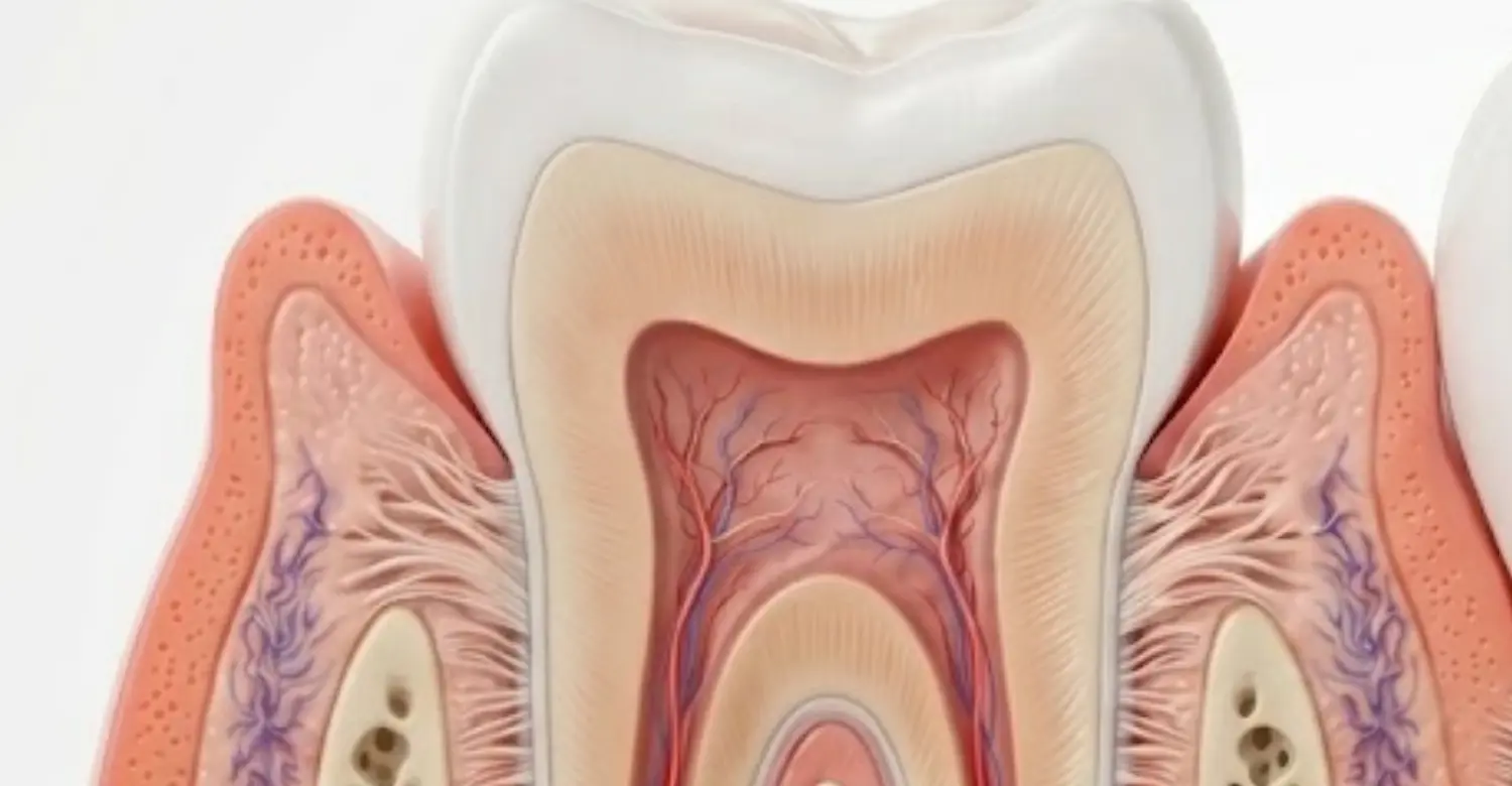
Gingival Sulcus Diagram: Understanding the Anatomy
A gingival sulcus diagram can simplify the relationship between the tooth and surrounding periodontal tissues. Typical diagrams show the tooth surface, gingival margin, sulcular epithelium, junctional epithelium, connective tissue, and alveolar bone. The diagram helps explain where periodontal probing occurs. It also demonstrates how the junctional epithelium attaches near the base of the crevice. Understanding these structures helps patients interpret periodontal terminology. Visual anatomy can make discussions about gum disease easier to follow.
Gingival Sulcus Diagram Explained
A typical diagram represents the crevice surrounding the tooth. The gingival margin appears at the coronal edge. The sulcular epithelium lines the inner surface facing the tooth. The junctional epithelium continues toward the deeper attachment region. Underneath these epithelial layers lies connective tissue. Alveolar bone supports the tooth farther apically. These structures form a coordinated periodontal attachment system. Damage to this system can contribute to periodontal disease.
Key Structures Shown in a Gingival Sulcus Diagram
Important structures include the tooth, gingival margin, sulcular epithelium, junctional epithelium, connective tissue, periodontal ligament, cementum, and alveolar bone. Each structure contributes to tooth support and periodontal defense. The junctional epithelium creates a specialized attachment to the tooth. The connective tissue contains collagen and vascular components. The periodontal ligament connects cementum with alveolar bone. Together, these structures maintain periodontal stability.
Gingival Margin, Sulcus, and Junctional Epithelium
The gingival margin marks the outer edge of the gingiva around the tooth. The sulcus lies immediately apical to this margin. The junctional epithelium forms the deeper epithelial attachment. Their relationship determines the clinical depth measured during probing. Inflammation can alter the position and condition of these tissues. Understanding their anatomical sequence makes periodontal charting easier to understand.
How to Read a Gingival Sulcus Diagram
Start by identifying the tooth surface and gingival margin. Follow the space between them toward the base. Identify the sulcular epithelium lining the crevice. Then locate the junctional epithelium attached to the tooth. Finally, identify connective tissue and supporting bone underneath. This sequence reflects the anatomical organization of the periodontium. A diagram should always be interpreted alongside clinical information when studying disease. Details and studies on Correct Brushing Technique
Gingival Sulcus and Junctional Epithelium
The relationship between the sulcus and junctional epithelium is central to periodontal health. The junctional epithelium forms a specialized attachment to the tooth. It also provides an important barrier between bacterial biofilm and deeper periodontal tissues. Its permeability allows immune components to participate in local defense. Gingival crevicular fluid passes through this interface and contributes to host protection. Inflammation can disrupt epithelial barrier function. Persistent periodontal inflammation may eventually compromise deeper supporting tissues.
What Is the Junctional Epithelium?
The junctional epithelium is a specialized epithelial tissue attached directly to the tooth surface. It forms a collar-like attachment around the tooth. Hemidesmosomes contribute to this epithelial attachment. The tissue also has distinctive cellular characteristics and rapid turnover. Its strategic location allows immune cells to respond to microbial challenges. This makes it essential for maintaining periodontal health.
Where Is the Junctional Epithelium Located?
It is located at the base of the gingival crevice. It extends between the sulcular epithelium and deeper periodontal connective tissues. The tissue attaches to the tooth surface through specialized cellular structures. Its exact position can vary according to age and periodontal condition. During periodontal disease, the attachment may migrate apically. This contributes to pathological pocket formation.
How the Junctional Epithelium Protects the Periodontium
The junctional epithelium creates a specialized barrier around the tooth. It allows controlled interaction between periodontal tissues and the oral environment. Immune cells can move through this tissue toward microbial challenges. Gingival crevicular fluid also reaches the crevice through this interface. These mechanisms contribute to local antimicrobial defense. Damage to this barrier can increase periodontal vulnerability.
Gingival Sulcus vs Junctional Epithelium
The sulcus is a space, while the junctional epithelium is a tissue. The sulcus lies between the tooth and marginal gingiva. The junctional epithelium forms the attachment at the deeper part. Confusing these terms can make periodontal anatomy difficult to understand. Their relationship is essential for interpreting probing measurements.
Dr. Rifat Alsaman’s Clinical Opinion
Dr. Rifat Alsaman emphasizes that periodontal health should be evaluated through multiple clinical findings. A single probing number cannot explain the entire condition of periodontal tissues. Bleeding, tissue appearance, attachment levels, recession, and bone support provide additional information. Patients should also understand that early gum inflammation can be subtle. Consistent professional monitoring helps detect changes before they become more difficult to manage. The complete article for Sedation Options For Anxious Patients
What Causes the Gingival Sulcus to Become Deeper?
Several factors can increase the measured depth around the tooth. Plaque-associated inflammation is one of the most common causes. Gingivitis can produce swollen tissue and temporary pseudopocketing. Periodontitis can create pathological pockets through attachment loss. Calculus can retain bacterial biofilm and complicate cleaning. Local restorations may also irritate gingival tissues. Identifying the underlying cause is more important than focusing on depth alone.
Plaque and Bacterial Biofilm
Dental plaque is a microbial biofilm that accumulates on tooth surfaces. When plaque remains around the gumline, it can trigger inflammatory responses. Poor plaque control is strongly associated with gingival disease. CDC identifies good oral hygiene as an important component of preventing gum disease. Regular brushing and interdental cleaning help reduce microbial accumulation. Professional cleaning may be necessary when deposits cannot be removed effectively at home.
Gingivitis and Gingival Inflammation
Gingivitis involves inflammation of gingival tissues without the bone destruction characteristic of periodontitis. Inflamed tissue may become red, swollen, and prone to bleeding. Swelling can increase probing measurements. Effective plaque control can often reverse gingivitis. Persistent inflammation should receive professional evaluation because untreated disease can increase periodontal risk.
Periodontitis
Periodontitis is a chronic inflammatory disease involving destruction of periodontal supporting tissues. Bone loss and attachment loss are important features. Periodontal pockets can develop as attachment migrates apically. Disease severity varies considerably among patients. Smoking, diabetes, oral hygiene, and other factors can influence periodontal risk. Professional treatment can slow and manage established disease.
Calculus and Tartar Buildup
Calculus is hardened dental plaque that cannot be removed effectively with routine brushing. Its rough surface can retain additional bacterial biofilm. Calculus can therefore contribute to persistent gingival inflammation. Professional dental instruments are generally required for its removal. Patients should not attempt aggressive scraping at home. Regular professional cleaning can help control deposits.
Improper Oral Hygiene
Inadequate brushing and interdental cleaning allow plaque to remain around gingival margins. This increases exposure to microbial biofilm. CDC recommends brushing twice daily and cleaning between teeth daily. Technique is also important because aggressive brushing can traumatize gingival tissue. Personalized oral hygiene instruction can improve plaque control.
Local Irritating Factors
Overhanging restorations, poorly contoured crowns, orthodontic components, and food impaction can complicate plaque control. These factors may create areas where biofilm accumulates more easily. Local irritation can contribute to persistent inflammation. Dentists evaluate restoration margins and anatomical factors during periodontal examinations. Correcting local contributors can improve the effectiveness of daily hygiene. Must-read guide to Veneers In Turkey
Signs of an Unhealthy Gingival Sulcus
Changes around the gumline can indicate inflammation or periodontal disease. Common warning signs include bleeding, redness, swelling, tenderness, persistent bad breath, and changes in gum contour. Deeper probing measurements may also require investigation. CDC identifies red, swollen, and bleeding gums as common gingivitis symptoms. Symptoms can remain mild even when disease is progressing. Regular dental examinations are therefore important.
Bleeding Gums
Bleeding during brushing or flossing should not automatically be considered normal. It commonly reflects gingival inflammation. Persistent bleeding warrants professional evaluation. CDC identifies easily bleeding gums as a common sign of gingivitis. Improved plaque control may reduce bleeding when inflammation is the underlying cause. Persistent bleeding can require periodontal assessment.
Red or Swollen Gingiva
Healthy gingiva generally appears firm and closely adapted around teeth. Inflammation can cause swelling and increased redness. Tissue swelling may also change the contour of the gumline. These signs should be considered alongside bleeding and probing measurements. Persistent swelling can indicate plaque-associated gingivitis or another local condition.
Gum Tenderness
Tender gums can occur with inflammation, trauma, infection, or localized irritation. Persistent tenderness should not be diagnosed from symptoms alone. Dentists can evaluate the area for plaque, calculus, restorations, periodontal disease, or other causes. Early assessment can prevent unnecessary discomfort and identify treatable factors.
Persistent Bad Breath
Persistent bad breath can have multiple causes. Oral bacteria, tongue coating, periodontal inflammation, dry mouth, and other conditions may contribute. Gum disease can produce unpleasant odors from bacterial activity and inflammatory changes. Persistent halitosis should receive dental evaluation when routine hygiene does not resolve it. Treatment depends on the underlying cause.
Increased Sulcus or Pocket Depth
Increasing measurements may indicate inflammation, tissue enlargement, attachment loss, or anatomical variation. Repeated measurements provide more useful information than one isolated reading. Dentists compare depth with recession and attachment levels. Radiographic bone assessment may be recommended when periodontal destruction is suspected.
Receding or Changing Gum Tissue
Gum recession exposes more of the tooth root and can change periodontal measurements. Recession may result from inflammation, traumatic brushing, thin tissue, tooth position, or other factors. It can also increase root sensitivity. Professional evaluation helps identify the cause and determine appropriate management.
Tips for Patients
Patients can support periodontal health through consistent daily plaque control. Brushing twice daily and cleaning between teeth are fundamental preventive measures. Regular professional examinations can identify early changes. Bleeding should not simply be ignored. Individual recommendations may vary according to periodontal risk and existing disease.
Brush Gently Along the Gumline
Use a soft-bristled toothbrush and gentle pressure around the gumline. Aim the bristles toward the area where teeth meet gingiva. Avoid aggressive horizontal scrubbing. Gentle technique reduces unnecessary mechanical trauma. Consistent plaque removal is more important than excessive brushing force.
Clean Between the Teeth Daily
Toothbrush bristles cannot effectively clean every interdental surface. Floss, interdental brushes, or other recommended devices can improve plaque removal. The appropriate method depends on tooth spacing and periodontal anatomy. Patients with periodontal pockets may benefit from professional instruction.
Schedule Regular Dental Checkups
Regular dental examinations allow clinicians to identify inflammation and periodontal changes early. CDC notes that dental checkups support early detection of gum disease. Visit frequency should reflect individual risk. Patients with periodontal disease may require more frequent monitoring.
Do Not Ignore Bleeding Gums
Bleeding can indicate inflammation and should not automatically be dismissed. Improving plaque control may reduce bleeding caused by gingivitis. Persistent bleeding requires professional assessment. Early treatment is generally easier than managing advanced periodontal destruction. In-depth look at Best Dental Clinic In Istanbul For A Full Smile Makeover
How Dentists Examine the Gingival Sulcus
Periodontal examination combines visual inspection, probing, and assessment of supporting tissues. Dentists evaluate multiple sites around each tooth. Probing identifies the depth of the clinically accessible crevice. Bleeding provides additional information about inflammatory activity. Recession and attachment levels help interpret probing measurements. Radiographs may be recommended when bone involvement needs evaluation.
Periodontal Probing
Periodontal probing uses a calibrated instrument to measure the depth around teeth. The probe is inserted gently along the tooth surface. Measurements are typically recorded at several sites per tooth. Technique and tissue characteristics can influence readings. Repeated measurements help establish periodontal patterns.
Measuring Gingival Sulcus Depth
Measurement begins at the gingival margin and extends toward the probeable base. Healthy sites commonly measure approximately 1–3 mm. Deeper measurements require interpretation within the complete periodontal examination. Dentists consider inflammation, recession, attachment, and bone support.
Checking for Bleeding on Probing
Bleeding on probing is an important clinical indicator of gingival inflammation. It can occur when inflamed tissues respond to gentle probing. The finding does not independently diagnose periodontitis. Instead, it contributes to the overall periodontal assessment. Patterns of bleeding can help clinicians identify sites requiring closer attention.
Assessing Gum Attachment and Bone Support
Clinical attachment levels help determine whether periodontal attachment has been lost. Bone support can be assessed through appropriate radiographic imaging. These findings provide information that probing depth alone cannot provide. Periodontal diagnosis therefore combines soft-tissue and hard-tissue measurements.
When Dental X-Rays May Be Recommended
Radiographs may be recommended when clinical findings suggest possible bone loss or other dental pathology. They can show supporting bone levels around teeth. Radiographic findings should always be interpreted alongside clinical examination. Routine imaging decisions should be individualized according to clinical need.
Clinical Note: Why Probing Depth Alone Does Not Tell the Whole Story
Probing depth measures the distance from the gingival margin to the probeable base. It does not directly measure attachment loss. Gingival enlargement can increase depth without destructive attachment loss. Recession can reduce depth while attachment loss remains significant. This distinction is fundamental in periodontal diagnosis. Clinical advice on Full Mouth Rehabilitation In Turkey
Can a Healthy Gingival Sulcus Become a Periodontal Pocket?
Disease-related changes can transform normal anatomy into a pathological pocket. Persistent inflammation can alter the junctional epithelium and periodontal attachment. In periodontitis, attachment loss and bone destruction accompany pocket formation. Gingivitis alone generally does not involve permanent periodontal attachment loss. Early detection and appropriate treatment can help control inflammatory disease.
From Gingival Inflammation to Periodontal Pocket Formation
Plaque accumulation can initiate gingival inflammation. Persistent inflammation can alter connective tissue and epithelial structures. In susceptible patients, periodontal attachment may subsequently become damaged. This process can create deeper pathological spaces around teeth. Periodontitis is distinguished from gingivitis by supporting tissue destruction.
Changes in the Junctional Epithelium
Periodontal inflammation can alter the structure and position of the junctional epithelium. During disease progression, attachment can migrate toward the root. The resulting tissue configuration contributes to periodontal pocket formation. These changes are accompanied by inflammatory cellular activity and connective tissue destruction.
Loss of Periodontal Attachment
Attachment loss means the periodontal attachment has moved apically relative to a stable anatomical reference. It is a key feature in diagnosing periodontitis. Probing depth and recession are used together to calculate clinical attachment levels. This measurement provides more meaningful information about periodontal destruction.
Bone Loss and Advanced Periodontal Disease
Advanced periodontitis can cause destruction of alveolar bone supporting the teeth. Bone loss can compromise tooth stability when disease becomes severe. CDC identifies bone loss around teeth as a major feature of periodontitis. Treatment aims to control inflammation and prevent further progression.
Dr. Rifat Alsaman’s Clinical Opinion
Dr. Rifat Alsaman recommends interpreting periodontal measurements within the entire clinical picture. Patients should not assume that every deeper reading represents irreversible disease. Likewise, normal-looking gums do not eliminate the need for professional assessment. Bleeding, attachment levels, recession, and bone support provide important additional information. Early evaluation can help distinguish reversible inflammation from destructive periodontal disease. Health impact of Gum Disease And Overall Health
How to Keep the Gingival Sulcus Healthy
Maintaining periodontal health begins with consistent plaque control. Daily brushing and interdental cleaning reduce microbial accumulation around the gumline. Professional cleaning can remove deposits that home care cannot eliminate. Regular periodontal monitoring helps identify changes early. CDC emphasizes oral hygiene and professional care as important measures for preventing and managing gum disease. Individual needs vary according to periodontal risk.
Effective Daily Brushing
Brush twice daily using fluoride toothpaste and a soft-bristled toothbrush. Focus carefully on the gumline without using excessive force. Replace the toothbrush when bristles become worn. Consistent technique is more important than aggressive pressure. Patients with special periodontal conditions may need personalized brushing instructions.
Interdental Cleaning
Cleaning between teeth removes plaque from surfaces that toothbrushes cannot reach effectively. Floss and interdental brushes are common options. Larger spaces may benefit from appropriately sized interdental brushes. A dental professional can recommend the best method for individual anatomy.
Professional Dental Cleaning
Professional cleaning removes calculus and plaque deposits that cannot be eliminated through routine home care. Cleaning frequency depends on individual periodontal risk. Patients with periodontal disease may require specialized periodontal maintenance. Regular professional care supports long-term plaque control.
Managing Plaque and Gingivitis
Plaque control is central to preventing gingival inflammation. Gingivitis is generally reversible when inflammation is effectively managed. Patients should continue daily hygiene even after symptoms improve. Professional examinations can identify persistent inflammation that may not be obvious at home.
Regular Periodontal Monitoring
Periodontal monitoring tracks changes in probing depth, bleeding, recession, and attachment. Comparing measurements over time can identify progression or improvement. Patients with previous periodontal disease often need structured maintenance. Monitoring allows clinicians to adjust treatment according to changing risk.
Tips for Patients
Healthy gums require consistency rather than occasional intensive cleaning. Brush gently twice daily and clean between teeth every day. Keep professional appointments according to your dentist's recommendations. Do not ignore persistent bleeding, swelling, or bad breath. Early evaluation can reduce the risk of untreated periodontal disease.
Use a Soft-Bristled Toothbrush
Soft bristles can effectively remove plaque when used with appropriate technique. Hard brushing does not necessarily provide better cleaning. Excessive pressure can contribute to gingival trauma and recession. Replace worn toothbrushes regularly. Ask your dentist for personalized technique advice.
Avoid Aggressive Brushing
Aggressive brushing can damage gingival tissue and contribute to recession in susceptible individuals. Use gentle movements and controlled pressure. Allow the toothbrush to clean rather than forcing it against the gumline. Electric toothbrush users should also avoid excessive pressure.
Clean the Gumline Carefully
Plaque frequently accumulates where teeth meet gingiva. Angle the toothbrush appropriately toward this region. Clean every tooth systematically. Interdental cleaning should complement brushing. Professional instruction can help patients improve difficult areas.
Seek Professional Advice for Persistent Bleeding
Persistent bleeding should receive dental assessment. It can indicate gingivitis, periodontal disease, trauma, or another local problem. Early evaluation helps identify the cause. Treatment can then target the underlying condition rather than only the symptom. Understanding the risks of Waterborne Diseases And Oral Health
Gingival Sulcus and Overall Periodontal Health
The condition of the dentogingival interface provides important information about periodontal health. Healthy tissues limit inflammatory exposure to deeper periodontal structures. Plaque accumulation can disrupt this balance. Gingivitis represents inflammation without the bone destruction characteristic of periodontitis. Long-term periodontal health depends on effective plaque control and appropriate professional monitoring.
Why the Gingival Sulcus Is Important for Gum Health
The sulcus is positioned directly beside dental biofilm. Its tissues therefore encounter microorganisms and their products regularly. The surrounding epithelial barrier and immune system help control this exposure. Gingival crevicular fluid also contributes protective components. Maintaining healthy tissues around the tooth reduces the risk of persistent inflammation.
The Role of Plaque Control
Plaque control reduces the microbial challenge presented to gingival tissues. Brushing and interdental cleaning are fundamental components. Professional cleaning addresses deposits that remain difficult to remove. CDC identifies good oral hygiene and professional care as important measures for gum health. Consistency is more important than occasional intensive cleaning.
Relationship Between Gingival Inflammation and Periodontitis
Gingivitis involves inflammation without periodontal bone destruction. Periodontitis involves deeper supporting tissue destruction and bone loss. Not every patient with gingivitis develops periodontitis. However, persistent inflammation increases concern when other risk factors exist. Professional evaluation helps distinguish these conditions.
Protecting the Periodontal Attachment
The periodontal attachment stabilizes the relationship between gingiva and tooth. Maintaining plaque control helps reduce inflammatory stress around this attachment. Periodontal disease can progressively damage attachment and supporting bone. Early diagnosis and appropriate management can slow disease progression. Long-term maintenance remains important after periodontal treatment.
What We Notice Clinically at Vitrin Clinic
At Vitrin Clinic, periodontal assessment focuses on the condition of the tissues surrounding each tooth. Clinicians evaluate gum appearance, probing measurements, bleeding, recession, and attachment. When appropriate, radiographs can provide additional information about supporting bones. This comprehensive approach helps identify both early inflammation and established periodontal disease. Clinical findings are interpreted according to each patient's individual anatomy and risk profile. Patients receive guidance based on their specific oral health needs.
Evaluating Gingival Sulcus Depth
Probing measurements are recorded around teeth to identify areas requiring closer evaluation. Healthy measurements are commonly around 1–3 mm. However, depth is interpreted alongside bleeding and attachment findings. Repeated measurements can help identify changes over time. This approach supports more reliable periodontal monitoring.
Identifying Early Signs of Gingival Inflammation
Clinicians look for redness, swelling, bleeding, tenderness, and changes in tissue contour. Early gingivitis may produce few symptoms outside the dental examination. CDC identifies bleeding and swelling as common gingivitis signs. Early detection allows patients to improve plaque control before inflammation becomes more difficult to manage.
Assessing the Relationship Between Gums and Teeth
The position of the gingival margin provides important information during periodontal assessment. Recession can expose root surfaces and alter probing measurements. Gingival enlargement can create deeper readings without attachment loss. Clinicians therefore evaluate both tissue position and periodontal attachment.
Personalized Periodontal Assessment
Every patient has different anatomy, hygiene habits, risk factors, and periodontal history. A personalized examination helps identify the factors contributing to inflammation. Treatment recommendations can then focus on the specific cause. This may include hygiene instruction, professional cleaning, periodontal therapy, or maintenance monitoring.
Dr. Rifat Alsaman’s Opinion
Dr. Rifat Alsaman emphasizes prevention and early periodontal assessment. He considers periodontal measurements most useful when interpreted alongside tissue appearance and attachment status. Patients should understand that bleeding is a warning sign rather than something to normalize. Consistent home care and professional monitoring remain central to maintaining healthy periodontal tissues.
Why Choose Vitrin Clinic for Comprehensive Gum and Dental Care?
Vitrin Clinic combines clinical examination with individualized dental planning. Periodontal health can influence the success and stability of many dental treatments. Comprehensive assessment helps identify gum inflammation before elective procedures begin. Patients can receive recommendations according to their periodontal condition and overall dental goals. Modern diagnostic approaches can support accurate clinical decision-making when indicated. The focus remains on long-term oral health rather than treating isolated symptoms.
Comprehensive Oral and Periodontal Assessment
A complete dental examination can evaluate teeth, gums, supporting tissues, and oral hygiene. Periodontal probing provides information about tissue conditions around individual teeth. Imaging may be used when clinically indicated. Combining these findings provides a more complete understanding of oral health.
Personalized Treatment Planning
Treatment should reflect each patient's diagnosis, anatomy, risk profile, and goals. Personalized planning can address inflammation before restorative or cosmetic procedures. Patients can also receive individualized maintenance recommendations. This approach supports healthier long-term treatment outcomes.
Modern Dental Technology and Clinical Evaluation
Modern dentistry can combine clinical examination with digital imaging and other diagnostic technologies. Technology does not replace clinical judgment. Instead, it can provide additional information when appropriate. The selected diagnostic tools should match the patient's clinical needs.
A Patient-Centered Approach to Long-Term Oral Health
Long-term periodontal health depends on patient participation and professional monitoring. Clear explanations help patients understand their diagnosis and treatment choices. Personalized hygiene instructions can improve daily plaque control. Regular follow-up allows clinicians to identify changes early.
Key Takeaways About Gingival Sulcus
Understanding periodontal anatomy helps patients recognize the difference between normal gum structures and disease. Healthy probing depths commonly fall around 1–3 mm. Deeper measurements require clinical interpretation rather than automatic disease labeling. Bleeding, attachment loss, recession, and bone loss provide additional diagnostic information. Persistent gum inflammation deserves professional evaluation. CDC data show that periodontal disease is common among adults, making prevention and monitoring important.
Healthy Gingival Sulcus: What to Remember
A healthy crevice is generally shallow and surrounded by firm periodontal tissue. Good brushing and interdental cleaning help control plaque. Bleeding should not be considered a normal permanent feature. Regular dental examinations can identify changes before they progress. A healthy periodontal environment supports long-term tooth stability.
When to See a Dentist or Periodontist
Seek professional evaluation for persistent bleeding, swelling, gum recession, bad breath, tenderness, or increasing periodontal measurements. Loose teeth and visible changes in supporting tissues also warrant prompt assessment. Patients with known periodontal disease may need periodontal maintenance. Early evaluation can help distinguish reversible inflammation from more advanced disease.
Reference
FAQs

Dr. Rifat Alsaman has more than 5 years of clinical experience in dentistry and currently serves as the Head of the Medical Team at Vitrin Clinic. He is dedicated to providing exceptional patient care, overseeing treatment planning, and ensuring the highest clinical standards across the team. His expertise, attention to detail, and commitment to continuous professional development have helped countless patients achieve healthier, more confident smiles.


