Periodontics
September 1, 2026
Periodontal Ligament: What Is It and Why Is It Important for Your Teeth?
 Dr. Rifat Alsaman
Periodontal ligament is a specialized connective tissue that surrounds the tooth root and connects it to the alveolar bone. It acts as a cushion between the tooth and the jaw, allowing small, controlled movement during chewing. Because of this, this tissue plays a central role in tooth stability, chewing comfort, and overall periodontal health.
Patients often raise concerns such as inflammation around the root area. A widening of the space seen on an X-ray is another common concern. So is a bruised feeling after an injury or heavy bite. These symptoms can sometimes resemble tooth decay, infection, or other dental problems. This overlap makes self-diagnosis unreliable. A clinical examination, combined with appropriate imaging, is required before any conclusion can be reached. Only this combination confirms the cause of discomfort in this structure.
Periodontal Ligament: Anatomy, Function, and Importance
This connective tissue is a thin but resilient layer that sits between the tooth root and the surrounding bone. Understanding its anatomy, fibre arrangement, and everyday function helps explain why small changes to it matter. Even minor changes can affect comfort while biting, tooth stability, and long-term periodontal health across the full dental arch.
What Is the Periodontal membrane?
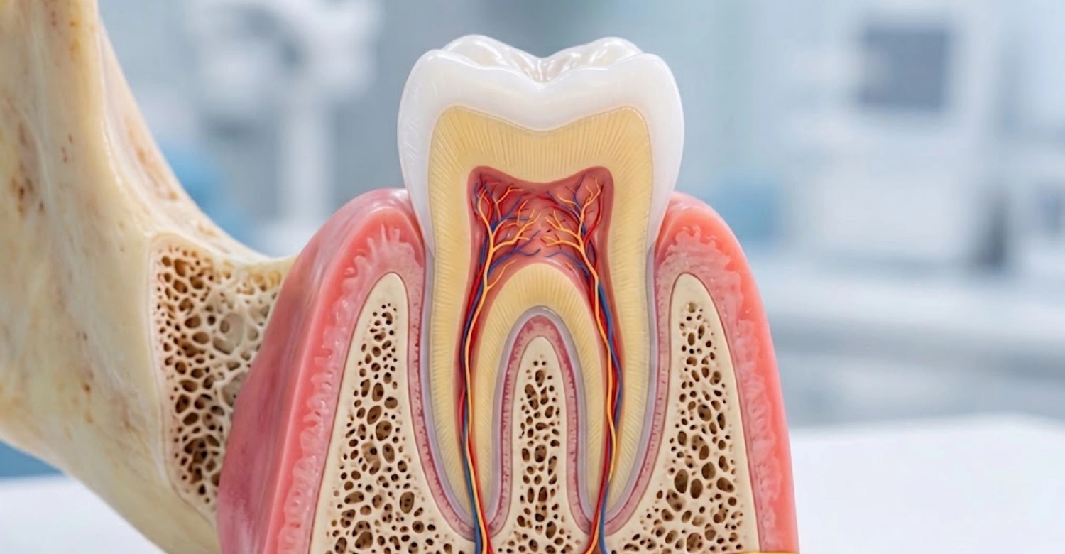
This tissue is a fibrous connective structure found between the cementum covering the tooth root and the alveolar bone socket. It is not a rigid attachment. Instead, it behaves like a flexible suspension system. This structure allows the tooth to absorb pressure rather than transmit it directly into bone. Beyond support, it also acts as a sensory structure. It detects pressure changes and helps the brain judge biting force during chewing.
Periodontal Ligament Anatomy
Its anatomy includes collagen fibre bundles, blood vessels, nerve endings, and specialized cells responsible for maintenance and repair. The vascular supply nourishes the tissue and supports healing after minor injury. The neural supply provides sensitivity to pressure and pain. This combination allows the tissue to respond dynamically to functional forces. It adjusts slightly with each bite rather than remaining fixed over time.
Fibers of Periodontal membrane
The fibers of periodontal ligament tissue are organized into distinct groups. Each group runs in a different direction between the root and bone. These Periodontal membrane fibers include alveolar crest, horizontal, oblique, apical, and interradicular groups. Together, they anchor the tooth firmly while still permitting slight, physiological movement. During chewing, different fibre groups become tense depending on the direction of force. This distributes pressure evenly instead of concentrating it at one point.
Function of Periodontal membrane
The function of periodontal ligament tissue extends beyond simple attachment. It supports the tooth within its socket, absorbs shock during chewing, and provides sensory feedback about pressure and position. It also contributes to ongoing remodeling of bone and cementum, helping the tooth adapt over time. Altogether, this activity helps maintain the entire periodontal attachment apparatus in a stable, functional state. Understanding the risks of Waterborne Diseases And Oral Health
What Problems Can Affect the Periodontal Ligament?
Several conditions can affect this tissue, ranging from short-term irritation to longer-term structural change. These include inflammation, visible widening on X-ray, bruising after trauma, and reduced tooth stability. Recognising these patterns helps patients understand when a symptom is likely minor. It also signals when professional assessment is appropriate for this structure and surrounding tissues.
Inflammation of Periodontal membrane
Inflammation of periodontal ligament tissue can result from dental trauma, excessive biting forces, periodontal disease, or infection around the tooth. When inflamed, the tissue becomes more sensitive to pressure. Patients often notice tenderness specifically when biting or chewing on the affected tooth, rather than constant pain. This pattern of pressure-related discomfort is a useful clue that points toward this area rather than another dental structure.
Widening of Periodontal membrane Space
A widening of periodontal ligament space refers to an increased gap around the tooth root. It is visible on a dental X-ray. This is a radiographic finding, not a diagnosis by itself. Possible causes include trauma from occlusion, inflammation, orthodontic forces, or periapical pathology near the root tip. Several conditions can produce a similar appearance. The surrounding clinical findings, including symptoms and examination results, must always be considered together.
Bruised Periodontal Ligament
Patients sometimes describe a "bruised periodontal ligament" after biting down unexpectedly hard or experiencing minor trauma. Excessive biting pressure can irritate the supporting tissues without causing lasting damage. Common symptoms include tenderness when biting and general discomfort around the tooth for several days. This usually settles as irritation resolves. Persistent symptoms should still be assessed to rule out a more significant underlying cause.
Periodontal membranes and Tooth Stability
Healthy periodontal ligaments help maintain the tooth firmly within its socket while still allowing controlled micro-movement. When periodontal support becomes compromised, whether through inflammation, bone loss, or trauma, tooth stability can be affected. It is important to distinguish these problems from general gum disease. Gum disease primarily affects surface tissue. Ligament-related issues relate more directly to the forces and structures supporting the root. Symptoms and relief for Rheumatoid Arthritis Tooth Pain
What Causes Periodontal Ligament Pain or Irritation?
Pain or irritation involving this tissue usually stems from identifiable causes. These include excessive biting forces, physical trauma, infection near the root, and periodontal disease affecting the supporting structures. Identifying the specific cause is essential, since treatment differs significantly depending on which factor is responsible for the discomfort.
Excessive Biting Forces
Excessive biting forces are a common contributor to irritation of this tissue. Bruxism and clenching place repeated, prolonged pressure on it. A high dental restoration or crown can concentrate force onto a single tooth. Sudden changes in the bite, such as after dental work, may also create uneven pressure. Over time, this excessive pressure on one tooth can lead to noticeable tenderness while chewing.
Dental Trauma
Dental trauma is another significant cause of this kind of irritation. Sports injuries and accidental impacts can stretch or bruise the supporting tissue suddenly. Biting unexpectedly hard objects, such as ice or bone, may produce similar irritation. Traumatic dental procedures, including difficult extractions of neighbouring teeth, can also place temporary stress on the ligament. This sometimes causes short-term tenderness that requires monitoring afterward.
Dental Infection
Inflammation around the root from dental infection can extend into the supporting tissue, producing tenderness on biting. It is important to differentiate this from pulp inflammation. The pulp involves the tooth's internal nerve, not its external support. When infection is confirmed as the underlying cause, root canal treatment may become necessary to remove the source. This allows the surrounding tissue to recover.
Periodontal Disease
Periodontal disease affects the tissues supporting the tooth, including the ligament connecting root to bone. Ongoing inflammation can contribute to bone loss and attachment loss around the root. Once supporting structures are significantly reduced, this kind of damage is not always fully reversible. Early identification and management of periodontal disease are therefore important for preserving as much of the surrounding attachment as possible.
Tips for Patients
Protect Your Periodontal Ligament
Maintain consistent brushing and interdental cleaning.
Attend regular dental examinations.
Avoid using teeth to bite hard non-food objects.
Wear appropriate protection during contact sports.
Address persistent clenching or grinding.
Do not ignore pain when biting.
What Patients Should Avoid
Self-diagnosing based only on an X-ray.
Delaying assessment because the pain is mild.
Repeatedly chewing on a painful tooth. Role and effect of Cortisol And Oral Health
Taking medication as a substitute for dental diagnosis.
What Symptoms Can Indicate a Periodontal membrane Problem?
Certain symptoms commonly point toward this kind of issue rather than another dental condition. These include pain specifically triggered by biting, tenderness, increased mobility, and general sensitivity. Recognizing these patterns helps patients describe symptoms accurately during a dental visit. This supports a more efficient and focused clinical assessment.
Pain When Biting or Chewing
Pressure may trigger discomfort when this tissue is inflamed or irritated, since biting directly loads it. This differs from spontaneous toothache, which can occur without any pressure at all. Pressure-related pain that appears mainly during chewing, and eases afterward, often points toward this area. This differs from pain from the tooth's internal pulp. A full examination is still needed to confirm the cause.
Tooth Tenderness
A tooth may feel "high" or uncomfortable when the tissue around it is inflamed, even slightly. This sensation can occur after a new restoration, following trauma, or alongside infection. Possible causes that require examination include occlusal interference, inflammation from periodontal disease, or pressure from a neighbouring dental procedure. Identifying the specific trigger helps determine the most appropriate next step for treatment.
Increased Tooth Mobility
Increased mobility can indicate that ligament support has been compromised, often alongside bone or attachment loss. However, mobility does not automatically mean the tooth must be extracted. Many cases of mild mobility improve once the underlying cause, such as excessive force or active inflammation, is addressed. A clinical assessment is needed to determine the extent of support loss and the most suitable management approach.
Sensitivity and Discomfort
Discomfort related to this tissue is usually triggered by pressure or biting force rather than temperature. This distinguishes it from sensitivity caused by the dental pulp, which typically reacts to hot or cold stimuli. Understanding this difference helps guide the clinical examination toward the correct structure. These two sources of discomfort often require different diagnostic tests and treatment approaches. Managing conditions like Sjogren S Syndrome Teeth Falling Out
How Is a Periodontal Ligament Problem Diagnosed?
Diagnosing this kind of problem requires more than a single test. A combination of clinical examination, radiographic imaging, and, when appropriate, three-dimensional scanning helps build an accurate picture. Each method contributes different information, and results are interpreted together to determine the underlying cause.
Clinical Dental Examination
A clinical dental examination typically includes several steps. Tooth mobility assessment checks for looseness, while percussion and bite testing identify pressure-related pain. Periodontal probing measures attachment levels around the tooth. Occlusal evaluation checks how the teeth meet during biting. Assessment of neighbouring teeth helps rule out contributing factors from adjacent structures. Together, these steps provide essential baseline information.
Dental X-Rays
Dental X-rays can reveal changes in the space around the root, including widening or irregularity. However, radiographic findings must always be interpreted alongside symptoms and clinical examination results. An X-ray alone cannot confirm the cause of a problem affecting this tissue. It provides one piece of information that, combined with other findings, helps guide an accurate diagnosis and appropriate next steps.
CBCT Imaging When Appropriate
Three-dimensional CBCT imaging may provide additional information when conventional radiographs do not fully explain the clinical picture. It is not necessary for every case. It is generally reserved for situations where standard X-rays leave uncertainty, such as complex root anatomy or unclear periapical findings. When used appropriately, CBCT can support more precise treatment planning without becoming a routine first step. Daily risks of Vaping And Oral Health
How Are Periodontal Ligament Problems Treated?
Treatment for these problems depends entirely on the underlying cause. Options range from adjusting biting forces to treating infection, managing periodontal disease, or addressing trauma. These approaches differ significantly, so an accurate diagnosis must come first. This ensures treatment targets the actual source of irritation rather than the symptoms alone.
Removing Excessive Occlusal Forces
When excessive force is identified as a contributing factor, bite adjustment may be carried out when clinically indicated. This helps redistribute pressure evenly. A high restoration contributing to localized force can be managed and refined. After correction, monitoring helps confirm that tenderness resolves. It also confirms the tissue is no longer under abnormal strain during normal chewing function.
Treating Dental Infection
When infection is confirmed as the underlying cause, endodontic treatment may be indicated to remove the source within the tooth. Treatment focuses on addressing the underlying infection rather than simply managing surface symptoms in isolation. Once the infection is resolved, inflammation affecting the surrounding tissue typically improves. This happens gradually as the area heals over the following weeks.
Managing Periodontal Disease
Managing periodontal disease involves professional periodontal cleaning to remove contributing bacteria and improved plaque control at home. Periodontal treatment may be recommended when indicated by examination findings, followed by long-term maintenance visits. This ongoing approach helps stabilize the tissues supporting the tooth. It also reduces further strain caused by continued inflammation or attachment loss.
Managing Dental Trauma
Managing dental trauma begins with assessment of the injured tooth to evaluate the extent of impact. Monitoring tooth vitality and periodontal support over time helps confirm recovery. Stabilization may be carried out when clinically necessary to protect a loosened tooth. Follow-up examinations are scheduled to track healing and detect any delayed complications affecting the supporting tissue.
Managing Bruxism and Clenching
Managing bruxism and clenching starts with identifying excessive forces contributing to irritation of the ligament. Occlusal protection, such as a night guard, may be recommended where appropriate to reduce pressure during sleep. Ongoing monitoring of symptoms and tooth wear helps confirm whether the approach is effective. Adjustments can be made if irritation or discomfort continues.
Clinical Note
A change in the periodontal ligament space on an X-ray is a finding, not a diagnosis. Dr. Rifat Alsaman, Head of the Medical Team at Vitrin Clinic and a cosmetic dentist, emphasizes this point. Radiographic appearance should be interpreted alongside clinical findings, including symptoms, periodontal measurements, tooth vitality, and occlusion. Other imaging findings are added when appropriate, rather than reading the X-ray in isolation.
When Should Patients Seek Professional Assessment?
Patients should seek professional assessment for persistent pain when biting, increasing tooth mobility, or swelling around a tooth. Pain following dental trauma also warrants prompt evaluation. Symptoms that continue despite improved oral hygiene suggest an underlying cause beyond plaque control. Unexplained changes seen on dental X-rays should likewise be reviewed clinically rather than left unaddressed.
What Can Patients Expect Before and After Treatment?
Understanding what to expect before and after treatment helps patients feel prepared. The process generally involves examination, diagnosis, and a discussion of options beforehand, followed by monitoring and preventive care afterward. Recovery timelines vary depending on the specific underlying cause.
Before Treatment
Before treatment, patients can expect a clinical examination and diagnosis to identify the underlying cause of their symptoms. Appropriate X-rays or additional imaging are taken when necessary to support this process. Once findings are reviewed, available treatment options are discussed clearly. This allows patients to understand the recommended approach before any procedure begins.
After Treatment
After treatment, many patients notice a reduction in inflammation and discomfort once the underlying cause has been successfully managed. Ongoing monitoring of tooth stability and periodontal tissues helps confirm steady improvement. Follow-up imaging may be arranged when clinically necessary. Continued preventive dental care remains important for maintaining long-term health of the supporting tissues going forward.
How Long Does Recovery Take?
Recovery time varies according to the underlying cause of the problem. Minor irritation from excessive biting forces may improve fairly soon after the cause is corrected. Infection, trauma, or periodontal disease may require longer-term treatment and monitoring over weeks or months. It would be inaccurate to promise a fixed recovery period, since individual healing responses differ. Safe procedures for Dental Implants And Osteoporosis
Periodontal membrane vs Other Tooth-Supporting Structures
This tissue works alongside other structures to support the tooth, including the gingiva, alveolar bone, and dental pulp. Understanding how these structures differ helps clarify why similar symptoms can sometimes arise from different sources. It also shows why an accurate diagnosis depends on examining more than one structure.
Periodontal Ligament vs Gingiva
The periodontal ligament and the gingiva are distinct tissues with different roles. The gingiva is the visible gum tissue surrounding the tooth at the surface. The deeper connective tissue lies beneath it, connecting the root to bone. Gum-related symptoms, such as surface swelling or bleeding, differ from pressure-related discomfort. That kind of discomfort is typically associated with irritation deeper around the root.
Periodontal membrane vs Alveolar Bone
This tissue connects the tooth root to the surrounding alveolar bone, acting as a flexible interface between the two. While the alveolar bone provides the rigid socket housing the tooth, this connective tissue allows controlled movement within that socket. Changes affecting either structure, such as bone loss or ligament inflammation, can influence overall tooth stability differently.
Periodontal Ligament vs Dental Pulp
The dental pulp is the tooth's internal nerve and blood supply. The periodontal ligament is an external supporting tissue outside the root surface. Because both structures can produce pain, pulp-related and ligament-related discomfort can sometimes feel similar to patients. Differentiating between the two requires specific clinical tests, since treatment approaches for each differ considerably.
What We Notice Clinically
Dr. Rifat Alsaman, Head of the Medical Team at Vitrin Clinic and a cosmetic dentist, notes something important. A widened space on an X-ray does not automatically mean severe disease. The underlying cause should always be identified before treatment is selected. Persistent pain on biting should be assessed rather than ignored. Treatment should focus on the actual cause, whether occlusal trauma, infection, periodontal disease, or dental trauma.
Why Clinical Context Matters
Two patients with similar X-ray findings may still require different treatments, depending on their individual symptoms and examination results. This is why individualized diagnosis matters so much in this area. This reflects Dr. Rifat Alsaman's approach. Treatment planning should always reflect the complete clinical picture rather than a single finding viewed on its own. Specialized guidelines for Dental Care For Special Needs And Circumstances
Why Professional Dental Assessment Matters
Professional dental assessment ensures that symptoms involving this tissue are correctly identified rather than assumed. Several conditions can present similarly. Relying on clinical expertise, appropriate imaging, and structured evaluation supports safer, more accurate outcomes. This matters most for patients with pain, tenderness, or unexplained changes around a tooth.
Accurate Diagnosis Before Treatment
Different conditions affecting this structure can produce similar symptoms, including tenderness or pain when biting. Without accurate diagnosis, treatment risks addressing the wrong cause entirely. Effective treatment should always address the underlying issue, whether it relates to force, infection, or periodontal disease. It should never simply target the discomfort itself in isolation.
Personalized Treatment Planning
Personalized treatment planning relies on combining clinical examination, imaging, bite analysis, and periodontal assessment into a single, coherent picture. This approach allows treatment to be tailored to each patient's specific findings. It avoids a generic solution that may not address the actual cause of their symptoms.
Modern Dental Technology
Modern dental technology supports more accurate assessment through digital radiography, which offers clearer imaging with lower exposure. CBCT is used when clinically justified for more complex cases. Digital treatment planning, along with computer-assisted diagnostics and restorative planning where appropriate, helps support more precise decisions. These stay individually tailored throughout the diagnostic and treatment process.
Why Choose Vitrin Clinic for Comprehensive Dental Care?
Vitrin Clinic takes a multidisciplinary approach to diagnosing and treating conditions affecting the periodontal ligament and surrounding structures. Modern dental technology and digital diagnostic tools support accurate assessment. Personalized treatment planning is based on individual findings rather than symptoms alone. International patients are supported through coordinated care and structured follow-up. Dr. Rifat Alsaman is Head of the Medical Team at Vitrin Clinic and a cosmetic dentist. He is part of the clinical team guiding this process. Recommendations are always based on individual examination and diagnosis, not on symptoms alone. Patients with persistent tooth pain, tenderness, mobility, or unexplained radiographic findings are encouraged to seek professional assessment at Vitrin Clinic. Post-treatment steps for Subperiosteal Implant Recovery And Aftercare
References:
FAQs
Dr. Rifat Alsaman has more than 5 years of clinical experience in dentistry and currently serves as the Head of the Medical Team at Vitrin Clinic. He is dedicated to providing exceptional patient care, overseeing treatment planning, and ensuring the highest clinical standards across the team. His expertise, attention to detail, and commitment to continuous professional development have helped countless patients achieve healthier, more confident smiles.


