General Dentistry
August 31, 2026
Tooth Anatomy: What Are the Parts and Structure of a Tooth?

Understanding Tooth Anatomy explains why teeth can withstand chewing, temperature changes, and daily pressure. A tooth contains several specialized tissues working together as one functional structure. These tissues include enamel, dentin, pulp, cementum, and supporting periodontal tissues. Each part has a different role in protection, sensation, nutrition, and stability. Understanding these structures also helps patients understand common dental problems and treatments. At Vitrin Clinic, dentists evaluate these structures before developing personalized treatment plans. A detailed examination helps determine whether treatment should preserve, restore, or replace tooth structure. This guide explains the anatomy of teeth using clear clinical and patient-friendly information.
What Is Tooth Anatomy?
Tooth anatomy describes the physical structure, tissues, surfaces, and supporting structures surrounding each tooth. A tooth has a crown, neck, and root. The crown is generally visible above the gumline. The root extends into the jawbone and anchors the tooth. Inside, enamel protects dentin, while dentin surrounds the dental pulp. The pulp contains nerves and blood vessels that support the living tooth. Cementum covers the root and connects with periodontal supporting tissues. NIDCR describes the pulp as containing nerves and blood vessels supplying the tooth. Understanding these components helps dentists identify structural damage more accurately.
Why Is Understanding Tooth Structure Important?
Knowing tooth structure helps patients understand why different dental problems require different treatments. A cavity affecting enamel differs from decay reaching dentin or pulp. Likewise, a cracked crown requires different management from a damaged root. The location and depth of damage influence treatment decisions. Dentists use clinical examinations and imaging to determine which tissues remain healthy. This approach supports conservative treatment whenever suitable. The American Dental Association emphasizes evidence-based management according to lesion type and tooth surface. Understanding the structure can therefore make treatment discussions easier for patients.
How Does Tooth Anatomy Affect Oral Health?
Tooth structure directly influences susceptibility to decay, sensitivity, fractures, and infection. Enamel provides strong external protection but cannot naturally regenerate after significant loss. Dentin is softer and contains microscopic tubules connected with the pulp. When dentin becomes exposed, sensitivity can become more noticeable. The pulp contains nerves, making inflammation inside this space potentially painful. Root surfaces become more vulnerable when gums recede and expose cementum. Healthy supporting tissues also help maintain tooth stability. At Vitrin Clinic, dentists consider these relationships during comprehensive dental assessments.
Clinical Note
Clinical diagnosis rarely depends on one anatomical feature alone. Dentists evaluate tooth surfaces, gum tissues, bite relationships, symptoms, and radiographic findings together. A small visible defect can sometimes hide deeper structural involvement. Conversely, sensitivity does not always indicate severe internal damage. The correct interpretation depends on clinical findings and diagnostic testing. Patients should avoid self-diagnosing the source of persistent dental pain. Professional examination can identify problems before they become more extensive.
What Are the Main Parts of a Tooth?
The three broad anatomical regions are the crown, neck, and root. They have different locations and functions. The crown performs most visible chewing and biting functions. The neck forms the transition between the crown and root. The root anchors the tooth within its supporting tissues. Inside these regions, several specialized tissues create the complete structure. The relationship between these parts allows teeth to withstand substantial mechanical forces.
The Crown of the Tooth
Tooth Anatomy classifies the crown as the coronal portion of the tooth. The crown is the portion generally visible above the gumline. Its outer surface is covered by enamel. Dentin lies underneath the enamel and forms much of the tooth's internal structure. The crown contains the chewing surfaces, contact areas, and visible facial surfaces. Different tooth types have differently shaped crowns. Incisors have cutting edges, while molars have broader chewing surfaces. The crown is frequently affected by cavities, fractures, erosion, and wear. Restorative treatments often aim to preserve their original structure whenever possible.
The Neck of the Tooth
Tooth Anatomy identifies the neck as the cervical transition between crown and root. The neck is the region connecting the crown with the root. It is also called the cervical region. The gumline commonly surrounds this area in a healthy mouth. Enamel ends near the cervical region before the root surface continues with cementum. Changes around the neck can become clinically important when gums recede. Root exposure may increase sensitivity because cementum provides less protection than enamel. Plaque accumulation around this region can also contribute to gum inflammation. Careful brushing and professional dental care help protect cervical tissues.
The Root of the Tooth
Tooth Anatomy places the root beneath the neck, anchoring the tooth in the jawbone. The root anchors a tooth inside the alveolar bone. It contains canals that connect the pulp with tissues surrounding the tooth. The root surface is covered by cementum. Periodontal ligament fibers connect cementum with the surrounding bone. Root shape differs between tooth types and individual teeth. Some teeth have one main root, while others have multiple roots. Root anatomy is particularly important during root canal treatment. Dentists may use radiographs or three-dimensional imaging when complex anatomy is suspected.
What Are the Layers of a Tooth?
Tooth Anatomy defines four principal tissue layers within each tooth. The major tissues include enamel, dentin, dental pulp, and cementum. Each tissue has a different composition and biological role. Together, they create a strong structure with an active living center. Enamel protects the crown from mechanical and chemical challenges. Dentin provides bulk and transfers sensory stimuli toward the pulp. The pulp provides nerves, blood vessels, and biological support. Cementum protects the root and provides attachment for periodontal fibers. These tissues work together rather than functioning independently.
Enamel
Within Tooth Anatomy, enamel is classified as the hardest and most mineralized layer. Enamel is the highly mineralized outer tissue covering the crown. It is considered the hardest tissue in the human body. Approximately 96% of mature enamel is mineral by weight. Its mineral structure provides exceptional resistance against chewing forces. However, enamel is not capable of biological regeneration after significant structural loss. Acids can dissolve minerals during dental erosion and demineralization. Frequent acidic exposure can progressively weaken dental surfaces. Protecting enamel therefore remains central to preventive dental care.
Dentin
Dentin lies beneath enamel and forms much of the tooth's volume. It is less mineralized than enamel and contains organic components. Dentin is approximately 70% mineral by weight. Microscopic dentinal tubules extend through its structure. These tubules help explain why exposed dentin can become sensitive. Dentin also provides important mechanical support beneath enamel. If enamel becomes severely damaged, underlying dentin becomes more vulnerable. Dentists therefore consider remaining dentin thickness during restorative treatment.
Dental Pulp
Dental pulp occupies the central internal space of the tooth. It contains nerves, blood vessels, connective tissue, and specialized cells. NIDCR describes the pulp as containing nerves and blood vessels supplying nutrients and oxygen. Inflammation of this tissue can produce significant dental pain. Deep decay, trauma, cracks, or repeated procedures can irritate the pulp. When irreversible inflammation occurs, root canal treatment may become necessary. The pulp is therefore central to tooth vitality and sensation. Early diagnosis can sometimes prevent disease from progressing toward severe pulpal damage.
Cementum
Cementum covers the external surface of the tooth root. It resembles bone in composition but serves a specialized dental function. Periodontal ligament fibers attach to cementum and surrounding alveolar bone. This attachment helps secure the tooth inside its socket. Cementum is thinner and less resistant to wear than enamel. Gum recession can expose cementum to the oral environment. Exposed root surfaces may become sensitive or develop root caries. Gentle hygiene and periodontal care help protect these areas.
Tooth Anatomy Diagram: What Does a Tooth Look Like?
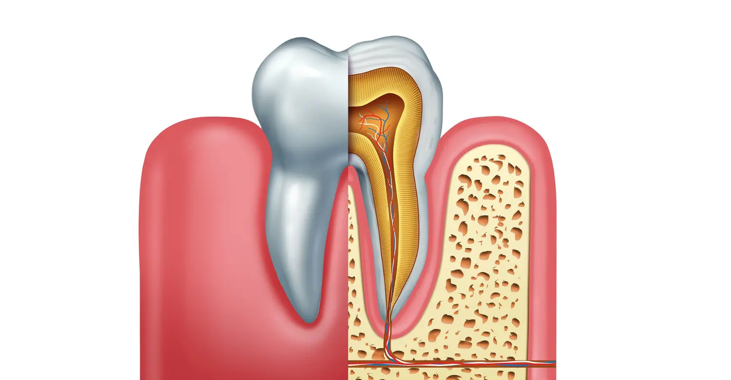
A tooth anatomy diagram typically presents a cross-section of a tooth. It may label enamel, dentin, pulp, cementum, crown, root, bone, and periodontal tissues. Diagrams simplify complex three-dimensional structures for educational purposes. They help patients understand where disease and treatment occur. A diagram can also clarify why symptoms differ according to affected tissues. At Vitrin Clinic, visual explanations can support patient discussions during treatment planning. However, diagrams cannot replace individualized clinical examination.
How to Read a Tooth Anatomy Diagram
Start by identifying the crown and root. Then locate the enamel surrounding the crown. Dentin appears beneath the enamel and surrounds the pulp. The pulp occupies the central chamber and root canals. Cementum appears over the root surface. Supporting bone surrounds the root, while periodontal fibers connect tooth and bone. Understanding these relationships makes dental terminology easier to follow. The same principles apply across most permanent teeth, despite differences in shape.
Crown vs. Root in a Tooth Anatomy Diagram
The crown and root perform different structural functions. The crown interacts directly with food and opposing teeth. The root remains embedded within supporting tissues. The crown primarily contains enamel and dentin externally. The root contains dentin covered by cementum. Root canals carry the pulp toward the root tip. The apical opening permits communication between the pulp and surrounding tissues. This distinction becomes especially important when discussing root canal treatment.
How the Different Tooth Layers Work Together
Tooth tissues operate as an integrated mechanical and biological system. Enamel provides a hard protective surface. Dentin supports enamel and helps distribute forces. The pulp supplies biological functions and sensory information. Cementum provides root attachment. Periodontal tissues absorb and distribute forces around the tooth. This layered design allows teeth to tolerate repeated chewing forces. Damage to one tissue can eventually affect neighboring structures. For example, untreated decay can progress from enamel toward dentin and pulp.
What Is the Anatomy of Teeth and Gums?
Teeth depend on surrounding tissues for stability and function. These tissues include gingiva, periodontal ligament, and alveolar bone. Together, they form important components of the periodontium. Healthy periodontal tissues help maintain tooth position and protect deeper structures. Inflammation or tissue destruction can compromise this support. NIDCR notes that healthy periodontal probing depths are usually around 1–3 millimeters. Deeper pockets can indicate periodontal disease. Therefore, tooth health and gum health cannot be considered completely separately.
Gingiva (Gums)
The gingiva surrounds and protects tissues near the teeth. Healthy gums are generally firm and closely adapted around the teeth. Inflamed gums can become red, swollen, tender, or prone to bleeding. Gum recession can expose previously protected root surfaces. NIDCR identifies bleeding and gum recession among common signs of periodontal disease. Good brushing and interdental cleaning help control plaque. Professional periodontal assessments can detect changes that patients may not notice. Vitrin Clinic includes gum evaluation when assessing comprehensive oral health.
Periodontal Ligament
The periodontal ligament is a specialized connective tissue surrounding tooth roots. Its fibers connect cementum with alveolar bone. It helps anchor teeth while allowing limited physiological movement. The ligament also helps distribute chewing forces. It contains sensory structures that contribute to pressure perception. Damage to periodontal tissues can reduce tooth stability. Periodontal disease can destroy supporting tissues and contribute to tooth mobility. Maintaining periodontal health therefore protects both function and tooth retention.
Alveolar Bone
Tooth Anatomy depends partly on the surrounding alveolar bone for long-term stability. Alveolar bone forms the bony sockets supporting tooth roots. It adapts to mechanical forces and changes throughout life. Severe periodontal disease can lead to loss of supporting bone. Bone loss may cause teeth to become mobile or shift position. NIDCR identifies supporting bone destruction as a feature of advanced periodontal disease. Dental imaging helps dentists evaluate bone levels. Bone assessment is also essential when planning dental implants. At Vitrin Clinic, imaging supports individualized treatment planning when clinically indicated.
Clinical Note
Healthy teeth require healthy supporting tissues. A structurally intact crown can still become unstable if periodontal support is severely compromised. Likewise, gum inflammation can complicate restorative treatment. Dentists therefore evaluate both teeth and surrounding tissues. This integrated approach is especially important for patients considering extensive dental rehabilitation.
How Does Tooth Anatomy Differ Between Different Types of Teeth?
Different teeth have different shapes because they perform different functions. Incisors cut food, while canines help tear it. Premolars and molars primarily crush and grind food. Their crown shapes and root configurations reflect these functions. Anatomical differences also influence dental treatment. For example, molars often have more complex root canal systems. Understanding tooth type helps dentists interpret radiographs and clinical findings accurately.
Incisors
Tooth Anatomy classifies incisors among the anterior teeth used for cutting food. Incisors are the front teeth used mainly for cutting food. They usually have relatively thin crowns with sharp incisal edges. Most permanent incisors have one root. Their visible position also makes them important for smiling aesthetics. Trauma can commonly affect their edges or crowns. Restorative treatment must often balance structural preservation with appearance. Vitrin Clinic considers both functional and cosmetic requirements when treating visible anterior teeth.
Canines
Canines are located beside the incisors. They have prominent pointed crowns designed for tearing food. Their roots are generally long compared with many other teeth. Canines also contribute to guidance during certain jaw movements. Their strong root support can make them important during orthodontic and restorative planning. Their position also contributes significantly to the appearance of a smile. Damage involving a canine should be evaluated carefully because of its functional role.
Premolars
Premolars are positioned between canines and molars. They usually have broader crowns with one or two prominent cusps. Their role includes tearing and crushing food. Premolar root anatomy varies among individuals. Some upper premolars can have multiple canals. This variation matters during endodontic treatment. Dental imaging can help identify unexpected root or canal anatomy. Accurate diagnosis reduces the risk of untreated anatomy during treatment.
Molars
Tooth Anatomy classifies molars among the posterior teeth responsible for grinding food. Molars are the largest posterior teeth in many adults. Their broad surfaces are designed for crushing and grinding. They often have multiple cusps and roots. Upper molars commonly have three roots, although anatomical variation exists. Lower molars commonly have two roots. Their complex anatomy can make root canal procedures more technically demanding. Deep grooves and pits can also retain plaque and food particles. Dental sealants can help protect susceptible chewing surfaces in appropriate patients.
How Is Baby Teeth Anatomy Different From Adult Teeth Anatomy?
Primary and permanent teeth share many fundamental anatomical features. However, their proportions and internal structures differ. Primary teeth generally have thinner enamel and relatively larger pulp chambers. These differences can influence the progression and treatment of dental decay. Primary teeth also have roots designed to eventually resorb before natural exfoliation. Permanent teeth develop with structures intended for long-term function. Understanding these differences helps dentists select age-appropriate treatments.
Primary Tooth Anatomy
Primary teeth are commonly called baby teeth or deciduous teeth. Humans normally develop 20 primary teeth. They begin emerging at approximately six months of age. Primary teeth have thinner enamel than permanent teeth. Their pulp chambers are also proportionally larger. These differences can allow decay to reach the pulp more quickly. Primary teeth support chewing, speech, and normal dental development. They also help maintain space for permanent successors.
Permanent Tooth Anatomy
Most adults have up to 32 permanent teeth, including third molars. Permanent teeth are designed for long-term chewing and speech functions. Their enamel is highly mineralized and provides strong surface protection. Permanent teeth have more developed roots than newly emerging primary teeth. Their root and canal anatomy varies according to tooth type. Permanent teeth require lifelong preventive care because significant enamel loss cannot naturally regenerate. Regular examinations can detect structural changes early.
Why Baby Teeth Still Matter
Primary teeth are important even though they eventually fall out. They support chewing, speech, and normal facial development. They also maintain appropriate space for permanent teeth. Premature loss can affect spacing and eruption patterns. Decay in primary teeth can also cause pain and infection. Parents should therefore treat primary teeth as important functional structures. Early preventive care can reduce avoidable dental disease. Vitrin Clinic encourages age-appropriate dental assessments for children.
How Does Tooth Anatomy Relate to Common Dental Problems?
Different dental conditions affect different anatomical tissues. Understanding the affected tissue helps explain symptoms and treatment options. Decay commonly begins in hard dental tissues. Sensitivity often involves exposed dentin or pulpal responses. Cracks can extend through several structural layers. Pulpitis affects the internal living tissue. Gum recession exposes root surfaces previously protected by periodontal tissues. Accurate diagnosis determines the most appropriate treatment approach.
Tooth Decay
Tooth Anatomy helps explain how decay can progress from the outer surface toward the pulp. Dental decay results from a complex interaction involving bacteria, diet, tooth tissues, and time. Acid produced by bacterial activity can cause mineral loss. Early lesions may affect enamel before progressing deeper. Advanced lesions can reach dentin and eventually the pulp. The ADA provides evidence-based recommendations for managing different caries lesions. Treatment depends on lesion activity, depth, location, and patient risk. Preventive strategies include oral hygiene, fluoride exposure, dietary control, and professional care. Early intervention can help preserve more natural tooth structure.
Tooth Sensitivity
Tooth sensitivity can occur when dentin becomes exposed. Gum recession and enamel wear can contribute to dentin exposure. Dentin contains microscopic tubules that communicate with the pulp. Temperature, touch, or chemical stimuli can trigger discomfort. Sensitivity can also arise from other dental conditions. Therefore, persistent or severe sensitivity deserves professional evaluation. Dentists first identify the underlying cause before recommending treatment. At Vitrin Clinic, diagnosis guides personalized sensitivity management.
Cracked or Broken Teeth
Teeth can crack because of trauma, heavy biting forces, or structural weakening. Cracks may involve enamel, dentin, pulp, or deeper structures. Their location and depth strongly influence prognosis. Some cracks produce pain during chewing or temperature changes. Others may remain difficult to detect without clinical testing. Recent NIDCR research highlights the importance of internal enamel organization in tooth strength. Treatment may include bonding, restorations, crowns, or more advanced procedures. Severe fractures may compromise the possibility of preserving the tooth.
Pulpitis and Tooth Infection
Pulpitis refers to inflammation of the dental pulp. It can develop after deep decay, trauma, cracks, or repeated procedures. Symptoms can include spontaneous pain or prolonged sensitivity. Untreated pulpal disease can progress toward infection around the root. Root canal treatment removes diseased pulp while preserving the remaining tooth structure. The treatment requires detailed knowledge of root and canal anatomy. Complex canal systems can contain additional branches or anatomical variations. Professional imaging and careful instrumentation help improve treatment accuracy.
Gum Recession
Gum recession exposes root surfaces that were previously covered by gingiva. Exposed roots can become sensitive and more vulnerable to root caries. Aggressive brushing, periodontal disease, anatomical factors, and other causes can contribute. NIDCR lists gums pulling away from teeth among periodontal disease symptoms. Treatment depends on the cause and severity. Improving hygiene techniques can reduce further irritation. Periodontal therapy may be necessary when disease is present. Some cases may require soft-tissue procedures for root coverage.
What We Notice Clinically
Tooth Anatomy can vary subtly from patient to patient, as clinicians often observe. Clinical assessment shows that anatomy varies between individual patients. Two teeth of the same type can have different root shapes or canal configurations. Symptoms also do not always correspond directly with visible damage. A small cavity can sometimes cause significant pain. A large structural defect may occasionally produce limited symptoms. This is why dentists combine examination, imaging, and patient history. Modern diagnosis aims to preserve healthy tissues whenever possible.
Clinical Note
One important clinical principle is tissue preservation. Every unnecessary removal of healthy tooth structure can reduce future restorative options. Dentists therefore consider the smallest effective intervention when clinically appropriate. The ADA's restorative guidance supports conservative approaches for suitable caries lesions. Treatment decisions should still reflect lesion characteristics and patient-specific risks. Vitrin Clinic uses comprehensive assessment to support individualized treatment planning.
Dr. Rifat Alsaman's Opinion
Dr. Rifat Alsaman emphasizes that successful dental treatment begins with accurate diagnosis. A tooth should be evaluated as a complete biological and functional structure. The visible crown is only one part of the clinical picture. Root anatomy, pulp condition, periodontal support, and bite forces also matter. Preserving healthy tissue should remain an important treatment objective. For cosmetic procedures, appearance should also support proper function and tooth stability. This balanced approach helps patients receive treatment designed around their individual needs.
How Do Dentists Examine Tooth Anatomy?
Dentists use multiple diagnostic methods to understand dental structures. No single method provides every piece of information. Visual examination identifies visible changes and surface abnormalities. Radiographs reveal internal structures and supporting bones. Pulp tests evaluate responses to specific stimuli. Digital scans provide detailed three-dimensional information in selected cases. Combining these methods improves diagnostic confidence.
Visual Dental Examination
A visual examination begins with inspection of the teeth and gums. Dentists look for discoloration, cavities, fractures, wear, swelling, and gum changes. They also evaluate tooth shape and existing restorations. Dental instruments can help assess surfaces and periodontal tissues. The dentist may also examine the patient's bite. Clinical findings help determine whether additional diagnostic tests are needed. Regular examinations can identify abnormalities before symptoms become severe.
Dental X-Rays
Dental radiographs allow dentists to evaluate structures that cannot be seen directly. They can show roots, pulp spaces, bone levels, and hidden decay. Different radiographic techniques provide different views. Periapical images can evaluate individual teeth and surrounding structures. Bitewing images are particularly useful for detecting certain interproximal lesions. Panoramic imaging provides a broader view of the jaws. Radiographs should be selected according to clinical need.
Pulp and Sensitivity Tests
Pulp tests help determine how a tooth responds to stimuli. Common methods include cold testing and electrical pulp testing. Percussion and biting tests can also provide useful information. The results must be interpreted alongside symptoms and imaging. A positive response does not automatically mean that the pulp is healthy. Likewise, a lack of response requires careful interpretation. Clinical diagnosis depends on the complete evidence rather than one test.
Digital Scans and 3D Imaging
Digital imaging can provide detailed information about complex dental structures. Three-dimensional imaging may help assess roots, bone, impacted teeth, and implant sites. Cone-beam computed tomography is used selectively when additional information is justified. It should not automatically replace conventional radiographs. The choice depends on diagnostic requirements and expected clinical benefit. At Vitrin Clinic, advanced imaging can support complex treatment planning when appropriate.
How Can Tooth Anatomy Affect Dental Treatment?
Dental treatment must respect the underlying anatomy. A filling must protect remaining tooth tissues without unnecessarily weakening them. A crown must restore function while maintaining appropriate structural support. Root canal treatment requires accurate identification of canals. Implant treatment requires assessment of bone and neighboring anatomical structures. Understanding anatomy therefore influences diagnosis, planning, treatment, and follow-up.
Tooth Anatomy and Dental Fillings
Fillings restore teeth affected by suitable cavities or structural defects. The dentist removes or manages diseased tissue according to the treatment approach. The remaining enamel and dentin provide the foundation for restoration. The size and location of the defect influence material selection. Deep lesions require particular attention to pulpal health. The ADA recommends evidence-based approaches for managing caries lesions. Preserving sound tissue can help maintain long-term structural strength. Vitrin Clinic develops restorative plans according to each tooth's condition.
Tooth Anatomy and Dental Crowns
Crowns cover and protect substantially damaged teeth. They can restore shape, function, and appearance when clinically indicated. The amount of remaining tooth structure affects crown planning. The dentist must also consider the bite and periodontal tissues. A crown cannot compensate for untreated periodontal disease or unresolved pulpal problems. Proper diagnosis should therefore precede definitive crown placement. Vitrin Clinic combines functional assessment with aesthetic planning for suitable patients. The goal is a stable restoration that integrates with the surrounding teeth.
Tooth Anatomy and Root Canal Treatment
Root canal treatment addresses diseased or irreversibly inflamed pulp. The dentist accesses the pulp chamber and cleans the root canal system. The canals are then shaped, disinfected, and sealed. Successful treatment depends partly on identifying the complete canal anatomy. Extra canals or unusual root configurations can complicate treatment. Dental imaging may help identify complex anatomy. After treatment, the tooth may require a restoration that protects the remaining structure. Treatment planning should consider both biological and mechanical factors.
Tooth Anatomy and Dental Implants
Dental implants replace missing tooth roots using biocompatible implant components. Unlike natural teeth, implants do not contain enamel, dentin, or dental pulp. They depend on surrounding bone and soft tissues for stability and health. Implant planning therefore requires assessment of bone volume and anatomical structures. Three-dimensional imaging may be appropriate for complex implant cases. The final restoration must also provide functional and aesthetic integration. Vitrin Clinic evaluates each patient's oral condition before implant planning. A personalized approach helps identify suitable treatment options.
Tips for Patients to Protect Their Tooth Structure
Protecting natural tooth structure is usually easier than replacing lost tissue. Daily prevention focuses on plaque control, fluoride exposure, dietary habits, and professional monitoring. Patients should also avoid unnecessary mechanical or chemical stress. Regular dental visits can identify early structural changes. Small problems are generally easier to manage than advanced damage. Good habits protect both teeth and surrounding periodontal tissues.
Maintain Good Oral Hygiene
Brush teeth twice daily using an appropriate fluoride toothpaste. Clean between teeth using floss or interdental cleaning devices. Pay particular attention to areas around the gumline. Plaque accumulation can contribute to caries and periodontal inflammation. Professional cleaning may be recommended according to individual risk. Avoid aggressive brushing that can contribute to cervical wear or recession. The ADA notes that appropriate toothpaste abrasivity can support effective cleaning while limiting wear. Vitrin Clinic can provide individualized hygiene guidance during routine visits.
Protect Your Tooth Enamel
Enamel protection depends on reducing repeated acid exposure and maintaining preventive care. Frequent acidic drinks can contribute to erosive tooth wear. The ADA identifies carbonated soft drinks as an important dietary risk factor. Fluoride can support enamel resistance and remineralization processes. Avoid using teeth as tools for opening packages or biting hard objects. Patients with grinding may benefit from professional evaluation. Once substantial enamel is lost, it cannot biologically regenerate. Prevention is therefore especially important.
Protect Your Gums and Supporting Tissues
Healthy gums protect the cervical and root areas of teeth. Clean gently along the gumline without excessive pressure. Bleeding during brushing should not simply be ignored. Persistent bleeding can indicate gingival inflammation or periodontal disease. NIDCR recommends professional evaluation for symptoms such as bleeding and recession. Smoking and other risk factors can also influence periodontal health. Individual risk assessment helps determine appropriate preventive care. Healthy periodontal tissues support long-term tooth stability.
When Should You See a Dentist?
See a dentist when pain, swelling, bleeding, sensitivity, or structural changes persist. Urgent assessment is appropriate for significant trauma or facial swelling. A broken tooth should also be evaluated promptly. Early diagnosis can prevent some problems from becoming more extensive. Routine examinations remain important even without symptoms. Some cavities and periodontal problems can progress without obvious discomfort. Vitrin Clinic provides comprehensive assessments designed to identify both visible and hidden concerns.
Why Choose Vitrin Clinic for Comprehensive Dental Care?
Vitrin Clinic focuses on individualized diagnosis and treatment planning. Comprehensive care begins with understanding the patient's oral condition. Dentists evaluate teeth, gums, supporting tissues, bite, and aesthetic concerns. Diagnostic information is then used to develop an appropriate treatment strategy. This approach helps connect preventive, restorative, and cosmetic dentistry. Patients can discuss different treatment options before making decisions. The objective is to create treatment plans based on clinical needs.
Personalized Dental Diagnosis
Tooth Anatomy forms the foundation of any individualized diagnostic process. Every patient's teeth and supporting tissues have individual characteristics. Root shapes, canal anatomy, bone levels, and restoration needs can vary considerably. A personalized diagnosis therefore provides more useful information than generic treatment recommendations. Clinical examination and appropriate imaging help identify relevant anatomical factors. Vitrin Clinic uses diagnostic findings to guide individualized treatment planning. The approach considers both immediate concerns and long-term oral health. This can help patients understand why a particular treatment is recommended.
Advanced Dental Technology
Modern dentistry uses digital tools to improve diagnosis and treatment planning. Digital imaging can provide detailed information about teeth and supporting structures. Intraoral scanning can capture digital representations of dental surfaces. Three-dimensional imaging can assist selected complex procedures. Technology should support clinical judgment rather than replace it. At Vitrin Clinic, appropriate technology can help improve treatment visualization and planning. The specific technology used depends on each patient's clinical requirements.
Dr. Rifat Alsaman's Clinical Perspective
Dr. Rifat Alsaman emphasizes diagnosis, preservation, and predictable treatment planning. Understanding the complete dental structure helps clinicians avoid treating only visible symptoms. The surrounding gums, bone, pulp, and bite can influence treatment outcomes. This perspective is particularly important for complex restorative and cosmetic cases. Treatment should aim for appropriate function while maintaining natural structures whenever possible. At Vitrin Clinic, this philosophy supports individualized dental care.
Why Choose Vitrin Clinic for Restorative and Cosmetic Dentistry?
Restorative and cosmetic dentistry often overlap when damaged teeth affect both function and appearance. Treatment should address the underlying structural condition before focusing on aesthetics. A beautiful restoration also needs appropriate fit, function, and periodontal compatibility. Vitrin Clinic combines restorative planning with aesthetic considerations when clinically suitable. Patients can receive treatment designed around their individual smile and dental condition.
Personalized Treatment Planning
Treatment planning begins by identifying the condition of each relevant tooth. Dentists assess structural damage, periodontal health, occlusion, and aesthetic requirements. The final plan should reflect the patient's clinical needs and expectations. Different teeth may require different treatment approaches within the same smile. Personalized planning helps avoid unnecessary procedures. It can also improve communication between patients and clinicians. Vitrin Clinic uses individualized planning for restorative and cosmetic treatment.
Restoring Damaged Tooth Structure
Restoration aims to replace or protect damaged tooth structure. The appropriate treatment depends on the extent and location of damage. Small defects may require conservative restorations. More extensive damage may require crowns or other restorative approaches. Root canal treatment may be necessary when the pulp is irreversibly affected. The goal is to preserve the tooth whenever a predictable outcome remains possible. Proper maintenance after treatment also supports restoration longevity.
Combining Function and Aesthetics
Cosmetic dentistry should not be separated from dental function. Restorations must interact properly with neighboring and opposing teeth. The bite, gumline, tooth proportions, and supporting structures all matter. Aesthetic treatment should therefore begin with comprehensive assessment. Vitrin Clinic considers smile appearance alongside functional and structural requirements. This approach can help create results that look natural while supporting oral function. The best outcome is usually one that balances appearance, health, and durability.
Final Takeaway: Why Understanding Tooth Anatomy Matters
Understanding the structure of a tooth makes dental health easier to understand. Enamel, dentin, pulp, and cementum each perform distinct functions. The crown, neck, and root also have different anatomical roles. Supporting tissues help keep teeth stable and functional. Dental problems become easier to understand when their affected structures are clear. This knowledge can also help patients understand diagnostic tests and treatment options. At Vitrin Clinic, anatomical assessment supports personalized dental care. Protecting natural tooth structure remains an important long-term goal.
Reference
FAQs

Dr. Rifat Alsaman has more than 5 years of clinical experience in dentistry and currently serves as the Head of the Medical Team at Vitrin Clinic. He is dedicated to providing exceptional patient care, overseeing treatment planning, and ensuring the highest clinical standards across the team. His expertise, attention to detail, and commitment to continuous professional development have helped countless patients achieve healthier, more confident smiles.
.webp&w=3840&q=75)


