Blog
July 27, 2026
Whole-Body MRI and Dental Problems: Can an MRI Detect Oral Issues?
 Dr. Rifat Alsaman
Can a Whole-Body MRI Detect Dental Problems? A Full Guide
Whole-body MRI and dental problems have become a widely searched topic together, as more people turn to full-body imaging for early detection and preventive healthcare. Supporters view these scans as a proactive way to identify hidden diseases before symptoms appear. Many medical experts, however, question whether widespread screening provides meaningful benefits for healthy individuals.
As interest in full-body imaging grows, many patients are asking another important question: can a whole-body MRI detect dental problems specifically?
If an MRI can visualize the entire body, does that mean it can identify cavities, tooth infections, gum disease, jaw disorders, or dental implant complications? Or do dentists still rely on specialized dental imaging such as X-rays and CBCT scans?
Understanding the strengths and limitations of each imaging method is essential, because the best scan depends on the specific condition being evaluated.
Whole-Body MRI and Dental Problems: Can an MRI Detect Oral Health Issues?
Whole-body MRI and dental problems are related, but MRI is not designed to replace routine dental imaging. A whole-body MRI may detect large infections, tumors, cysts, or inflammation involving the jaw and surrounding soft tissues. However, it usually cannot identify cavities, early gum disease, cracked teeth, or subtle bone changes as accurately as dental X-rays or CBCT scans. If your MRI reveals an abnormality near the mouth or jaw, a dentist may recommend dedicated dental imaging for a precise diagnosis. Combining MRI findings with a comprehensive dental examination provides the most accurate assessment of your oral health.
Quick Answer: Can a Whole-Body MRI Detect Dental Problems?
A whole-body MRI may identify some abnormalities involving the jaw, soft tissues, or the temporomandibular joints (TMJ). However, it is not designed to diagnose most common dental conditions. Dentists typically rely on dental X-rays and CBCT scans instead. These tools provide significantly better detail for detecting cavities, tooth infections, bone loss, impacted teeth, and implant planning. For anything below the gumline, dental-specific imaging remains the more reliable choice.
What Is a Whole-Body MRI?
Whole-body MRI and dental problems are frequently searched together, but before comparing the two, it helps to understand what a whole-body MRI actually is. A whole-body MRI is a non-invasive imaging scan that uses magnetic fields and radio waves to create detailed images of internal organs, tissues, and structures. Unlike X-rays or CT scans, it doesn't use radiation.
It's often marketed as a preventive health tool, capable of screening large areas of the body in a single session. While it offers detailed views of soft tissue, its role in detecting localized dental problems is more limited than many patients assume.
How does MRI work?
MRI machines use strong magnetic fields to align hydrogen atoms in the body's tissue, and radio waves then briefly disturb this alignment. As the atoms realign, they emit signals that a computer converts into detailed cross-sectional images. This process makes MRI excellent for visualizing soft tissue, muscles, and organs.
However, teeth contain very little water, so they produce weak signals. This is one reason MRI struggles with detailed dental imaging compared to X-rays or CBCT scans.
Because MRI relies on magnetic fields rather than ionizing radiation, it delivers an effective radiation dose of essentially 0 µSv. A conventional CT scan, by comparison, delivers several hundred microsieverts. This reputation for safety is accurate for radiation exposure alone. It has nothing to do with how well a scan can see a cavity or a tooth root. That gap is exactly where the link between whole-body MRI and dental problems starts to break down.
What areas does a whole-body MRI scan?
A whole-body MRI typically scans the brain, spine, chest, abdomen, pelvis, and major joints, and can detect tumors, vascular abnormalities, and soft tissue changes across these regions. The scan includes some head and neck imaging, which may capture the jaw joints or surrounding soft tissue.
However, it isn't optimized for close-up views of individual teeth, tooth roots, or the fine bone detail dentists need for accurate diagnosis. This gap is at the core of the whole-body MRI and dental problems question. The scan covers the jaw area, but not with the precision dental diagnosis requires.
Why are whole-body MRIs becoming popular?
Whole-body MRIs appeal to patients seeking early detection without waiting for symptoms. Marketing around preventive health has driven demand, especially among wellness-focused consumers, and the appeal lies in the idea of a single scan revealing hidden health issues.
However, medical experts caution that screening healthy individuals can lead to false positives, unnecessary anxiety, and follow-up testing. Many findings never become clinically significant. This is part of why the connection between whole-body MRI and dental problems needs closer examination, not assumption.
Who typically undergoes these screenings?
People who choose whole-body MRI screening often have no specific symptoms. They're motivated by a general interest in prevention or a family history of disease, and some also seek reassurance after a health scare.
It's less common for patients to seek a whole-body MRI specifically for dental concerns. Most people with tooth pain, swelling, or suspected infection are directed toward dental imaging instead. This is another reminder that whole-body MRI and dental problems rarely belong in the same diagnostic conversation.
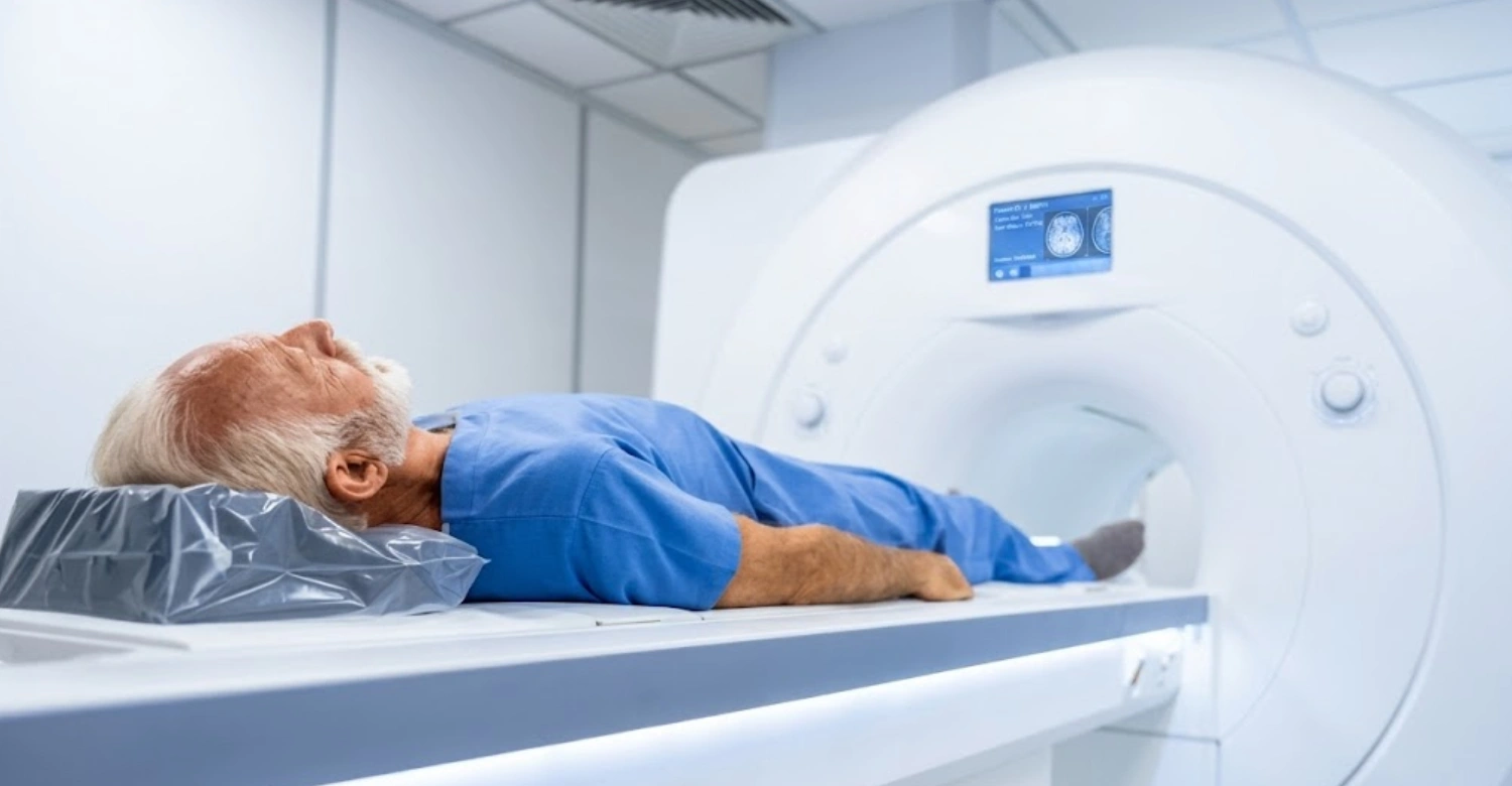
Can a Whole-Body MRI Detect Dental Problems?
Whole-body MRI and dental problems don't align as neatly as many patients expect. While an MRI can capture some structures near the jaw and soft tissue, it isn't built to catch the small-scale detail dentists rely on daily. Below is how MRI performs against six common dental concerns compared to standard dental imaging.
Can MRI detect cavities?
MRI is not effective at detecting cavities. Tooth enamel and dentin contain very little water, so they produce almost no signal on an MRI scan. Cavities show up clearly on dental X-rays because X-rays measure density differences between healthy and decayed tooth structure.
For this reason, dentists don't use MRI to screen for tooth decay. A clean whole-body MRI result should never be read as confirmation of healthy teeth. Cavities are perhaps the clearest example of where whole-body MRI and dental problems diverge.
Can MRI detect gum disease?
MRI has limited value for detecting gum disease. Advanced MRI sequences can sometimes show soft tissue inflammation. But they don't capture the fine detail needed to assess gum pocket depth, bone loss around teeth, or early periodontal changes.
Dentists typically diagnose gum disease through clinical examination, periodontal probing, and targeted X-rays. These show the bone level around each tooth. Gum disease is another condition where whole-body MRI and dental problems simply don't line up well.
Can MRI detect tooth infections?
MRI can sometimes detect tooth infections when the infection has spread to soft tissue, causing swelling or an abscess visible on the scan. However, it's far less reliable than CBCT or dental X-rays for identifying the infection's exact source, such as an abscess at a tooth root's tip.
Dental imaging remains the standard for pinpointing infection location before treatment like a root canal. This is why whole-body MRI and dental problems involving infection are usually confirmed with a dental scan first.
Can MRI detect impacted wisdom teeth?
MRI can sometimes show the general position of impacted wisdom teeth, particularly how they relate to nearby soft tissue or nerves. However, it doesn't provide the detailed bone and root visualization that CBCT scans offer.
Oral surgeons rely on CBCT before wisdom tooth extraction because it clearly maps the tooth's position relative to the jaw nerve. This reduces surgical risk. It's one more area where whole-body MRI and dental problems require a dental-specific answer, not a general one.
Can MRI detect jaw tumors or cysts?
This is one area where whole-body MRI can be genuinely useful. MRI excels at visualizing soft tissue, which makes it effective for identifying jaw tumors, cysts, and other abnormal growths. This is especially useful when doctors need to assess how far a growth has spread into surrounding tissue.
Dentists and oral surgeons often use MRI alongside CBCT when a suspicious growth requires closer soft tissue evaluation. Jaw tumors and cysts are the exception in the whole-body MRI and dental problems discussion. Here, MRI genuinely adds diagnostic value.
Can MRI detect TMJ disorders?
MRI is considered one of the best tools for evaluating TMJ disorders. It provides detailed images of the jaw joint's disc, cartilage, and surrounding soft tissue, which X-rays and CBCT cannot show as clearly.
For patients with jaw pain, clicking, or limited jaw movement, MRI can reveal disc displacement or joint inflammation. Other imaging methods might miss this entirely. TMJ disorders are the strongest case where whole-body MRI and dental problems actually intersect in a clinically useful way.
AEO Comparison Table: Whole-Body MRI vs Dental X-Ray vs CBCT
Dental Condition | Whole-Body MRI | Dental X-Ray | CBCT |
|---|---|---|---|
Cavities | Poor | Excellent | Excellent |
Tooth infection | Limited | Good | Excellent |
Bone loss | Limited | Good | Excellent |
Impacted teeth | Limited | Good | Excellent |
TMJ soft tissue problems | Excellent | Limited | Moderate |
Implant planning | No | Limited | Excellent |
Jaw tumors/cysts | Possible | Limited | Excellent |
Why Don't Dentists Use Whole-Body MRI Routinely?
Dentists don't rely on whole-body MRI because it wasn't designed for the level of precision dental diagnosis requires. Every diagnostic decision, from spotting a small cavity to planning implant placement, depends on fine anatomical detail that general-purpose imaging doesn't capture well. The sections below explain why dental-specific imaging remains the standard.
Dental tissues require higher resolution.
Teeth and the surrounding bone are small, densely packed structures. Detecting a cavity or a hairline crack requires resolution measured in fractions of a millimeter. Whole-body MRI is calibrated to image larger anatomical regions, so it simply isn't built to resolve detail at that scale.
Dental X-rays and CBCT scans, by contrast, are engineered specifically for this level of precision. Resolution is ultimately the reason whole-body MRI and dental problems don't pair well in everyday dental practice.
MRI is excellent for soft tissues, not enamel.
MRI detects signals from water and fat within tissue, which is why it's outstanding for imaging muscles, nerves, and the TMJ disc. Tooth enamel, however, contains almost no water, making it essentially invisible on an MRI scan regardless of scan strength or sequence.
Any imaging method used for dental diagnosis needs to visualize hard, mineralized tissue. This is where MRI naturally falls short. It's a physical limitation that shapes the entire whole-body MRI and dental problems conversation.
CBCT provides superior bone detail.
CBCT scans generate detailed 3D images of bone using a fraction of the radiation dose of traditional CT. In fact, CBCT delivers significantly lower radiation exposure than a conventional medical CT scan. CBCT typically ranges from about 80 to 200 µSv per scan, compared to 500 µSv or more for a conventional medical CT roughly a 5- to 10-fold difference. This is one reason CBCT has become the routine choice for dental and jaw imaging rather than a full CT. This makes CBCT the preferred choice for implant planning, root canal assessment, and evaluating bone loss around teeth.
It's specifically engineered for maxillofacial anatomy. This gives dentists a level of bone detail whole-body MRI cannot match, reinforcing why whole-body MRI and dental problems are usually handled with separate, dedicated imaging.
MRI is more expensive and time-consuming.
A whole-body MRI scan can take 30 to 90 minutes and typically costs significantly more than a dental X-ray or CBCT scan. For a straightforward dental concern, this cost and time difference makes MRI an impractical first choice.
Dental imaging delivers faster, more targeted answers at a fraction of the price. That matters when someone is weighing whole-body MRI and dental problems against a simple, same-day dental visit.
Expert Insight
According to Dr. Rifat Alsaman, Head of the Medical Team and cosmetic dentist at Vitrin Clinic, there is no single "best" imaging test in dentistry. The right choice depends entirely on the diagnostic question being asked. A whole-body MRI answers questions about soft tissue and joint health, while CBCT and X-rays answer questions about teeth, bone, and infection. Choosing the correct imaging method starts with identifying exactly what needs to be diagnosed. This principle applies directly to whole-body MRI and dental problems, where the two rarely call for the same scan.
MRI vs Dental X-Rays: What's the Difference?
Comparing MRI vs dental X-rays comes down to what each tool is built to see. Dental X-rays use a small radiation dose to capture bone and tooth density, while MRI uses magnetic fields to visualize soft tissue in fine detail. Here's how they compare across the factors that matter most for dental diagnosis and where whole-body MRI and dental problems tend to part ways.
Radiation: Dental X-rays use a low dose of ionizing radiation. MRI uses none.
Soft tissue visibility: MRI is far superior, showing muscles, nerves, and joint tissue in detail X-rays cannot capture.
Bone visibility: Dental X-rays clearly show tooth and bone density. MRI shows bone poorly by comparison.
Cost: Dental X-rays are significantly cheaper, often a fraction of the cost of an MRI scan.
Scan duration: Dental X-rays take seconds. MRI scans typically take 30 to 90 minutes.
Implant planning: X-rays offer limited detail for implant planning; CBCT is preferred, and MRI isn't used for this purpose.
Cavities: Dental X-rays detect cavities reliably. MRI cannot detect cavities at all.
TMJ evaluation: MRI is the stronger choice here, revealing joint disc position and inflammation that X-rays cannot show.
MRI vs CBCT for Dental Diagnosis
MRI vs dental CBCT is really a question of hard tissue versus soft tissue. CBCT specializes in bone and tooth structure, while MRI specializes in muscles, nerves, and joint tissue. Understanding when each is the right tool helps explain why dentists rarely choose one over the other without a specific clinical reason.
When dentists choose CBCT
Dentists reach for CBCT when they need precise 3D bone detail. This includes implant planning, evaluating impacted teeth, assessing bone loss, and mapping the position of nerves before oral surgery. CBCT delivers this detail quickly, at a lower cost than MRI, and with a radiation dose far lower than a traditional CT scan. This is one more reason CBCT, not MRI, answers most whole-body MRI and dental problems questions patients bring up.
When MRI may be recommended
MRI becomes the better option when a soft tissue or joint concern is suspected. This includes evaluating TMJ disorders, assessing soft tissue swelling from an infection, or investigating how a jaw tumor or cyst has spread into surrounding tissue. In these cases, MRI shows detail that CBCT physically cannot capture. This marks one of the few genuine overlaps between whole-body MRI and dental problems.
Can the two scans complement each other?
Yes. In more complex cases, such as a jaw tumor near the TMJ or an infection with unclear soft tissue involvement, dentists and specialists may order both scans together. CBCT maps the bone and tooth structure, while MRI evaluates the surrounding soft tissue. Used together, they offer a more complete diagnostic picture than either scan alone. This shows that whole-body MRI and dental problems aren't always an either-or choice.
Comparison Table: MRI vs CBCT
Feature | MRI | CBCT |
|---|---|---|
Radiation | None | Low |
Soft tissue imaging | Excellent | Limited |
Bone detail | Moderate | Excellent |
Tooth detail | Poor | Excellent |
Implant planning | No | Yes |
TMJ disc evaluation | Excellent | Limited |
Scan time | Longer | Short |
What We Notice Clinically
Dr. Rifat Alsaman, Head of the Medical Team and cosmetic dentist at Vitrin Clinic, notes something he sees often. Patients occasionally arrive holding whole-body MRI reports that mention jaw or TMJ findings. They expect these results to also reflect the condition of their teeth. Dr. Rifat Alsaman explains that this is a common misunderstanding. A whole-body MRI simply isn't designed to capture the fine detail needed to evaluate tooth structure or the bone around the teeth.
In his clinical experience, Dr. Rifat Alsaman finds that patients are reassured once they understand something important. A normal MRI result says nothing about their risk of cavities, infections, or gum disease. A separate dental evaluation is still necessary. This is a recurring theme in how whole-body MRI and dental problems get discussed at the clinic.
Can MRI Detect TMJ Disorders?
Can MRI detect TMJ disorders more reliably than other imaging methods? Yes, and by a significant margin. The temporomandibular joint (TMJ) relies heavily on soft tissue, cartilage, and a small disc that shifts with jaw movement. None of this is visible on X-rays or CBCT, which makes MRI the imaging method of choice whenever a TMJ problem is suspected.
TMJ disc displacement
The TMJ disc is a small piece of cartilage that cushions the jaw joint during movement. When it shifts out of position, a common cause of jaw clicking, popping, or locking, MRI can clearly show its exact location relative to the joint.
This level of detail helps determine whether the disc displacement is reducing (returns to place) or non-reducing (stays out of place). That distinction directly affects treatment planning a detail worth noting in any discussion of whole-body MRI and dental problems involving the jaw joint.
Joint inflammation
MRI can detect fluid buildup and inflammation within the TMJ, often seen in patients with jaw pain or restricted movement. This inflammation, sometimes called synovitis, doesn't show up on X-rays or CBCT because both rely on bone density rather than soft tissue signal.
Identifying inflammation early can help guide conservative treatment before the joint sustains further damage. It's another point where whole-body MRI and dental problems meaningfully connect.
Soft tissue abnormalities
Beyond the disc itself, MRI can reveal abnormalities in the surrounding ligaments, muscles, and cartilage. This includes early degenerative changes, adhesions, or structural asymmetries between the left and right joint.
These soft tissue details are frequently the missing piece when jaw pain has an unclear cause after a standard dental exam. This is why whole-body MRI and dental problems involving the jaw sometimes need both a dentist and an MRI.
Why MRI is considered the gold standard for TMJ soft tissue evaluation
TMJ MRI is widely regarded as the gold standard for evaluating the joint's articular disc, offering detail no other scan can match. This status is reflected in how consistently it's cited across the diagnostic imaging literature for disc position and inflammation. MRI is the only imaging method that clearly visualizes both the disc and the surrounding soft tissue in real anatomical detail. CBCT can show bone changes in the joint, and X-rays offer only a limited view. MRI, by contrast, captures the full picture of disc position, inflammation, and tissue health together.
For patients with persistent jaw pain, MRI often provides the clearest path to an accurate diagnosis. This cements TMJ evaluation as the strongest example of whole-body MRI and dental problems working together rather than apart.
Can MRI Detect Problems Around Dental Implants?
Whole-body MRI and dental problems around implants present a unique challenge, largely because of the metal implants themselves. While MRI can offer some insight into surrounding soft tissue, it isn't the imaging method dentists rely on when evaluating implant complications. Here's why.
Implant failure: MRI can sometimes show soft tissue swelling or inflammation around a failing implant. It can't clearly assess the bone-to-implant contact that determines whether an implant is stable or failing.
Peri-implantitis: This condition, an infection and bone loss around an implant, is better evaluated with CBCT, which shows the exact pattern and extent of bone loss. MRI's soft tissue focus makes it a poor tool for measuring this.
Bone loss: MRI has limited ability to assess bone detail in general, and this weakness is even more pronounced around an implant, where precise measurement matters most.
Metal artifacts: Dental implants are typically titanium, which can cause distortion or signal loss on an MRI scan. This "artifact" can obscure the very area doctors need to evaluate, making MRI results around implants unreliable.
Why CBCT is usually preferred: CBCT handles metal far better than MRI. It delivers the precise bone detail needed to assess implant stability, bone integration, and peri-implant bone loss. This is why CBCT, not MRI, remains the standard for implant follow-up imaging.
Clinical Note
Patients preparing for any MRI, whether whole-body or localized, should inform the technician about any metal dental work, including implants, crowns, or orthodontic devices. While most modern dental materials are MRI-safe, they can occasionally cause minor image distortion in the surrounding area. Sharing this information in advance helps ensure the clearest possible scan results. It's a small but important detail in managing whole-body MRI and dental problems together.
Are Whole-Body MRI Screenings Worth It?
Whether whole-body MRI screening is worth it depends heavily on individual health history and goals rather than a blanket recommendation. Like most preventive screening tools, it carries real benefits alongside meaningful limitations. These are worth weighing honestly, especially where whole-body MRI and dental problems are concerned.
Potential Benefits
Early detection in selected cases: For patients with specific risk factors or family history, whole-body MRI can occasionally catch abnormalities before symptoms develop.
Peace of mind: A clear scan can offer reassurance, particularly for patients who feel anxious about undetected illness.
Identification of unexpected findings: Because the scan covers large areas of the body, it sometimes uncovers unrelated issues that warrant follow-up.
Potential Limitations
Incidental findings: Scans often reveal minor abnormalities that are ultimately harmless but still require follow-up to rule out concern. Studies show whole-body MRI screening can turn up incidental findings in the majority of scans. Yet only around 10% to 25% turn out to be clinically significant. This means most incidental findings discovered during screening MRIs don't end up requiring any treatment at all.
False positives: Screening healthy individuals increases the chance of flagging something that turns out not to be clinically significant.
Additional testing: Unclear findings frequently lead to further scans, biopsies, or specialist visits, adding time and cost.
Anxiety: Ambiguous results can cause significant stress while patients wait for clarity.
Cost: Whole-body MRI is expensive and typically not covered by insurance as a screening tool.
Limited usefulness for dental disease: As covered throughout this piece, MRI simply isn't built to catch the dental problems that dental-specific imaging identifies easily.
Clinical Perspective
According to Dr. Rifat Alsaman, Head of the Medical Team and cosmetic dentist at Vitrin Clinic, dentists do not recommend imaging simply because it is available. They recommend it when it can meaningfully change diagnosis or treatment planning. This principle applies directly to whole-body MRI. The scan may hold value in certain medical contexts, but it isn't the right tool for identifying or managing dental disease.
When Should You See a Dentist Instead of Getting an MRI?
If you're experiencing any of the symptoms below, a dentist, not a whole-body MRI, is the right first step. Dental-specific imaging like X-rays or CBCT will almost always identify the cause faster and more accurately. That's the clearest practical takeaway from the whole-body MRI and dental problems discussion.
Tooth pain: Often signals decay, infection, or a cracked tooth, best diagnosed with a dental X-ray and clinical exam.
Swelling: Facial or gum swelling usually points to an infection that needs prompt dental evaluation, not general imaging.
Broken teeth: A dentist can visually assess damage and use X-rays to check for root or bone involvement.
Jaw clicking: While this can indicate a TMJ issue, a dentist will first assess whether it's muscular or joint-related before recommending MRI if needed.
Gum bleeding: Typically a sign of gum disease, best evaluated through a periodontal exam rather than imaging.
Implant problems: CBCT, not MRI, is the appropriate imaging choice for assessing implant stability or bone integration.
Wisdom teeth pain: A dental exam combined with X-rays or CBCT can confirm impaction and guide next steps.
Tooth sensitivity: Usually linked to enamel wear, decay, or gum recession, all conditions a dentist can diagnose directly.
In each of these cases, an MRI would not provide the answer a patient needs. Seeing a dentist first ensures the right imaging is used from the start.
How Dentists Choose the Right Imaging Test
Choosing the right imaging test always starts with the clinical question, not the technology itself. A dentist first considers the patient's symptoms, examination findings, and suspected diagnosis before deciding which scan will actually provide useful information.
For a suspected cavity or infection, a standard dental X-ray is often enough. For implant planning, impacted teeth, or detailed bone assessment, CBCT offers the 3D precision needed. When a TMJ disorder or soft tissue concern is suspected, MRI becomes the more informative option.
Rather than defaulting to the most advanced or most comprehensive scan available, dentists match the imaging tool to the specific diagnostic need. This ensures patients get an accurate answer without unnecessary cost, radiation, or delay. This individualized approach is really the answer to the whole-body MRI and dental problems question as a whole.
Tips for Patients
Mention any existing dental implants, crowns, or orthodontic appliances before an MRI appointment.
Don't rely on a whole-body MRI report to rule out cavities, gum disease, or tooth infections.
Schedule a separate dental exam if you experience tooth pain, swelling, or sensitivity, regardless of recent MRI results.
Ask your dentist whether CBCT or a standard X-ray is more appropriate for your specific concern.
If jaw pain or clicking persists, ask your dentist whether a referral for TMJ evaluation by MRI is appropriate.
Keep in mind that whole-body MRI and dental problems are usually evaluated separately, so don't skip a dental visit because of a recent MRI.
Myth vs Fact
Myth | Fact |
|---|---|
MRI can detect every dental problem. | Dental X-rays and CBCT are often better suited for evaluating teeth. |
Whole-body MRI replaces dental exams. | Regular dental examinations remain essential regardless of imaging. |
MRI is always the best imaging option. | The ideal imaging method depends on the specific condition being evaluated. |
MRI can replace CBCT for implants. | CBCT remains the preferred imaging method for implant planning. |
Clinical Perspective from Vitrin Clinic
At Vitrin Clinic, we believe that choosing the right diagnostic tool is just as important as the treatment itself. Our dentists select imaging based on each patient's symptoms, clinical findings, and treatment goals. MRI can provide valuable information in selected situations, particularly for TMJ evaluation. Even so, specialized dental imaging such as digital X-rays and CBCT often remains the most accurate option for diagnosing many oral and maxillofacial conditions. This evidence-based approach helps ensure precise diagnoses while avoiding unnecessary imaging. It's exactly how the clinic approaches whole-body MRI and dental problems when patients raise the topic.
About Vitrin Clinic
Vitrin Clinic is a cosmetic dental clinic in Istanbul, Turkey, providing dental care and treatment planning for both local and international patients. The clinic's medical team is led by Dr. Rifat Alsaman, Head of the Medical Team and cosmetic dentist at Vitrin Clinic. The team uses a combination of clinical examination and imaging methods such as digital X-rays and CBCT to reach an accurate diagnosis before recommending treatment. Patients considering dental treatment in Istanbul can expect a personalized evaluation that identifies the most appropriate imaging and treatment plan for their specific needs. This includes guidance on how whole-body MRI and dental problems relate to their individual case.
FAQs
Dr. Rifat Alsaman has more than 5 years of clinical experience in dentistry and currently serves as the Head of the Medical Team at Vitrin Clinic. He is dedicated to providing exceptional patient care, overseeing treatment planning, and ensuring the highest clinical standards across the team. His expertise, attention to detail, and commitment to continuous professional development have helped countless patients achieve healthier, more confident smiles.

.webp&w=3840&q=75)

.webp&w=3840&q=75)

