General Dentistry
October 7, 2026
What Is Abfraction, and What Are Its Symptoms, Causes, Treatment, and Prevention?
 Dr. Rifat Alsaman
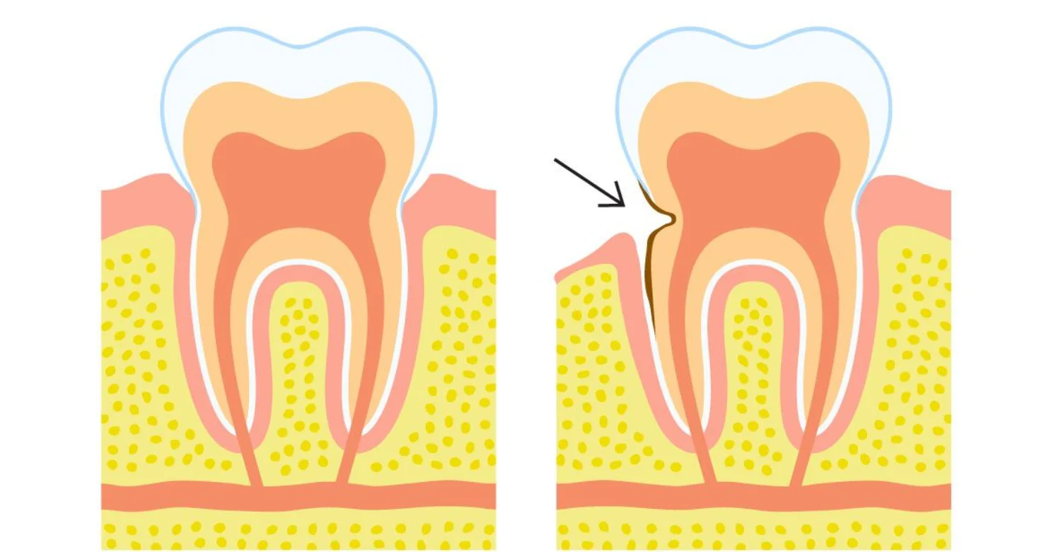
Abfraction condition is a non-carious cervical tooth-wear pattern often found near the gumline. It may appear as a notch, groove, or wedge-shaped defect on the tooth surface. Many patients first notice it as a small dip close to the gum. Its development is usually influenced by several mechanical, chemical, and behavioral factors working together over time. Treatment depends on symptoms, progression, tooth structure, function, and the underlying risk pattern. Some lesions need careful monitoring only, while others benefit from sensitivity care or restoration. This guide explains how these defects form and how dentists diagnose them. It also covers prevention, costs, warning signs, and the questions worth asking before treatment begins. The aim is to help patients make calm, informed decisions about their teeth.
What Is Abfraction?
This condition describes a non-carious cervical lesion involving loss of tooth structure near the cervical area. The term historically refers to a proposed stress-related mechanism involving tooth flexure under load. Modern literature generally considers cervical tooth wear multifactorial rather than purely mechanical. Abrasion, erosion, occlusal loading, anatomy, behavior, and tissue properties may interact on the same tooth. A dentist should therefore evaluate the entire wear pattern before assigning one specific cause. This approach helps avoid unnecessary treatment and improves prevention planning. It also helps patients understand which daily habits matter most for protecting their remaining tooth structure. In simple terms, the word describes where and how the tooth is wearing, not a single proven cause.
Abfraction as a Non-Carious Cervical Lesion
Non-carious cervical lesions involve mineralized tooth loss near the gingival margin. They occur without the bacterial decay process responsible for conventional cavities. These lesions can result from mechanical wear, chemical degradation, or combined mechanisms. The clinical appearance can vary considerably between patients and between individual teeth. Some defects remain shallow and stable for many years. Others become deeper, particularly when multiple contributing factors persist without change. Diagnosis should consider location, shape, surface texture, dentin exposure, and surrounding tissues. Within this broader group, this condition is the label linked to the stress-based theory of cervical tissue loss. Dentists often use the neutral term "non-carious cervical lesion" until the contributing factors are clearer.
Where Does Abfraction Occur on a Tooth?
These lesions commonly occur on buccal or facial surfaces near the cementoenamel junction. This junction is where the enamel crown meets the cementum covering the root. Premolars frequently show cervical wear patterns in clinical studies. However, similar defects can occur on canines, incisors, and molars. Distribution may reflect bite forces, brushing habits, acid exposure, anatomy, and gum recession. The exact location does not prove one specific mechanism. A dentist should compare affected teeth with unaffected teeth across both arches. Patterns across the mouth may reveal repeated mechanical or chemical influences. For example, lesions on one side may match the direction of a patient's dominant brushing hand.
Why Abfraction Often Appears Near the Gumline
The cervical region has anatomical and structural characteristics that make it clinically important. Enamel becomes thinner here and transitions toward dentin and cementum. These underlying tissues are softer and less resistant to wear than full-thickness enamel. Mechanical loading can concentrate stress in this region under certain conditions. Acid exposure can also soften these mineralized tissues. Brushing can then remove softened or weakened material more readily. This interaction may create defects that resemble classic wedge-shaped lesions. Therefore, location alone cannot establish a definitive cause for the defect.
What Does an Abfraction Lesion Look Like?
Cervical lesions can appear as grooves, notches, saucer-shaped defects, or angular depressions. Classic descriptions include sharply defined wedge-shaped defects with clear internal angles. However, systematic evidence shows substantial morphological variation between lesions. A 2024 review identified wedge-shaped and saucer-shaped patterns among reported presentations. Color changes may occur when dentin becomes exposed. Dentin can appear darker or more yellow than surrounding enamel. Surface texture can also differ depending on the dominant wear mechanisms. A glossy, smooth surface may suggest chemical involvement. Sharp, well-defined margins may suggest a stronger mechanical contribution. Dentists record these features but interpret them together with the patient's history.
Wedge-Shaped and Grooved Cervical Defects
A sharp internal angle can create a recognizable clinical appearance. Some lesions have clearly defined borders and relatively narrow openings. Others become broader and smoother as additional processes influence their shape. The final appearance does not necessarily identify the original cause. Mechanical wear may combine with acid-related tissue softening. Brushing can further modify the defect over months and years. A narrow groove today may become a wider, rounded defect later. Clinicians therefore describe the shape carefully but interpret it cautiously.
How Abfraction Tooth Damage Can Expose Dentin
Enamel loss can uncover the dentin beneath the cervical surface. Dentin contains microscopic tubules that communicate with the underlying pulp. External stimuli can therefore trigger short, sharp sensitivity. Cold drinks, touch, airflow, and sweet foods may become uncomfortable. The severity varies between people and between individual teeth. Exposed surfaces can also become more vulnerable to additional wear. Dentin wears faster than enamel, so progression may speed up once it is exposed. Protecting exposed dentin is therefore a practical goal of early management.
Is Abfraction the Same as a Cavity?
No, the two conditions are different. A cavity results from dental caries and bacterial biofilm activity. A cervical wear lesion is classified as non-carious when decay is absent. Their appearances can sometimes overlap near the gumline. Brown discoloration does not automatically mean decay. Stains from coffee, tea, or tobacco can also darken exposed dentin. A professional examination helps distinguish caries from non-carious tooth wear. Radiographs may assist when decay extends below visible surfaces. Clinical examination, including gentle probing and visual assessment, remains central to diagnosis. Patients who confuse cervical wear with a cavity may worry unnecessarily or delay proper care.
Is the Abfraction Theory Proven?
The classic stress-based theory remains scientifically debated. It proposes that repeated loading bends teeth and concentrates stress near the cervical area. That stress could contribute to microscopic structural damage and subsequent tissue loss. However, evidence does not establish stress as the sole cause. One systematic review included 69 publications examining this relationship. About 81% reported an association between occlusal stress and cervical lesions. Yet no clinical study demonstrated stress alone as the cause. This gap explains why many researchers now prefer broader terms for these defects. The theory remains useful as one possible explanation within a wider clinical assessment.
What Research Says About Tooth Flexure and Occlusal Stress
Laboratory studies can demonstrate cervical stress concentration under controlled loading. Finite-element studies have frequently identified stress concentrations near the cervical region. Clinical evidence is more complex because patients experience many simultaneous exposures. A systematic review found inconsistent clinical associations with occlusal factors. Study designs and diagnostic criteria varied substantially between publications. The authors concluded that causation remained uncertain. Statistics and insight: 56 of 69 reviewed publications reported an association with occlusal stress. However, association does not establish a direct cause-and-effect relationship. Future clinical research with consistent definitions may clarify the true role of tooth flexure.
Why Modern Literature Describes Cervical Tooth Wear as Multifactorial
Current literature increasingly emphasizes interactions between mechanical and chemical influences. Brushing force, acidic exposure, occlusal loading, and individual anatomy can overlap. Behavioral habits can change the intensity and timing of these exposures. A 2024 systematic review also reported wide morphological variation. Its included studies mainly supported multifactorial explanations. Therefore, treatment planning should identify contributing factors rather than rely on appearance alone. In practice, stress-related wear is best viewed as one possible piece of a larger wear picture. This view encourages prevention plans that cover diet, brushing, and bite at the same time.
What Causes Abfraction?
The causes of cervical tooth wear are rarely explained by one factor. Mechanical loading may contribute to some patients. Chemical exposure and brushing may also influence progression. Individual tooth structure and behavioral patterns can modify susceptibility. Saliva quality, diet, and gum position add further layers of variation. Understanding these influences helps a dentist build a realistic prevention plan. The sections below look at each contributor and how it may interact with the others.
Abfraction Causes and the Role of Mechanical Stress
Mechanical stress may contribute to cervical tissue loss in susceptible teeth. Repeated loading can produce stress concentration around structurally vulnerable regions. The effect probably depends on the magnitude, direction, frequency, and duration of force. However, mechanical stress should not automatically be considered the primary cause. Clinical evidence remains mixed and affected by confounding factors. Researchers still debate how much this wear depends on biting forces alone. A careful history helps separate mechanical influences from chemical and behavioral ones.
Occlusal Forces, Tooth Flexure, and Cervical Stress
Teeth can experience different forces during chewing and parafunctional activity. Lateral forces may produce different stress patterns than axial forces. Some models suggest that cervical regions experience increased stress during loading. Clinical interpretation remains more complicated than laboratory simulations. Patients have different diets, saliva, tooth shapes, brushing habits, and periodontal conditions. These variables can influence observed lesions independently or together. A model can show where stress concentrates, but not everything happening inside a real mouth.
Bruxism and Repeated Tooth Loading
Bruxism involves repeated grinding or clenching during wakefulness or sleep. It can increase mechanical loading throughout the dentition. The presence of bruxism does not prove that it caused a cervical lesion. Dentists may look for wear facets, muscle tenderness, tooth mobility, or reported grinding. A night guard may be considered when clinically appropriate. Management should address symptoms and overall occlusal risk. Stress, sleep quality, and certain medications may also influence grinding behavior.
Malocclusion and Uneven Bite Forces
Uneven contacts can alter how force is distributed across teeth. Some clinicians associate heavy contacts with cervical wear patterns. Scientific evidence, however, does not establish a universal causal relationship. Orthodontic treatment should therefore have a clear clinical indication. Treatment should not be prescribed solely because cervical defects exist. A careful bite assessment helps decide whether contacts deserve attention at all.
How Abrasion and Erosion Can Occur Alongside Abfraction
Mechanical and chemical processes can operate simultaneously. Acid exposure may soften dental surfaces before brushing contacts them. Brushing can then contribute to additional material loss. This interaction can blur the clinical distinction between wear categories. Modern reviews describe combined mechanisms as common considerations. Treatment should target the dominant modifiable risk factors. A lesion that started with stress-related wear may later be shaped by abrasion and erosion. This is one reason older lesions often look different from newly formed ones.
Causes of Dental Abfraction: What Can Increase Risk?
Risk can increase when several exposures occur repeatedly. Potential contributors include heavy loading, aggressive brushing, acidic diets, and exposed roots. Grinding and clenching may increase mechanical forces. Reduced salivary protection may also increase vulnerability to acidic challenges. Dry mouth from medications is one example of a risk that patients often overlook. Existing tooth wear can change local stress distribution. A patient may therefore have several interacting risk factors simultaneously. Listing these factors honestly helps the dentist tailor advice to real daily habits.
Aggressive or Improper Tooth Brushing
Excessive pressure can contribute to cervical tooth wear. Hard bristles may increase mechanical abrasion on vulnerable surfaces. Incorrect technique can also repeatedly traumatize exposed root surfaces. Highly abrasive toothpaste may add to the problem over many years. A soft-bristled brush and gentle technique are generally preferable. Brushing twice daily remains important for plaque control. The goal is to clean thoroughly without scrubbing the gumline.
Acid Exposure From Food, Drinks, or Gastric Sources
Acids can chemically soften dental mineral surfaces. Common dietary sources include acidic beverages, citrus products, and frequent sour foods. Sports drinks, carbonated drinks, and some flavored waters also lower oral pH. Gastric acid can also expose teeth during recurrent reflux or vomiting. The overall frequency and duration of exposure matter. Rinsing with water can reduce prolonged acid contact. Aggressive brushing immediately after acid exposure should be avoided. Patients with reflux symptoms may benefit from medical review alongside dental care.
Tooth Grinding and Clenching
Repeated clenching can expose teeth to substantial mechanical loading. Grinding may additionally create visible wear facets on biting surfaces. Some patients remain unaware of nocturnal activity. A partner may notice grinding sounds during sleep. Morning jaw stiffness or headaches can be further clues. A dentist may assess symptoms, wear patterns, and muscle findings. Protection may be considered when bruxism creates clinically meaningful risk.
Existing Tooth-Wear Patterns
Existing wear can indicate repeated exposure to mechanical or chemical stresses. Flattened biting surfaces may suggest attrition. Cupped areas on molars may suggest acid involvement. Cervical defects may occur alongside these changes. The complete pattern can provide more information than one isolated lesion. Photographs and periodic measurements can support monitoring. Comparing records over time shows whether wear is active or stable.
Abfraction Symptoms and Signs
Symptoms depend on lesion depth, dentin exposure, and individual sensitivity. Some people notice visible notches without experiencing pain. Others experience sharp sensitivity during everyday activities. These signs can also overlap with other dental problems. A careful examination separates expected findings from symptoms that need further investigation.
Common Abfraction Symptoms
Common findings include cervical grooves, notches, discoloration, and dentin exposure. Sensitivity may occur when cold, heat, touch, or airflow contacts exposed dentin. Some patients notice the toothbrush catching on a small edge near the gum. Some lesions remain completely asymptomatic. Symptoms alone cannot establish the diagnosis. A dentist must rule out decay, cracks, pulp problems, and other causes.
What Abfraction Near the Gumline May Look and Feel Like
A cervical defect may appear close to the gingival margin. It can feel rough, indented, or sensitive when touched with a fingernail or tongue. The visible shape may be narrow or broad. Gum recession can make the defect easier to see. Exposed root surfaces may also contribute to sensitivity. Food and plaque may collect inside deeper notches. Clinical evaluation determines whether the defect is actively progressing.
Tooth Sensitivity From Abfraction
Sensitivity commonly occurs when dentin becomes exposed. Dentin exposure allows external stimuli to affect structures associated with sensory signaling. Cold air and drinks are common triggers. Modern reviews support several desensitizing toothpaste ingredients. These include potassium-based, stannous, and arginine-containing formulations. Consistent daily use usually matters more than the specific brand chosen. When cervical wear exposes dentin, sensitivity care often becomes the first practical step.
Sensitivity to Cold, Heat, Sweet Foods, or Touch
Short, sharp discomfort is typical of dentin hypersensitivity. It usually occurs immediately after a triggering stimulus and fades quickly. Persistent spontaneous pain deserves further investigation. Pain during biting may suggest another dental problem. Lingering pain after hot drinks may indicate pulp involvement. A dentist should investigate symptoms that do not behave like ordinary sensitivity.
Why Exposed Dentin Can Trigger Sensitivity
Dentin contains microscopic tubules extending toward the pulp. Fluid movement within these tubules can stimulate sensory responses. Temperature and touch can therefore produce brief pain. The response varies according to tubule exposure and individual sensitivity. Open tubules usually cause more discomfort than tubules blocked by natural mineral deposits. Other conditions can produce similar symptoms. Diagnosis remains important before assuming cervical wear is responsible.
Can Abfraction Cause Pain Without a Cavity?
Yes, cervical tooth wear can produce pain without dental caries. Exposed dentin may react strongly to cold or touch. However, pain can have many dental causes. A cracked tooth, pulp inflammation, recession, or decay may produce similar complaints. Professional assessment is important when symptoms persist or worsen. Pain from cervical wear is usually brief, so lasting pain often points elsewhere.
Can Abfraction Be Present Without Symptoms?
Yes, many cervical defects cause no noticeable symptoms. A dentist may discover them during routine examination. Asymptomatic lesions may be suitable for observation. Monitoring can help determine whether the defect is stable or progressing. Intervention is not automatically required for every visible defect. Natural sclerosis of dentin may explain why some deep lesions remain painless. In sclerotic dentin, mineral deposits gradually narrow or close the tubules.
Abfraction Tooth Damage: Early Versus More Advanced Changes
Early lesions may appear as shallow grooves or small surface changes. More advanced defects may show deeper structural loss and obvious dentin exposure. Deep lesions can create functional and restorative challenges. They may also trap plaque and complicate cleaning. Research supports careful monitoring when symptoms and function remain satisfactory. One clinical review recommends observation before invasive care for suitable asymptomatic lesions. Identifying these lesions early gives patients more conservative options.
Abfraction
Other Types of Tooth Wear
Cervical tooth wear can have overlapping clinical appearances. Accurate terminology requires consideration of both morphology and contributing factors. No single appearance always identifies the exact mechanism. Understanding the main wear categories helps patients follow targeted prevention advice.
Abfraction vs Abrasion
Abrasion refers to mechanical tooth wear caused by external contact. Toothbrushing is one commonly discussed contributor. The distribution often reflects the location and direction of repeated contact. Cervical lesions may involve abrasion together with other mechanisms. Therefore, the terms should not be treated as mutually exclusive. Abrasive toothpaste, toothpicks, and pipe stems are other possible sources. Unlike abrasion, stress-related wear is linked mainly to forces acting inside the tooth.
Differences in Location, Appearance, and Contributing Factors
Abrasion often shows surface changes associated with repeated external friction. Chemical exposure can produce smoother and broader defects. Mechanical loading may contribute to angular cervical changes. A lesion on one tooth may suggest a local factor, such as a heavy contact. Widespread lesions may suggest habits affecting the whole mouth. Dentists evaluate shape, location, surface texture, and patient behavior together. This approach is more reliable than visual classification alone.
Abfraction vs Erosion
Erosion refers to chemical loss caused by acids. External dietary acids and internal gastric acids can contribute. No bacterial process is required for this type of wear. Chemical softening may make teeth more vulnerable to subsequent mechanical forces. Mixed lesions can therefore display characteristics of several mechanisms. Erosion often affects broad surfaces, while stress-related lesions are described as more localized.
Chemical Tooth Wear Versus Mechanical Factors
Chemical wear can smooth dental surfaces and reduce surface hardness. Mechanical brushing may then remove softened material. Occlusal loading may influence stress distribution at the same time. The combined process explains why clinical categories can overlap. Modern literature emphasizes multifactorial evaluation for these lesions. A short dietary diary can help reveal hidden acid exposures during the day.
Abfraction vs Tooth Abrasion
Tooth abrasion specifically emphasizes external mechanical contact. Common examples include abrasive brushing habits and repeated contact with objects. Holding pens, pins, or nails between the teeth can also cause localized wear. Clinical history can reveal relevant behaviors. A dentist may ask about brushing force, tools, habits, and occupational exposures. These answers help separate habit-related abrasion from load-related cervical wear.
Abfraction vs Tooth Erosion
Tooth erosion primarily involves chemical dissolution from acids. It may affect multiple tooth surfaces, depending on exposure patterns. The surface commonly appears smooth rather than sharply notched. However, mixed mechanisms can alter the final appearance. Restorations may appear to stand above the surrounding eroded tooth surface. Careful history talking about diet and reflux remains essential.
Can Abfraction, Abrasion, and Erosion Happen Together?
Yes, multiple processes can affect the same tooth. Acids may soften tissue before brushing contributes to material loss. Mechanical loading may further influence local stress. This interaction supports a risk-based approach to diagnosis. Treating only one exposure may fail to control progression. Most clinicians now accept that stress-related wear rarely acts alone.
Who Is More Likely to Develop Abfraction Lesions?
Cervical lesions may occur across different age groups. Risk depends more on cumulative exposure than on age alone. Bruxism, brushing habits, acid exposure, recession, and existing tooth wear can contribute. Patients who combine several of these factors may face higher overall risk. Identifying vulnerable patients early allows dentists to focus prevention advice where it matters most.
Bruxism and Clenching
Patients who grind or clench may experience increased tooth loading. Wear facets may provide supporting clinical evidence. However, the presence of grinding does not prove causation. Management should consider symptoms, tooth damage, muscle findings, and overall risk. Jaw muscle tension and morning headaches can be additional clues. Some studies link bruxism with cervical lesions, but the connection remains inconsistent.
Patients With Significant Occlusal Stress
Some patients experience strong or repetitive bite forces. Certain loading patterns may increase cervical stress. Evidence remains insufficient for assuming occlusion is always the dominant factor. Bite findings should be combined with the rest of the clinical history.
Patients With Gum Recession and Exposed Root Surfaces
Gum recession exposes cementum and root dentin. These tissues are less resistant to mechanical and acidic challenges. Sensitivity may become more pronounced after root exposure. A combined periodontal and restorative assessment may be appropriate. Recession can result from periodontal disease, thin gum tissue, or traumatic brushing.
Dietary and Oral-Hygiene Behaviors That May Add to Cervical Wear
Frequent acidic drinks can repeatedly challenge tooth minerals. Sipping slowly throughout the day extends acid contact time. Aggressive brushing can add mechanical stress. Whitening products may aggravate sensitivity in susceptible patients. The goal is balanced prevention rather than reduced oral hygiene. Good plaque control remains essential.
Why Age Alone Does Not Explain Every Cervical Lesion
Age reflects cumulative exposure, but it does not identify the cause. Older teeth may simply have experienced more years of wear. Younger patients can also develop cervical defects when risk factors are strong. A personalized assessment remains more informative than age alone. Athletes who sip sports drinks frequently are one example of a younger risk group.
How Is Abfraction Diagnosed?
Diagnosis begins with a clinical examination. The dentist evaluates lesion shape, depth, location, dentin exposure, and surrounding tissues. The bite and tooth contacts may also be assessed. A detailed history of diet, brushing, and habits completes the picture. Diagnosing these lesions is largely a process of weighing several possible contributors.
What a Dentist Looks for During a Clinical Examination
The dentist examines the affected tooth under appropriate lighting and magnification. Surface texture and lesion margins can provide useful clues. Other teeth are compared to identify broader wear patterns. Medical and dental history can reveal behavioral or dietary contributors. Medications that cause dry mouth may also be noted during history. Signs of stress-related wear are then weighed against signs of abrasion and erosion.
Assessing Lesion Shape, Depth, Location, and Surface Characteristics
Dentists may record approximate lesion dimensions during monitoring. Sharp angles, smooth surfaces, and irregular borders provide descriptive information. No single feature proves a specific etiology. Repeated photographs can help document changes over time. Digital scans may also allow precise comparison between visits.
Checking for Dentin Exposure and Dentin Hypersensitivity
The clinician checks whether dentin is visible or accessible. Air, touch, or thermal tests may help reproduce symptoms. The response should be compared with neighboring teeth. Other painful conditions must remain part of the differential diagnosis. A brief response suggests dentin hypersensitivity, while lingering pain needs further investigation.
Evaluating Bite, Tooth Contact, and Possible Bruxism
Occlusal contacts can be examined during routine dental assessment. The dentist may also ask about daytime or nighttime clenching. Wear facets and muscle symptoms can support clinical interpretation. Occlusal findings should be interpreted cautiously because evidence remains mixed. A bite finding is a useful clue, not automatic proof of cause.
Checking for Gingival Recession and Other Periodontal Findings
Recession can expose root surfaces and increase sensitivity. Bleeding, inflammation, attachment changes, and plaque accumulation may also need evaluation. Periodontal findings can influence restorative and preventive planning. Complex recession may require periodontal treatment alongside restorative care.
When Dental X-Rays May Be Useful
Radiographs can help investigate suspected decay or other internal problems. They may also assess deeper structural changes when clinically justified. Routine radiography is not required for every superficial cervical lesion. The dentist should choose imaging according to clinical need. Previous radiographs can sometimes be reused to avoid unnecessary exposure.
When Additional Imaging Is Clinically Appropriate
Advanced imaging may be useful when conventional assessment leaves important questions unanswered. It can help evaluate complex anatomy or suspected structural problems. CBCT should not be performed automatically. Radiation exposure and diagnostic benefits should always be considered. Most cervical wear cases never need three-dimensional imaging.
Why Diagnosis Must Rule Out Caries and Other Causes of Tooth Damage
Cervical discoloration can resemble decay. Cracks can also create localized sensitivity and visible structural defects. Root caries may occur near areas affected by recession. Correct diagnosis prevents inappropriate or delayed treatment. Labeling decay as simple wear could allow an active infection to progress.
Abfraction Versus Root Caries
Root caries involve bacterial demineralization and cavitation. It commonly affects exposed root surfaces. The lesion may feel softer than a non-carious wear defect. Clinical examination and radiographic assessment can assist when diagnosis is uncertain. Caries often appear matte, with plaque collecting on the surface. By contrast, a non-carious wear lesion usually feels hard and smooth when gently probed.
Abfraction Versus Cracked or Fractured Tooth Structure
Cracks can cause pain during chewing or release. Their patterns differ from ordinary superficial cervical wear. Pain on biting deserves particular attention. A cracked tooth can require different treatment. Do not assume every cervical defect explains biting pain. Magnification, transillumination, and bite tests can help locate cracks.
Abfraction Versus Cervical Wear From Brushing or Acid Exposure
Brushing-related wear emphasizes mechanical abrasion. Acid-related wear emphasizes chemical surface dissolution. Many patients have overlapping mechanisms. Therefore, clinicians should identify the dominant contributors rather than force a single label. The prevention plan then targets the habits that matter most for each patient.
Dr. Rifat Alsaman : Why identifying the contributing factors matters before deciding to restore a cervical lesion
Dr. Rifat Alsaman Clinical Note: A restoration can repair lost structure. However, it does not automatically remove the forces or behaviors contributing to the defect. Assessment should therefore consider symptoms, bite, brushing, acid exposure, gum position, and tooth structure. This helps make treatment more targeted and conservative. A filling placed without addressing these risks may simply delay the next problem.
Abfraction Treatment: Does Every Lesion Need Treatment?
No, every cervical lesion does not require immediate restoration. Treatment depends on symptoms, progression, function, structural risk, and patient preferences. Stable and asymptomatic defects may require monitoring only. A major clinical review emphasizes individualized decision-making. This conservative approach preserves natural tooth structure for as long as possible. Many patients with cervical wear never need a filling at all.
When Monitoring May Be More Appropriate Than Immediate Restoration
Monitoring may suit shallow, asymptomatic, and stable defects. The dentist can record photographs and lesion dimensions. Periodic review can reveal whether progression is occurring. Avoiding unnecessary intervention can preserve remaining tooth structure. Every restoration eventually needs maintenance or replacement. Delaying treatment, when safe, can therefore reduce long-term restorative cycles.
When Abfraction Treatment May Be Recommended
Treatment may be considered when sensitivity persists despite conservative measures. Significant structure loss may also justify restoration. Functional concerns or esthetic concerns can influence treatment decisions. The treatment plan should match the clinical problem. Plaque retention inside deep notches is another practical reason to restore.
Persistent Tooth Sensitivity
Persistent sensitivity can interfere with eating and oral hygiene. Desensitizing measures may provide adequate relief for some patients. Restoration may be considered when conservative measures fail. The cause of sensitivity should still be confirmed first. Some patients avoid brushing sensitive areas, which increases plaque and decay risk.
Significant Tooth Structure Loss
Deep defects can weaken remaining tooth structure. They may also create plaque-retentive areas or esthetic concerns. Restorative treatment can recreate a smoother contour when appropriate. The clinician must assess whether restoration provides meaningful benefit. Very deep notches may gradually approach the pulp over time.
Functional Problems
Some lesions interfere with comfort during chewing or brushing. Sharp edges may irritate the tongue or surrounding soft tissues. Functional symptoms can support treatment consideration. Food impaction within a deep notch can also be bothersome.
Esthetic Concerns
Visible cervical defects can alter tooth contours. Color differences may become noticeable when dentin is exposed. Cosmetic restoration can improve appearance when clinically suitable. Patient expectations should be discussed before treatment. Front teeth and smile-zone premolars often attract the most concern.
How to Treat Abfraction Lesions
Management ranges from prevention and monitoring to restorative intervention. Sensitivity control can be attempted before invasive treatment. Contributing behaviors should be addressed simultaneously. Restorative selection depends on tooth location, lesion characteristics, moisture control, and esthetic requirements. For cervical lesions, the best results usually combine careful repair with risk control.
Desensitizing Toothpaste and Fluoride-Based Measures
Desensitizing toothpaste can help reduce dentin sensitivity. Common active ingredients include potassium salts, stannous compounds, and arginine. Evidence supports twice-daily use of several formulations. Fluoride toothpaste also supports general caries prevention. These measures do not physically replace missing tooth structure. Spitting without rinsing may leave more active ingredients on the teeth.
Fluoride or Other Professional Desensitizing Treatments
Dentists can provide stronger professional sensitivity-management options. The choice depends on the sensitivity mechanism and clinical findings. Professional measures may be useful when home products provide insufficient relief. Persistent sensitivity still requires diagnostic evaluation. Examples include fluoride varnish, sealing agents, and thin bonding resins.
Composite Resin Restoration and Cervical Bonding
Composite resin can recreate lost cervical contour. It can also cover exposed dentin and improve appearance. Adhesion can be challenging because cervical dentin may be sclerotic. Recent research continues to examine adhesive strategies and retention. Careful isolation and selective enamel etching may improve bonding performance. Shade matching allows the restoration to blend with natural tooth color.
Glass Ionomer or Other Restorative Materials
Glass ionomer materials can also restore suitable cervical defects. Material selection depends on moisture control, location, function, and clinical goals. A 2025 meta-analysis found no significant clinical performance difference between composite and glass ionomer. Individual clinical factors still determine the most suitable material. Glass ionomer releases fluoride and bonds chemically to tooth structure.
Managing Contributing Occlusal or Bruxism-Related Factors
Potential mechanical contributors should be assessed rather than assumed. Bruxism management may include behavioral strategies and protective appliances. Routine irreversible bite adjustment should not be performed automatically. Evidence does not support universal occlusal intervention for cervical lesions. Reversible measures are usually preferred before any permanent change.
Periodontal or Soft-Tissue Treatment When Gingival Recession Is Involved
Gingival recession can expose vulnerable root surfaces. Periodontal treatment may therefore form part of comprehensive care. Some cases combine restorative treatment with periodontal procedures. Evidence suggests restoration can reduce sensitivity without necessarily increasing root coverage. Gum grafting may cover part of the defect in selected patients.
Is Occlusal Adjustment Necessary for Abfraction?
Occlusal adjustment is not automatically required. Current evidence does not support routine irreversible adjustment for every cervical lesion. Treatment should have a clear indication supported by clinical findings. Unnecessary adjustment can permanently alter tooth contacts. Patients should ask why adjustment is proposed and what it aims to achieve.
When a Night Guard May Be Considered
A night guard may be considered when clinically significant grinding is suspected. It can help protect teeth from repeated nocturnal loading. The device should be properly evaluated and fitted. It does not correct every cause of cervical wear. Over-the-counter guards may not fit as accurately as custom appliances. A guard protects teeth from grinding but does not reverse existing wear.
When Orthodontic Treatment May Be Relevant
Orthodontics may be relevant when malposition contributes to a broader functional problem. Treatment should have an established orthodontic indication. Cervical wear alone does not justify orthodontic treatment. Diagnosis should consider overall bite, function, aesthetics, and periodontal health.
Why Treatment Should Address the Whole Pattern of Tooth Wear
Treating a single lesion without addressing repeated exposures may provide incomplete management. Acid exposure, brushing habits, clenching, and recession can continue afterward. A whole-mouth assessment can identify patterns across multiple teeth. This supports prevention alongside restorative care. It also helps predict which teeth may develop lesions next.
Clinical Note : Choosing treatment based on symptoms, tooth structure, function, and contributing factors
Treatment decisions should balance conservative care with meaningful clinical benefit. Small stable lesions may need observation only. Sensitive or structurally compromised teeth may benefit from restoration. The objective should be controlled disease management rather than automatic filling.
Relief for Tooth Sensitivity From Abfraction
Sensitivity management begins with identifying the cause. Dentin exposure is common, but other conditions can mimic sensitivity. Treatment therefore combines diagnosis, prevention, and symptom control. Sensitivity linked to cervical wear often improves with simple, consistent daily measures.
What Can Relieve Abfraction-Related Sensitivity?
Sensitive toothpaste can help some patients reduce painful responses. Gentle brushing can limit additional mechanical irritation. Professional desensitizing treatments may help persistent symptoms. Restoration can also cover exposed dentin when clinically indicated.
Short-Term Relief While Waiting for a Dental Appointment
Avoid known triggers when symptoms are severe. Choose lukewarm foods and beverages temporarily. Use a soft toothbrush and gentle pressure. Do not stop brushing completely. Plaque accumulation can create additional oral-health problems. Applying a small amount of desensitizing toothpaste to the area may help.
Desensitizing Toothpaste and Fluoride Measures
Use a fluoride toothpaste consistently unless your dentist recommends otherwise. Desensitizing ingredients can reduce sensitivity with repeated use. Potassium nitrate, stannous compounds, and arginine have supporting evidence. Improvement can take time rather than occurring after one application. Many products need two to four weeks of regular use before relief is noticeable.
Foods, Drinks, and Habits to Limit When Sensitivity Is Active
Reduce frequent exposure to acidic foods and drinks. Water can help reduce prolonged acid contact. Avoid habits that repeatedly expose sensitive surfaces to extreme temperatures. The goal is to reduce triggers without unnecessarily restricting a balanced diet. Using a straw may limit contact between acidic drinks and front teeth.
When Sensitivity Relief Is Not Enough and the Tooth Needs Evaluation
Professional evaluation is important when sensitivity continues or worsens. Persistent symptoms may indicate deeper tooth damage or another diagnosis. Pain without an obvious trigger also deserves assessment. Biting pain requires additional attention because cracks may present differently.
How to Brush Without Increasing Mechanical Wear
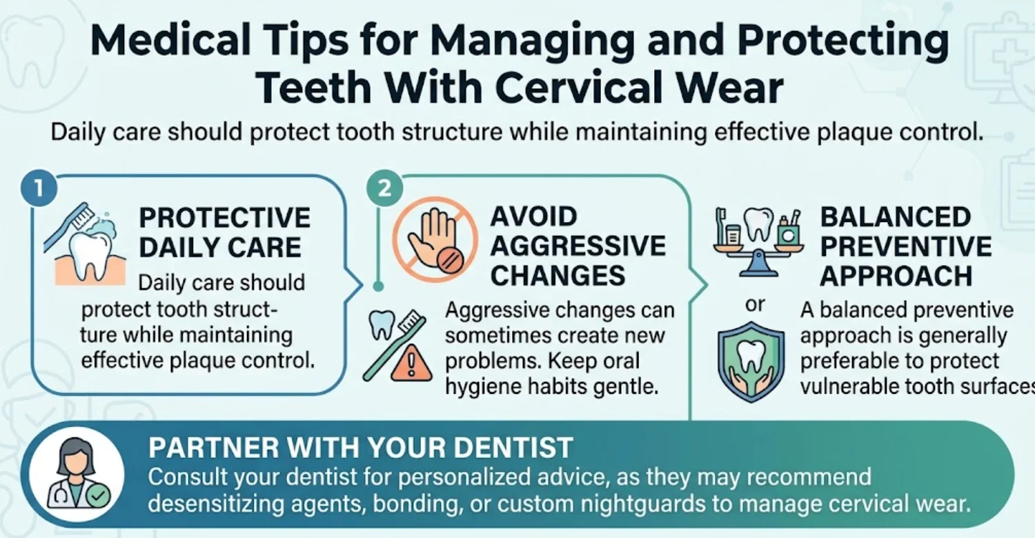
Use gentle pressure with controlled short movements. Avoid scrubbing aggressively across exposed cervical surfaces. Brush twice daily with fluoride toothpaste. The ADA also recommends soft bristles and gentler brushing techniques for sensitivity. Angle the bristles toward the gumline and use small, gentle circular motions. Brushing this way also protects any area affected by cervical wear.
Choosing a Soft-Bristled Toothbrush and Appropriate Toothpaste
Soft bristles can reduce unnecessary mechanical trauma. A fluoride toothpaste should remain part of regular oral hygiene. Sensitivity toothpaste may be appropriate when dentin hypersensitivity is present. Avoid excessive abrasive whitening products when sensitivity is active. Replace worn toothbrushes about every three months.
How to Reduce Acid Exposure Without Overcorrecting Oral Hygiene
Limit frequent acidic drinks throughout the day. Rinse with water after acidic exposure. Avoid immediately brushing after strong acid exposure. Continue regular brushing because plaque control remains essential. Waiting a short period allows saliva to begin buffering the acid.
What to Discuss With Your Dentist if You Grind or Clench
Tell your dentist about jaw tension, morning soreness, or suspected grinding. Mention any partner observations about nighttime activity. Your dentist can assess wear patterns and functional symptoms. A protective appliance may be considered when appropriate.
Why Self-Filing, Aggressive Polishing, or Home Repairs Should Be Avoided
Home filing can permanently remove healthy tooth structure. Aggressive polishing can also increase surface wear. Unsupervised filling materials may fail or irritate surrounding tissues. Professional diagnosis should come before structural modification.
Potential Treatment Side Effects and Limitations
Every treatment has benefits, limitations, and possible complications. Cervical restorations can be technically demanding. Long-term success depends on material, technique, lesion characteristics, and ongoing risk factors.
Temporary Sensitivity After Cervical Restorations
Some patients experience temporary sensitivity after adhesive procedures. This may resolve as tissues recover. Persistent or worsening symptoms need clinical assessment. The dentist should consider marginal leakage, pulp status, occlusion, and other causes. Mild sensitivity often settles within a few days or weeks.
Restoration Wear, Staining, Chipping, or Debonding
Cervical restorations can lose retention or develop marginal changes. Material selection and adhesive technique influence performance. Moisture control can be challenging near the gingival margin. Recent studies continue evaluating methods for improving restoration retention. Regular checkups allow early repair of small defects.
Why Restoring the Defect Does Not Automatically Remove Its Underlying Cause
A restoration replaces a missing structure but does not change every risk factor. Acid exposure, brushing pressure, and grinding can continue afterward. Long-term management therefore requires prevention and monitoring. This principle is central to conservative treatment planning. Fillings placed over active wear may therefore loosen sooner.
Why Treatment Choice Depends on Lesion Characteristics and Individual Risk Factors
No single restoration works identically for every cervical defect. Depth, width, moisture control, dentin sclerosis, symptoms, and aesthetics matter. Patient habits can also affect maintenance and longevity. Personalized treatment is therefore more appropriate than a universal protocol.
Note : All images used are for editorial and illustrative purposes only and may not originate from the original news provider or associated company.
How to Prevent Abfraction and Further Cervical Tooth Damage
Prevention focuses on reducing modifiable mechanical and chemical exposures. Protective habits should begin before substantial structural loss occurs. Existing lesions should also be monitored over time. Preventing lesion progression is usually simpler than repairing advanced damage.
How to Prevent Tooth Abfraction Through Safer Brushing Habits
Use a soft-bristled toothbrush with gentle pressure. Avoid forceful horizontal scrubbing at the gumline. Maintain twice-daily fluoride brushing for plaque and decay prevention. Hold the brush lightly, almost like a pen, to limit force.
How to Prevent Abfraction Tooth Damage From Acid Exposure
Reduce frequent acidic drinks and prolonged sipping. Drink water after acidic foods or beverages. Avoid brushing immediately after substantial acid exposure. This strategy reduces chemical and mechanical challenges simultaneously. Chewing sugar-free gum can also stimulate protective saliva.
Managing Teeth Grinding and Clenching
Discuss persistent clenching or grinding with your dentist. Stress management may help some patients reduce daytime clenching. A dentist may consider a protective appliance where indicated. Management should focus on overall tooth protection. Simple reminders to keep teeth apart during the day can help.
Protecting Exposed Dentin and Root Surfaces
Gentle brushing helps reduce additional mechanical wear. Fluoride toothpaste supports general enamel protection. Desensitizing products can help manage exposed dentin sensitivity. Your dentist can recommend additional protection when root surfaces are significantly exposed.
Regular Dental Monitoring for Existing Cervical Lesions
Regular examinations help identify progression before significant structural loss occurs. Photographs and measurements can support comparison between visits. Stable lesions may continue under observation. Progressive lesions may require preventive or restorative intervention. Monitoring lesions this way helps avoid both overtreatment and neglect.
Preventing Combined Abrasion, Erosion, and Mechanical Wear
Prevention should address every relevant exposure. Reducing only brushing pressure may not solve frequent acid exposure. Likewise, reducing acidic intake may not address severe clenching. A combined approach can better control the overall wear pattern.
When Should You See a Dentist for an Abfraction Lesion?
Any new cervical defect deserves professional assessment when its cause is uncertain. Early evaluation can identify decay, cracks, recession, or other conditions. Routine checkups are a good time to mention new notches or sensitivity.
Warning Signs That Need Professional Evaluation
Increasing sensitivity, visible progression, biting pain, and significant structural loss require assessment. Swelling or unusual gum changes also deserve professional attention.
Increasing Sensitivity or Pain
Increasing sensitivity may indicate ongoing dentin exposure. Persistent pain may also indicate another dental problem. A dentist should evaluate changes rather than relying on home treatment alone.
A Lesion That Appears to Be Getting Deeper
Visible progression can indicate ongoing structural loss. Periodic measurement helps confirm whether changes are occurring. Deepening lesions deserve assessment even when discomfort remains mild. A defect that progresses quickly may signal an uncontrolled risk factor.
A Tooth That Hurts When Biting
Pain during biting can suggest a crack or other structural problem. This symptom should not automatically be attributed to cervical wear. Prompt examination can help prevent delayed diagnosis. Untreated cracks can spread and may eventually split the tooth.
Visible Cracks, Fractures, or Major Tooth Structure Loss
Major structural changes may compromise tooth strength. A dentist should determine whether restorative or other treatment is required.
Swelling, Bleeding, or Other Concerning Oral Changes
Swelling can indicate infection or another significant oral condition. Persistent bleeding can also indicate periodontal disease. These findings require professional evaluation rather than self-treatment. Fever with facial swelling needs urgent care.
When a Cervical Defect May Need Urgent Dental Assessment
Urgent evaluation is appropriate for severe pain or rapidly worsening symptoms. Swelling, trauma, uncontrolled bleeding, or suspected tooth fracture also warrants prompt attention. The specific urgency depends on symptoms and clinical findings.
How Much Does Abfraction Treatment Cost in ?
US
UK
Turkey
Prices vary according to diagnosis, materials, number of teeth, and additional procedures. Cervical defects may need monitoring instead of restoration. Therefore, filling prices should only serve as planning benchmarks.
Average Abfraction Treatment Cost by Country
Country | Indicative Cost Per Tooth | Treatment Reference |
United States | Around $250 | Composite filling planning benchmark |
United Kingdom | Around $150 | Private composite filling planning benchmark |
Turkey | Around $97 | Composite filling planning benchmark |
NHS England | $76.60 | Band 2 course-of-treatment charge, including fillings |
These figures are indicative planning benchmarks, not fixed prices or official national averages. Actual costs vary by clinic, diagnosis, materials, and treatment complexity.
United States: Around $250 Per Tooth as a Composite-Filling Planning Benchmark
A planning benchmark of about $250 per tooth can be used for composite care. Actual prices vary substantially between locations and dental practices. Complex lesions may require additional diagnostic or periodontal care. Treatment should be quoted after clinical examination.
United Kingdom: Around $150 Per Tooth for Private Composite Filling Care
Private composite care can be planned around $150 per tooth as a broad reference. Private dental prices vary according to region and practice. Material selection and appointment complexity can also change the final fee.
NHS England Band 2: $76.60 for a Course of Treatment Including Fillings
From April 2026, NHS England Band 2 costs $76.60. It covers Band 1 services plus treatments such as fillings. The charge applies per course of treatment, not per filling.
Turkey: Around $97 Per Tooth for Composite Filling Care
A broad planning benchmark can be placed around $97 per tooth. Actual private-clinic pricing varies by dentist, material, and treatment complexity. International patients should confirm inclusions before booking.
Average Cost Depending on the Treatment Approach
Treatment Approach | Relative Cost | Typical Purpose |
Monitoring | Lowest | Small or stable lesions |
Desensitizing treatment | Low | Sensitivity management |
Composite restoration | Moderate | Repairing suitable cervical defects |
Additional periodontal care | Higher | When gum or supporting-tissue problems require treatment |
Multiple restorations | Higher | Several affected teeth or extensive tooth-structure loss |
Why There Is No Single Standard Price for Abfraction Treatment
Not every lesion needs restoration. Some patients require only monitoring or sensitivity management, while others need composite restoration or periodontal treatment. The final price therefore depends on the individual clinical plan.
Monitoring Versus Desensitizing Treatment Versus Composite Restoration
Monitoring can involve fewer procedures than restorative care. Desensitizing treatment may require products or professional applications. Composite restoration adds preparation, bonding, and finishing, resulting in different costs.
Number of Teeth and Extent of Tooth Structure Loss
Treating one shallow defect differs from treating multiple deep lesions. Larger defects can require additional chair time and materials. Several affected teeth may also require broader diagnostic evaluation.
Additional Diagnostics or Treatment for Bruxism, Gingival Recession, or Other Conditions
Treatment can become more complex when additional conditions are present. Periodontal procedures, protective appliances, or orthodontics may add separate costs. Imaging can also increase the total fee when clinically necessary.
Abfraction Treatment Costs Are Planning Ranges, Not Fixed Quotes
These figures should never be treated as guaranteed prices. The final quote should follow an appropriate clinical examination. Packages can differ significantly in included diagnostics and aftercare.
What Patients Should Confirm Is Included in a Dental Treatment Quote
Quote Item | Confirm Whether Included |
Dental examination | Yes / No |
X-rays or other imaging | Yes / No |
Local anesthesia | Yes / No |
Composite material | Yes / No |
Restoration and polishing | Yes / No |
Follow-up appointment | Yes / No |
Treatment for gum recession or bruxism | Yes / No |
Repair or replacement policy | Yes / No |
What We Notice Clinically
Cervical wear should be evaluated as a pattern rather than a single shape. Visual appearance can provide clues but cannot establish causation alone. Clinical context remains essential. Many patients referred for cervical wear first believed they had an ordinary cavity. A calm explanation of the findings usually reduces their anxiety considerably.
Why a V-Shaped Lesion Does Not Automatically Prove Abfraction
A V-shaped defect can resemble the classic presentation. However, morphology alone does not identify the responsible mechanism. Research demonstrates considerable variation among cervical lesions. Multiple processes can create overlapping appearances. Brushing abrasion, for example, can also produce sharp and angular notches.
Why Multiple Cervical Wear Mechanisms May Exist in the Same Tooth
One tooth can experience brushing, acidic exposure, and mechanical loading. These factors can influence one another over time. This explains why some lesions become broader or smoother. Management should address the complete exposure pattern.
Common Patient Misconceptions About “Cavities” Near the Gumline
Not every dark spot near the gumline is tooth decay. Some cervical defects are non-carious wear lesions. Conversely, genuine root caries can occur in the same location. Only professional examination can establish the diagnosis. Another common misconception is that whitening toothpaste can repair a notch.
Why Treating the Lesion Without Finding Contributing Factors May Not Solve the Problem
A restoration cannot eliminate ongoing grinding or acid exposure. It also cannot correct aggressive brushing habits. Continued exposure may contribute to future wear or restoration failure. Therefore, cause management should accompany restorative treatment.
.webp)
.webp)
.webp)
.jpg)
.jpg)
.jpg)
Evidence-Based Guidance and EEAT Opportunities
Medical dental content should distinguish established findings from debated theories. Source quality is especially important when discussing etiology and treatment. Readers deserve clear signals about what is proven and what remains uncertain.
Use PubMed and Peer-Reviewed Literature for Abfraction Etiology and Diagnosis
Peer-reviewed literature provides stronger support for clinical explanations. Systematic reviews are especially useful when evidence is inconsistent. The evidence base should be presented with appropriate uncertainty. Overstating causation can reduce medical credibility. Content about cervical lesions should cite reviews rather than isolated laboratory findings.
Explain the Evidence Debate Around Occlusal Stress and Tooth Flexure
The classic theory remains clinically influential but scientifically debated. Some studies associate occlusal stress with cervical lesions. Other reviews report insufficient evidence for direct causation. This distinction strengthens EEAT and reduces misleading claims.
Use Current Dental Guidance for Dentin Hypersensitivity, Prevention, and Restorative Care
Sensitivity recommendations should reflect current clinical evidence. Recent evidence supports several desensitizing toothpaste formulations. Preventive advice should also support fluoride use and gentle brushing. Guidance should be reviewed regularly as new studies appear.
Support General Oral-Health Advice With Authoritative Sources Such as ADA, NIH, NIDCR, CDC, NHS, and Relevant Dental Organizations
Authoritative organizations strengthen patient-facing medical content. They can support general hygiene, prevention, sensitivity, and treatment information. Country-specific information should use the relevant national health authority. For England, NHS and NHSBSA provide current dental charge information.
Cite Current Clinical Reviews When Discussing Restorative Materials and Management of Non-Carious Cervical Lesions
Clinical reviews should support claims about restoration indications and longevity. Recent systematic reviews also address adhesive strategies and restorative materials. This approach creates stronger evidence alignment for medical SEO content.
How Vitrin Clinic Evaluates and Treats Abfraction Lesions
At Vitrin Clinic, treatment planning begins with an individualized clinical assessment. The team considers teeth, gums, oral tissues, symptoms, and contributing factors. Treatment recommendations depend on clinical findings rather than appearance alone. Suspected cervical wear is always evaluated within the full picture of oral health.
Individualized Assessment of Teeth, Gums, and Oral Tissues
Vitrin Clinic evaluates the affected teeth alongside surrounding gum and oral tissues. The goal is to understand the broader oral condition. This helps distinguish isolated cervical wear from a wider tooth-wear pattern.
Evaluating Possible Sources of Cervical Tooth Wear and Sensitivity
Potential mechanical, chemical, and behavioral contributors are reviewed. The assessment can include brushing habits, acidic exposure, clenching, recession, and sensitivity patterns. Understanding contributors can improve preventive recommendations.
Digital X-Rays, CBCT, and 3D Scanning When Clinically Appropriate
Digital X-rays may assist when decay or deeper structural problems require evaluation. CBCT can be considered when three-dimensional information is clinically justified. Three-dimensional scanning can support digital assessment when treatment planning benefits from it. Imaging should be selected according to clinical need.
Personalized Treatment Planning for Sensitive or Structurally Compromised Teeth
Treatment planning considers symptoms, lesion depth, remaining structure, and functional requirements. Conservative options may be appropriate when intervention offers limited benefit. Restorative care can be considered when symptoms or structural concerns justify it.
Restorative and Cosmetic Options When a Cervical Lesion Requires Treatment
Cervical composite restoration may improve contour and cover exposed dentin. Material selection depends on clinical conditions and treatment objectives. Cosmetic considerations can also influence restoration design. Shade and contour are planned to blend naturally with neighboring teeth.
Internal-Link Opportunity: Relevant Vitrin Clinic Article on Tooth Sensitivity or Related Oral Symptoms
Add an internal link to a Vitrin Clinic article addressing tooth sensitivity. The anchor should describe the specific symptom naturally. Example anchor: “tooth sensitivity causes and treatment.”
Internal-Link Opportunity: Relevant Vitrin Clinic Treatment Information for Restorative or Cosmetic Dental Care
Link to the relevant restorative treatment page from this section. Use descriptive anchor text related to composite restoration or cosmetic dental treatment. This strengthens topical relationships and improves internal navigation.
How Vitrin Clinic Supports Your Treatment Journey
Vitrin Clinic can support international patients throughout the treatment journey. Clinical planning remains individualized before treatment begins. Coordination can help patients understand appointments and practical arrangements.
Consultation and Clinical Assessment Before Treatment
Patients receive clinical assessment before definitive treatment recommendations. The goal is to understand symptoms, tooth condition, and treatment needs. A treatment plan should reflect the actual findings. Previous dental records and radiographs can be shared to support this review.
Digital Treatment Planning Based on Individual Findings
Digital tools can support documentation and communication when clinically appropriate. Planning may incorporate digital imaging and scanning where useful. The purpose is greater clarity during treatment preparation.
Treatment Coordination for International Patients
International patients may require coordination across several appointments. Treatment scheduling should account for clinical sequencing and recovery needs. Clear communication can improve the overall experience.
Appointment, Transport, Accommodation, and Treatment Scheduling Support
Vitrin Clinic can coordinate practical elements around scheduled treatment. Transport and accommodation support may help international patients organize their visits. Clinical decisions remain guided by the dental team.
Follow-Up and Aftercare After Treatment
Follow-up helps assess healing, symptoms, restorations, and preventive progress. Patients should receive appropriate aftercare instructions. Ongoing monitoring can help identify changes early. Patients returning home can arrange later reviews with a local dentist.
Ready to transform your smile?
Talk to our team and get a personal FREE treatment plan.
Key Takeaways
Cervical tooth wear should be assessed individually. The correct approach depends on symptoms, structure, function, and contributing factors. The points below summarize what patients should remember about cervical tooth wear.
Abfraction Is a Type of Cervical Tooth-Wear Lesion With a Multifactorial Background
The term describes a proposed pattern within non-carious cervical tooth wear. Modern evidence supports multifactorial explanations rather than one universal cause.
Symptoms Can Include Visible Notches and Tooth Sensitivity, but Some Lesions Are Asymptomatic
Patients may notice grooves, discoloration, or sharp sensitivity. Others may have no symptoms at all. Asymptomatic defects can sometimes be monitored conservatively.
Diagnosis Should Distinguish Abfraction From Abrasion, Erosion, Caries, and Other Tooth Damage
Clinical appearance alone cannot reliably establish etiology. Dentists should consider several differential diagnoses. This prevents incorrect treatment and delayed detection of decay or cracks.
Treatment May Range From Monitoring and Sensitivity Relief to Restorative Care When Clinically Indicated
Conservative management may be sufficient for stable lesions. Restoration may help when symptoms, structure, function, or aesthetics justify intervention.
Prevention Focuses on Reducing Contributing Mechanical, Chemical, and Behavioral Factors
Gentle brushing can reduce unnecessary abrasion. Reducing frequent acid exposure may limit chemical wear. Bruxism assessment may help when excessive loading is suspected.
Persistent Symptoms, Progressive Tooth Damage, or Unusual Changes Should Be Evaluated by a Dental Professional
Worsening pain requires proper diagnosis. Biting pain, swelling, cracks, or major structural loss deserve particular attention.
Professional Evaluation Is More Important Than Assuming Every Gumline Defect Is Abfraction
Similar defects can have different causes. Professional assessment provides safer and more accurate treatment planning.
At Vitrin Clinic
Vitrin Clinic evaluates cervical defects based on the teeth, gums, symptoms, bite, and overall oral condition. Our treatment may include monitoring, sensitivity management, or restoration, depending on the clinical findings. International patients can also receive support with appointments and treatment scheduling.
Reference
FAQs
Dr. Rifat Alsaman has more than 5 years of clinical experience in dentistry and currently serves as the Head of the Medical Team at Vitrin Clinic. He is dedicated to providing exceptional patient care, overseeing treatment planning, and ensuring the highest clinical standards across the team. His expertise, attention to detail, and commitment to continuous professional development have helped countless patients achieve healthier, more confident smiles.


.webp&w=3840&q=75)

