Oral Surgery
September 29, 2026
Bone Spicule: What Is the Hard or Sharp Bone Fragment in Your Gum?
 Dr. Rifat Alsaman
A bone spicule is a small, hard piece of bone that can work its way through the gum. You may notice something sharp, hard, or white pushing through, especially after a dental procedure or tooth extraction. Sometimes this is healing bone. However, similar sensations can have other causes, and appearance alone cannot confirm what it is. This guide explains what it is and why it appears. It also covers symptoms, safe home care, when to see a dentist, treatment options, and how it differs from similar problems.
What Is a Bone Spicule?
An oral bone fragment is a small projection or fragment of bone that becomes exposed through the gum tissue. Many people describe it as a bony protrusion on gums that was not there before. You may also hear it called a bone fragment in gums, or in some cases, bone sequestra. You might feel a sharp point with your tongue or a hard area under the gum. Some people see a white piece of bone or feel irritation when eating or brushing. Small exposed fragments may resolve naturally. Persistent, painful, or infected areas need dental assessment.
What Does a Bone Spicule Feel Like?
Most people feel it before they see it. You might notice a tiny sharp edge that catches your tongue. Something hard may seem stuck in the gum. The area can feel tender when touched or irritated when chewing. Some describe a rough, pointed spot that was not there before. The tongue is far more sensitive than the fingertips, with dense nerve endings and constant movement. That is why a very small fragment can feel much larger than it really is.
Where Can a Bone Fragment Appear in the Mouth?
The location depends on the underlying cause and your dental history. Most often, spicules appear at extraction sites, where the socket is still healing. They can also show up in areas treated with dental surgery. Regions where bone was reshaped or smoothed are another common spot. Occasionally, a small fragment becomes exposed elsewhere along the jaw ridge. Still, location alone does not establish the diagnosis. A hard area in a typical spot could have another explanation, so examination remains important.
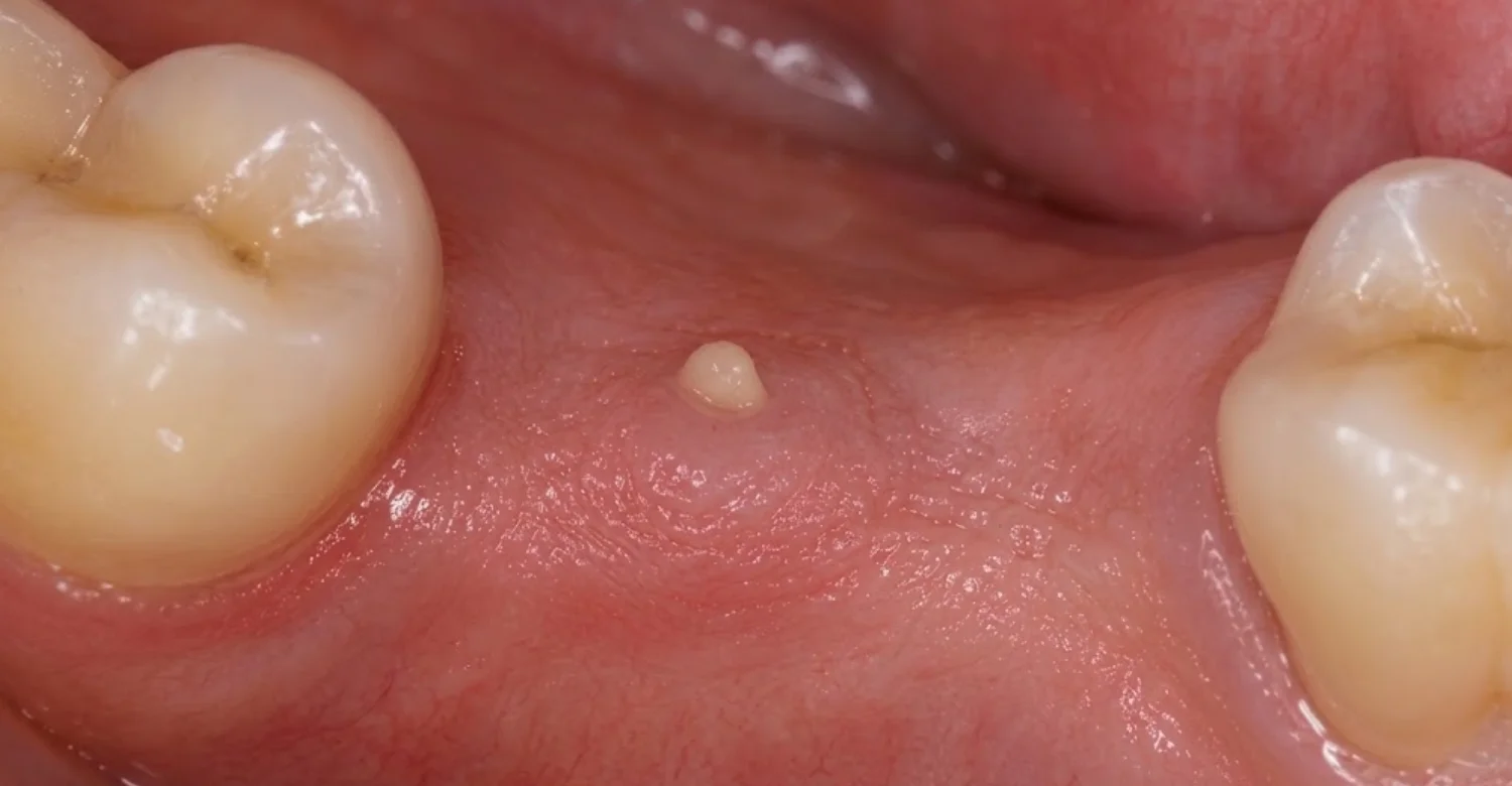
What Causes a Bone Spicule in the Gum?
Understanding dental bone spicule causes helps you judge whether what you feel is likely part of normal healing. The most common setting is recovery after a dental procedure, particularly a tooth extraction. During healing, the bone around the treated area remodels. Small pieces of bone can loosen and move toward the surface. Other situations, such as surgery involving the jawbone, can also contribute. The sections below explain each cause in more detail, including when a fragment deserves closer attention.
Bone Spicule After Tooth Extraction
When a tooth is removed, the surrounding socket begins to heal and remodel. This is a normal process. During it, tiny pieces of bone may remain under the healing gum. As the tissue changes over the following weeks, a fragment can gradually move toward the surface. Your tongue often detects it before you can see anything. However, not every sharp spot after an extraction is a bone fragment. The edge of the socket itself can sometimes feel sharp during healing.
Bone Spicule After a Dental Procedure
Noticing a bone fragment after dental procedure recovery begins is not unusual. It can happen when bone has been disturbed, reshaped, or exposed during treatment. Examples include surgical dental procedures, bone contouring, and extraction-related procedures. Treatments that involve manipulating the jawbone may also play a role. That does not mean every procedure causes spicules. Most treated areas heal smoothly. When a fragment does appear, it usually reflects the body clearing small pieces of bone during normal repair.
Bone Sequestra: When a Small Piece of Bone Loses Its Blood Supply
Bone sequestra is the technical term for pieces of bone that have separated from the healthy bone around them. Once a piece loses its blood supply, the body treats it as something to push out. This is why it may become loose over time. Routine healing usually settles within a predictable timeframe. A fragment that stays exposed, keeps hurting, or shows signs of infection is different. It deserves professional assessment, because the underlying cause shapes the treatment decision.
Can Alveoloplasty Lead to a Bone Spicule?
Alveoloplasty is a procedure that reshapes or smooths the jawbone. It is often performed around an extraction site or before dentures are fitted. The aim is a smooth, even ridge. During healing, small bone irregularities may still become noticeable as the gum settles. You might feel a sharp point or a slight edge. This does not automatically mean there is a complication. Many such areas settle naturally, although any persistent or painful spot should be checked by your dentist.
What Symptoms Can a Bone Spicule Cause?
Bone spicule symptoms can range from barely noticeable to locally irritating. Many people only feel a small point with the tongue. Others experience tenderness when eating or brushing. The most common symptoms of an oral bone spicule are local and mild. They include a sharp or hard spot, a visible white fragment, and discomfort when the area is touched. Knowing which signs are typical, and which are not, helps you decide how urgently to seek care.
A Sharp or Hard Spot You Can Feel With Your Tongue
This is often the first sign. You notice a rough or pointed area and keep checking it with your tongue. It is a natural reflex, since the tongue is drawn to anything unfamiliar in the mouth. Unfortunately, constant contact can make things worse. Repeated rubbing may irritate the surrounding gum and the tongue itself. The spot can then feel sore or larger than it is. Leaving the area alone often makes it feel less noticeable within days.
A White Bone Fragment Stuck in the Gums
A white bone fragment stuck in gums is a common concern after an extraction. Exposed bone often looks white or pale because no gum tissue covers it. However, a white spot is not automatically bone. Healing tissue can look white or yellowish in the first days. Food debris can lodge in a socket. Some oral lesions also appear pale. Because several things can look similar, a dentist should confirm what you are seeing before any decision is made.
Tenderness, Irritation, or Pain
Discomfort usually appears when the sharp area rubs against something. That might be your tongue, the inside of your cheek, food while chewing, or a toothbrush. This kind of pain is generally mild and localized. It tends to ease when the area is left alone. Increasing pain is different. Swelling, discharge, or feeling generally unwell may suggest a different or additional problem. Those signs need assessment rather than being dismissed as part of a simple exposed fragment.
What Symptoms Are Less Typical?
Some symptoms point beyond a simple spicule and call for a broader evaluation. Watch for:
Increasing swelling
Significant or worsening pain
Pus or drainage
Persistent bleeding
Fever
Difficulty opening the mouth
Difficulty swallowing or breathing
These signs may indicate infection or another condition. A small fragment rarely causes widespread symptoms, so their presence changes the picture. Difficulty swallowing or breathing needs urgent attention. Do not assume the problem is only a small fragment when these symptoms appear.
Tips for Patients
A few simple habits can keep you comfortable while you wait for professional advice.
Do not pull at a sharp fragment: Let a dentist determine whether it needs removal.
Keep the area gently clean: Avoid aggressive brushing over healing tissue.
Watch the trend: Increasing pain or swelling matters more than a single uncomfortable moment.
Avoid repeated tongue checking: Constant contact may increase irritation.
Keep your procedure details: Tell your dentist when and why the area was treated.
What Other Problems Can Feel Like a Bone Fragment ?
Not every hard or sharp area in the gums is actually bone. The mouth contains natural bony ridges, healing tissue, and occasionally foreign material. Any of these can feel similar to the tongue. A root tip, a piece of old filling, or a firm lesion may also create a hard sensation. This is why self-diagnosis is unreliable. Comparing your symptoms with other possibilities helps you understand why a dentist may want to examine the area before recommending anything.
Bone Spur in Mouth vs. Bone Spicule
The phrase bone spur in mouth usually describes a bony projection or overgrowth. A spicule more often refers to a small fragment working its way through the gum. In everyday conversation, people and even clinicians sometimes use the terms loosely. A dental bone spur may feel just as sharp as a loose fragment. Because the appearance and sensation overlap, the label matters less than the finding. An examination shows whether the bone is attached, loose, or part of normal anatomy.
Bone Fragment in Gums vs. Other Oral Growths
A hard area could have several explanations, depending on its location and your history. A professional assessment can help distinguish between:
Healing bone fragments
Normal bony anatomy
Bone irregularities after treatment
Foreign material, such as filling pieces
Other oral lesions
Calcified tissues, such as tooth fragments or roots
Not every hard area needs removal. Many are harmless and simply need monitoring. The goal of examination is to identify what the area is, then choose the least invasive suitable approach.
Can You Treat a Bone Fragment Yourself?
In short, not the fragment itself. You should avoid trying to pull, cut, dig out, or forcefully remove a suspected fragment at home. Doing so can tear healing gum, push debris deeper, or introduce bacteria. You also cannot be sure it is bone. What you can do is support healing and reduce irritation. Gentle care often keeps the area comfortable while it settles or until a dentist examines it. The next sections explain what is safe.
Bone Spicule Home Treatment: What Can You Safely Do?
Home care focuses on comfort and cleanliness, not removal. Low-risk measures include:
Keeping the area clean with gentle oral hygiene
Avoiding touching it with your tongue or fingers
Choosing softer foods if chewing causes irritation
Following your post-procedure instructions
Avoiding smoking during healing
Using only recommended rinses or medications
These steps may reduce irritation. However, they do not confirm what the area is or remove the underlying cause. If discomfort continues, arrange an examination.
What Should You Avoid Doing at Home?
Some reactions are understandable but risky. Avoid the following:
Do not dig into the gum with tweezers or sharp objects
Do not deliberately break the fragment
Do not repeatedly press the area with your fingers
Do not stop prescribed medication without professional advice
Do not assume every white or hard area is bone
These actions can damage healing tissue and increase infection risk. They can also hide important signs your dentist needs to see during an examination.
When Should You See a Dentist About a Bone Fragment?
Knowing when to see a dentist about a bone spicule depends on your symptoms, timing, previous procedures, and whether the area is changing. A small, painless point that is shrinking may only need monitoring. An area that grows, hurts more, or looks inflamed is different. The direction of change matters most. If you are unsure, an examination is reasonable. A clear answer usually relieves worry and prevents unnecessary self-treatment. It also means problems are found early.
Signs You Should Arrange a Dental Examination
Book an examination if you notice any of the following:
The sharp area persists or becomes more noticeable
Pain is increasing rather than improving
The gum becomes swollen or increasingly red
You notice pus or unusual drainage
The area repeatedly cuts or irritates your tongue
Healing does not seem to be progressing normally
You are unsure whether the fragment is actually bone
None of these is necessarily an emergency. Still, each deserves a professional look rather than waiting indefinitely.
When Is Urgent Dental or Medical Attention Needed?
Some symptoms need prompt care, regardless of their cause. Seek urgent dental or medical attention if you have significant swelling, fever, or rapidly worsening pain. Facial swelling that spreads is also a warning sign. Difficulty swallowing or breathing requires emergency care immediately. These signs can indicate a spreading infection or another serious problem. Do not try to manage them at home or wait for a routine appointment. Your safety comes first, and the cause can be identified afterwards.
Clinical Note
Dr. Rifat Alsaman, Head of the Medical Team and cosmetic dentist at Vitrin Clinic, notes that the most important step is identifying what the hard or sharp area actually represents. A suspected spicule may be part of normal healing. However, similar symptoms can have other explanations. Treatment should therefore follow an examination rather than appearance alone. If discomfort is increasing, healing seems delayed, or infection is suspected, professional evaluation becomes more important. In many cases, a careful look is enough to decide the next step.
How Are Oral Bone Spicules Treated?
Treatment depends on what the examination finds. A small fragment that is resolving naturally may need nothing more than monitoring. A piece that stays exposed, causes pain, or shows signs of infection may need active care. The options range from simple observation to gentle removal or smoothing of the bone. When infection or another dental problem is present, treatment focuses on that cause. In every case, the approach should match the actual finding, not just the sensation.
Observation During Normal Healing
Observation is often appropriate when the fragment is small and discomfort is minimal. It suits cases with normal healing after an extraction, no signs of infection, and no significant problems with eating or speaking. In these situations, the body often pushes the fragment out or covers it with gum tissue. However, monitoring should follow professional advice when the diagnosis is uncertain. Your dentist can tell you what to watch for and when to return if things change.
Gentle Clinical Removal of a Loose Bone Fragment
If a fragment is clearly loose and irritating, a dentist can often remove it quickly using suitable clinical instruments. Local anesthetic may be used for comfort, depending on the site. The procedure is usually brief. Afterwards, the gum can heal over a smoother surface. This is very different from trying to remove it yourself. A dentist can see the area properly, control bleeding, and confirm that no further fragments remain. Removal should always be based on examination.
Smoothing or Reshaping an Irregular Bone Area
Sometimes the issue is not a loose fragment but an uneven bony edge. In these cases, alveoloplasty may be considered where clinically appropriate. The dentist smooths or reshapes the bone so the gum can heal over a more even contour. This may be recommended when an irregular edge interferes with healing, comfort, or future prosthetic treatment such as dentures or implants. It is not needed for every sharp spot. Many small irregularities settle without intervention.
Treatment When Infection or Another Problem Is Present
When infection or another issue is involved, treatment may focus on the underlying cause rather than the fragment itself. Depending on the findings, this may involve:
Cleaning the area
Managing inflammation
Treating infection when clinically indicated
Addressing an underlying dental problem
Further examination when the appearance is atypical
Antibiotics are not automatically required. Your dentist will decide whether they are needed based on the examination findings and your overall health.
What Happens Before and After Bone Spicule Treatment?
Knowing what to expect can make the process feel simpler. Before treatment, your dentist gathers information to understand what the area is. After treatment, the focus shifts to comfortable healing. Most visits for a small fragment are short and straightforward. Recovery is usually quick, although it depends on what was done. The two sections below explain the assessment steps and the changes you may notice in the following days and weeks. Asking questions at each stage is always welcome.
Before Treatment: What Will Your Dentist Check?
Your dentist will usually review:
Your recent dental history
Any previous extraction or procedure
Your pain and symptom timeline
A visual examination of the area
Gentle palpation to check whether it is loose
The condition of the surrounding gum tissue
Dental imaging when indicated
Imaging is not necessarily required for every small exposed fragment. It is more useful when the cause is unclear, symptoms are significant, or deeper problems are suspected.
After Treatment: What Should You Expect?
Most people notice improvement fairly soon. You may experience less irritation from the sharp area and easier chewing. Local tenderness usually reduces, and the gum heals gradually over the treated spot. Mild soreness for a short time is common. Healing time varies according to the procedure, the size and location of the area, and your overall oral health. Follow your aftercare instructions, and contact your dentist if pain increases instead of easing.
What We Notice Clinically
From a practical clinical perspective, Dr. Rifat Alsaman, Head of the Medical Team and cosmetic dentist at Vitrin Clinic, emphasizes that a sharp area after an extraction should not automatically be interpreted as a complication. Common observations include:
Patients often feel a sharp point before it becomes visible.
Timing after an extraction provides important context.
A fragment may become more noticeable as the gum changes.
A white appearance does not confirm its identity.
Repeated manipulation irritates the surrounding tissue.
Observation or removal should depend on symptoms and findings.
Persistent symptoms justify examination, even for small fragments.
How Can You Reduce the Risk of Problems During Healing?
You cannot always prevent a small fragment from surfacing, since remodeling is a natural part of healing. However, you can lower the chance of irritation, infection, or delayed recovery. Good aftercare supports the body's own repair process. The most helpful steps are simple: follow instructions, keep the area clean, leave it alone, and attend follow-up visits. Each is explained below. Together, they help the area heal smoothly and make any issue easier to spot early.
Follow Your Post-Procedure Instructions
Your dentist gives aftercare instructions based on the specific procedure and your health. These may cover eating, rinsing, medication, physical activity, and smoking. Following them closely supports uncomplicated healing. For example, avoiding forceful rinsing early on helps protect a healing socket. Taking medication as prescribed helps manage inflammation. General advice online cannot account for your individual situation. If anything is unclear, or you are unsure whether a symptom is expected, ask your dentist directly.
Keep the Area Clean Without Irritating It
Cleanliness helps prevent infection, but healing tissue is delicate. Brush the rest of your mouth normally, and use a soft-bristled brush near the treated site. Avoid scrubbing directly over it, especially in the first days. Gentle rinses, if recommended, can help clear food particles. Try to avoid very hard, crunchy, or sharp foods that may catch on the area. The goal is a clean environment that lets the gum recover without repeated friction.
Avoid Unnecessary Manipulation
It is tempting to check a sharp spot constantly. However, repeatedly touching it with your tongue, fingers, or objects can irritate the tissue. It may also delay healing and introduce bacteria. Attempting to remove a suspected fragment carries additional risks, including bleeding and gum damage. Leaving the area alone gives the body a chance to cover the fragment or push it out naturally. If it bothers you significantly, a dentist can deal with it safely.
Attend Follow-Up When Recommended
Follow-up visits let your dentist check whether healing is progressing as expected. Problems such as a persistent fragment, delayed closure, or early infection are easier to manage when found early. Even if you feel fine, keep the appointment when it has been recommended. Mention any sharp spots or changes you have noticed since treatment. A quick examination can confirm that everything is on track, or identify a small issue before it becomes more uncomfortable.
Care at Vitrin Clinic
Dental care at Vitrin Clinic begins with understanding the cause of your symptoms and assessing the condition of your teeth, gums, and surrounding tissues. If you notice a hard or sharp area in your gum, the dental team can evaluate whether it may be related to healing, a bone fragment, or another oral concern.
Depending on your individual needs, care may include:
A comprehensive dental examination
Assessment of the gums and surrounding tissues
Review of your dental and treatment history
Dental imaging when clinically indicated
Monitoring during the healing process when appropriate
Treatment of a confirmed bone fragment or related dental issue
Individualized follow-up based on your symptoms and clinical findings
The goal is to identify the underlying cause and provide care that is appropriate for your specific oral health needs.
.jpg)
.jpg)
.jpg)
.jpg)
Takeaway: What Should You Do If You Feel a Bone Fragment?
A hard or sharp area in the gum, particularly after an extraction or dental procedure, may be a small bone fragment surfacing during healing. However, the sensation alone does not confirm the diagnosis. The practical next steps are simple. Avoid manipulating the area, keep up gentle oral hygiene, and watch how it changes. Arrange a dental examination if the area persists, becomes painful, or develops swelling or other warning signs. Early assessment brings clarity and peace of mind.
Ready to transform your smile?
Talk to our team and get a FREE treatment plan.
References:
FAQs
Dr. Rifat Alsaman has more than 5 years of clinical experience in dentistry and currently serves as the Head of the Medical Team at Vitrin Clinic. He is dedicated to providing exceptional patient care, overseeing treatment planning, and ensuring the highest clinical standards across the team. His expertise, attention to detail, and commitment to continuous professional development have helped countless patients achieve healthier, more confident smiles.


.webp&w=3840&q=75)