General Dentistry
September 14, 2026
What Is Mastication and How Does the Process of Chewing Work?
 Dr. Rifat Alsaman
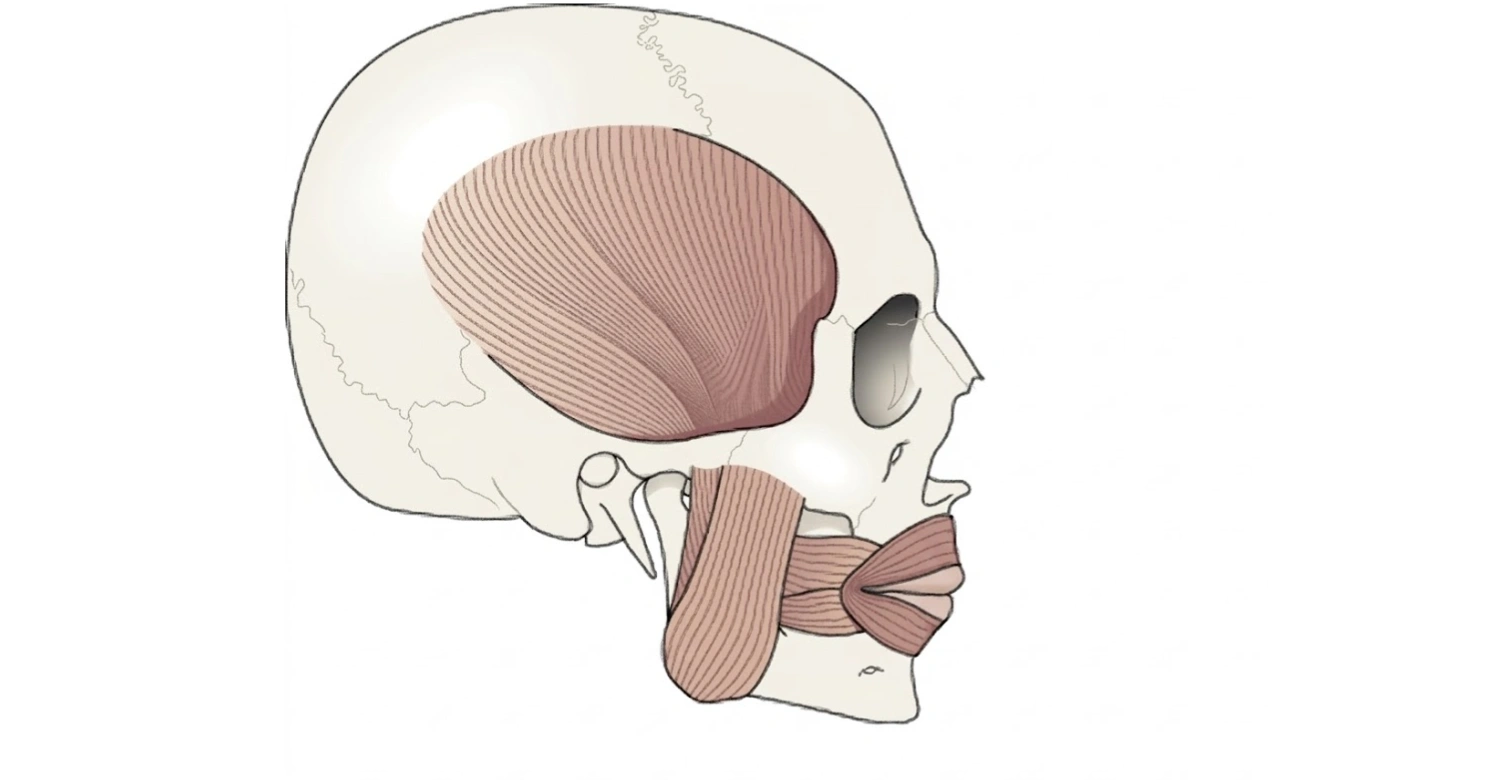
Mastication is the coordinated process that breaks food into smaller pieces before swallowing. It combines jaw movement, tooth contact, tongue control, saliva, and muscular activity. The process of mastication also prepares food for easier swallowing and supports the first stage of digestion. Four primary jaw muscles control many important movements involved in chewing. These include the masseter, temporalis, medial pterygoid, and lateral pterygoid muscles. The mandibular division of the trigeminal nerve provides their motor supply. Mastication also depends on healthy teeth and stable jaw relationships. Missing, damaged, or painful teeth can change chewing patterns and affect oral function. Vitrin Clinic considers these functional factors when evaluating patients who need restorative or reconstructive dental treatment. A proper assessment can help identify whether dental treatment is affecting comfort, bite, or everyday eating.
What Is Mastication?
Mastication describes the mechanical breakdown of food inside the mouth. Teeth cut, crush, and grind food while the tongue continuously repositions it. Jaw muscles generate controlled movements that bring the teeth together repeatedly. Saliva moistens food and helps transform separate particles into a cohesive mass. This coordinated activity prepares food for swallowing and subsequent digestive processes. The movement is not simply an opening-and-closing action. It includes elevation, depression, protrusion, retraction, and lateral movement. These movements depend on coordinated muscle activity and temporomandibular joint function. Healthy chewing therefore requires several anatomical systems to operate together. Problems involving teeth, muscles, nerves, or the jaw joint may change this coordination.
What Does Masticate Mean?
The verb “masticate” means to chew food using the teeth and coordinated movements of the jaw. It describes an active mechanical process occurring before swallowing. During this process, teeth reduce larger food particles into smaller pieces. The tongue moves food between different tooth surfaces during repeated chewing cycles. Saliva also becomes incorporated into the food as chewing continues. The resulting mixture becomes easier to control inside the mouth. The term can therefore describe both ordinary chewing and the physiological process studied in dental anatomy. Understanding this terminology can make dental information easier to follow. It is especially useful when reading explanations about jaw muscles, occlusion, chewing efficiency, or oral function.
Mastication vs. Chewing
Mastication and chewing generally describe the same functional activity. “Chewing” is the common everyday term used by patients and the general public. “Mastication” is the anatomical and clinical term commonly used in healthcare education. Both describe the mechanical processing of food inside the mouth. The scientific term emphasizes coordinated muscle, tooth, tongue, and jaw activity. Chewing can therefore be understood as a practical description of a complex physiological process. Dental professionals may use the scientific terminology when discussing oral function or treatment planning. Patients usually encounter simpler language during routine explanations. Understanding both terms helps patients interpret dental information without confusing them as separate processes.
What Is the Purpose of Mastication?
The primary purpose is to reduce food particle size and prepare it for swallowing. Teeth apply mechanical forces that cut, crush, and grind different types of food. The tongue continually positions food between appropriate tooth surfaces. Saliva moistens particles and helps create a cohesive bolus. These actions make swallowing more controlled and efficient. Chewing also exposes more food surface area to saliva and digestive enzymes. However, digestion does not begin exclusively after food reaches the stomach. Oral processing contributes to the earliest stage of gastrointestinal preparation. Efficient chewing depends on healthy teeth, appropriate jaw movement, muscle coordination, and sensory feedback. Research also links chewing ability with the number and distribution of remaining functional teeth.
How Mastication Helps Digestion
Chewing supports digestion primarily through mechanical food breakdown. Smaller food particles provide greater surface area for subsequent digestive processes. Saliva additionally lubricates food and contributes enzymes that begin chemical processing. The tongue mixes food with saliva while controlling its position within the mouth. This creates a manageable bolus before swallowing. Effective chewing can therefore make the transition from eating to swallowing smoother. However, chewing should not be considered the complete digestive process. Most chemical digestion occurs later within the gastrointestinal tract. The mouth provides an important preparation stage that combines mechanical and chemical functions. Poor dental health may interfere with this preparation when pain or tooth loss limits chewing.
How Teeth, Tongue, and Jaw Work Together During Mastication
Teeth provide the hard surfaces required to cut, crush, and grind food. The tongue moves food between tooth surfaces and helps prevent particles from escaping prematurely. The mandible provides the moving framework that positions the lower teeth against the upper teeth. Jaw muscles generate the forces needed for repeated movement. The temporomandibular joints allow controlled mandibular motion during opening, closing, and excursion. Saliva lubricates food while helping create a cohesive bolus. These structures operate through continuous sensory feedback. The nervous system adjusts movement according to food texture, resistance, and position. Effective chewing therefore represents a coordinated neuromuscular activity rather than an isolated tooth function.
Clinical Note
Chewing performance can change when dental structures become painful, unstable, or absent. Patients may unconsciously avoid one side when a tooth causes discomfort. Repeated one-sided chewing can alter normal movement patterns and increase functional strain. Tooth loss can also reduce the number of functional tooth contacts available during eating. A systematic review found that chewing ability is associated with the number and distribution of remaining teeth. Dental assessment should therefore consider function rather than appearance alone. Vitrin Clinic evaluates the condition of teeth, bite relationships, and supporting structures before recommending restorative treatment. The appropriate solution depends on the individual clinical findings.
Mastication Definition and Anatomy
The anatomy involved in chewing includes the mandible, teeth, temporomandibular joints, muscles, tongue, and associated nerves. The mandible performs several movements that position the lower teeth against the upper teeth. These movements occur across multiple directions rather than through simple vertical opening and closing. The four principal jaw muscles are masseter, temporalis, medial pterygoid, and lateral pterygoid. Accessory muscles also contribute to mandibular opening and stabilization. The temporomandibular joints connect the mandible with the temporal bones. Sensory and motor nerves provide information and control throughout the process. Understanding this anatomy helps explain why dental pain can sometimes involve muscles or jaw joints.
Structures Involved in Mastication
Several anatomical structures contribute to efficient chewing. The mandible provides the movable foundation for the lower teeth. The maxilla supports the upper teeth and remains comparatively stationary. The temporomandibular joints guide mandibular movement against the skull. The masseter, temporalis, and pterygoid muscles generate and control movement. The tongue continuously positions food during chewing. Salivary glands contribute fluid that moistens and lubricates food. Sensory nerves provide information about pressure, movement, texture, and discomfort. The trigeminal nerve plays a major role in sensory and motor control. Damage involving any important component may affect chewing efficiency. Dental diagnosis therefore requires consideration of the entire functional system.
How the Mandible Moves During Mastication
The mandible performs several coordinated movements during chewing. Elevation closes the jaw and brings opposing teeth together. Depression lowers the mandible and contributes to mouth opening. Protrusion moves the mandible forward relative to the maxilla. Retraction moves it backward toward its resting position. Lateral movement shifts the mandible toward either side. These movements combine during normal chewing cycles. The pterygoid muscles are particularly important for forward and lateral movements. The masseter and temporalis contribute strongly to jaw elevation. Accessory muscles assist opening and stabilization. The nervous system continuously coordinates these movements according to food resistance and position.
Elevation
Elevation moves the mandible upward toward the maxilla. This movement closes the mouth and creates tooth contact. The masseter, temporalis, and medial pterygoid contribute substantially to elevation. These muscles work together to generate controlled closing forces. Elevation is essential when crushing or grinding food between opposing teeth. The amount of force required depends on food texture and resistance. Harder foods generally require greater muscular force than softer foods. Pain or weakness affecting these muscles may reduce comfortable chewing. Dental professionals may consider jaw-closing function when evaluating patients with chewing difficulties.
Depression
Depression moves the mandible downward and contributes to mouth opening. Gravity assists this movement, while accessory muscles also participate. The lateral pterygoid has an important role in mandibular depression. Suprahyoid muscles can assist when greater opening is required. Normal depression allows food to enter the mouth and permits subsequent chewing cycles. Restricted opening can interfere with eating and dental procedures. Causes may include muscular problems, temporomandibular disorders, inflammation, or other conditions. Persistent difficulty opening the mouth should receive professional evaluation.
Protrusion
Protrusion moves the mandible forward relative to the upper jaw. Both lateral pterygoid muscles contribute strongly to this movement. The medial pterygoids can also assist forward movement. Protrusion helps position the lower teeth during certain grinding movements. It also contributes to coordinated jaw excursions during chewing. The movement depends on controlled contraction rather than simple forward displacement. Abnormal restriction may affect chewing patterns and jaw comfort. Dental examination can determine whether teeth, muscles, or temporomandibular structures contribute to the problem.
Retraction
Retraction moves the mandible backward toward its normal posterior position. The posterior fibers of the temporalis are especially important for this action. Retraction complements protrusion during controlled mandibular excursions. It helps return the jaw after forward movement. Normal retraction should occur smoothly without significant pain or restriction. Changes in occlusion can sometimes alter the way patients position their mandible. Persistent discomfort during backward movement may warrant assessment of the temporomandibular joints and associated muscles.
Lateral Movement
Lateral movement shifts the mandible toward either side. It is particularly important for grinding and processing food. Coordinated activity between the medial and lateral pterygoids produces these movements. When one side contracts differently from the other, the mandible can deviate laterally. This alternating movement helps expose food to different tooth surfaces. Effective grinding therefore depends on bilateral muscular coordination. Pain, tooth loss, or joint dysfunction can change these movements. Clinicians may observe mandibular excursions when evaluating functional chewing problems.
What Are the Muscles of Mastication?
Four primary muscles are traditionally identified in chewing anatomy. They are the masseter, temporalis, medial pterygoid, and lateral pterygoid. These muscles work in coordinated pairs on the right and left sides. Their combined activity produces elevation, depression, protrusion, retraction, and lateral movement. Accessory muscles also assist mandibular opening and stabilization. Each primary muscle has distinct anatomical attachments and functional roles. Their motor supply comes from the mandibular division of the trigeminal nerve. Understanding these muscles is useful when assessing jaw pain, chewing problems, and temporomandibular disorders. It also helps explain why pain around the cheek or temple can sometimes originate from muscular structures.
Masseter Muscle
The masseter is a powerful muscle positioned along the lateral surface of the mandibular ramus. It originates primarily from the zygomatic arch and inserts along the lateral mandible. Its main function is elevation of the mandible. It can also assist with mandibular protrusion. The muscle contains superficial and deep portions with different fiber orientations. Strong contraction helps generate the force needed for crushing food. Because of its superficial position, tenderness can sometimes be detected externally near the cheek. Excessive clenching may increase muscular workload and discomfort. Clinical evaluation can distinguish muscular tenderness from pain originating inside a tooth or from the jaw joint.
Temporalis Muscle
The temporalis is a broad, fan-shaped muscle covering much of the temporal region. Its fibers converge toward a tendon that attaches to the coronoid process. Anterior and middle fibers contribute to mandibular elevation. Posterior fibers contribute strongly to retraction. The muscle helps stabilize mandibular position during chewing. It works with other jaw-closing muscles to produce controlled force. Temporalis tenderness may be noticed around the temple when the muscle is overactive. Dental professionals may assess this region when patients report headaches associated with clenching or chewing.
Medial Pterygoid Muscle
The medial pterygoid lies on the inner side of the mandibular ramus. It has superficial and deep portions with different anatomical origins. Its main functions include mandibular elevation and assistance with protrusion. It also contributes to lateral jaw movements. The muscle works closely with the masseter during jaw closure. Their anatomical arrangement creates an important functional relationship around the mandibular angle. Abnormal activity may contribute to muscular discomfort. Clinical assessment considers the broader jaw system rather than evaluating one muscle independently.
Lateral Pterygoid Muscle
The lateral pterygoid is located deep within the infratemporal region. It has superior and inferior heads with distinct anatomical relationships. Bilateral contraction contributes to mandibular protrusion. Unilateral activity contributes to lateral deviation. The muscle also participates in mandibular depression and temporomandibular joint movement. Its relationship with the joint capsule and articular disc makes it clinically important. Dysfunction involving this region may influence jaw movement. However, muscle activity should be interpreted alongside joint and dental findings.
Clinical Note
The four primary muscles do not operate independently. They work as a coordinated system with the temporomandibular joints and teeth. Nerve signals regulate their activation according to movement and sensory feedback. Pain may develop when muscles are overloaded or when movement becomes abnormal. Dental problems can also cause patients to change their chewing pattern. These adaptations may place additional demand on certain muscles. A clinical examination can help determine whether symptoms originate from teeth, muscles, joints, or combinations of these structures.
Muscles of Mastication: Origin, Insertion, Function, and Innervation
Understanding origin, insertion, function, and innervation provides a complete anatomical picture. The four primary muscles have different attachments and movement patterns. Their motor supply comes through branches associated with the mandibular division of cranial nerve V. This arrangement reflects their development from the first pharyngeal arch. Their combined actions allow complex mandibular movement. Anatomical knowledge is particularly useful for dentists, dental students, surgeons, and patients seeking understandable explanations. It also helps clarify why jaw symptoms can involve several structures simultaneously.
Masseter: Origin, Insertion, Function, and Innervation
The masseter originates primarily from the zygomatic arch and adjacent zygomatic structures. It inserts along the lateral surface of the mandibular ramus and angle. Its principal function is mandibular elevation. It can also contribute to protrusion. The masseteric nerve, a branch of mandibular nerve V3, provides motor innervation. Its powerful contraction helps generate forces required during chewing. Different portions of the muscle contribute according to movement direction. Clinical examination may include palpation when patients report cheek or jaw soreness. Muscle symptoms should still be differentiated from dental and joint causes.
Temporalis: Origin, Insertion, Function, and Innervation
The temporalis originates broadly from the temporal fossa and surrounding temporal fascia. Its fibers converge into a tendon that attaches to the coronoid process. Anterior fibers mainly elevate the mandible. Posterior fibers contribute to mandibular retraction. Deep temporal branches of V3 provide motor innervation. The muscle helps maintain mandibular position during chewing. Its broad structure allows different fiber groups to contribute to different movements. Tenderness around the temple may sometimes reflect increased muscular activity.
Medial Pterygoid: Origin, Insertion, Function, and Innervation
The medial pterygoid has superficial and deep heads. Its origins involve the maxillary tuberosity and medial surface of the lateral pterygoid plate. It inserts on the medial surface of the mandibular ramus and angle. The muscle assists elevation and protrusion. It also contributes to lateral mandibular movement. The medial pterygoid nerve provides motor innervation through V3. Its activity complements the masseter during jaw closure. Coordinated function allows controlled chewing rather than simple vertical biting.
Lateral Pterygoid: Origin, Insertion, Function, and Innervation
The lateral pterygoid contains superior and inferior heads. The superior head originates from the greater wing of the sphenoid. The inferior head originates from the lateral pterygoid plate. Fibers attach to the mandibular condylar region and temporomandibular joint structures. Bilateral contraction promotes protrusion. Unilateral contraction contributes to contralateral mandibular movement. The muscle also participates in opening and joint coordination. Its motor supply comes from a branch of mandibular nerve V3.
What Nerve Innervates the Muscles of Mastication?
The primary muscles receive motor innervation from the mandibular division of the trigeminal nerve. This division is known as cranial nerve V3. The trigeminal nerve has three major divisions, but V3 uniquely carries motor fibers. These motor fibers supply the four principal muscles involved in chewing. V3 also supplies several additional muscles associated with mandibular movement. Sensory branches provide information from relevant oral and facial structures. This combined sensory and motor system supports coordinated jaw function.
Trigeminal Nerve and Mandibular Division
The trigeminal nerve is the fifth cranial nerve. It contains both sensory and motor components. Its three divisions are ophthalmic, maxillary, and mandibular. Only the mandibular division carries the motor fibers that supply the primary chewing muscles. V3 exits the skull through the foramen ovale. It then gives branches that reach individual muscles. Sensory information helps the nervous system monitor oral conditions during chewing. This system allows jaw movement to adapt to changing food texture and resistance.
Motor Innervation of the Muscles of Mastication
The masseter receives motor supply from the masseteric nerve. The temporalis receives deep temporal branches. The medial pterygoid receives the medial pterygoid nerve. The lateral pterygoid receives lateral pterygoid branches. All originate from the mandibular division of the trigeminal nerve. These branches allow precise activation of individual muscles. Their coordinated firing creates controlled mandibular movement. Damage affecting these pathways can interfere with normal chewing function.
Clinical Note
Nerve function is important when jaw weakness or unusual movement occurs. Neurological problems can affect the muscles directly supplied by V3. However, chewing difficulties are not automatically neurological. Tooth pain, missing teeth, joint disorders, and muscular overload can produce similar functional complaints. A complete dental examination helps distinguish these possibilities. When neurological findings are suspected, appropriate medical assessment may also be required. Vitrin Clinic can evaluate dental and structural factors that may contribute to altered chewing function.
How Do the Muscles of Mastication Work Together?
The muscles work as coordinated groups rather than isolated structures. Jaw elevation requires combined activity from the masseter, temporalis, and medial pterygoid. Protrusion depends strongly on bilateral pterygoid activity. Retraction involves the posterior temporalis and supporting muscles. Lateral movement requires asymmetric contraction between the right and left sides. The nervous system continuously adjusts these patterns during chewing. Food consistency influences the amount and direction of muscular force required. This coordination allows efficient processing without excessive movement.
Muscles Responsible for Jaw Elevation
Jaw elevation closes the mandible against the maxilla. The masseter is one of the strongest contributors to this movement. The temporalis provides powerful elevation and stabilization. The medial pterygoid also assists closure. These muscles can contract together to generate substantial biting force. Their coordinated activity is essential for crushing tougher food. The temporalis can also stabilize the mandible during controlled movement. Muscle discomfort may occur when clenching or grinding increases activity beyond normal functional demands.
Muscles Responsible for Jaw Protrusion
Protrusion moves the lower jaw forward. The lateral pterygoids are major contributors when they contract bilaterally. The medial pterygoids can assist the forward movement. Protrusion contributes to grinding patterns and mandibular positioning. It also helps coordinate movement of the condyles within the joints. Smooth protrusion requires balanced muscle activity on both sides. Unequal activation may produce deviation or altered movement.
Muscles Responsible for Jaw Retraction
Retraction moves the mandible backward. The posterior fibers of the temporalis are particularly important for this action. Retraction returns the mandible after forward excursions. It also contributes to mandibular stability during certain functional movements. The movement must remain coordinated with temporomandibular joint positioning. Abnormal discomfort during retraction can have dental, muscular, or joint-related causes. Persistent symptoms should receive professional evaluation.
Muscles Responsible for Lateral Jaw Movement
Lateral movement requires different activation between the two sides. Unilateral pterygoid contraction shifts the mandible laterally. This movement allows food to move across different tooth surfaces. It is particularly important during grinding. The tongue simultaneously controls food position. Balanced bilateral function supports smooth chewing cycles. Dental pain or missing teeth may cause patients to avoid particular movements.
What Happens During the Process of Mastication?
Chewing progresses through several overlapping stages. Food first enters the mouth and becomes positioned between the teeth. The jaw then performs repeated movements that reduce particle size. The tongue continuously repositions food throughout the cycle. Saliva mixes with the particles and improves lubrication. Eventually, the material becomes a cohesive bolus suitable for swallowing. Each stage depends on coordinated sensory, muscular, dental, and neurological function. Problems at any stage may reduce eating comfort or efficiency.
Food Intake and Positioning
Food enters the mouth during biting or other forms of intake. The lips and tongue help control the initial position of the food. The tongue then moves portions toward the posterior teeth. Teeth begin cutting or crushing larger pieces. Sensory receptors provide information about texture, size, and resistance. This information helps regulate subsequent jaw movements. The process changes continuously depending on the food being eaten. Softer foods require different movement patterns than hard or fibrous foods.
Crushing and Grinding Food
Teeth mechanically reduce food through repeated contact. Incisors are particularly suited to cutting. Premolars and molars provide broader surfaces for crushing and grinding. Jaw muscles generate force while the mandible moves through different directions. Lateral movements help grind food against opposing surfaces. The tongue repeatedly returns particles toward functional tooth contacts. Efficient grinding reduces particle size and prepares food for swallowing.
Mixing Food With Saliva
Saliva enters the food during repeated tongue and jaw movements. It lubricates particles and helps create a cohesive mixture. Salivary enzymes also begin certain aspects of chemical digestion. The tongue spreads saliva throughout the food mass. Adequate lubrication supports comfortable swallowing. Reduced saliva can make eating dry or difficult. Persistent dry mouth should therefore be discussed with an appropriate healthcare professional.
Formation of the Food Bolus
Continued chewing transforms separate food particles into a cohesive bolus. The tongue collects and shapes the material before swallowing. Saliva contributes moisture and cohesion. Teeth continue processing any remaining larger pieces. Sensory feedback helps determine when the bolus is ready for swallowing. The swallowing process then transfers the material toward the pharynx. This transition demonstrates the close relationship between chewing and swallowing.
What We Notice Clinically
Clinicians often observe changes in chewing patterns when teeth, muscles, or joints become compromised. Patients may avoid painful areas or favor one side of the mouth. Missing teeth can reduce available functional contacts. Tooth damage may also change how forces are distributed. Research indicates that chewing ability is associated with the number and distribution of remaining teeth. These changes may be subtle during everyday eating. A professional assessment can identify functional changes before they become more disruptive.
Common Functional Changes During Mastication
Common changes include slower chewing, one-sided chewing, reduced bite force, and difficulty processing certain foods. Patients may also report fatigue after prolonged eating. These symptoms can arise from dental, muscular, joint, or neurological factors. Tooth loss can reduce functional tooth contacts. Pain can cause protective movement patterns that change normal jaw use. Clinicians consider the patient's symptoms alongside oral examination findings. Treatment should address the underlying cause rather than simply the chewing complaint.
Pain During Mastication
Pain during chewing can originate from several structures. A tooth may hurt because of decay, fracture, inflammation, or infection. Muscles can become tender after excessive clenching or overuse. Temporomandibular disorders can also produce pain during jaw movement. The location and timing of pain provide useful diagnostic information. Pain on biting one specific tooth may suggest a dental source. Broader jaw or facial pain may involve muscles or joints. Persistent or worsening pain should be professionally evaluated.
Changes Caused by Missing or Damaged Teeth
Missing teeth reduce the number of available contacts for chewing. Damaged teeth may become painful when exposed to pressure. Patients can compensate by shifting food toward healthier areas. This adaptation may change normal chewing patterns. A systematic review found that chewing ability relates closely to remaining tooth number and distribution. Treatment planning should therefore consider functional tooth distribution. Vitrin Clinic evaluates individual dental conditions before recommending restorative or implant treatment.
Dr. Rifat Alsaman’s Clinical Opinion
Dr. Rifat Alsaman, Head of the Medical Team at Vitrin Clinic and cosmetic dentist, emphasizes that chewing should be considered alongside appearance. A smile restoration should support comfortable and stable oral function. Tooth position, bite relationships, supporting tissues, and existing dental conditions all matter. A cosmetic improvement alone does not replace a complete functional assessment. Patients with missing or damaged teeth may require restorative planning that considers everyday eating. Digital diagnostics can help clinicians evaluate these factors before treatment. The final approach should always reflect individual clinical findings rather than a standardized treatment formula.
Note : All images used are for editorial and illustrative purposes only and may not originate from the original news provider or associated company.
What Can Cause Pain During Mastication?
Pain during chewing has many possible causes. Dental problems are common, but muscles and temporomandibular joints can also produce symptoms. The exact location, duration, intensity, and trigger provide useful diagnostic information. A tooth that hurts specifically during biting may require dental examination. Jaw soreness may instead involve muscular or joint structures. Swelling, fever, trauma, or severe pain requires prompt professional assessment. Patients should avoid self-diagnosing persistent oral pain. Professional examination is necessary to identify the underlying cause.
Dental Causes
Dental causes include decay, cracked teeth, pulp inflammation, periodontal problems, and dental abscesses. A damaged filling or crown can also affect biting comfort. Tooth sensitivity may cause discomfort when pressure or temperature changes occur. The symptoms often vary according to the underlying condition. Dental imaging may be needed when clinical examination cannot fully identify the problem. Early diagnosis can help prevent progression. Patients experiencing persistent chewing pain should arrange an appropriate dental assessment.
Muscle-Related Causes
Muscular causes can include excessive clenching, grinding, fatigue, or overuse. The masseter and temporalis may become tender when activity increases. Pain may feel like soreness around the cheek, jaw, or temple. Some patients notice symptoms after stressful periods or prolonged clenching. However, muscular pain can resemble dental or joint pain. A clinician should evaluate the complete presentation. Treatment depends on the identified cause and may involve conservative management or dental intervention.
Temporomandibular Joint Causes
Temporomandibular joint problems can cause pain during opening, closing, or chewing. Patients may also experience clicking, limited movement, or jaw locking. The joint contains an articular disc and several sensory structures. Symptoms vary considerably between individuals. Joint sounds without pain do not necessarily indicate serious disease. Persistent pain, locking, or significant movement limitation warrants professional assessment.
When Should You See a Dentist?
See a dentist when chewing pain persists, worsens, or repeatedly returns. Professional care is particularly important when swelling, fever, trauma, or significant tooth damage occurs. Difficulty opening the mouth should also receive appropriate assessment. A dentist can determine whether the source involves teeth, gums, muscles, or joints. Delaying evaluation can allow some dental conditions to progress. Vitrin Clinic provides diagnostic assessment and individualized treatment planning for patients with functional dental concerns.
Mastication and Oral Health
Healthy teeth and supporting tissues contribute substantially to effective chewing. Oral health problems can change how forces are distributed across the dental arches. Pain may encourage patients to avoid certain teeth or chewing directions. Tooth loss can reduce functional contacts and alter chewing efficiency. Research supports an association between chewing ability and the number and distribution of remaining teeth. Maintaining teeth therefore supports more than appearance. Regular dental examinations can help identify problems before they significantly affect oral function.
The Relationship Between Mastication and Occlusion
Occlusion describes the relationship between upper and lower teeth when they contact. Stable tooth contacts help distribute chewing forces across the dental arches. Changes in tooth position, restorations, or tooth loss can alter these relationships. The muscles and temporomandibular joints adapt to changing mandibular positions. However, not every occlusal difference causes symptoms. Clinical assessment should consider the patient's symptoms and functional needs. Treatment should not be based solely on an isolated bite measurement.
How Missing Teeth Can Affect Mastication
Missing teeth reduce the number of available contacts during chewing. The effect depends on the location and number of missing teeth. Losing posterior teeth can particularly affect grinding surfaces. Patients may compensate by using remaining teeth more frequently. A systematic review found that chewing ability is associated with remaining tooth number and distribution. Dental implants, bridges, or other restorations may restore selected functional contacts. Treatment choice depends on bone, gum health, bite, general dental condition, and patient goals.
How Tooth Damage Can Affect Chewing Function
Cracks, severe decay, fractures, and worn surfaces can alter chewing. A damaged tooth may become painful under pressure. Patients may then avoid placing food on that side. This protective behavior can change normal chewing patterns. Untreated dental damage can sometimes progress and compromise additional structures. Early evaluation helps determine whether conservative restoration or more extensive treatment is appropriate. Vitrin Clinic can assess damaged teeth using clinical and digital diagnostic approaches.
Mastication After Tooth Loss or Dental Treatment
Tooth loss can change chewing patterns by reducing available functional tooth contacts. Dental treatment aims to restore oral structures while considering function and long-term stability. Restorations may include implants, crowns, bridges, or larger reconstructive approaches. The appropriate solution depends on the patient's clinical anatomy and treatment goals. Digital planning can help evaluate existing structures before treatment. Successful restoration requires more than replacing visible tooth surfaces. It should also consider bite relationships, supporting tissues, and functional movement.
How Missing Teeth Affect Chewing
Missing teeth can make certain foods more difficult to process. Patients may compensate by changing where they place food. The remaining teeth may then carry a greater proportion of chewing activity. Research indicates that both tooth number and distribution influence chewing ability. The clinical impact varies according to individual circumstances. A missing front tooth may primarily affect cutting. Missing posterior teeth may have a greater impact on grinding. Dental evaluation determines whether replacement would provide meaningful functional benefit.
Restoring Teeth to Support Oral Function
Restorative treatment can replace damaged tooth structure or missing teeth. Crowns protect and restore individual teeth when sufficient supporting structure remains. Bridges replace selected missing teeth using neighboring structures for support. Dental implants replace missing tooth roots and support prosthetic teeth. Larger tooth loss may require comprehensive rehabilitation. Treatment planning should consider function, aesthetics, hygiene, bone, gums, and long-term maintenance. Vitrin Clinic uses individualized planning rather than assuming every patient requires the same restoration.
Clinical Note
Restoration does not automatically guarantee normal function immediately. Patients may need time to adapt to changes in tooth shape or bite. Follow-up appointments can identify areas requiring adjustment. The surrounding muscles and joints may also adapt to the restored dental relationship. Treatment success should therefore be evaluated over time. Patients should follow their dentist's aftercare instructions. Any persistent pain or difficulty chewing should be reported rather than ignored.
How Vitrin Clinic Evaluates Oral Function and Mastication
Vitrin Clinic combines clinical examination with digital diagnostic tools when planning appropriate dental treatment. The evaluation considers teeth, gums, bone, bite relationships, and overall treatment objectives. Digital technology can improve visualization and communication during treatment planning. CBCT and digital X-rays can provide information about relevant hard tissues. Intraoral scanning can capture detailed digital records of oral structures. These tools do not replace clinical judgment. Instead, they support a more comprehensive assessment of individual anatomy.
Digital Dental Examination and Treatment Planning
A digital examination can help organize information about the patient's current oral condition. Photographs, scans, radiographs, and clinical findings can be considered together. Treatment planning can then address both functional and aesthetic objectives. Digital Smile Design may help visualize potential restorative changes. The clinician still determines whether proposed changes are clinically appropriate. Vitrin Clinic uses digital workflows to support communication between clinicians and international patients. This approach can make complex treatment plans easier to explain before treatment begins.
CBCT and Digital X-Rays
CBCT provides three-dimensional imaging of selected oral and maxillofacial structures. It can help clinicians assess bone anatomy and other structures when appropriate. Digital dental X-rays provide detailed two-dimensional images for specific diagnostic purposes. Neither imaging method is required for every patient. The appropriate imaging technique depends on the clinical question. Dentists should use imaging when the expected diagnostic benefit justifies exposure. Vitrin Clinic incorporates digital imaging into treatment planning when clinically indicated.
3D Intraoral Scanning and Digital Dentistry
A 3D intraoral scanner captures detailed digital information about oral surfaces. This information can support restorative planning and laboratory communication. Digital records can reduce dependence on traditional physical impressions in appropriate cases. Scanning may also help document tooth positions and restorative spaces. Digital workflows can improve communication between clinical and laboratory teams. At Vitrin Clinic, digital dentistry supports personalized treatment planning for international patients. The technology remains a clinical tool rather than a replacement for professional assessment.
Dr. Rifat Alsaman’s Clinical Perspective
Dr. Rifat Alsaman considers functional stability an important part of comprehensive dental planning. His clinical perspective emphasizes examining the patient's teeth, supporting structures, bite, and treatment objectives together. Cosmetic treatment should be designed around the patient's overall oral condition. Digital diagnostics can provide valuable information before restorative decisions are finalized. The exact treatment approach depends on individual findings. Patients should receive a plan based on clinical examination rather than assumptions based only on photographs.
Dental Treatments That Can Help Restore Oral Function
Different treatments can replace missing teeth or restore damaged structures. Dental implants can replace individual missing teeth and support larger restorations. Crowns can restore damaged individual teeth. Bridges can replace selected missing teeth without implant placement. Full-mouth rehabilitation can address multiple functional and restorative problems. Full-arch implant treatment may be considered for extensive tooth loss. Treatment selection depends on bone availability, periodontal health, tooth condition, bite, and patient goals. A comprehensive assessment should occur before selecting any treatment.
Dental Implants
Dental implants replace missing tooth roots with biocompatible fixtures. They can support individual crowns, bridges, or larger prosthetic restorations. Implant treatment requires assessment of bone, gums, medical factors, and restorative requirements. Digital planning may help evaluate implant positioning. The final restoration should provide appropriate function and maintainability. Patients should also understand that implants require long-term oral hygiene and professional maintenance. Vitrin Clinic provides implant treatment planning according to individual anatomical and restorative requirements.
Dental Crowns and Bridges
Crowns restore teeth that have significant structural damage while retaining the remaining tooth structure when possible. Bridges replace missing teeth using adjacent teeth or implants for support. Both approaches can restore tooth shape and contribute to functional contacts. Their suitability depends on the condition of the supporting structures. Properly planned restorations should consider appearance, bite, hygiene, and durability. Patients should receive individualized advice before choosing between restorative options.
Full-Mouth Rehabilitation
Full-mouth rehabilitation addresses multiple dental problems affecting function and appearance. It may combine crowns, implants, periodontal treatment, or other restorative procedures. The exact sequence depends on the patient's diagnosis. Comprehensive planning is important because changing several teeth can influence bite relationships. Digital records may support visualization and communication. Vitrin Clinic develops individualized treatment plans according to clinical findings and patient objectives. Complex rehabilitation should always involve detailed examination and appropriate follow-up.
Full-Arch Dental Implants
Full-arch implant treatment can replace an entire dental arch using strategically positioned implants. Treatment may provide support for a fixed prosthetic arch in suitable patients. Assessment typically considers bone volume, gum health, occlusion, and restorative space. The number and positioning of implants depend on the treatment design. Patients should understand that full-arch treatment involves significant planning and maintenance. Vitrin Clinic evaluates individual anatomy before recommending full-arch implant treatment.
Tips for Patients
Healthy chewing depends on maintaining teeth, gums, muscles, and supporting structures. Daily oral hygiene helps reduce the risk of common dental disease. Regular examinations can identify problems before they significantly affect function. Patients should also pay attention to changes in chewing comfort. New pain, swelling, sensitivity, or difficulty eating should not be ignored. Appropriate dental treatment can protect or restore function when problems develop.
How to Support Healthy Mastication
Brush teeth twice daily using fluoride toothpaste unless a dental professional advises otherwise. Clean between teeth regularly to reduce plaque accumulation. Maintain regular dental examinations according to individual risk factors. Avoid using damaged teeth to open packages or bite hard objects. Seek evaluation when pain develops during eating. A balanced diet can also support general oral and systemic health. Patients with missing teeth should discuss restorative options when chewing becomes difficult. Maintaining oral structures is usually easier than treating advanced damage.
When to Seek Professional Dental Care
Seek dental care when chewing becomes painful or noticeably less effective. Persistent sensitivity, tooth mobility, swelling, or broken teeth also require evaluation. Jaw locking or significant difficulty opening the mouth should not be ignored. Urgent assessment may be necessary when severe swelling, fever, or trauma occurs. Early diagnosis can reduce complications associated with some dental conditions. Vitrin Clinic can assess dental structures and develop an individualized treatment plan when appropriate.
How to Pronounce Mastication and Masticate
The words “mastication” and “masticate” are related to the act of chewing. They are commonly encountered in dental, medical, and anatomical education. Correct pronunciation can make professional communication easier. The emphasis falls on different syllables depending on the word. Patients do not need medical terminology to understand treatment. However, learning common dental terms can make consultations and educational materials easier to follow.
Mastication Pronunciation
The standard English pronunciation uses four main syllable groups. The primary stress falls on the third syllable. It can be represented phonetically as it . Regional accents can produce small differences in vowel quality. The spelling may initially appear more difficult than the pronunciation. Breaking the word into syllables can make it easier to learn.
How to Pronounce Masticate
The word “masticate” contains three syllables. The primary stress falls on the final syllable. The pronunciation can be represented as It is commonly used as a verb meaning to chew. Dental and medical writing may use this term when describing chewing actions. Understanding the relationship between the words can also help with vocabulary learning.
What Is a Masticator?
A masticator is a person or organism that chews. The word comes from the same linguistic root associated with chewing. In clinical anatomy, related terminology often describes the structures involved in the process. The term is less common in everyday dental conversations. “Chewer” is usually easier for general communication. However, understanding the term can help when reading specialized anatomical or physiological literature.
Conclusion About Mastication
Chewing is a coordinated physiological process involving teeth, tongue, jaw muscles, nerves, saliva, and temporomandibular joints. The mandible performs elevation, depression, protrusion, retraction, and lateral movements. Four primary muscles generate and control much of this activity. The mandibular division of the trigeminal nerve provides their motor innervation. Healthy teeth remain important because tooth number and distribution influence chewing ability. Pain, tooth loss, and jaw disorders can therefore affect everyday eating. Vitrin Clinic approaches restorative care by considering both oral appearance and functional requirements. Digital diagnostics can support individualized treatment planning. Patients experiencing persistent chewing problems should seek professional dental assessment rather than relying on self-diagnosis.
Reference
FAQs
Dr. Rifat Alsaman has more than 5 years of clinical experience in dentistry and currently serves as the Head of the Medical Team at Vitrin Clinic. He is dedicated to providing exceptional patient care, overseeing treatment planning, and ensuring the highest clinical standards across the team. His expertise, attention to detail, and commitment to continuous professional development have helped countless patients achieve healthier, more confident smiles.


