Dental Implant
September 27, 2026
How Is Peri Implant Bone Loss Detected and Diagnosed?
 Dr. Rifat Alsaman
.webp&w=3840&q=75)
Peri Implant Bone Loss Detected describes a clinical finding confirmed through clinical examination, measurements, and imaging. Early bone changes often develop without discomfort. Dentists measure peri-implant pockets, assess bleeding, and compare radiographs against baseline records. At Vitrin Clinic, implant evaluations combine clinical checks with digital diagnostics when appropriate.
What Is Peri-Implant Bone Loss?
Peri Implant Bone Loss Detected means bone around an implant has decreased beyond expected physiological remodeling. Some remodeling is normal after placement and loading. However, progressive loss can signal inflammation or mechanical stress. Because it rarely causes pain, dentists rely on probing, bleeding assessment, and current radiographs.
What does bone loss around a dental implant mean?
It means the anchoring bone has reduced beyond normal adaptation. Implants need adequate surrounding bone for long-term stability and function. Radiographs show changes around implant threads. Clinical findings reveal active inflammation. A single image rarely proves active progression. Earlier records and implant history are essential for accurate interpretation. This distinction helps patients avoid unnecessary worry while addressing real changes.
How peri-implant bone loss differs from normal bone remodeling
Initial remodeling involves limited crestal bone changes during healing and functional adaptation. Pathological loss is progressive and usually linked to inflammation or adverse factors. Dentists compare sequential radiographs, probing depths, and bleeding. A radiographic change is never automatically labeled peri-implantitis without a full examination.
When does bone loss become clinically concerning?
Bone changes become concerning when they progress over time. They also matter when paired with bleeding or deeper pockets. Vertical or circumferential defects may need specific clinical management. Mobility is a serious but late finding. Early disease is identified through combined radiographic, periodontal, and prosthetic assessment.
Difference Between Peri-Implantitis and Implant Bone Loss
Peri Implant Bone Loss Detected can be part of peri-implantitis. However, the terms are not interchangeable. Peri-implantitis is an inflammatory condition with progressive bone loss. Implant bone loss simply describes reduced bone level without naming a cause. Remodeling, surgical factors, or mechanical stress can also reduce bone. Inflammation must be confirmed clinically.
Discover treatments for Enamel Hypoplasia
What Causes Bone Loss Around Dental Implants?
Peri Implant Bone Loss Detected usually reflects biological, mechanical, and patient factors. Plaque-driven inflammation is the primary contributor. Excessive forces, prosthetic design, implant position, smoking, and periodontitis history also play key roles. Dentists evaluate every possible cause before planning management.
Peri-implant inflammation and peri-implantitis
Bacterial biofilm on implant surfaces can inflame surrounding soft tissues. In susceptible patients, it leads to bone destruction. Signs include bleeding on probing, swelling, redness, and suppuration. Previous periodontitis increases individual susceptibility. Regular checks provide a vital window for simpler intervention.
Plaque accumulation and inadequate oral hygiene
Implants collect biofilm just like natural teeth. Restoration contours can make daily cleaning harder. Persistent plaque keeps surrounding tissues inflamed. This often shows as bleeding, swelling, or unpleasant taste. Tailored interdental cleaning and professional maintenance remain vital even when implants feel comfortable.
Excessive biting forces and implant overload
Implants transmit chewing forces directly into bone. Heavy occlusion, clenching, or grinding may stress surrounding tissues. Fractured restorations or repeated screw loosening can be clues. Overload rarely acts as the sole cause. It often coexists with inflammation. Prosthetic adjustment is considered after careful examination.
Implant position, prosthetic factors, and restoration design
Implant placement and angulation affect force distribution and cleaning access. Bulky emergence profiles, deep margins, or poor contacts trap plaque. Dentists inspect restorations, review treatment records, and request imaging. Correcting unfavorable prosthetic features improves plaque control and supports healthier tissues.
Smoking and other modifiable risk factors
Smoking is associated with poorer periodontal and peri-implant outcomes. It alters inflammatory responses and delays healing. Irregular maintenance, weak plaque control, and uncontrolled periodontitis raise overall risk. Sharing habits with your dentist allows personalized prevention and closer monitoring.
History of periodontal disease and other patient-related factors
A history of periodontitis signals increased susceptibility to inflammatory disease. Plaque control, smoking, diabetes, and maintenance attendance shape your overall risk profile. Dentists review previous periodontal records during assessment. Structured long-term monitoring benefits both natural teeth and implants.
Find out how CT Scans Detect Medication-Related Osteonecrosis of the Jaw
What Are the Symptoms of Dental Implant Bone Loss?
Peri Implant Bone Loss Detected often produces few symptoms in early stages. Possible signs include bleeding, redness, swelling, tenderness, deeper pockets, recession, exposed threads, or discharge. Mobility is a late, serious finding. Persistent changes should be reported even when chewing remains comfortable.
Early Signs of Peri-Implant Bone Loss
Peri Implant Bone Loss Detected at an early stage is usually found by professionals. Comfort does not guarantee stable underlying bone. Early inflammatory signs include bleeding, redness, swelling, tenderness, and deeper probing depths. Comparing radiographs with earlier scans reveals hidden changes before extensive damage develops.
Bleeding or tenderness around an implant
Bleeding during brushing or probing indicates peri-implant inflammation. Tenderness can have several clinical causes. Dentists measure pocket depths and bleeding at multiple sites. Repeated bleeding at the same implant deserves prompt attention. Plaque or prosthetic design may be responsible.
Redness, swelling, or changes in gum tissue
Persistent redness or swelling suggests inflammation that needs professional assessment. The gum's appearance cannot reveal how much bone remains, so dentists evaluate tissue color, contour, consistency, and bleeding response, adding radiographs when bone changes are suspected. Reporting swelling early, rather than waiting for pain, allows simpler management of many inflammatory problems.
Increasing pocket depth around the implant
Rising probing depths over time may indicate inflammation or changes in supporting structures. However, readings must be interpreted with bleeding, suppuration, bone levels, restoration contours, and earlier measurements. One deeper reading does not prove progressive loss. Standardized, repeated professional measurements create meaningful trends, and patients should never attempt to probe implants themselves.
Later Symptoms That May Indicate Progressive Bone Loss
Advanced changes may include gum recession, persistent inflammation, suppuration, food trapping, or altered restoration appearance. Implant mobility indicates a more serious problem involving supporting structures. These findings need professional evaluation, including radiographs to determine the extent and pattern of bone loss, and prompt treatment may help prevent further complications.
Gum recession or visible changes around the implant
Recession can expose parts of the implant or change tissue appearance, but it does not always mean active bone loss. Tissue thickness, positioning, surgical history, and inflammation all influence gum levels. Dentists correlate soft-tissue findings with radiographs and plaque control. Aggressive brushing should be avoided, and changes are monitored for stability or progression.
Pus or persistent inflammation
Pus from an implant site signals an active inflammatory or infectious process and needs prompt assessment. It does not show how much bone has been lost, so dentists evaluate probing depths, bleeding, restoration, and radiographs. Patients should never squeeze or drain the area; professional cleaning, prosthetic correction, or other treatment may be required.
Implant mobility as a late warning sign
A healthy implant should not move. Mobility may indicate loss of osseointegration, although a loose crown or abutment can feel similar. Dentists test the implant separately from its restoration and use radiographs to assess surrounding bone. Until examined, patients should avoid chewing hard foods on the affected side. Keeping the loose crown or component, if it comes off, can also help the dentist identify the mechanical problem more quickly.
Can Dental Implant Bone Loss Occur Without Symptoms?
Peri Implant Bone Loss Detected without any pain, swelling, or visible change is common, which is why routine maintenance matters. Patients may chew normally and assume the implant is healthy. Probing, bleeding assessment, and radiographs compared with baseline images reveal silent changes. The absence of pain never guarantees the absence of disease.
Read about Vitamin B1 Deficiency and Tooth Damage
How Do Dentists Detect Bone Loss Around Implants?
Peri Implant Bone Loss Detected through a combined approach: clinical inspection, probing at multiple sites, recording bleeding and suppuration, checking stability, and taking standardized periapical radiographs. Comparing images with earlier records reveals change over time. CBCT is reserved for specific questions conventional imaging cannot answer, because symptoms alone cannot identify early bone loss.
Clinical examination of the implant and surrounding tissues
Peri Implant Bone Loss Detected begins with a systematic examination of the gum, restoration, and accessible components. Dentists note tissue color, swelling, plaque, bleeding, suppuration, and mechanical problems, and they assess mobility when appropriate. Clinical findings alone cannot measure bone, so they guide whether radiographs or further diagnostic steps are needed.
Probing and peri-implant pocket measurements
A periodontal probe, used gently to avoid tissue trauma, measures pocket depth around accessible implant surfaces. Bleeding on probing indicates inflammatory activity. Because restoration contours and implant position affect readings, measurements are most useful when compared with previous standardized records and interpreted alongside radiographic and clinical findings.
Checking bleeding, suppuration, and soft-tissue changes
Bleeding on probing suggests inflammation, while suppuration points to an active process needing further evaluation. Redness, swelling, recession, and altered tissue consistency add context but cannot quantify bone loss. Dentists also check plaque, cleaning access, and prosthetic contours, then interpret everything within the patient's broader implant history.
Assessing implant stability and prosthetic factors
Dentists distinguish movement of the implant from movement of the crown, abutment, or screw. A loose component is usually a mechanical issue, whereas implant mobility may reflect compromised support. Occlusion, contacts, margins, and restoration contours are inspected, helping determine whether the main problem is biological, mechanical, or combined.
Comparing current findings with previous examinations
One radiograph shows a single moment, not a trend. Baseline images taken after healing or restoration establish the reference level, and follow-up images taken with consistent technique reveal change. Probing and bleeding records are compared similarly. Patients should keep their implant records, especially when treatment happens at different practices. Digital files are ideal, because they preserve image detail and can be measured accurately against new radiographs.
How Dentists Detect Bone Loss Around Implants Without Relying on Symptoms
Peri Implant Bone Loss Detected does not depend on whether the patient feels anything. Dentists rely on objective findings: probing depths, bleeding, suppuration, tissue changes, and radiographic bone levels compared with earlier records. CBCT adds three-dimensional detail only when justified, balancing diagnostic value against radiation exposure for each individual patient.
Learn How to Stop Acid Erosion on Teeth
How Is Peri-Implant Bone Loss Diagnosed?
Peri Implant Bone Loss Detected is diagnosed by interpreting clinical and radiographic findings together. Dentists assess pockets, bleeding, suppuration, stability, plaque, restoration design, occlusion, and periodontal history, then compare radiographs with earlier images while accounting for early remodeling. No single test settles every case; diagnosis rests on the complete clinical picture.
Peri-Implant Bone Loss Diagnosis Methods
Peri Implant Bone Loss Detected requires combining clinical examination with suitable diagnostic methods, including probing, bleeding and suppuration checks, standardized radiographs, and occasionally three-dimensional imaging. Previous records matter because diagnosis often depends on change over time. Implant history, restoration design, and risk factors complete the assessment before treatment is recommended.
Clinical measurements
Standardized measurements include probing depths at several sites, bleeding, suppuration, recession, and implant or prosthetic stability. They become most valuable when compared with earlier records. Probing cannot directly measure total bone loss, so radiographs complement it, and careful interpretation is needed around implants with unusual anatomy or restoration contours.
Standardized dental radiographs
Periapical radiographs show crestal bone levels around most implants. Reproducible positioning improves comparison between baseline and follow-up images, using implant features as reference points. Two-dimensional images cannot display every surface, and angulation can distort measurements, so radiographs are interpreted with clinical findings and taken only when clinically justified. Film holders and consistent exposure settings are commonly used to make each follow-up image as comparable as possible with the last.
Three-dimensional imaging when clinically indicated
CBCT provides three-dimensional views of bone around an implant, helping evaluate buccal, lingual, or complex defects and nearby anatomical structures. It is not routine for every examination. Dentists weigh diagnostic value against radiation exposure, and CBCT findings are always interpreted together with the clinical examination and treatment requirements.
Peri-Implant Bone Level Assessment
Peri Implant Bone Loss Detected can be quantified by measuring the distance from a defined implant reference point to the crestal bone. Consistent radiographic technique makes comparisons reliable. Measurements are interpreted according to implant system, restoration stage, and imaging timing, because early remodeling differs from later pathological change accompanied by inflammation.
Dental Implant Bone Loss Measurement
Peri Implant Bone Loss Detected on radiographs is estimated by comparing crestal bone height across standardized images. Accuracy depends on image quality, angulation, implant geometry, and chosen reference points, and small differences may be technical. Probing, bleeding, and recession provide context, and a single number should never be interpreted without the full clinical picture.
Why baseline records are important for detecting changes over time
Baseline radiographs and probing measurements create the reference for all later comparisons. Without them, dentists may not know whether a bone level is new or longstanding. Useful baseline documentation includes implant type, placement date, restoration details, and clinical findings. Patients changing providers should request copies of these records.
How dentists distinguish true bone loss from differences in imaging or measurement
Angle, positioning, exposure, and image quality can all change apparent bone levels. Dentists compare images carefully, standardize technique, and look for supporting clinical evidence such as deeper pockets or persistent inflammation. Poor-quality images may be repeated, and CBCT can clarify inconclusive findings, preventing unnecessary treatment based on measurement variation.
Understand the link in Sinonasal Disease and Dental Health
How Is X-Ray Detection of Implant Bone Loss Performed?
Peri Implant Bone Loss Detected on X-rays typically relies on standardized periapical radiographs showing the crestal bone around the implant. Dentists compare the image with earlier ones using consistent positioning and implant reference points. Because these images are two-dimensional, facial or lingual defects may be hidden, so probing findings and occasional CBCT complete the assessment.
What can a dental X-ray show around an implant?
Periapical X-rays show crestal bone height, the implant's surroundings, and other findings relevant to treatment planning. Comparing previous images reveals change. However, they flatten three-dimensional anatomy, so buccal and lingual defects may be missed and angulation can distort the bone-implant relationship. Clinical examination remains essential for interpretation.
Periapical radiographs for monitoring implant bone levels
Periapical images provide detailed views of the implant and adjacent bone and are the standard monitoring tool during maintenance. Reproducible positioning and implant threads as reference features support longitudinal measurements. Small changes need careful evaluation, and routine radiographs should follow a justified monitoring plan rather than being repeated automatically.
Radiographic Evaluation of Dental Implants
Peri Implant Bone Loss Detected through radiographic evaluation requires consistent imaging and careful interpretation. Dentists assess crestal bone for vertical or horizontal change and review implant position, angulation, and restoration. Poor positioning reduces reliability. Radiographs never replace clinical examination, but together with bleeding and probing data they give a comprehensive evaluation of implant health.
How dentists compare bone levels on follow-up X-rays
Dentists identify the same implant or anatomical reference points on each image and check whether crestal bone appears stable or changed. Differences in projection can create apparent changes that are not biological, so technical quality is assessed first. Clinical findings then help confirm whether any radiographic difference is clinically meaningful.
Limitations of X-rays for detecting peri-implant bone loss
Conventional radiographs show complex three-dimensional anatomy in two dimensions, so facial or lingual defects may remain hidden, and angulation affects apparent bone height. Small differences may be technical. CBCT can help when a specific question remains, but it has its own limitations and does not automatically replace conventional imaging.
Check the effects of Secondhand Smoke and Tooth Enamel Development
When Is CBCT Used for Peri-Implant Bone Loss?
Peri Implant Bone Loss Detected calls for CBCT mainly when conventional radiographs cannot answer a specific clinical question, such as suspected facial or lingual defects, complex defect shapes, or planning before surgery. In these situations, it clarifies the defect and the implant position relative to nearby anatomy. Metal artifacts and radiation exposure are considered, and findings are always combined with clinical examination and conventional imaging.
What CBCT can reveal that conventional X-rays may not
CBCT shows bone on all implant surfaces, including buccal and lingual walls that periapical films cannot display. It helps characterize complex defects and the implant's relationship to nearby structures, supporting surgical planning. Because exposure is higher than with many intraoral radiographs, it is chosen only when its information is expected to influence management.
CBCT for Peri-Implant Bone Loss Assessment
Peri Implant Bone Loss Detected with CBCT can be characterized in shape and extent when three-dimensional information is clinically necessary. Metal from the implant may produce artifacts that reduce image quality nearby, and clinicians account for this. CBCT complements, rather than replaces, clinical examination and periapical imaging, and its use should answer a defined question.
When three-dimensional imaging may be clinically useful
Three-dimensional imaging helps with complex or uncertain defects, suspected facial or lingual loss, and planning of surgical access or regenerative procedures. The clinician should first define a specific diagnostic question. In many cases, clinical findings and conventional radiographs are sufficient, so CBCT is justified only when it adds meaningful information.
Why CBCT is not automatically required for every implant
Many routine implant assessments need only clinical examination and conventional radiographs. CBCT involves more radiation than many intraoral images, so it should have a clear purpose, such as a complex defect or surgical planning. Patients can ask why a particular imaging method is recommended; the answer should relate to the clinical question.
Radiation considerations and appropriate imaging selection
Both dental radiographs and CBCT use ionizing radiation, and CBCT generally involves greater exposure than selected intraoral images. Dentists weigh diagnostic benefits before recommending imaging, review previous scans to avoid repetition, and choose protocols suited to the clinical purpose. Telling your dental team about earlier imaging helps keep exposure justified.
Compare treatments in Professional Teeth Whitening vs Whitening Strips
How Much Bone Loss Around an Implant Is Normal?
Peri Implant Bone Loss Detected must be interpreted against the implant's history and baseline bone level. Some early remodeling after placement and restoration is expected, whereas progressive destruction is not. Timing, magnitude, location, and inflammation all matter, and implant design influences interpretation, so individual assessment is more reliable than one universal numerical threshold.
Expected early remodeling versus progressive pathological bone loss
Bone adapts during healing and functional loading, and this physiological remodeling does not mean disease. Progressive reduction accompanied by inflammation carries greater significance. Dentists consider when the implant was placed and loaded, use baseline radiographs as the reference, and check later images to see whether bone levels stabilize or continue changing.
Why implant bone levels must be interpreted over time
A single radiograph cannot show whether change is ongoing. Sequential standardized images reveal biological trends, while probing, bleeding, suppuration, recession, and stability add clinical context. Differences in imaging technique can mimic change, so consistent positioning is important, and treatment decisions should rest on the complete longitudinal picture. For this reason, many dentists take a baseline radiograph once the final restoration is fitted and use it as the reference for all future comparisons.
Factors that influence acceptable bone-level changes
Surgical technique, implant design and surface, restoration timing, loading, and position all influence early remodeling. Periodontal history, plaque control, and imaging technique also affect what is measured. A stable bone level with healthy tissues differs clinically from progressive loss with inflammation, so dentists evaluate trends rather than applying one fixed number.
Why there is no single measurement that determines implant health for every patient
Implant health depends on clinical, radiographic, biological, and mechanical findings interpreted together. Different implant designs and imaging methods produce different measurements, and early remodeling differs from pathological loss. A number without context can mislead, so longitudinal records and inflammatory findings guide individualized monitoring and treatment decisions.
Explore the difference between Hollywood Smile vs Veneers
What Is the Difference Between Peri-Implantitis and Peri-Implant Mucositis?
Peri Implant Bone Loss Detected alongside inflammation points toward peri-implantitis, whereas peri-implant mucositis affects only the soft tissues without progressive bone loss. Both may cause bleeding, redness, or swelling, often without much pain. Probing and radiographs distinguish them, and identifying mucositis early allows management before it can progress to bone involvement.
How peri-implant mucositis affects the soft tissues
Mucositis is inflammation of the soft tissue around an implant, marked mainly by bleeding on probing, sometimes with redness, swelling, or tenderness. Bone loss beyond remodeling is absent. Professional cleaning and improved home care are usually recommended, followed by monitoring, because mucositis does not reliably resolve on its own. Mucositis is generally considered reversible when plaque control improves, which makes early detection particularly worthwhile for patients.
How peri-implantitis involves supporting bone
Peri-implantitis combines inflamed soft tissues with progressive loss of supporting bone. Findings may include bleeding, deeper pockets, and suppuration, while radiographs compared with earlier images confirm bone change. It can progress quietly, so maintenance matters. Treatment aims to control inflammation and local factors, with surgery reserved for suitable advanced defects.
Why early diagnosis can change the management approach
When problems are found early, improved plaque control and professional cleaning may be enough, while advanced defects often need surgical intervention. Early assessment also allows prosthetic and mechanical contributors to be corrected. Early attention does not guarantee reversal of lost bone, but it supports timely disease control and simpler care.
Clinical and radiographic findings used to distinguish the conditions
Mucositis shows soft-tissue inflammation, while peri-implantitis adds progressive bone loss. Clinicians assess bleeding, probing depth changes, swelling, and suppuration, then use radiographs and prior images to judge bone change. Implant function and prosthetic factors are considered too, because no single finding should be interpreted in isolation.
See what works best for you with Fixed Retainers vs Removable Retainers
What We Notice Clinically
Peri Implant Bone Loss Detected in the clinic is often a surprise to patients, who expect pain as the warning sign. In reality, bone changes can develop quietly while the implant functions and remains stable. Probing and radiographs reveal what visual inspection cannot, which is why Vitrin Clinic pairs clinical findings with digital diagnostics.
Why Patients May Not Notice Early Peri-Implant Bone Loss
Implants lack the sensory ligament that surrounds natural teeth, so inflammation may cause little discomfort. The implant continues working normally, and bleeding may appear only during probing. Radiographic changes develop out of sight. Comfort should never be the only measure of implant health, particularly for patients with periodontal risk factors.
Common Patient Misconceptions About Implant Symptoms
Many patients expect severe pain before anything is wrong, believe bleeding around implants is normal, or assume a stable implant cannot be losing bone. Others think antibiotics alone fix implant problems. In reality, persistent bleeding needs evaluation, early disease occurs around stable implants, and treatment often requires mechanical or surgical care. Another common belief is that implants cannot develop disease because they cannot decay, yet the surrounding gum and bone remain vulnerable to inflammation.
Why a Healthy-Looking Implant Does Not Replace Professional Monitoring
Healthy-looking gums can hide bone changes beneath them, and an implant may stay comfortable while inflammation progresses. Radiographs and probing provide information that the eye cannot. Professional maintenance allows dentists to track plaque, bleeding, pocket depth, and bone level against earlier records, so it remains necessary after successful treatment.
The Importance of Comparing Bone Levels With Previous Records
Baseline radiographs and measurements make it possible to tell whether bone is stable or declining. Consistent imaging technique improves reliability, and periodontal records add information about inflammation. Patients moving between clinics or countries should request digital copies, which Vitrin Clinic can incorporate into its assessment when available.
Clinical Note
Peri Implant Bone Loss Detected should always be interpreted through a complete clinical assessment. Dr. Rifat Alsaman, Head of the Medical Team at Vitrin Clinic and cosmetic dentist, emphasizes individualized evaluation of teeth, gums, and oral tissues, supported by digital X-rays, CBCT, or intraoral scanning when appropriate. No single scan replaces professional examination and clinical judgment.
Learn what treatment you need in Dental Inlays vs Onlays
What Happens After Peri-Implant Bone Loss Is Detected?
Peri Implant Bone Loss Detected leads next to an assessment of severity and contributing factors, including inflammation, bone levels, stability, and prosthetic conditions. Initial care may involve professional cleaning, better plaque control, and prosthetic or occlusal adjustment. Advanced defects may need surgery or regeneration, and implant removal is considered only when the implant cannot be maintained.
How dentists determine the severity and likely cause
Dentists review clinical findings, radiographs, earlier records, and implant history, recording probing depths, bleeding, suppuration, recession, and stability. Prosthetic design, occlusion, periodontal history, and plaque control are examined. The pattern and extent of bone loss, sometimes clarified with CBCT, determine whether inflammation, mechanical factors, or both are responsible.
Treating inflammation and controlling local risk factors
Treatment typically begins with professional removal of bacterial deposits and individualized cleaning instructions. Plaque-retentive restoration contours may be modified, smoking cessation discussed, and occlusion checked. Controlling inflammation does not regenerate lost bone, but it aims to stop progression, which is then confirmed through regular follow-up examinations.
Non-surgical management of peri-implant disease
In selected cases, treatment starts with mechanical debridement, hygiene instruction, and correction of local factors, sometimes with antiseptic measures. Antibiotics are not automatically appropriate. Follow-up visits check whether tissues respond, and persistent disease may need further intervention. Choices depend on severity, access, implant surface, and patient factors. Patient commitment to daily cleaning strongly influences how well the tissues respond during this phase of treatment.
When surgical treatment may be considered
Surgery may be considered when significant defects persist despite appropriate non-surgical care. It gives direct access to contaminated implant surfaces. Suitability depends on defect shape, implant stability, prosthetic design, and the patient's plaque control. Risks include swelling, infection, and recession, and some advanced cases are better managed by implant removal.
Access to the implant surface and management of affected tissues
Surgical access lets the clinician see and decontaminate the implant surface and remove inflamed tissue. The approach depends on defect location, depth, and surface characteristics, and surrounding tissues may be reshaped. Surgery does not guarantee bone regeneration, and careful postoperative maintenance remains essential for long-term stability.
Regenerative approaches in selected cases
Regenerative procedures aim to rebuild supporting tissue where the defect shape is favorable, typically contained defects with controlled inflammation and a stable implant. Not every defect qualifies. Clinical examination and, when indicated, CBCT helps assess suitability. Complete restoration of original bone levels is not guaranteed, and maintenance remains important.
When implant removal may be considered
Removal may be considered when severe bone loss, persistent disease, mobility, or mechanical complications make the implant unmaintainable. It is not automatic whenever bone loss is found. After a comprehensive assessment, alternatives are discussed, and a replacement implant may require healing time or bone reconstruction first.
Why treatment depends on the amount and pattern of bone loss
Defects differ in depth, width, and the number of surfaces involved, and some shapes suit regeneration better than others. Implant stability, inflammation, prosthetic access, and treatment history also shape the plan. Radiographs or CBCT characterize the defect, allowing treatment that controls disease without unnecessary procedures.

Can Peri-Implant Bone Loss Be Prevented?
Peri Implant Bone Loss Detected less often in patients who maintain consistent preventive care. Daily plaque control, professional implant maintenance, managed periodontal disease, smoking cessation, and attention to excessive forces all reduce risk. Prevention cannot eliminate every biological risk, but it supports long-term implant health and earlier recognition of problems.
Maintaining daily plaque control
Daily cleaning limits biofilm around implant restorations. Depending on design, patients may use toothbrushes, interdental brushes, implant floss, or water flossers as recommended. Gentle technique is better than aggressive pressure. Professional visits identify areas that remain hard to clean, and home care never replaces regular examinations. Many patients find that a short demonstration at the clinic makes it much easier to reach every surface of their restoration effectively.
Professional implant maintenance and periodontal monitoring
Maintenance visits allow professionals to monitor pocket depths, bleeding, plaque, and tissue changes, with radiographs when appropriate. Intervals are set according to individual risk; patients with previous periodontitis often need closer follow-up. Professional cleaning removes deposits home care misses and helps detect restoration problems early. Many patients attend maintenance every three to six months, although the right interval is always decided individually.
Managing periodontal disease before and after implant treatment
Active periodontal disease should be treated before implant placement and kept under control afterwards. A history of periodontitis does not prevent implant success, but it makes long-term maintenance especially important. Dentists create monitoring plans based on periodontal findings, supporting a healthier environment around both natural teeth and implants.
Avoiding smoking and other modifiable risk factors
Smoking is linked to worse periodontal and peri-implant outcomes, so patients who smoke should discuss cessation support with a healthcare professional. Oral hygiene and maintenance attendance should also be reviewed regularly. Reducing risk factors does not guarantee prevention, but it improves the conditions for stable implant health.
Protecting implants from excessive mechanical forces
Dentists check occlusion and prosthetic contacts at maintenance visits. Patients who clench or grind may need further evaluation and sometimes a protective night guard. Restorations are inspected for mechanical complications. Excessive force is not the cause of every bone change, but avoiding it supports long-term function.
Monitoring Bone Levels Around Dental Implants Over Time
Peri Implant Bone Loss Detected more reliably through long-term monitoring, which lets dentists compare clinical findings and radiographs across appointments. Baseline images are especially valuable. Monitoring covers inflammation, probing depths, restoration condition, and stability, with schedules adapted to each patient's risk. It cannot prevent every problem but supports earlier recognition.
Find your ideal smile with Teeth Whitening vs Veneers
When Should You See a Dentist About Possible Implant Bone Loss?
Peri Implant Bone Loss Detected early usually starts with a patient reporting persistent change. See a dentist for bleeding, swelling, redness, tenderness, discharge, recession, exposed implant surfaces, or any mobility. Pain is not necessary for bone loss to develop, so scheduled maintenance should also be kept even when everything feels normal.
Warning signs that should prompt a dental evaluation
Warning signs include bleeding, swelling, redness, tenderness, suppuration, increasing pocket depth, gum recession, visible threads, and any loose restoration or implant. Pain is not required for a problem to matter. Report persistent symptoms rather than waiting for them to fade, so the dentist can decide on examination and imaging.
When bleeding around an implant should be investigated
Occasional minor bleeding can have simple causes, but repeated bleeding needs professional evaluation because bleeding on probing is a key inflammatory sign. Dentists check whether plaque or restoration design contributes and may take radiographs if bone change is suspected. A functional implant can still be inflamed.
When swelling, pus, pain, or persistent inflammation requires assessment
These symptoms can indicate inflammation or infection and warrant prompt examination, though they cannot show how much bone is lost. Dentists perform clinical measurements and radiographs as needed. Pus should never be squeezed at home, and pain may also have mechanical or prosthetic causes worth identifying.
When implant movement requires prompt professional evaluation
Any implant movement needs prompt assessment. Dentists first determine whether the implant or only the restoration is moving, since a loose component can mimic implant mobility. True mobility requires careful evaluation of bone and osseointegration with radiographs. Avoid heavy chewing on the implant until it has been examined.
Why patients should not attempt to diagnose bone loss at home
Looking at the gum cannot reveal bone levels, and comfort or pain says little about the underlying bone. Professional probing, radiographs, and comparison with earlier images provide the objective information needed. Home care remains vital for prevention, but persistent bleeding, swelling, discharge, or mobility calls for professional evaluation.
Tips for Patients
Peri Implant Bone Loss Detected is assessed more efficiently when patients bring complete records, including previous X-rays, CBCT scans, implant details, and maintenance notes. Report bleeding, swelling, discharge, or loosening. Avoid aggressive brushing, tightening components yourself, or taking unprescribed antibiotics, and ask whether your current bone levels differ from earlier images.
What to bring to an implant evaluation
Bring previous radiographs and CBCT scans, implant and restoration records, periodontal maintenance notes, and details of any past complications. Together these show how things have changed over time. International patients should request digital copies before travelling, as organized records support continuity between different dental providers.
Previous X-rays and CBCT scans
Earlier periapical radiographs can provide baseline bone levels, and previous CBCT scans help when three-dimensional comparison is needed. Keep original digital files, since screenshots may lack detail. Dentists decide whether older images are suitable for comparison, and providing them can sometimes avoid unnecessary repeat imaging. Where possible, ask your previous clinic to send images in their original digital format directly to your new dental team.
Implant and restoration records
Useful details include implant placement dates, implant system, restoration type (crown, bridge, denture, or full-arch prosthesis), and abutment information. Previous complications or repairs should also be reported. These details help clinicians evaluate biological and mechanical factors and plan treatment more accurately, particularly for patients visiting a new clinic.
Previous periodontal or maintenance records
Maintenance records may show earlier bleeding or pocket depths, and periodontal history informs risk assessment. Cleaning history reveals how the implant has been cared for. Such information helps dentists separate new findings from longstanding conditions and is particularly useful when care has been provided by several different clinics.
Questions to Ask Your Dentist About Implant Bone Levels
Ask whether current bone levels differ from previous records, which findings support the diagnosis, and whether additional imaging is truly necessary. Ask whether inflammation or mechanical factors contribute, how severe the loss is, whether the implant is stable and maintainable, and what monitoring intervals and treatment alternatives exist. Clear answers help patients participate confidently in decisions and compare information across future appointments.
What patients should avoid before receiving professional advice
Avoid squeezing swollen tissue, tightening or removing prosthetic components, brushing aggressively over inflamed gums, or taking antibiotics without professional advice. Do not delay assessment just because the implant is comfortable, and avoid heavy chewing if it feels mobile. A professional evaluation should determine the correct treatment.
How to maintain the area around a dental implant safely
Follow the cleaning technique recommended for your specific restoration, including interdental cleaning where appropriate. Choose tools that suit the restoration design and tissue anatomy, and use gentle pressure. Report persistent bleeding or swelling despite good home care, and let maintenance visits identify areas that remain difficult to clean.
Compare options in Snap-On Veneers vs Porcelain Veneers
What Relief Is Possible After Implant-Related Inflammation Is Identified?
Peri Implant Bone Loss Detected with inflammation may cause discomfort that improves once professional cleaning, better plaque control, and prosthetic adjustments are carried out. However, symptom relief does not prove that bone loss has stopped. Follow professional instructions, avoid hard chewing near painful areas, and report severe or worsening symptoms promptly.
Managing discomfort while waiting for professional evaluation
Arrange an examination rather than relying on symptom relief. Gentle hygiene reduces plaque without further irritating tissues, and avoiding hard chewing on the painful side limits mechanical stress. Some adults may use over-the-counter pain relief if suitable for their health, confirmed with a pharmacist. Symptom control never replaces diagnosis.
Why symptom relief does not necessarily mean bone loss has stopped
Pain and swelling reflect inflammation or irritation, not bone level, so bone loss can continue after symptoms settle. Follow-up clinical measurements and appropriate radiographs are needed to confirm stability. Patients should complete recommended maintenance even after feeling better, and recurring symptoms deserve particular professional attention.
Professional treatment options for inflammation and peri-implant disease
Options include professional debridement, improved plaque control, management of local risk factors, and prosthetic adjustments that improve cleaning access. Advanced defects may require surgery, with regeneration in selected cases, and removal may be discussed for unmaintainable implants. Treatment addresses both inflammation and mechanical factors, followed by long-term maintenance.
What Are the Possible Side Effects and Risks of Peri-Implant Bone Loss Treatment?
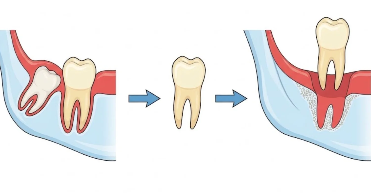
Peri Implant Bone Loss Detected may call for treatments with different recovery periods and risks. Non-surgical cleaning can leave gums temporarily tender, while surgery may cause swelling, soreness, bleeding, or tissue changes. Regeneration and implant removal involve extra healing. Dentists should explain expected benefits and complications, and postoperative instructions must be followed carefully.
Note : All images used are for editorial and illustrative purposes only and may not originate from the original news provider or associated company.
Temporary soreness and swelling after dental treatment
Cleaning inflamed tissues can cause short-term tenderness, while surgical procedures usually produce more noticeable swelling and discomfort. The degree depends on the procedure and individual healing. Following the aftercare instructions supports comfortable recovery, and severe, worsening, or unexpected symptoms should be reported to the dental team. Cold compresses, a soft diet, and careful cleaning are often advised during the first days, depending on the procedure performed.
Sensitivity and soft-tissue irritation
Treatment around inflamed or surgically managed tissue may cause temporary sensitivity or changes in gum appearance. The extent depends on the location and scale of treatment. Dentists provide cleaning instructions for the recovery period, and any irritation that persists or worsens should be professionally checked.
Risks associated with surgical treatment
Surgical risks include pain, swelling, bleeding, infection, and soft-tissue recession, with additional considerations for graft materials in regenerative procedures. Anatomy may affect complexity and recovery. Surgery is not suitable for every defect, so patients should receive individualized information before consenting and attend all follow-up appointments.
Why individual risks depend on the procedure and clinical findings
Non-surgical cleaning carries different risks from surgical or regenerative care. Defect size, implant location, tissue condition, stability, and patient health all influence complexity and healing. Dentists should explain benefits, limitations, and potential complications, and patients should ask questions before agreeing to any treatment plan.
How Much Does Peri-Implant Bone Loss Diagnosis and Treatment Cost?
Peri Implant Bone Loss Detected treatment costs depend on diagnosis, severity, and required procedures. Examination, periodontal measurements, radiographs, and possibly CBCT form the diagnostic cost, while debridement, prosthetic adjustment, surgery, or regeneration add treatment fees. Prices vary between countries and clinics, so an individualized estimate after examination is more reliable than generic figures.
Average Peri-Implant Evaluation and Diagnostic Costs in the US
In the United States, consultation fees vary widely by practice and region, and periodontal assessment may be billed separately from imaging. Periapical radiographs generally cost less than CBCT. Treatment costs depend on severity and the number of affected implants, and insurance coverage can change the final out-of-pocket amount. Published averages should be treated as rough guidance rather than fixed prices.
Clinical examination, periodontal assessment, X-rays, and possible CBCT
Diagnosis may involve a clinical examination, periodontal measurements, periapical radiographs, and CBCT when three-dimensional information is needed. Each can carry a separate fee, so total diagnostic cost varies. Ask which examinations are included in a quoted consultation, and expect any additional imaging to have a clear clinical purpose.
Average Peri-Implant Evaluation and Diagnostic Costs in the UK
UK costs differ between private practices and specialist periodontal services. Radiographs may be charged separately, and CBCT usually costs more than intraoral imaging. Treatment fees reflect the number of implants and disease severity. Published prices change and may exclude procedures, so request a written estimate after assessment.
Consultation, radiographic assessment, and additional diagnostic imaging
Some patients need only a standard examination, while complex cases may require periodontal charting and three-dimensional imaging, each with its own cost. Ask whether imaging is included in the consultation fee. Treatment planning should follow diagnostic findings, and a personalized estimate is more accurate than online averages.
Average Peri-Implant Evaluation and Diagnostic Costs in Turkey
Peri Implant Bone Loss Detected evaluation costs in Turkey vary by clinic, imaging needs, and case complexity. Some clinics include examination and digital imaging within a consultation, with CBCT charged separately when indicated. International patients should confirm package contents, including accommodation, transport, restorations, and aftercare, and request a plan based on actual findings.
Dental examination, digital imaging, and implant assessment
Evaluation may combine clinical examination, digital X-rays, and CBCT when three-dimensional detail is needed, alongside assessment of surrounding tissues and restoration components. The exact process depends on the clinical presentation. Ask which imaging is included, since an accurate estimate requires examination rather than symptoms alone.
What Can Change the Cost of Peri-Implant Bone Loss Treatment?
Costs are shaped by the number of affected implants, the severity of bone loss, whether treatment is surgical, diagnostic imaging requirements, regenerative materials, and any prosthetic replacement. For international patients, accommodation and transportation may also apply. A detailed, itemized estimate should be requested before treatment begins.
Severity and extent of bone loss
Mild inflammatory disease usually needs simpler management than advanced defects, which may require surgery, more clinical time, and extended monitoring. Several affected implants increase complexity further. The final cost therefore depends on clinical findings and chosen strategy, and an accurate estimate requires a detailed assessment first.
Number of affected implants
Treating one implant differs considerably from managing several. Multiple affected sites may need broader periodontal or prosthetic assessment and additional imaging. A good treatment plan identifies each affected implant and its recommended intervention, and patients should request an itemized estimate when more than one implant is involved.
Imaging and diagnostic requirements
Standard radiographs are sufficient for many evaluations, while CBCT adds cost when three-dimensional information is necessary. The number of scans and regions examined influences price. Imaging should be chosen according to clinical need, and patients can ask whether diagnostic imaging is included in their consultation package.
Non-surgical versus surgical treatment
Non-surgical care involves professional debridement and personalized hygiene instruction, whereas surgery requires more clinical time, materials, and follow-up. The type of surgery depends on defect shape and treatment goals. A detailed estimate should follow diagnosis, and cost alone should never decide whether a procedure is clinically appropriate.
Bone grafting or regenerative procedures when clinically appropriate
Regeneration adds materials, surgical fees, and follow-up visits, and is suitable only for certain defect shapes with controlled inflammation. Patients should receive individualized information about expected benefits and limitations, and costs are best discussed after the defect has been properly evaluated clinically and radiographically.
Why Peri-Implant Treatment Prices Are Not Fixed
Peri Implant Bone Loss Detected in one patient may need only non-surgical care, while another requires surgery, regeneration, or prosthetic replacement. The number of implants, defect severity, inflammation, and imaging needs all vary. At Vitrin Clinic, pricing follows clinical and digital assessment, so each patient receives an individualized plan.
How Vitrin Clinic Evaluates Implant Bone Levels
Peri Implant Bone Loss Detected at Vitrin Clinic is evaluated through clinical examination of the implant, tissues, and restoration, including probing, bleeding, and stability checks. Digital X-rays are used when appropriate and CBCT when three-dimensional information is required. Previous records reveal change over time, and planning follows findings rather than symptoms alone.
Clinical Assessment of Implant and Surrounding Tissues
The dental team evaluates the implant, gums, oral tissues, and restoration, recording plaque, inflammation, bleeding, probing depths, stability, and prosthetic factors. Previous records provide baseline information. This examination determines whether additional imaging is needed, building an individualized understanding of implant health rather than relying on symptoms.
Digital Imaging for Dental Implant Bone Loss Assessment
When clinical findings call for more information, Vitrin Clinic can use digital X-rays and, where three-dimensional detail is required, CBCT. Imaging supports evaluation of implant position and surrounding bone and is interpreted with the examination and previous records. Not every implant needs CBCT, which avoids unnecessary exposure.
Digital X-rays when clinically appropriate
Digital periapical radiographs show crestal bone levels and allow comparison with earlier images when consistent records exist. The dentist checks image quality and positioning before interpreting changes. Radiographs form only part of the diagnostic picture, but digital imaging supports efficient documentation and reliable long-term monitoring.
CBCT when three-dimensional assessment is clinically indicated
CBCT can characterize complex defects and areas hidden on two-dimensional images when conventional imaging cannot answer a clinical question. At Vitrin Clinic, scans are used when indicated and interpreted alongside the clinical examination and other records. CBCT is not automatically required for every implant.
Personalized Evaluation of Bone Levels and Implant Health
Bone levels are assessed together with implant stability, tissue condition, and restoration factors, using previous radiographs and current clinical measurements to identify change. Digital diagnostics are added when appropriate. The resulting plan reflects the severity and pattern of findings, avoiding one-size-fits-all treatment for every radiographic change.
Treatment Options for Peri-Implant Disease and Bone Loss
Depending on severity, Vitrin Clinic may recommend non-surgical management, surgical evaluation for advanced defects, prosthetic correction where cleaning or function is compromised, or regeneration for selected defects. Removal is discussed only when an implant cannot be predictably maintained, and long-term maintenance follows active treatment.
Internal-Link Opportunity: Dental Implant Treatment
Patients considering or maintaining implants can learn more through Vitrin Clinic's dental implant services, covering diagnostic planning and restoration options. Treatment begins with an individualized assessment, appropriate imaging of available bone and anatomy, and a discussion of long-term maintenance so that placement and future care are planned together. Discussing maintenance before treatment helps patients understand that implant care continues long after the restoration is fitted.
Internal-Link Opportunity: Full-Mouth Implant Treatment
Patients with multiple implants or full-mouth restorations need careful planning of implant position, prosthetic design, and maintenance. Existing implants may require evaluation before additional treatment, and previous bone loss should be considered when designing future restorations. Vitrin Clinic assesses individual findings before recommending a strategy.
Internal-Link Opportunity: All-on-4, All-on-6, or All-on-8 Treatment
Full-arch systems such as All-on-4, All-on-6, and All-on-8 need long-term professional maintenance, because plaque beneath the prosthesis can affect peri-implant tissues. Prosthetic design should allow cleaning access. Patients with symptoms around full-arch implants should be evaluated, and Vitrin Clinic can assess existing conditions before further treatment.
How Vitrin Clinic Supports Your Treatment Journey
Peri Implant Bone Loss Detected in international patients often requires coordinated assessment and planning. Vitrin Clinic reviews previous X-rays, CBCT scans, and implant records, examines tissues and restorations, and adds digital diagnostics when appropriate. Recommendations reflect bone levels, inflammation, stability, and prosthetic factors, with follow-up planned for long-term maintenance.
Initial Consultation and Review of Previous Dental Records
Evaluation begins by reviewing available X-rays, CBCT scans, implant documentation, and previous periodontal and maintenance findings. Historical records help identify changes in bone levels over time. Patients should provide as much relevant documentation as possible, since it supports a more complete and accurate clinical assessment.
Clinical and Digital Diagnostic Assessment
The team may assess teeth, gums, oral tissues, implant stability, and restoration condition, adding digital X-rays or CBCT when appropriate and three-dimensional intraoral scanning for digital workflows. Imaging decisions depend on specific clinical findings, with the goal of gathering exactly the information needed for accurate treatment planning.
Personalized Treatment Planning Based on Clinical Findings
Plans are tailored to the number and location of affected implants and may address inflammation, plaque control, mechanical factors, or bone defects, with surgery considered when appropriate. Patients can discuss treatment stages, expected monitoring, and aftercare, and individual findings determine the final strategy.
Coordinating Appropriate Implant or Peri-Implant Treatment
Management may need to address both biological and prosthetic factors. Vitrin Clinic organizes care according to the identified problem, which may include professional cleaning, periodontal management, prosthetic adjustment, or surgery. The specific approach depends on disease severity and implant stability.
Follow-Up and Monitoring of Implant Bone Levels
Monitoring continues after treatment, with clinical findings compared against previous measurements and radiographs taken when indicated. Follow-up confirms whether inflammation has improved and bone levels remain stable, making long-term maintenance an essential part of implant care at Vitrin Clinic.
Supporting International Patients Through the Assessment and Treatment Process
Vitrin Clinic supports international patients through consultation, planning, and coordinated care. Previous records can be reviewed before or during assessment, and digital communication helps organize diagnostic information. Each patient receives an individualized plan, and appropriate follow-up is discussed before returning home.
Evidence-Based Guidance for Peri-Implant Bone Loss Detection
Peri Implant Bone Loss Detected should be evaluated with evidence-based clinical and radiographic methods. Contemporary implant dentistry emphasizes probing, bleeding and suppuration assessment, radiographs compared with baseline records, and CBCT in selected cases. Literature also highlights individualized risk assessment, and studies should be interpreted according to their design and limitations.
What Current Peer-Reviewed Evidence Says About Peri-Implant Disease Diagnosis
Current literature supports diagnosis based on clinical and radiographic findings rather than symptoms. Probing, bleeding assessment, and radiographic evaluation are central, while baseline and follow-up records improve interpretation of bone change. Risk factors are considered, and guidelines favor individualized evaluation over any single isolated measurement.
Recommendations From Periodontal and Implant Dentistry Literature
Professional guidance emphasizes regular assessment of peri-implant tissues through probing, bleeding and suppuration checks, and radiographic evaluation of bone. Longitudinal monitoring is stressed because disease can progress silently. Management depends on severity and defect characteristics, and recommendations continue to evolve as research develops.
Role of NIH, NIDCR, PubMed, and Peer-Reviewed Research
PubMed indexes peer-reviewed research on implant dentistry and peri-implant disease, while NIH and NIDCR offer evidence-based oral health information. Research informs diagnostic methods and treatment outcomes, but individual studies must be read in light of design, population, and limitations. Systematic reviews and guidelines provide broader context. Clinical decisions should never rest on a single study alone.
How Evidence Supports Clinical and Radiographic Assessment
Evidence favors combining clinical findings on inflammation with radiographic information on bone levels, and longitudinal comparison improves detection of progression. CBCT helps in selected complex cases. No single imaging method replaces clinical assessment, and the approach should match each patient's clinical question and risk profile.
Key Takeaways About Peri Implant Bone Loss Detected
Peri Implant Bone Loss Detected relies on clinical findings, radiographic assessment, and comparison with previous records, because early changes are often painless. Bleeding, swelling, suppuration, recession, and deeper pockets need evaluation. Treatment depends on severity and cause, while prevention rests on plaque control, maintenance, periodontal care, and managing modifiable risk factors.
Peri-Implant Bone Loss Detection in Brief
Peri Implant Bone Loss Detected involves more than noticing symptoms. Dentists assess soft tissues, probing depths, bleeding, suppuration, stability, and supporting bone, using standardized radiographs and CBCT when indicated. Previous records reveal progression, and biological, mechanical, and patient factors are all considered. Regular maintenance catches problems before they become advanced.
The Importance of Clinical Examination and Appropriate Imaging
Clinical examination identifies inflammation, pocket changes, bleeding, and prosthetic factors before imaging is interpreted. Radiographs show supporting bones, and CBCT adds three-dimensional detail when necessary. Choosing imaging according to the clinical question, and combining methods, provides a far more complete assessment than symptoms alone.
Early Warning Signs Patients Should Not Ignore
Bleeding, swelling, redness, suppuration, recession, and increasing pocket depth all warrant attention, and implant mobility needs prompt evaluation. Early disease can also be completely silent, so regular professional examinations remain essential. Persistent changes should never be dismissed simply because the implant still feels comfortable.
When Professional Evaluation Is Appropriate
Seek evaluation when persistent inflammation, bleeding, swelling, discharge, recession, or mobility develops around an implant. Dentists decide whether clinical measurements or imaging are needed, and early assessment can identify inflammation before extensive tissue damage occurs. Self-diagnosis of bone loss is not reliable.
Relief, Medical Tips, and Treatment Considerations
Treatment may address inflammation, plaque, mechanical factors, or bone defects, guided by professional diagnosis. Non-surgical care suits selected cases, while surgical or regenerative treatment may be considered for advanced defects. Remember that relief of symptoms does not prove that bone loss has stopped.
Why Long-Term Monitoring Helps Protect Dental Implants
Consistent monitoring combines clinical measurements of tissue health with radiographs of supporting bone, compared against earlier records. Maintenance visits also address plaque accumulation and prosthetic problems early. Monitoring remains important for the life of the implant, even when it feels completely comfortable.
At Vitrin Clinic
Dental implant treatment begins with a detailed evaluation of your teeth, gums, bone levels, and existing restorations. Clinical examination is combined with digital X-rays and CBCT when three-dimensional assessment is clinically appropriate. The team evaluates bone volume, implant position, surrounding tissues, and the condition of existing implants. These findings help determine whether implant placement, implant restoration, or additional treatment may be appropriate. Previous dental records can also help identify changes in bone levels over time. Each treatment plan is developed according to individual clinical findings, implant needs, and restorative goals. Vitrin Clinic provides personalized dental implant solutions, including single implants, full-mouth implant treatment, and All-on-4, All-on-6, or All-on-8 options when clinically suitable.
Reference
FAQs
Dr. Rifat Alsaman has more than 5 years of clinical experience in dentistry and currently serves as the Head of the Medical Team at Vitrin Clinic. He is dedicated to providing exceptional patient care, overseeing treatment planning, and ensuring the highest clinical standards across the team. His expertise, attention to detail, and commitment to continuous professional development have helped countless patients achieve healthier, more confident smiles.


