Endodontics
September 30, 2026
Salivary Viscosity and Root Canal Retreatment Healing: Can Thick Saliva Affect Recovery?
 Dr. Rifat Alsaman
Recovery after root canal retreatment depends on several connected factors. These include infection control, canal sealing, tooth anatomy, and restoration quality. The tissue response around the root also shapes the final outcome. Researchers are now studying saliva as a possible biological marker in this process. Salivary Viscosity and Root Canal Retreatment Healing is therefore a newly explored clinical topic. A prospective cohort published on September 29, 2026, examined this relationship in retreatment patients. It found that higher viscosity was associated with less favorable healing. However, it did not prove that thick saliva directly causes retreatment failure. This distinction matters when patients describe their symptoms. Thick saliva can also follow dehydration, reduced flow, medications, or dry-mouth conditions. This guide explains the current evidence and its practical meaning.
What Is Salivary Viscosity and Why Does It Matter in Endodontics?
Salivary Viscosity and Root Canal Retreatment Healing links a saliva property with a dental healing outcome. Viscosity describes how strongly saliva resists flowing. It changes with water content, proteins, mucins, and gland activity. Thick saliva may feel stringy, sticky, or hard to swallow. Endodontics usually focuses on the treated tooth, canal system, bone, and restoration. Saliva becomes relevant mainly through contamination risk and broader biology. The 2026 cohort suggests viscosity may carry prognostic value. However, the underlying mechanism remains uncertain. This approach avoids overinterpreting an easily noticed symptom.
What does salivary viscosity mean?
Salivary viscosity is the resistance of saliva to movement or flow. Higher viscosity makes saliva feel thicker, stickier, or more string-like. Mucin-rich secretions contribute strongly to these properties. Different glands produce secretions with different water and protein proportions. Whole saliva mixes these secretions and changes throughout the day. Food, hydration, stimulation, and medications can all alter consistency. Laboratory devices measure viscosity more objectively than visual inspection. A dentist may therefore ask about thirst, medications, and daily fluid intake.
What is the difference between normal saliva, thick saliva, and dry mouth?
Normal saliva supports lubrication, swallowing, speech, buffering, and tissue protection. Thick saliva describes a noticeable change in consistency. Dry mouth describes a symptom of oral dryness. Hyposalivation refers to objectively reduced salivary flow. These concepts overlap, but they are not identical. A person may feel dry without a major measured flow reduction. Another may have reduced flow with obvious stickiness. It also does not automatically indicate a root canal complication.
How do saliva flow, mucins, hydration, and oral conditions influence viscosity?
Several physical and biological factors interact to shape viscosity. Hydration controls how much water reaches oral secretions. Mucins add lubrication and the stringy feel of thicker saliva. Resting and stimulated saliva differ in concentration. Mouth breathing can increase dryness, especially overnight. Stress, medications, systemic conditions, and gland dysfunction may also contribute. Saliva texture cannot show whether bacteria remain inside a treated canal.
Why is saliva important for oral microbial balance and tissue protection?
Saliva protects the mouth through several functions. It lubricates tissues and supports chewing, speaking, and swallowing. It provides buffering capacity and minerals for tooth surfaces. Salivary proteins influence how microorganisms attach to oral surfaces. Flow clears food debris, acids, and microbes from exposed areas. Reduced flow can increase discomfort and dental disease risk. Larger research is needed before viscosity becomes a validated biomarker.
Saliva flow versus saliva consistency
Flow and consistency describe different properties. Flow is the amount of saliva produced over a set time. Consistency describes physical behavior, such as thin, foamy, or sticky saliva. A patient may have normal flow yet feel thick saliva. Another may have low flow without noticing thickness. Endodontic decisions rarely depend on either measurement alone.
Why thick saliva does not automatically mean an endodontic problem
Thick saliva is a mouth-level observation, not a tooth diagnosis. It may follow dehydration, mouth breathing, medications, stress, or reduced flow. Temporary changes can occur while teeth heal normally. Root canal disease requires evidence from symptoms, examination, or imaging. Swelling, drainage, and persistent lesions provide more direct information. Research in this field does not change this basic principle. Patients should report saliva changes without self-diagnosing failure.
See which suits you in Maryland Bridge vs Traditional Bridge
Is There a Link Between Salivary Viscosity and Root Canal Retreatment Healing?
Salivary Viscosity and Root Canal Retreatment Healing gained attention after a new prospective cohort study. It included 56 patients with asymptomatic apical periodontitis in mandibular molars. Researchers measured salivary viscosity with a digital viscometer. Clinical and CBCT assessments occurred at baseline, six months, and twelve months. Fifty teeth were evaluated at twelve months. The combined success rate, counting healed and healing cases, was 88%. Each 1-cP increase gave an adjusted odds ratio of 0.68. The findings justify further research rather than a new clinical rule.
What does the latest research show about salivary viscosity and root canal healing?
The most relevant evidence is the cohort published on September 29, 2026. Viscosity was measured objectively rather than estimated from descriptions. All 50 evaluable teeth showed clinical success at twelve months. Radiographically, 16% were healed and 72% were healing. The remaining 12% were classified as diseased. They called for larger multicenter studies with longer follow-up.
Can higher salivary viscosity be associated with less favorable periapical healing?
Yes, the 2026 cohort reported this association. Higher viscosity predicted lower odds of twelve-month success. It also predicted smaller reductions in lesion volume. A mixed-effects analysis found a significant viscosity-by-time interaction. This suggests the association strengthened across follow-up. Viscosity may act as a marker rather than a direct cause.
Does salivary viscosity directly cause root canal retreatment failure?
Current evidence does not establish direct causation. The cohort was observational rather than interventional. Unmeasured confounding may still exist after adjustment. Treatment used strict rubber dam isolation. Definitive coronal restorations were placed shortly afterward. Patients should not assume thick saliva damages their retreatment.
Association versus causation in the available evidence
An association means two variables change together more than chance predicts. Causation means one variable directly produces the other. Observational studies can show associations without proving cause. The 2026 cohort adjusted for tooth type and baseline lesion volume. Viscosity remained significantly associated with success afterward. The findings guide research, not direct intervention.
What the 2026 prospective cohort study actually measured
The study enrolled 56 patients with asymptomatic apical periodontitis in mandibular molars. Salivary viscosity was measured with a digital viscometer. Intracanal bacteria were identified with the VITEK 2 system. Clinical and CBCT outcomes were recorded before treatment. Follow-up occurred at six and twelve months. Outcomes were graded as healed, healing, or diseased. It remains the core source on Salivary Viscosity and Root Canal Retreatment Healing.
Why larger and longer-term studies are still needed
A single-center cohort gives a signal, not a universal rule. This study had a modest sample and narrow disease profile. Results may differ for anterior teeth or symptomatic disease. Primary treatment and surgical retreatment may also behave differently. The authors noted bacterial analyses may have been underpowered. Periapical repair can continue beyond twelve months. Such work would clarify Salivary Viscosity and Root Canal Retreatment Healing more reliably.
How Could Saliva Influence Bacterial Infection After Root Canal Treatment?
Salivary Viscosity and Root Canal Retreatment Healing must be separated from direct salivary contamination. During treatment, saliva can carry microbes into an open tooth if barriers fail. A finished, sealed canal should no longer communicate with the mouth. Persistent infection usually involves microbes that survived or entered through a failed seal. Thick saliva may matter biologically, but it does not prove contamination. Rubber dam isolation controls salivary exposure during procedures. Patients should focus on treatment quality, sealing, and restoration follow-up.
How can salivary contamination contribute to intracanal bacterial infection?
Saliva contains many microorganisms that normally live in the mouth. An open canal becomes vulnerable if contamination control is interrupted. Bacteria may reach instruments, the operative field, or the access cavity. Research has found contamination of files, gloves, and rubber dam surfaces. These findings support strict aseptic technique during endodontics. The focus then shifts toward preventing coronal leakage.
Can saliva introduce bacteria into a poorly sealed root canal?
Yes, a compromised coronal seal can create a bacterial pathway. Gaps, defective fillings, decay, or fractures may expose the access area. Over time, bacterial penetration can reinfect the canal system. Classic research examined coronal leakage as a route to failure. Retreatment assessment therefore includes restoration quality. Imaging may reveal findings that need further treatment.
How do coronal leakage, defective restorations, and recurrent decay affect reinfection?
Coronal leakage lets oral bacteria travel toward the treated canal. Defective restorations can admit fluids and microorganisms. Recurrent decay may undermine restorations and expose treated structure. These problems can matter even without pain. A sound restoration protects the endodontic filling. The restoration may need repair or replacement after endodontic care.
What role may saliva play in bacterial retention and biofilm formation?
Saliva shapes the environment where oral bacteria attach and form biofilms. Its proteins form acquired pellicles on exposed surfaces. Flow helps clear microbes and dietary sugars. Higher viscosity may change how secretions move across surfaces. However, the 2026 cohort found no significant bacterial prevalence differences. Anatomy, oxygen, nutrients, and irrigation shape canal ecology.
Saliva contamination during treatment versus salivary characteristics during healing
These are two different clinical concepts. Contamination concerns microbes entering an open canal system. Salivary characteristics describe the oral fluid environment afterward. Rubber dam isolation addresses the first problem. The 2026 cohort limited direct exposure through isolation and early restorations. Its viscosity association may therefore reflect indirect biological factors.
Why rubber dam isolation and a durable coronal seal matter
Rubber dam isolation creates a controlled field during treatment. It reduces exposure to saliva and oral microbes. Research has documented contamination risks when treatment surfaces become soiled. A durable coronal seal then limits microbial access afterward. These protections work regardless of saliva thickness. Success depends on both canal treatment and coronal protection.
Find the differences in Root Canal vs Apicoectomy
What Do We Know?
Salivary Viscosity
Intraradicular Bacteria
Salivary Viscosity and Root Canal Retreatment Healing includes an important microbiological question. Researchers asked whether viscosity matched different bacterial findings inside canals. The 2026 cohort identified intracanal organisms with the VITEK 2 system. Prevalence was compared between lower and higher viscosity groups. After false discovery rate correction, no significant differences remained. Viscosity therefore did not simply track detected bacteria in this cohort. This challenges a straightforward contamination explanation. Larger molecular studies could examine these possibilities.
Are bacteria more common in people with higher salivary viscosity?
The retreatment cohort did not show a significant prevalence difference. Researchers compared lower and higher viscosity groups. Multiple-comparison correction was applied to all tests. All adjusted p values were above 0.05. The study therefore does not support linking thicker saliva with more canal bacteria. Future work could measure bacterial quantity and molecular signatures.
Which bacterial findings have been studied in endodontic retreatment?
Retreatment research examines microbes that persist after earlier treatment. Studies use culture, sequencing, or automated identification systems. Reported organisms include Gram-positive and Gram-negative taxa. Fungi such as Candida have also been studied in persistent disease. The 2026 cohort used VITEK 2 for identification. A negative comparison does not prove identical microbial ecosystems.
Why bacterial presence does not always explain differences in healing
Healing reflects microbial burden, anatomy, treatment quality, host response, and time. Two teeth with detectable bacteria may heal differently. Bacterial location matters when complex anatomy limits access. Host inflammatory responses also vary between patients. A 2024 systematic review identified several clinical prognostic factors. Bacterial presence is important but not the only determinant.
Findings from the latest retreatment cohort
The 2026 cohort compared viscosity, bacteria, and healing together. Higher viscosity was linked with lower odds of twelve-month success. It was also linked with smaller lesion volume reduction. Yet bacterial prevalence did not differ significantly after correction. It remains central to Salivary Viscosity and Root Canal Retreatment Healing discussions. The results support a research signal, not a treatment prescription.
Limits of bacterial identification and small-sample studies
Sample size, detection method, and sampling depth limit microbiological studies. Culture-based or automated systems may miss parts of the community. Molecular methods detect more but need careful interpretation. The 2026 study was powered for success, not bacterial outcomes. The authors acknowledged limited power for modest taxonomic differences. Future studies need larger, multicenter samples and advanced molecular tools.
Compare options in Traditional Bridge vs Cantilever Bridge
What Factors Really Affect Root Canal Retreatment Healing Outcomes?
Salivary Viscosity and Root Canal Retreatment Healing is one emerging question among many. Established predictors remain central to retreatment decisions. Persistent infection is a major reason for retreatment. Missed canals can leave infected spaces untreated. Poor cleaning, shaping, or filling can let microbes persist. Coronal leakage can cause secondary infection. A 2024 systematic review found pooled healing near 78.8% under strict criteria. Looser criteria produced about 87.5% healing. These figures show why outcome definitions matter.
Persistent or recurrent intracanal infection
Persistent infection occurs when microbes survive the original treatment. Recurrent infection develops when microbes later re-enter the canal. Both can maintain inflammation around the root tip. Retreatment removes old filling material for renewed disinfection. Irrigation and shaping should reach accessible anatomy thoroughly. The goal is controlling infection while preserving a restorable tooth.
Missed canals and complex root anatomy
Root canal systems vary widely in anatomy. A missed canal can retain tissue, bacteria, or debris. Molars are usually more complex than single-rooted front teeth. Magnification and appropriate imaging improve canal identification. CBCT helps when conventional images leave key questions unresolved. The right option depends on restorability and lesion location.
Inadequate cleaning, shaping, or root filling
Canal preparation aims to reduce microbial load in complex spaces. Cleaning depends on instrumentation, irrigation, and access. Shaping creates room for irrigation and effective filling. Voids, short fillings, or overextensions complicate prognosis. The 2024 review linked outcomes with the extent of existing root filling. Healing is clearly multifactorial.
Coronal leakage and restoration quality
The restoration above the canal is part of long-term protection. Defective restorations let fluids and microbes reach the access area. Recurrent decay and fractures create similar risks. The coronal seal should be checked during retreatment assessment. Planning should consider remaining tooth structure and chewing forces. Endodontic healing cannot be separated from restoration integrity.
Pre-existing periapical lesion size and severity
A periapical lesion reflects inflammation around a root tip. Larger lesions usually take longer to show improvement. Baseline severity influences how studies classify success. The 2024 review listed lesion characteristics among key prognostic factors. The 2026 cohort measured lesion volume with CBCT. It found greater reduction at twelve months than at six.
Tooth fracture, perforation, and other structural problems
Retreatment cannot correct every structural cause of symptoms. Vertical root fractures may cause periodontal defects or drainage. Perforations can compromise sealing depending on location. Extensive cracks may affect restorability and prognosis. Magnification and imaging help evaluate suspicious findings. Treatment should target the real source of disease.
Patient-related and biological factors
Patients differ in healing response, habits, and treatment history. Age alone does not decide whether retreatment heals. Hygiene, smoking, medications, and systemic health influence oral conditions. These factors should be reviewed without assuming causation. Healing also needs time after infection is controlled. Salivary Viscosity and Root Canal Retreatment Healing research may eventually add biological information here.
Oral hygiene, smoking, medications, and dry mouth
Oral hygiene controls plaque around treated teeth. Smoking can affect oral tissues and healing responses. Medications may reduce saliva flow or alter mouth feel. Persistent dry mouth raises discomfort and dental disease risk. A medication history helps explain oral dryness. Concerns should go to the prescribing professional.
Why salivary viscosity should be considered alongside established prognostic factors
Salivary Viscosity and Root Canal Retreatment Healing may eventually add value to prognosis. The 2026 cohort found a significant association with twelve-month outcomes. Established variables still guide treatment planning directly. These include periapical status, lesion size, and filling quality. Anatomy, restoration integrity, and follow-up duration also matter. It should not replace clinical diagnosis or imaging.
.jpg)
.jpg)
.jpg)
.jpg)
What Are the Signs That a Previously Root-Treated Tooth May Not Be Healing?
Salivary Viscosity and Root Canal Retreatment Healing should never be judged from saliva alone. Mild tenderness can occur during early recovery. Persistent or worsening pain deserves clinical assessment. Swelling, drainage, or a recurring sinus tract may indicate ongoing infection. Some lesions remain visible while tissues continue healing. Radiographic disease may also persist without symptoms. A dentist can confirm whether recovery follows the expected course.
Can pain after root canal retreatment be normal?
Mild tenderness can occur after retreatment. Surrounding tissues respond temporarily to instrumentation. Biting sensitivity may appear while these tissues recover. Symptoms should fade rather than worsen over time. Severe pain or increasing swelling needs professional review. Imaging may be needed if symptoms persist.
When can tenderness or discomfort indicate delayed healing?
Tenderness becomes concerning when it outlasts the expected recovery pattern. A stable or worsening trend deserves evaluation. Biting pain may reflect periapical inflammation, occlusal irritation, or structural problems. Examination separates periodontal, restorative, and endodontic sources. Radiographs provide a baseline for later comparison. Saliva consistency should be mentioned but not a dominant assessment.
What do swelling, a gum boil, drainage, or a sinus tract suggest?
Swelling near a treated tooth can signal active inflammation or infection. A gum boil may be a draining sinus tract. Persistent drainage should not be dismissed because pain is mild. Drainage can relieve pressure and mask disease severity. Examination and radiographs help trace the source. Antibiotics are not routine for localized conditions without systemic involvement.
Can a patient have a persistent periapical lesion without obvious symptoms?
Yes, a lesion may persist with little or no pain. Endodontic inflammation does not always cause daily symptoms. Follow-up imaging is therefore important after retreatment. A lesion may shrink gradually rather than disappear quickly. Strict criteria require complete resolution for healing. Serial imaging is more informative than one isolated image.
How should thick saliva be interpreted when a treated tooth is also symptomatic?
Thick saliva should be recorded as a separate symptom. The dentist can ask about hydration, dryness, medications, and timing. The treated tooth is then assessed with dental-specific findings. Pain, percussion, swelling, restoration quality, and imaging give direct evidence. Salivary Viscosity and Root Canal Retreatment Healing evidence shows association, not proven causation. Treating dehydration may improve comfort without fixing a failed canal.
Learn more about Rodent-Borne Disease and Dental Health
How Is Root Canal Retreatment Healing Diagnosed?
Diagnosis of healing begins with a structured clinical review. Salivary Viscosity and Root Canal Retreatment Healing informs research, but diagnosis stays tooth-centered. The dentist reviews treatment history, pain, swelling, biting, and drainage. The restoration is checked for fractures, leakage, and decay. Periapical radiographs show two-dimensional information about the tooth and bone. CBCT adds three-dimensional detail when results could change management. Healing is a process, not a single moment.
What happens during a clinical examination?
The examination starts with symptoms and treatment history. The dentist may test percussion and palpation tenderness. Biting tests help locate symptoms to a specific tooth. Gums are checked for swelling, sinus tracts, and pockets. The restoration is assessed for defects, decay, and fractures. Objective examination prevents blaming symptoms on saliva alone.
Which dental X-rays are used to monitor periapical healing?
Periapical radiographs are standard for following treated teeth. They show changes around the root apex over time. Consistent positioning improves serial comparisons. A shrinking lesion may represent ongoing healing. Complete disappearance indicates radiographic resolution. CBCT is not needed for every routine follow-up.
When is CBCT useful for assessing root canal retreatment healing?
CBCT is useful when three-dimensional information could change management. It shows roots, canals, cortical bone, and periapical structures. Complex anatomy may justify a scan. Suspected missed canals or unusual lesions are common indications. The AAE describes CBCT as valuable in selected endodontic cases. CBCT supports, but never replaces, clinical judgment.
What can CBCT show that conventional radiographs may not?
CBCT gives three-dimensional views of teeth and surrounding bone. It reveals anatomy that overlaps on standard radiographs. It can show canal configuration and cortical involvement. It helps evaluate suspected missed anatomy in retreatment cases. The 2026 cohort used this method to track healing. Benefits must outweigh radiation and incidental findings.
Periapical lesion size and volume
Lesion size describes the extent of radiographic change near a root. Traditional radiographs estimate size in two dimensions. CBCT supports three-dimensional volume measurement. Volumetric data may detect subtle changes over time. The 2026 study on Salivary Viscosity and Root Canal Retreatment Healing used this approach. Consistent imaging improves long-term comparison.
Healing, persistent disease, and changes over time
Periapical healing is gradual rather than immediate. A lesion can shrink without disappearing completely. Researchers separate complete healing from ongoing healing. The 2024 review showed how definitions change success rates. A shrinking, symptom-free lesion may warrant observation. Serial evaluation reveals the direction of change.
Why CBCT should be used selectively rather than routinely
CBCT uses ionizing radiation and needs a clear purpose. Routine scanning may not change management. Focused scans reduce unnecessary exposure. The AAE notes CBCT greatly enhances diagnosis in certain cases. Field of view and resolution should match the question. Selective use supports both safety and usefulness.
Explore Electrolyzed Saline and Airborne Bacteria in Dental Procedures
What does “healed” mean after endodontic retreatment?
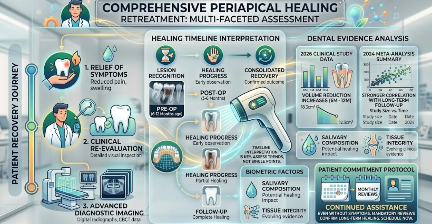
“Healed” usually means symptoms resolved and periapical disease disappeared radiographically. Definitions vary across studies and clinics. Strict classifications require complete radiographic resolution. Loose classifications also accept substantial lesion reduction. The 2024 review reported about 78.8% strict and 87.5% loose healing. Patients can ask how their dentist defines healing.
What does “healing” mean when a lesion has not completely disappeared?
“Healing” describes an improving lesion that has not fully resolved. Radiographic reduction suggests biological repair is progressing. The 2026 cohort classified 72% of cases as healing. Another 16% were healed at twelve months. Six of 50 teeth, or 12%, were classified as diseased. Complete disappearance should not be expected immediately.
How long can periapical healing take after retreatment?
Healing time varies with disease extent, treatment quality, and biology. Some improvement appears within months. Complete resolution can take longer than one year. The 2026 cohort saw more reduction at twelve months than six. Follow-up duration also influenced outcomes in the 2024 review. Worsening symptoms or enlarging lesions should still be addressed.
Why are 6-month and 12-month assessments useful?
Six-month reviews show early clinical and radiographic direction. Twelve-month reviews capture further bone remodeling. The Salivary Viscosity and Root Canal Retreatment Healing cohort evaluated both points deliberately. Lesion reduction was greater at twelve months. Healing clearly continues after early reviews. Consistent imaging technique makes comparisons meaningful.
Clinical symptoms versus radiographic healing
Clinical and radiographic outcomes change at different speeds. Pain often improves before bone changes appear. A comfortable tooth may still show a visible lesion. Symptom relief therefore does not prove complete healing. In the 2026 study, all 50 teeth succeeded clinically. Both dimensions must be considered together.
Why a persistent radiolucency does not always mean immediate treatment failure
A radiolucency can remain visible while healing continues. Bone repair is slower than symptom relief. Research separates complete resolution from lesion reduction for this reason. A stable or shrinking symptom-free lesion may justify observation. Enlargement, drainage, or worsening pain needs reassessment. New images should always be compared with baseline records.
What Treatment Options Are Available When a Root Canal Is Not Healing?
Salivary Viscosity and Root Canal Retreatment Healing should be separated from the decision to intervene. Treatment depends on why the tooth is not healing. Nonsurgical retreatment suits correctable infection or technical problems. Endodontic microsurgery suits disease near the root tip. Extraction is discussed when the tooth cannot be predictably retained. Treatment should target the specific reason for persistent disease.
When is nonsurgical root canal retreatment considered?
Nonsurgical retreatment suits persistent or recurrent apical disease with correctable causes. Suspected missed canals or poor previous fillings are common reasons. Recurrent decay or coronal leakage may also prompt reassessment. The tooth must first be structurally suitable. A crack or fracture can change the plan entirely. Retreatment is diagnosis-driven, not automatic.
What happens during endodontic retreatment?
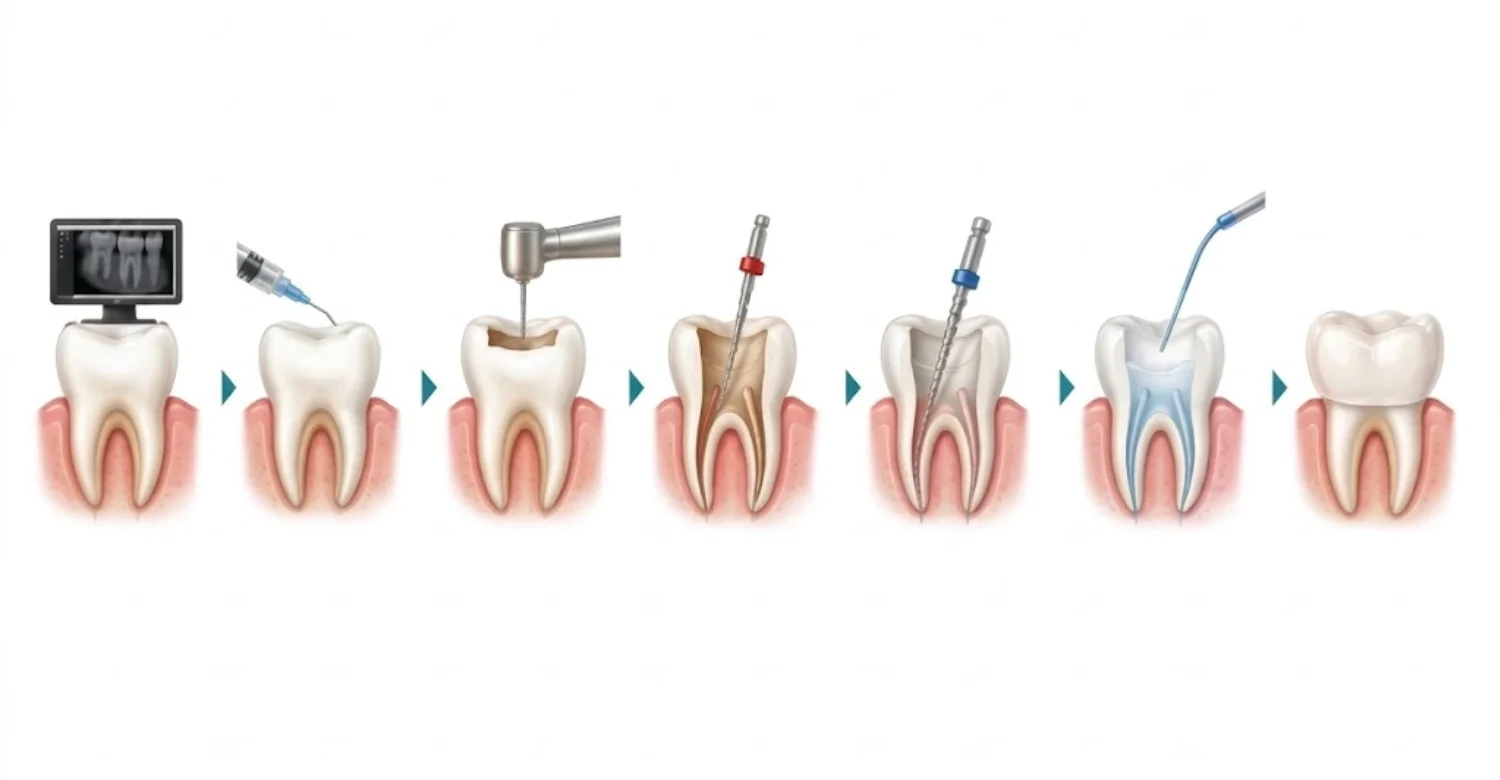
The dentist first reviews the tooth, restoration, symptoms, and imaging. Local anesthesia keeps the patient comfortable. Isolation is established before accessing the canal. Old filling materials are removed with specialized instruments. Canals are checked for missed anatomy or debris. Cleaning, shaping, and irrigation disinfect accessible spaces. Complex cases may need more than one appointment.
When might endodontic microsurgery be considered?
Microsurgery is considered when nonsurgical treatment cannot address the disease. It suits persistent apical infection near inaccessible canal segments. Separated instruments or anatomical limits may also justify it. The procedure accesses the root tip through surrounding tissue. Inflamed tissue is removed and the root end is sealed. The goal is preserving a restorable natural tooth.
When is extraction discussed?
Extraction is discussed when a tooth cannot be predictably restored. Vertical root fractures are a major structural reason. Severe tooth structure loss also limits options. Persistent disease alone does not automatically require extraction. Retreatment or microsurgery may still be possible. The decision should reflect the whole oral treatment plan.
Assessing restorability before choosing retreatment
Restorability asks whether enough tooth remains for a durable restoration. A successful canal cannot save a tooth that cannot be sealed. The dentist evaluates cracks, walls, gum support, and existing restorations. Tooth position affects functional demands. Patients should understand benefits, limits, and alternatives.
Treating missed canals, persistent infection, or coronal leakage
Each cause requires a different strategy. Missed canals are located through better access and magnification. Persistent infection needs deeper disinfection and correction of old fillings. Coronal leakage needs restoration repair or replacement. Recurrent decay must be removed before sealing. Identifying the failure mechanism avoids unnecessary procedures.
Can Improving Saliva and Oral Moisture Help During Recovery?
Salivary Viscosity and Root Canal Retreatment Healing concerns saliva texture, not a proven cure. Improving hydration has not been shown to heal a failed canal. Thick saliva often reflects changes in oral moisture. Drinking fluids can relieve temporary dryness when dehydration contributes. Persistent dry mouth needs separate evaluation. The 2026 study did not test hydration as an intervention. Moisture measures support comfort, not endodontic treatment.
Can dehydration make saliva feel thicker after dental treatment?
Yes, low fluid intake can make saliva more concentrated and sticky. Patients often drink less after procedures because of soreness. Mouth breathing can further increase dryness. The change may be most noticeable after waking. Salivary Viscosity and Root Canal Retreatment Healing research did not test hydration as treatment. Persistent symptoms deserve a separate dry-mouth assessment.
What can help relieve thick or sticky saliva?
Regular water intake can relieve a sticky mouth. Small, frequent sips may be easier than large amounts. Sugar-free gum can stimulate flow when chewing is comfortable. Saliva substitutes lubricate when natural secretion is low. Limiting caffeine may help some patients. Thick saliva is not proof of root canal failure.
Does drinking water improve saliva consistency?
Water can improve comfort when dehydration contributes to thick saliva. It supports normal body fluid balance. The effect is strongest when dehydration is actually present. More water does not guarantee normal measured viscosity. Flow, mucins, proteins, and gland function also matter. Hydration does not treat persistent intracanal infection.
What oral-care measures are appropriate after retreatment?
Continue gentle but thorough toothbrushing after retreatment. Keep flossing unless given a specific temporary restriction. Chew carefully until the final restoration is placed. Use fluoride toothpaste, especially with dry mouth. Follow temporary-restoration instructions closely. Report pain, swelling, or drainage promptly.
Sugar-free gum and saliva stimulation
Chewing sugar-free gum stimulates saliva flow. More flow can reduce dryness and lubricate tissues. Non-acidic, sugar-free products are best for teeth. Patients with jaw pain may need another option. Gum should not mask persistent swelling or pain. Persistent dryness still needs its cause identified.
Fluoride and routine oral hygiene
Fluoride strengthens enamel and lowers decay risk. This protection matters more when saliva flow drops. Brush twice daily with fluoride toothpaste. Include interdental cleaning in your routine. Dry mouth increases plaque retention and discomfort. Fluoride protects teeth but does not decide retreatment success.
When persistent dry mouth needs separate medical or dental evaluation
Dry mouth deserves evaluation when it continues despite good hydration. Trouble swallowing, speaking, eating, or sleeping signals a significant problem. Frequent cavities or oral soreness may also appear. Medication effects should be reviewed without stopping drugs independently. Salivary gland disorders may need medical assessment. The treated tooth still needs separate evaluation.
Understand the effects of Frequent Snacking and Tooth Decay
Relief After Root Canal Retreatment: What Medical Tips Are Safe?
Salivary Viscosity and Root Canal Retreatment Healing can cause anxiety when soreness and thick saliva coincide. Mild tenderness is usually managed with the dental team's plan. Protecting the tooth until final restoration reduces mechanical stress. Soft foods can improve comfort early on. Gentle oral hygiene should continue around the treated area. The ADA recommends dental treatment and pain relievers for most pulpal and periapical conditions. Every medication plan should follow professional advice.
What can help relieve mild soreness after retreatment?
Soreness occurs because tissues around the tooth were manipulated. A dentist may suggest a suitable over-the-counter pain reliever. The choice should consider allergies, conditions, and other medicines. Taking extra doses does not speed healing. Avoid chewing hard foods on a tender tooth. Severe or worsening pain warrants a call to the clinic.
How should patients protect the treated tooth while the final restoration is completed?
A treated tooth is more fracture-prone before definitive restoration. Follow the dentist's chewing instructions carefully. Avoid hard, sticky, or tough foods on that side. Keep the temporary restoration intact and clean. Contact the clinic if it loosens or breaks. Protection means reducing avoidable stress, not stopping normal eating.
What eating and oral-hygiene habits may make recovery more comfortable?
Soft foods reduce chewing pressure during early recovery. Chewing on the opposite side can help temporarily. Very hard foods may overload a tender tooth. Gentle brushing controls plaque without aggressive pressure. Careful flossing can continue around the tooth. These habits support comfort but do not guarantee success.
When should pain relief measures be discussed with a dentist or doctor?
Seek advice when pain is severe, worsening, prolonged, or uncontrolled. Medication questions matter for people taking several medicines. Kidney, liver, stomach, or bleeding conditions affect pain-reliever choices. Pregnancy and allergies also require individual advice. A dentist decides whether pain is expected. Never increase doses simply because pain persists.
Avoiding self-prescribed antibiotics
Antibiotics do not treat every dental pain. The ADA advises against them for most pulpal and periapical conditions. Dental treatment should address the infection source directly. Antibiotics matter more when fever or malaise appears. Leftover antibiotics risk side effects and inappropriate treatment. Use antibiotics only when prescribed for your situation.
Following the prescribed pain-control plan
A written plan helps prevent accidental medicine overuse. Follow dose, interval, and duration instructions exactly. Check whether products share the same active ingredient. Combining products blindly can cause overdose. The dental team can recommend the safest option. Changing symptoms are more informative than extra doses.
Maintaining hydration when saliva feels thick
Regular hydration eases sticky saliva from low fluid intake. Small sips through the day may be easier after treatment. Saliva often feels thicker after an overnight dry mouth. Sugar-free gum can add stimulation when appropriate. Persistent dryness should be investigated separately. Its role is moisture and general comfort.
What Side Effects Can Occur After Root Canal Retreatment?
Salivary Viscosity and Root Canal Retreatment Healing can overlap with temporary postoperative mouth changes. Mild tenderness is a common concern after retreatment. Chewing sensitivity may occur while tissues settle. The gum near the tooth may feel irritated. Some patients notice dryness or thicker saliva. These changes may reflect reduced drinking, mouth breathing, or medications. The course varies with infection severity and procedure complexity.
Temporary tenderness and chewing sensitivity
Tenderness occurs because tissues around the root are temporarily inflamed. Chewing pressure may make the tooth feel sensitive. Mild symptoms are usually monitored for improvement. Avoid excessive force while chewing remains uncomfortable. A bite adjustment may help if the tooth feels high. The symptom trend matters more than one moment.
Mild swelling or local irritation
Mild local irritation can follow endodontic procedures. The gum may feel tender after tissue manipulation. Minor swelling should be monitored, not ignored. Spreading swelling needs prompt assessment. The ADA reserves antibiotics for cases with systemic signs. Contact the dental team if swelling does not improve.
Changes in the sensation of saliva or dry mouth
Saliva may feel thick, sticky, or foamy during recovery. Reduced fluid intake often contributes. Medication effects can also reduce salivary flow. Dryness may cause discomfort without any tooth problem. Salivary Viscosity and Root Canal Retreatment Healing research measured viscosity objectively, not by perception. Perceived thickness does not diagnose endodontic disease.
What symptoms are expected to improve rather than persist?
Mild tenderness should gradually become less noticeable. Temporary chewing sensitivity usually improves as tissues recover. Minor irritation should not progressively worsen. Persistent or increasing symptoms change the picture. Recurrent swelling, drainage, or a sinus tract needs review. Attend follow-up even when symptoms disappear.
Side effects that should be monitored
Monitor pain intensity, swelling, chewing ability, and drainage. A simple symptom diary shows whether trends improve. Note saliva texture without treating it as a test. Record medication changes that could affect dryness. Tell the dental team about symptoms lasting longer than expected. Monitoring works best when focused on trends.
Symptoms that require prompt dental assessment
Severe or worsening pain requires contacting the dental team. Facial or gum swelling needs prompt assessment. Fever, malaise, or spreading swelling suggests systemic involvement. Difficulty swallowing or breathing is an urgent medical warning. Do not wait for saliva changes to settle first.
See the risks of Purposely Avoiding Dental Checkups and Tooth Loss
What We Notice Clinically
Salivary Viscosity and Root Canal Retreatment Healing confuses patients because saliva is easier to notice than bone. Thick saliva is felt immediately. Bone healing around a root is invisible to the patient. This makes saliva seem more diagnostic than it is. In practice, the tooth is assessed through symptoms, examination, restoration, and imaging. The 2026 cohort showed association without causal proof.
Why patients often connect thick saliva with treatment failure
Patients often explain new symptoms by their timing. Thicker saliva after retreatment may seem procedure-related. Reduced drinking often follows uncomfortable chewing. Pain medicines can alter how the mouth feels. Stress may change oral habits and perception. Salivary Viscosity and Root Canal Retreatment Healing research shows association, not proof that saliva caused failure.
The difference between a salivary symptom and a root canal complication
A salivary symptom usually affects the whole mouth. A root canal complication is typically localized to one tooth. Thick saliva may occur everywhere at once. A leaking restoration or apical lesion affects a specific site. This distinction prevents mixing unrelated symptoms. Saliva descriptions remain useful for identifying dryness.
Why persistent symptoms should be assessed with the tooth, restoration, and surrounding tissues in mind
Endodontic symptoms can arise from several sources. Persistent infection is only one possibility. A leaking or fractured restoration can admit bacteria. Periodontal disease can cause localized tenderness. Occlusal trauma can cause biting pain without infection. Assessment must include the whole tooth and supporting tissues.
Why treatment history and imaging usually matter more than saliva appearance alone
Previous treatment shapes what problems may exist today. The dentist needs to know how canals were prepared and filled. Earlier radiographs reveal changes over time. Current images show whether lesions are stable, shrinking, or growing. Saliva appearance cannot show missed canals or fractures. Imaging and history provide more direct endodontic evidence.
Note : All images used are for editorial and illustrative purposes only and may not originate from the original news provider or associated company.
Common misconception: thicker saliva automatically means more bacteria inside the root canal
Thicker saliva does not mean more bacteria inside a treated canal. The Salivary Viscosity and Root Canal Retreatment Healing cohort compared bacteria across viscosity groups. No significant prevalence differences survived correction. A treated canal differs greatly from oral surfaces. Objective dental assessment remains necessary.
Common misconception: no pain means complete periapical healing
Absence of pain is encouraging but not proof of complete healing. Lesions can remain visible after symptoms disappear. Research separates clinical success from radiographic resolution. In the 2026 cohort, all evaluated teeth were clinically successful. Symptom-free teeth may therefore need imaging follow-up.
Clinical Note
Careful discussion separates what a patient feels from what imaging shows. Salivary Viscosity and Root Canal Retreatment Healing evidence remains an observed research association. It is not a validated diagnosis for failed root canal treatment. The 2026 cohort linked higher viscosity with less favorable outcomes. Its limits include sample size, observational design, and unclear mechanisms. This distinction helps patients understand new evidence without alarm.
Dr. Rifat Alsaman
At Vitrin Clinic, salivary changes form part of the overall oral history. Dr. Rifat Alsaman emphasizes separating saliva changes from direct evidence of endodontic disease. The treated tooth is evaluated through clinical findings and appropriate imaging. The clinic's workflow emphasizes rubber dam isolation during root canal procedures. Individual findings determine which examinations are appropriate.
Read about Alveolar Ridge Preservation After Tooth Extraction
Tips for Patients
Salivary Viscosity and Root Canal Retreatment Healing is easier to understand when symptoms are documented separately. Tell your dentist when saliva became thicker. Mention reduced drinking, mouth breathing, new medicines, or dryness. Describe pain location, intensity, triggers, swelling, and drainage. Bring previous radiographs and treatment records if available. Attend follow-up visits even when symptoms improve quickly.
What should you tell your dentist about thick or sticky saliva?
Describe when the change began and whether it lasts all day. Mention thirst or ongoing mouth dryness. Report recent dental treatment and new medicines. Note whether saliva looks thick, feels sticky, or turns stringy. Explain whether symptoms worsen after sleep or exercise. Clear notes make these questions easier to discuss.
What information should you bring to a retreatment consultation?
Bring previous dental records whenever available. Earlier radiographs provide valuable baseline information. Share when the original root canal was completed. Describe earlier flare-ups, swelling, or treatments. List current medicines and supplements. Good records can reduce unnecessary repeat imaging.
How can you protect the treated tooth during healing?
Follow the chewing restrictions your dentist provides. Avoid very hard or sticky foods before final restoration. Keep the temporary restoration clean and intact. Never use the tooth to open packages. Keep brushing and interdental cleaning gentle but regular. Complete the planned final restoration on schedule.
What should you avoid while monitoring recovery?
Avoid pressing or chewing on the tooth to test it. Do not self-prescribe antibiotics or raise medication doses. Never stop prescribed medicines because saliva feels thicker. Do not delay care when swelling or drainage appears. Keep every scheduled follow-up appointment.
When should thick saliva be discussed with a doctor as well as a dentist?
Medical review helps when dry mouth persists without a dental explanation. Consider medication effects after a prescription change. Trouble swallowing, persistent thirst, or widespread dryness warrants evaluation. A dentist assesses oral findings and saliva flow. Both problems can be assessed in parallel.
When Should You Contact a Dentist Urgently?
Salivary Viscosity and Root Canal Retreatment Healing should never delay care for warning signs. Severe or worsening pain matters more than saliva texture. Facial or gum swelling may indicate active infection. Fever, malaise, or spreading swelling suggests systemic involvement. Difficulty swallowing or breathing needs immediate medical attention. Follow your clinic's emergency instructions.
Severe or worsening dental pain
Steadily increasing pain deserves professional review. Causes include persistent inflammation, bite problems, or structural damage. The dentist should find the cause rather than raise medication. Pain with swelling makes assessment more urgent. Do not wait for saliva texture to normalize.
Facial or gum swelling
Swelling near the treated tooth may signal ongoing infection. Facial swelling can spread beyond the tooth. Gum swelling may represent a draining dental abscess. The clinician can inspect the tooth, gums, and tissues. Antibiotics alone do not replace dental treatment.
Fever, spreading swelling, or difficulty swallowing
Fever may indicate systemic spread of dental infection. Swelling can extend into facial or neck tissues. Difficulty swallowing signals potentially serious spread. The ADA supports antibiotics when systemic signs accompany dental infection. These symptoms should never be monitored at home alone.
Persistent drainage or a recurring sinus tract
A sinus tract drains infection from an underlying dental source. Recurrence suggests the source remains unresolved. Pain may stay mild because drainage relieves pressure. Examination can trace the tract to the responsible tooth. Treatment depends on the identified cause.
Symptoms that continue or worsen after expected recovery
Symptoms that fail to improve deserve reassessment. Tenderness can have endodontic, periodontal, restorative, or structural causes. New swelling or drainage is especially important. Persistent radiolucencies need comparison with earlier images. The clinician decides between observation and further treatment.
Find out the link in Peripheral Artery Disease and Periodontitis
How Much Does Root Canal Retreatment Cost in the US, UK, and Turkey?
Salivary Viscosity and Root Canal Retreatment Healing does not determine treatment price. Fees depend on tooth type, canal number, complexity, and imaging. Specialist care usually costs more than general dental treatment. Extra procedures may include microscopy, post removal, or surgery. Published 2026 listings vary widely between countries and clinics.
Country | Published 2026 example | Note |
United States | About 1,200–2,500 per tooth | Specialist cases may cost more |
United Kingdom | About 955–1,205 per tooth | Other guides cite around $600 |
Turkey | From $150 | $349–$599 |
These figures are planning references, not treatment quotes.
Average root canal retreatment cost in the United States
US prices vary by region and clinician type. Complex anatomy or advanced imaging raises specialist fees. Always ask what the quote includes.
Cost factor | What to expect |
US | About 1,200–2,500 per tooth |
Molar with several canals | Usually toward the higher end |
Post or instrument removal | May add extra fees |
CBCT imaging | Often charged separately |
Final crown | Usually priced separately |
Surgical retreatment | Priced differently from nonsurgical care |
The final amount should be confirmed after examination.
Average root canal retreatment cost in the United Kingdom
UK private fees depend on location and clinician type. Endodontist-led care usually costs more than general dentist care.
Cost factor | What to expect |
UK | About 955–1,205 per tooth |
More states | Around $600, higher for complex care |
Microscope or specialist use | Often raises the fee |
Crown or final restoration | Commonly a separate charge |
Follow-up after travel abroad | Should be planned in advance |
Confirm which imaging and restoration services are included.
Average root canal retreatment cost in Turkey
Turkish clinics offer different service levels and pricing structures. A single national average can therefore mislead patients.
Provider or category | Published 2026 price |
Turkey | From $150 |
DentMax anterior tooth | $349 |
DentMax premolar | $479 |
DentMax molar | $599 |
Final restoration | Often separate |
Extra imaging or procedures | May add cost |
International patients should confirm exactly what each quote includes.
What can change the final retreatment price?
The final fee can change once examination reveals complexity. Tooth position and canal number affect treatment time. Existing crowns, posts, and materials may need removal. Separated instruments require magnification and special techniques. CBCT may add a diagnostic fee. An itemized estimate after assessment is the safest approach.
Tooth type and number of canals
Front teeth usually have simpler anatomy than multicanal molars. Molars take longer because several canals need disinfection. Curved canals add technical difficulty. Anatomical surprises can complicate a seemingly simple case. Confirm whether prices are per tooth and include restoration.
CBCT, specialist consultation, and microscope use
Advanced technology can add cost depending on the clinic. CBCT is justified when three-dimensional detail matters. Microscopes improve visualization in difficult retreatments. Specialist consultations may carry higher fees. Ask whether they are included in the quote.
Post or instrument removal
Earlier treatment may leave posts, cores, or separated instruments. Removing them increases difficulty and time. Specialized techniques help avoid unnecessary tooth damage. Clinicians weigh retrieval benefits against structural risks. These steps can change the final fee.
Final crown or other restoration
Endodontic treatment and final restoration are separate stages. Weakened teeth often need a protective crown. Restoration type depends on remaining structure and function. Vitrin Clinic notes retreatment prices may exclude crowns or fillings. Request a complete plan, not only the endodontic fee.
Surgical retreatment when nonsurgical retreatment is unsuitable
Endodontic surgery carries separate professional and facility fees. It may need different imaging and aftercare. Its indication depends on the location of persistent disease. Surgery is not simply a costlier form of nonsurgical care. Patients should hear all alternatives before choosing.
Indicative 2026 planning figures for root canal retreatment
Published 2026 examples differ widely across countries and providers.
Country | Published figure |
United States | About 1,200–2,500 per tooth |
United Kingdom | About 955–1,205 per tooth |
United Kingdom | Around $600, higher if complex |
Turkey | From $150 |
Turkey | $349–$599 by tooth category |
These are planning ranges, not an official international average. Exchange rates can change apparent differences. Compare like-for-like inclusions before choosing on price.
US: approximately 950–1,600 per tooth, with specialist cases potentially higher
This heading reflects a general US planning range. Geography and provider type change costs substantially.
Scenario | Planning range |
General planning estimate | About 950–1,600 per tooth |
Turkey | About 1,200–2,500 per tooth |
Complex specialist cases | May exceed both ranges |
No single US figure is universal. Obtain an itemized estimate from the treating clinic.
UK: approximately $480–$950 per tooth in published private fee schedules
UK private fees vary by clinician, location, and complexity.
Source | Published figure |
Private fee schedules | About $480–$950 per tooth |
UK-facing comparison guides | Around $600 typical |
Turkey | About 955–1,205 per tooth |
Endodontist-led retreatment often sits at the higher end. Sources use different currencies and inclusions. Always request a current written quote.
Turkey: approximately 260–760 per tooth, with published clinic prices varying by case
Turkish retreatment prices vary between providers and tooth categories.
Source | Published figure |
General planning range | About 260–760 per tooth |
Turkey | From $150 |
U.S. average | $349–$599 by tooth category |
A broad range is more realistic than one national number. Confirm restoration, imaging, and extra procedure fees before travel.
Why these figures are estimates rather than fixed treatment quotes
Online prices cannot reflect every clinical finding. Photos may miss fractured roots, complex anatomy, or difficult posts. Imaging can change the planned approach. Some teeth need microsurgery rather than nonsurgical care. Final restorations may be charged separately. Obtain a written, itemized plan after assessment.
Vitrin Clinic’s Approach to Root Canal Retreatment and Healing Assessment
At Vitrin Clinic, Salivary Viscosity and Root Canal Retreatment Healing sits within a broader diagnostic process. The priority is understanding why a treated tooth is symptomatic or not healing. Assessment covers teeth, gums, oral tissues, and existing restorations. Digital X-rays or CBCT are used when clinically appropriate. The endodontic workflow emphasizes rubber dam isolation. Individual findings determine the final plan.
How is a previously treated tooth assessed before retreatment?
Assessment begins with a structured review of previous treatment. Symptoms, history, restorations, and old images are discussed. Examination checks tenderness, swelling, gums, and structure. The existing restoration is checked for leakage or fracture. Digital radiographs show the root filling and periapical tissues. The goal is finding the specific cause of persistent disease.
When may digital X-rays or CBCT be considered?
Digital periapical radiographs establish the current status of a treated tooth. CBCT is considered when conventional images leave key uncertainty. It visualizes roots and surrounding structures in three dimensions. Vitrin Clinic selects imaging according to the clinical situation. Each scan should answer a question that affects treatment.
How are teeth, gums, oral tissues, and existing restorations evaluated?
Assessment covers the whole tooth and its supporting tissues. Teeth are checked for tenderness, cracks, decay, and function. Gums are inspected for swelling, sinus tracts, and defects. Restorations are checked for marginal integrity and stability. This separates endodontic problems from restorative or periodontal causes.
How does individualized digital treatment planning support complex endodontic cases?
Digital planning organizes findings into a tooth-specific pathway. Imaging is reviewed alongside symptoms and restorations. Complex anatomy can be mapped before treatment. Posts, missed canals, or unusual roots can be anticipated. Digital records support communication between clinicians. Software supports, but never replaces, clinical judgment.
How is Dental Treatment planned when retreatment is part of a wider restorative need?
Retreatment may be one stage in broader dental care. Endodontic work often precedes definitive restorative treatment. Gum health may need attention around the tooth. Vitrin Clinic plans treatment around the tooth and surrounding tissues. The final restoration is considered from the start.
What should international patients discuss before traveling for retreatment?
Share previous radiographs and records before traveling. Describe current pain, swelling, drainage, and chewing problems. Ask about likely diagnostic steps and alternatives. Confirm how follow-up works after returning home. Discuss the final restoration before booking travel. Remote assessment cannot replace in-person examination.
How Vitrin Clinic Supports Your Treatment Journey
Salivary Viscosity and Root Canal Retreatment Healing is discussed within a coordinated journey at Vitrin Clinic. International patients often need diagnostics, endodontic care, restoration, and follow-up. The clinic can review previous records before appointments. Imaging is used when clinical assessment indicates a need. Planning considers the tooth, surrounding tissues, and healing status. Every case remains individualized to the examination findings.
Consultation and review of previous root canal treatment
A retreatment consultation starts with the original treatment. Old X-rays show how canals were shaped and filled. The dentist compares older images with current findings. Symptoms reveal when the problem developed. This history helps decide whether nonsurgical retreatment is practical.
Diagnostic imaging and clinical assessment when indicated
Imaging is selected according to the diagnostic question. Digital X-rays give initial information about root and bone. CBCT is added when three-dimensional detail could change treatment. Clinical examination remains essential alongside imaging. No single diagnostic source is relied on alone.
Personalized treatment planning based on tooth condition and healing status
Planning considers restorability and why disease persists. Options include retreatment, surgery, restoration, or observation. Stable healing lesions may need monitoring rather than intervention. Vitrin Clinic tailors plans to each tooth's findings. Timing aligns with biological healing and restorative needs.
Coordination between endodontic, restorative, and other dental needs when clinically appropriate
A treated tooth may need both endodontic and restorative care. Coordination prevents the final restoration being planned too late. Periodontal findings can affect timing and prognosis. Vitrin Clinic coordinates relevant disciplines when indicated. This creates one coherent plan for limited travel time.
Treatment coordination and communication for international patients
International patients need clear information before traveling. The clinic reviews photos, records, and goals before arrival. Appointment timing allows for diagnosis and plan changes. Patients should know exactly what planned care includes. Clinical decisions still rest on in-person assessment.
Follow-up planning after retreatment and return to home care
Follow-up continues after the patient leaves the clinic. Timing depends on symptoms, lesion status, and complexity. Patients can share updates with the treating team. A local dentist may perform scheduled exams or imaging. Persistent symptoms always need in-person assessment.
Ready to transform your smile?
Talk to our team and get a personal FREE treatment plan.
Key Takeaways
Salivary Viscosity and Root Canal Retreatment Healing now has an important early research finding. Higher measured viscosity was linked with less favorable healing in 2026. The study did not prove that thick saliva causes failure. Infection control, anatomy, restoration quality, and lesion status still drive outcomes. Symptoms and imaging remain central to diagnosis. Seek care for worsening pain, swelling, fever, drainage, or swallowing difficulty.
What is the main connection between salivary viscosity and root canal retreatment healing?
Salivary Viscosity and Root Canal Retreatment Healing are linked by a statistical association in one cohort. Higher viscosity meant lower odds of twelve-month success. It also meant less lesion volume reduction. No direct causal pathway was demonstrated. Larger studies must confirm the finding.
Why bacterial infection and coronal leakage remain important considerations
Persistent infection remains a leading reason for retreatment. Coronal leakage lets oral microbes reach the treated system. Missed canals and poor cleaning can leave bacteria behind. A durable restoration reduces later contamination risk. These factors have established links with treatment failure. They matter more than any Salivary Viscosity and Root Canal Retreatment Healing signal.
Why CBCT can help assess healing in selected cases
CBCT produces three-dimensional images of teeth and bone. It clarifies anatomy that standard radiographs may hide. It supports measurement of periapical lesion volume. Scans should be used when results could change management. Routine scanning without indication is unnecessary.
What relief and medical tips may help with thick saliva and post-treatment discomfort?
Hydration can ease saliva thickened by low fluid intake. Sugar-free gum stimulates flow when suitable. Gentle oral hygiene should continue throughout recovery. Mild soreness is managed through the prescribed plan. The ADA favors dental treatment for most localized conditions.
Which side effects can be temporary and which warning signs need professional care?
Temporary tenderness and mild chewing sensitivity can follow retreatment. These should trend toward improvement. Worsening pain, facial swelling, drainage, or fever needs assessment. Trouble swallowing or breathing requires urgent medical care. Antibiotics may be considered with systemic involvement.
When should a dentist or endodontist evaluate a previously treated tooth?
Seek evaluation when pain, swelling, drainage, or recurring symptoms appear. Evaluation is also needed for persistent or enlarging lesions on imaging. Assess the tooth before assuming another retreatment is needed. Examination, imaging, restorability, and history guide decisions. Early evaluation clarifies the best pathway forward.
At Vitrin Clinic
At Vitrin Clinic, Salivary Viscosity and Root Canal Retreatment Healing sits within a complete diagnostic context for Endodontic Treatment. Root canal procedures use rubber dam isolation, while obturation is verified before sealing. Definitive coronal restoration is planned as part of the treatment sequence. Assessment covers teeth, gums, oral tissues, existing restorations, and appropriate imaging. Recommendations remain individualized to each tooth, with Endodontic Treatment selected according to the clinical findings and the specific cause of persistent disease.
Reference
FAQs
Dr. Rifat Alsaman has more than 5 years of clinical experience in dentistry and currently serves as the Head of the Medical Team at Vitrin Clinic. He is dedicated to providing exceptional patient care, overseeing treatment planning, and ensuring the highest clinical standards across the team. His expertise, attention to detail, and commitment to continuous professional development have helped countless patients achieve healthier, more confident smiles.


