Endodontics
September 30, 2026
Gutta Percha Solvents and Root Canal Retreatment: How Is Old Filling Material Removed?
 Dr. Rifat Alsaman
Gutta Percha Solvents and Root Canal Retreatment is about regaining safe access to a previously treated canal. A solvent can soften gutta percha and make selected removal steps easier. Softened material can also smear across canal walls and remain as residue. A scoping review of 41 studies raised frequent cleanliness concerns after routine solvent use. A 2023 systematic review found no significant pain difference with solvent use, although evidence certainty was very low. Solvent use should therefore follow anatomy, material, access, and clinical judgment. When a solvent is selected, the quantity should stay small and controlled. Extrusion beyond the root tip must be avoided because surrounding tissues are sensitive.
What Is Gutta Percha and Why Is It Removed During Root Canal Retreatment?
Gutta percha is the rubber-like core material that fills a cleaned and shaped root canal. It is placed together with a sealer to close the canal space after the first treatment. Retreatment removes it so the dentist can reach, clean, and reseal the canal system again. Understanding Gutta Percha Solvents and Root Canal Retreatment starts with understanding what the old filling does.
What role does gutta percha play in a completed root canal?
Gutta percha fills the canal space after the dentist has cleaned and shaped it. It works together with a thin layer of sealer to close gaps along the canal walls. This seal limits the space where bacteria could grow again. A good filling also supports a stable base for the final crown or restoration. Its stability also makes it removable later if retreatment ever becomes necessary.
Why must previous gutta percha be removed before retreatment?
Old filling material blocks files and irrigants from reaching the canal walls. Bacteria may also live beneath or beside the previous filling. The dentist must therefore remove it before the canal can be disinfected again. Only then can the canal be reshaped, cleaned properly, and sealed with fresh material. This step is central to Gutta Percha Solvents and Root Canal Retreatment planning.
What can cause a root canal to fail or fail to heal?
Failure usually reflects bacteria that survived, returned, or reached areas the first treatment never cleaned. The cause may lie inside the canal, at the crown, or within the tooth structure itself. Identifying that cause is the first step in planning any retreatment. The right treatment depends on which problem is actually present. A clear diagnosis protects patients from procedures that cannot address the real problem.
Missed canals and complex root canal anatomy
Some teeth have extra canals that are difficult to detect on standard X-rays. An untreated canal can harbor bacteria that keep inflammation active around the root tip. Upper molars and some premolars are especially known for hidden canals. Magnification and three-dimensional imaging can help locate them during retreatment.
Persistent or recurring microbial contamination
Some bacteria survive initial cleaning and remain inside dentinal tubules or irregular spaces. These organisms can later sustain a low-grade infection around the root. Contamination can also return if the tooth is exposed to saliva again. Retreatment aims to reduce this bacterial load through thorough cleaning and disinfection.
Inadequate sealing or coronal leakage
A leaking crown or filling can let saliva and bacteria travel down the canal. This process is called coronal leakage and can undo an otherwise good root filling. Gaps may develop because restorations age, wear, or break over time. A tight coronal seal is therefore as important as the root filling itself.
New decay, a damaged restoration, or tooth fracture
Fresh decay beneath a crown can create a new pathway into the canal. A broken restoration can expose the root filling to the mouth. Cracks can also allow bacteria to reach deeper parts of the tooth. Each problem changes whether retreatment is suitable and how it should be planned.
Is every previously treated tooth suitable for nonsurgical retreatment?
No, not every previously treated tooth is suitable for another nonsurgical procedure. Deep fractures, severe structural loss, or inaccessible anatomy may favor surgery or extraction instead. Poor gum and bone support can also limit the expected benefit. The dentist weighs these factors before recommending retreatment. Gutta Percha Solvents and Root Canal Retreatment is only recommended when the tooth can be predictably restored.
Learn how to prevent enamel loss from Sports Drinks and Dental Erosion
What Is Gutta Percha Removal During Root Canal Retreatment?
Gutta percha removal is the stage where the old root filling is taken out of each canal. It reopens the pathway for cleaning, disinfection, and a new seal. The process is gradual and controlled to protect the tooth. Instruments, heat, ultrasonics, and occasionally solvents may be combined depending on the case.
What happens when the dentist reopens a previously treated tooth?
The dentist first creates access through the crown or existing filling. Each canal entrance is then located and cleaned of overlying material. The existing root filling is assessed for density, length, and quality. This inspection helps the dentist choose the safest removal method for each canal. Any cracks or decay found during access are also recorded at this stage.
How is old gutta percha removed from the canal?
Removal usually begins in the upper part of the canal and progresses deeper. Files, heat, ultrasonic tips, and sometimes a small amount of solvent are combined. Each step removes material gradually rather than forcing instruments through. Irrigation flushes loosened debris out of the canal throughout the process. This layered approach is the practical core of Gutta Percha Solvents and Root Canal Retreatment.
Why is complete removal important for cleaning and re-instrumentation?
In Gutta Percha Solvents and Root Canal Retreatment, leftover filling can shield bacteria and block full-length cleaning. Irrigants cannot disinfect walls that remain covered by old material. Residual filling may also prevent files from reaching the root tip. Thorough removal therefore supports more effective disinfection and a better new seal.
What factors make gutta percha removal more difficult?
Several factors influence how challenging removal will be in a specific tooth. Canal anatomy, filling density, obstructions, and sealer type are the most important. A combination of these factors can make one tooth far harder than another. The dentist evaluates each factor before starting treatment. Knowing them in advance helps set realistic expectations for treatment time.
Curved, narrow, or calcified canals
Curved canals restrict how freely instruments can move toward the root tip. Narrow or calcified spaces make canals harder to locate and follow. These conditions raise the risk of ledges, transportation, or file separation. Flexible instruments and patient technique help reduce these risks. Three-dimensional imaging can show curvature before treatment begins.
Deeply compacted or densely packed gutta percha
Dense, well-condensed filling resists file penetration more than loosely packed material. Removal may require heat to soften the upper portion first. Selective solvent use may help in areas where files cannot progress. Patience is important to avoid pushing material beyond the root tip. Gutta Percha Solvents and Root Canal Retreatment research supports this careful, stepwise approach in dense fillings.
Posts, separated instruments, and other obstructions
Posts placed for crown support often sit on top of the root filling. Broken instruments from earlier treatment may also block the canal. These obstructions must usually be removed or bypassed before deeper work continues. Ultrasonics and magnification are commonly used for this delicate step. Some obstructions cannot be safely removed, which may change the treatment plan.
Different root canal sealer formulations
Gutta Percha Solvents and Root Canal Retreatment planning must also consider sealer, because sealers respond differently to solvents. Traditional solvents soften gutta percha but do not reliably dissolve bioceramic sealers. A 2026 laboratory study found a newer solvent with EDTA dissolved more bioceramic sealer than chloroform. Laboratory findings do not automatically establish a routine clinical protocol.
Understand the connection between Circulatory System Diseases and Periodontal Disease
Which Techniques Are Used to Remove Gutta Percha During Retreatment?
Several removal techniques exist, and most cases combine more than one. The choice depends on anatomy, filling quality, sealer type, and access. Mechanical methods usually do most of the work. Heat, ultrasonics, and solvents are added where they offer a clear advantage.
What are the main root canal retreatment gutta percha removal techniques?
The main approaches are hand files, engine-driven files, heat, ultrasonics, and selective solvent use. Each method has strengths in particular parts of the canal. Dentists often begin with heat or engine-driven files in the upper canal. Hand files and irrigation then help finish the deeper sections. The final combination depends on what the dentist finds inside each canal.
Hand files and manual instrumentation
Hand files give the dentist tactile feedback while moving through the canal. They are useful for negotiating the delicate apical portion near the root tip. Manual work is slower but allows careful control in curved canals. It is often combined with faster engine-driven instruments. Precurved hand files are especially useful for bypassing ledges and small obstructions.
Rotary and reciprocating retreatment files
Engine-driven retreatment files remove bulk filling efficiently. They are commonly used in the upper and middle thirds of the canal. Their design helps pull material upward and out of the canal. Careful use reduces the risk of file separation or excessive dentin removal. Frequent irrigation keeps these instruments working efficiently and safely.
Heat-assisted removal and thermoplastic techniques
A heated instrument can soften and lift upper gutta percha from the canal. This reduces the workload on files used later in the procedure. Heat is most useful near the canal entrance where access is easier. Temperature must be controlled to protect the tooth and surrounding tissues. Once the upper filling is softened, deeper removal becomes more predictable.
Ultrasonic-assisted removal
Ultrasonic tips vibrate rapidly to loosen filling material and sealer. They are especially helpful in restricted areas and around obstructions. Good magnification allows the dentist to see exactly where the tip works. Ultrasonic activation of irrigants can also improve cleaning. In Gutta Percha Solvents and Root Canal Retreatment, ultrasonics are valued for precision rather than speed.
Solvent-assisted removal
Gutta Percha Solvents and Root Canal Retreatment research shows solvents soften filling but may leave smeared residue. They are therefore used selectively rather than routinely. A few drops can help files advance through dense material. The softened filling still needs mechanical removal and thorough irrigation afterward.
When is mechanical removal preferred over chemical softening?
Mechanical removal is often preferred when files can progress well through the filling. It avoids leaving a solvent-softened film on the canal walls. It also limits chemical exposure for the patient and clinical team. Solvents are then kept for areas where mechanical progress stalls.
Can a combination of techniques improve access without over-preparing the canal?
Yes, combining heat, files, and ultrasonics can clear material efficiently. Each method handles the part of the canal where it works best. This approach reduces the need to enlarge the canal aggressively. Preserving dentin helps keep the root stronger after treatment. This balanced method is common in modern Gutta Percha Solvents and Root Canal Retreatment.
Learn about the causes and solutions for Habitual Jaw Clenching and Tooth Wear
What Are Gutta Percha Solvents and How Do They Work?
Gutta percha solvents are chemicals that soften the old filling so instruments can penetrate it. They do not remove material on their own. Understanding Gutta Percha Solvents and Root Canal Retreatment means knowing both their benefits and their limits. Their use should always be controlled and case-specific.
What are solvents used to soften gutta percha?
Commonly studied options include chloroform, eucalyptol, orange oil, and xylene. Each differs in speed of action, safety profile, and residue. Some act quickly but raise toxicity concerns. Others are gentler but soften the filling more slowly. In Gutta Percha Solvents and Root Canal Retreatment, the choice depends on the case and clinical preference.
How does gutta percha dissolution help during endodontic retreatment?
Softening reduces how tightly the filling binds to the canal walls. Files can then advance more easily through compacted material. This helps the dentist reach the original working length sooner. It may also reduce the force needed on instruments in dense fillings. The softened material is then removed with files and irrigation.
Do solvents dissolve gutta percha completely?
No, solvents mainly soften gutta percha rather than fully dissolving it. The softened filling still needs mechanical removal with files. Irrigation is then used to flush remaining debris away. Solvents are therefore a support tool, not a complete removal method. Gutta Percha Solvents and Root Canal Retreatment therefore always combines chemical and mechanical steps.
Why can softened gutta percha still leave canal remnants?
Softened material becomes sticky and can spread across canal walls. It may collect in fins, isthmuses, and irregular spaces. Files and irrigants struggle to reach these hidden areas. This is one reason solvents are not used routinely in every case. Extra irrigation and ultrasonic activation help reduce these remnants.
Is there one “best” solvent for gutta percha removal?
Current evidence in Gutta Percha Solvents and Root Canal Retreatment does not identify one solvent as best for every tooth. Each option balances softening speed against safety and residue. The dentist chooses based on the case and clinical experience. In many cases, no solvent is needed at all.
Chloroform and its historical role in retreatment
Chloroform softens gutta percha quickly and was popular for many years. Its speed made it useful in dense or difficult fillings. However, toxicity concerns have led many clinicians to limit its use. When used, it is applied in very small, controlled amounts.
Eucalyptol and other essential-oil-based solvents
Eucalyptol is derived from eucalyptus oil and softens gutta percha more slowly. It is generally considered better tolerated by tissues than chloroform. Warming may improve its softening effect. Its slower action requires more patience during removal. It is often chosen when clinicians prefer a milder option.
Orange oil and limonene-based options
Orange oil contains limonene, which can soften gutta percha. It is studied as a lower-toxicity alternative to traditional solvents. Its effectiveness is usually described as moderate. Results vary between studies and testing methods. Its pleasant smell and lower toxicity make it attractive to some clinicians.
Xylene and other chemical solvents
Xylene softens gutta percha effectively in laboratory settings. However, its toxicity has reduced its use in modern dental practice. Handling requires strict safety precautions. Many clinicians now prefer safer alternatives when a solvent is needed. Its role in modern Gutta Percha Solvents and Root Canal Retreatment is therefore limited.
Why solvent efficacy must be considered alongside safety and canal cleanliness
Fast softening is not useful if it leaves more residue behind. A solvent must also avoid exposing tissues to unnecessary chemical risk. Canal cleanliness affects how well the new filling will seal. The best choice balances efficiency, safety, and a clean final result.
Find out how thick saliva impacts recovery in Salivary Viscosity and Root Canal Retreatment Healing
Is Chemical Removal of Gutta Percha Safe in Endodontics?
Chemical removal can be used safely when the dentist controls quantity, isolation, and exposure. Safety within Gutta Percha Solvents and Root Canal Retreatment depends on technique, not only on the chemical. Small volumes and careful length control are essential. Rubber dam isolation also protects the patient throughout the procedure.
What are the main safety considerations when using gutta percha solvents?
The key safeguards are small quantities, rubber dam isolation, and good ventilation. The dentist also avoids pushing solvent beyond the root tip. Exposure time is kept as short as clinically possible. Each of these steps reduces the chance of irritation or residue.
What are the potential side effects or complications of solvent use?
Possible issues include canal residue, tissue irritation, and fume exposure during handling. Residue may interfere with disinfection and the new seal. Irritation can increase short-term discomfort after treatment. Most risks are reduced through careful technique and controlled amounts. Your dentist should explain these possibilities before treatment begins.
Softened filling material and residual debris
Softened filling can remain as a thin film on canal walls. This film may block irrigants from reaching bacteria beneath it. It can also affect how well the new sealer adapts. Thorough irrigation and mechanical cleaning help reduce this problem. Magnified inspection helps confirm that walls are as clean as possible.
Irritation from inappropriate exposure or extrusion
Solvent pushed beyond the root tip can irritate surrounding tissues. This may increase tenderness or swelling after treatment. Careful length control helps keep solvent inside the canal. Using minimal volumes also lowers the chance of extrusion. Such irritation is usually temporary but can increase short-term discomfort.
Toxicity, sensitization, and handling considerations
Some solvents carry toxicity or sensitization concerns for patients and staff. Clinics follow material safety guidance during storage and handling. Containers are kept closed to limit evaporation. Staff protection and ventilation form part of routine clinical safety. Patients with known chemical sensitivities should inform the dentist beforehand.
Why solvent quantity, exposure, and clinical control matter
Using the least effective amount limits residue and chemical exposure. Short exposure times reduce the risk of tissue irritation. Controlled delivery keeps the solvent where it is needed. These measures together make chemical softening safer and more predictable. They reflect the cautious principles behind modern Gutta Percha Solvents and Root Canal Retreatment.
Is chloroform still used during root canal retreatment?
Some clinicians still use chloroform in small, controlled amounts. Others prefer alternative solvents or avoid solvents entirely. The decision depends on the case and clinical judgment. Safety measures remain essential whenever chloroform is selected. Patients can ask their dentist which solvent, if any, will be used.
Why should patients never try to use dental solvents themselves?
Dental solvents can burn the gums, tongue, and lips. They cannot reach filling material sealed inside the tooth. Home use therefore causes harm without any benefit. Only a dentist can remove root canal filling material safely. Home attempts can also delay proper treatment and worsen an infection.
How does the dentist protect surrounding tissues during retreatment?
A rubber dam isolates the tooth from the rest of the mouth. Careful length control keeps instruments and chemicals inside the canal. Small solvent volumes reduce the risk of extrusion. Suction and ventilation further limit exposure during treatment. These protections are standard in careful Gutta Percha Solvents and Root Canal Retreatment.
Explore how liver health influences gum disease in Cirrhosis and Periodontics
How Does Root Canal Sealer Affect Gutta Percha Removal?
Sealer fills the gaps between gutta percha and the canal wall. Its chemistry strongly influences how easily the old filling can be cleared. Some sealers soften easily, while others set very hard. Knowing the sealer type helps the dentist plan an effective removal strategy.
Why must the dentist consider both gutta percha and root canal sealer?
A solvent that softens gutta percha may leave the sealer untouched. Sealer residue can then remain on canal walls after the core is removed. This residue may shelter bacteria and affect the new seal. Both materials therefore need their own removal plan.
Are all root canal sealers equally easy to remove?
No, root canal sealers differ widely in how they respond to removal. Some soften readily alongside gutta percha. Others bond to dentin or set hard and resist common solvents. These differences can significantly change treatment time and technique. The sealer type is therefore an important part of treatment planning.
How does sealer type change the retreatment strategy?
Known sealer chemistry helps the dentist choose methods in advance. Softer sealers may respond well to solvents and files. Hard-set sealers often need more ultrasonic and mechanical work. Previous treatment records can reveal which sealer was used. Gutta Percha Solvents and Root Canal Retreatment plans are adjusted when sealer type is unknown.
Conventional sealers with gutta percha
Zinc oxide–eugenol type sealers are common in older root canal treatments. They usually soften alongside gutta percha when solvents are used. This makes them comparatively easier to clear from the canal. Irrigation helps remove the remaining softened material. Most older retreatment cases involve this type of sealer.
Resin-based sealers
Resin sealers can bond firmly to canal walls. They may resist some solvents more than conventional sealers. Removal often relies more on mechanical and ultrasonic action. Careful technique helps avoid removing excessive dentin. Magnification helps the dentist identify remaining resin on canal walls.
Calcium silicate and bioceramic sealers
Bioceramic sealers set hard and resist traditional gutta percha solvents. This makes mechanical removal especially important in these cases. Ultrasonics and irrigation help loosen remaining sealers. Newer removal agents are still being studied in laboratories. Removal may therefore take longer than with conventional sealers.
Can bioceramic sealer be dissolved with traditional gutta percha solvents?
Within Gutta Percha Solvents and Root Canal Retreatment evidence, traditional solvents do not reliably dissolve bioceramic sealers. Mechanical and ultrasonic methods remain the main approach. Newer agents show promise mainly in laboratory research. Clinical confirmation is still needed before routine use. Dentists therefore plan extra time for these cases.
Compare procedure steps in Simple vs Surgical Tooth Extraction
How Is a Failed Root Canal Diagnosed Before Retreatment?
Diagnosis combines the patient's history, clinical tests, and imaging. The goal is to find the actual cause before any filling is removed. Symptoms alone rarely give the full picture. A careful diagnosis guides whether retreatment, surgery, or another option is best. This approach is the foundation of responsible Gutta Percha Solvents and Root Canal Retreatment.
What symptoms can suggest a previously treated tooth needs evaluation?
Pain, tenderness, swelling, or a small gum bump near a treated tooth all warrant assessment. Some patients notice discomfort only when biting. Others have no symptoms but show changes on X-rays. Any of these findings is a reason to book a dental review.
Persistent or recurring tooth pain
Pain that never fully settled after treatment may signal ongoing infection. Pain that returns later may indicate a new problem. The pattern and timing of pain help guide diagnosis. The dentist also checks whether pain comes from a neighboring tooth. A clinical examination is needed to confirm the source.
Pain when biting or chewing
Biting pain can reflect inflammation around the root tip. It may also point to a crack in the tooth. A high restoration can cause similar symptoms. Bite tests help the dentist separate these possible causes. Each cause requires a different treatment approach.
Gum tenderness, swelling, or a draining sinus tract
A small draining bump on the gum often signals chronic infection. The body creates this pathway to release pressure from the infection. Tenderness or swelling nearby may accompany it. The dentist can trace the tract to find its source. This finding usually supports an endodontic diagnosis.
Symptoms that return months or years after treatment
Late symptoms may follow new leakage, decay, or fracture. They do not always mean the original treatment was poor. Restorations can age and allow bacteria back in. The cause must be found before choosing the next step. Timely assessment can prevent the problem from becoming more serious.
Can a failed root canal be present without obvious pain?
Yes, a chronic lesion can remain silent for a long time. It may appear only on a routine X-ray. The absence of pain does not always mean healing has occurred. Regular check-ups help detect these hidden problems early. Comparing new and old images helps the dentist judge whether a lesion is changing.
How does the dentist distinguish endodontic failure from other causes of tooth pain?
The dentist tests the treated tooth and its neighbors. Gums, sinuses, and bites are also examined carefully. Imaging helps confirm whether the problem is at the root tip. This process separates endodontic pain from other sources. Accurate diagnosis prevents unnecessary Gutta Percha Solvents and Root Canal Retreatment.
What X-rays are used before root canal retreatment?
Gutta Percha Solvents and Root Canal Retreatment planning uses periapical X-rays showing root length, filling quality, and bone. These images reveal how well the previous filling reached the root tip. They also show dark areas that may indicate infection. Angled views can separate overlapping roots.
When might a limited-field CBCT scan be appropriate?
CBCT may help when two-dimensional images cannot answer the clinical question. It shows canals, fractures, and lesions in three dimensions. An AAE commentary reported CBCT changed diagnosis in 42% of one studied group. The scan should always be clinically justified. Limited-field scans focus on one area to reduce radiation exposure.
See what fits your needs in Dental Implants vs Implant Supported Dentures
What We Notice Clinically
Clinical experience shows that retreatment decisions are rarely as simple as they first appear. Symptoms, images, and previous records must be read together. Each tooth presents its own combination of challenges. The following observations shape how cases are assessed. They also reflect lessons from Gutta Percha Solvents and Root Canal Retreatment research.
Why “the root canal failed” does not always mean the tooth must be extracted
Many teeth with failed root canals heal after careful retreatment. Extraction is not automatically the next step. The decision depends on restorability, anatomy, and the cause of failure. Saving a natural tooth is often worth considering when prognosis is favorable. The dentist explains the realistic chances before any decision is made.
Why persistent symptoms do not automatically prove that gutta percha is the problem
Symptoms may come from a crack, the gums, or the bite. Neighboring teeth can also refer to pain in the treated tooth. Removing gutta percha will not fix these other problems. Accurate diagnosis must come before any retreatment. A full examination therefore comes before any decision to retreat.
Why removing every visible material is not the same as achieving a clean, disinfectable canal
A canal can look clear while bacteria remain hidden. Microbes may persist in tubules, fins, and isthmuses beyond view. Irrigation and disinfection are needed to address these areas. Visual cleanliness alone does not confirm a disinfected canal. This is why irrigation protocols are so important in retreatment.
Why the previous treatment record can change the retreatment approach
Records may reveal the original sealer, post type, and canal count. This information helps the dentist choose safer removal methods. It can also highlight earlier procedural problems. Patients should share any available records before treatment. Even partial records can provide useful clues.
Clinical Note: Dr. Rifat Alsaman
A retreatment decision should begin with the reason the tooth is being reconsidered. The clinician reviews symptoms, examination findings, imaging, restoration, and previous treatment. Removal technique is selected only after this assessment. A solvent may help in a difficult area, but it does not replace mechanical removal or disinfection. Sealer type can change the strategy because some modern materials resist traditional solvents. At Vitrin Clinic, endodontic planning can be coordinated with restorative needs after the canal is assessed. Dr. Rifat Alsaman's clinical perspective centers on diagnosis, controlled technique, and individualized sequencing.
Practical clinical perspective on assessing the reason for retreatment before choosing a removal technique
The cause of failure decides the plan, and the removal technique follows that decision. A missed canal requires different steps from a leaking crown. A cracked tooth may not benefit from retreatment at all. Starting with diagnosis prevents unnecessary procedures and protects tooth structure.
Clinical distinction between gutta percha removal, sealer management, canal disinfection, and final re-obturation
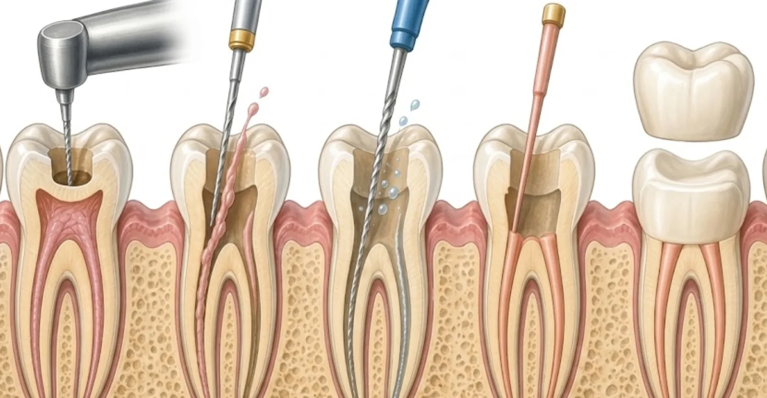
Gutta percha removal clears the old core filling from the canal. Sealer management addresses the separate material bonded to the canal walls. Disinfection then targets the bacteria that caused the problem. Re-obturation finally restores the seal, and each step has its own goal.
Note : All images used are for editorial and illustrative purposes only and may not originate from the original news provider or associated company.
What Happens During Gutta Percha Removal in Root Canal Retreatment?
Retreatment follows a structured sequence from diagnosis to final restoration. Each step prepares the tooth for the next one. Skipping steps can reduce the chance of successful healing. The sequence below outlines what patients can generally expect. Timing may vary depending on anatomy and infection.
Step 1 : Clinical examination and review of previous treatment
The dentist records your symptoms and dental history in detail. The tooth is tested for tenderness, mobility, and gum health. Previous treatment notes are reviewed when available. This information helps identify why the tooth needs attention. Findings from this step guide every later decision.
Step 2 : Imaging and assessment of canal anatomy
X-rays show the existing filling, root length, and surrounding bone. CBCT may be added when three-dimensional detail is clinically justified. Imaging reveals canal number, curvature, and possible obstructions. These findings shape the removal plan for each canal. Imaging also helps predict how difficult the case may be.
Step 3 : Accessing the previously treated canals
The dentist opens through the crown or existing filling. As much sound tooth structure as possible is preserved during access. Each canal entrance is then located and cleaned. A rubber dam keeps the working area isolated throughout. Conservative access helps keep the tooth strong for its final restoration.
Step 4 : Removing the coronal and deeper gutta percha
Upper filling is usually removed first with heat or engine-driven files. Deeper material is then cleared gradually with smaller instruments. A small amount of solvent may help in dense areas. Irrigation flushes loosened material out during each stage. This gradual method is central to safe Gutta Percha Solvents and Root Canal Retreatment.
Step 5 : Managing residual sealer and filling material
Ultrasonic tips help loosen remaining sealer from canal walls. Magnification allows the dentist to see residual material clearly. Irrigation is repeated to remove debris from irregular spaces. This step prepares the walls for effective disinfection. Clean walls allow irrigants and the new sealer to work properly.
Step 6 : Re-establishing working length and canal patency
The dentist confirms the canal's full length to the root tip. An electronic apex locator and X-rays support this measurement. Patency means a small file can pass through the canal end. This allows cleaning to reach the entire canal length. Without full length, bacteria near the root tip may remain untreated.
Step 7 : Cleaning, shaping, and disinfecting the canal system
Files shape the canal so irrigants can flow effectively. Sodium hypochlorite kills bacteria and dissolves organic tissue. EDTA helps remove mineral debris from the canal walls. Activated irrigation can reach fins and side canals that files miss. Thorough disinfection is the step most closely linked to healing.
Step 8 : Re-obturation and coronal sealing
Gutta Percha Solvents and Root Canal Retreatment ends with new gutta percha, sealer, and a tight coronal seal. The new filling is placed to the confirmed working length. An X-ray checks density and length before closing the tooth. A temporary or permanent seal is then placed on top.
Step 9 : Planning the definitive restoration when required
A crown or durable filling protects the tooth after retreatment. It also prevents new leakage into the canal system. Many back teeth need crowns to reduce fracture risk. The restoration should be placed promptly after endodontic work. Delaying the restoration increases the risk of contamination and fracture.
Evaluate your choices in Implant Supported Bridge vs Traditional Bridge
Is root canal retreatment painful?
Local anesthesia keeps the procedure comfortable for most patients. Pressure or vibration may be felt, but sharp pain is uncommon. Mild tenderness afterward is common for a few days. Most patients return to normal activities the same or next day. Your dentist can explain what to expect for your specific tooth.
Can solvent use reduce pain after gutta percha removal?
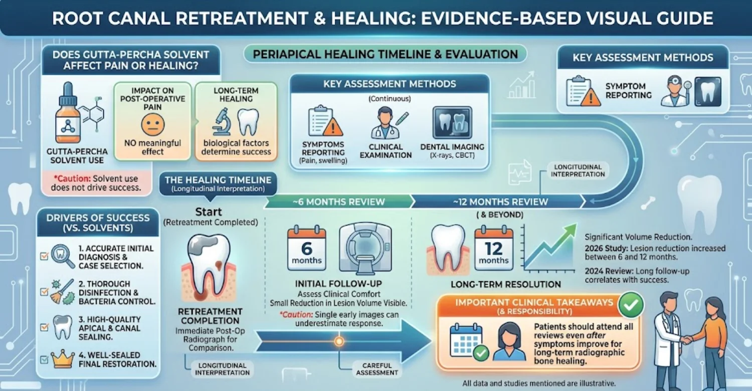
Studies have not shown that solvents reliably reduce postoperative pain. They also have not shown a meaningful increase in pain. Pain after retreatment depends on many factors beyond solvent use. Inflammation, infection, and bite pressure are usually more important. Gutta Percha Solvents and Root Canal Retreatment comfort depends mostly on inflammation control.
What does current research say about postoperative pain and solvent use?
A randomized trial of 90 patients found no significant pain difference with solvent use. A 2023 systematic review reached a similar conclusion with very low certainty. More high-quality clinical trials are still needed. For now, solvent choice is guided by clinical need rather than pain control.
What is normal discomfort after retreatment?
Mild tenderness when biting is a common and expected response. The tooth may feel slightly sore or raised for several days. Discomfort usually eases gradually as inflammation settles. Simple pain relief advised by your dentist is often enough. Tenderness that steadily improves is generally a reassuring sign.
What symptoms suggest that postoperative problems need dental review?
Worsening pain after the first few days should prompt a call. New swelling or fever also needs dental assessment. A loose or missing temporary filling should be reported quickly. These signs may indicate infection or a restoration problem. Early review usually makes problems easier to manage.
How Can You Get Relief After Root Canal Retreatment?
Most soreness settles with simple care and your dentist's advice. The aim is comfort while protecting the treated tooth. Following instructions closely supports smoother healing. Contact the clinic if symptoms do not improve as expected. Most patients find recovery simpler than they expected.
What can help with mild soreness after retreatment?
Resting the tooth and chewing on the other side can reduce pressure. Eating soft foods for a few days also helps. Dentist-recommended pain relief usually controls mild discomfort. Symptoms should ease gradually over the following days. Keeping the area clean with gentle brushing also supports comfort.
How should patients follow their dentist’s pain-relief instructions?
Take medicines exactly as your dentist advises. Tell the dentist about allergies, health conditions, and other medicines you use. Never increase doses or combine medicines without professional guidance. Ask the clinic if you are unsure about any instructions. Keep the written instructions provided after treatment for easy reference.
What should you avoid while the tooth is being restored?
Avoid chewing hard, sticky, or very chewy foods on the treated side. Temporary fillings are weaker than permanent restorations. Heavy biting can crack the temporary or the weakened tooth. Wait until the final restoration is placed before normal chewing. Your dentist will tell you when normal chewing can resume.
Why is the temporary filling or restoration important?
The temporary filling keeps saliva and bacteria out of the cleaned canal. Without it, the canal could become contaminated again. It protects the tooth until the permanent restoration is ready. Report any loss or damage to your dentist promptly. It is a small but essential part of Gutta Percha Solvents and Root Canal Retreatment.
When does postoperative pain become a reason to contact the dentist?
Contact the dentist if pain increases after a few days. Severe pain that medicines do not control also needs review. Pain combined with swelling or fever requires prompt attention. Early contact allows problems to be managed quickly. When in doubt, calling the clinic is always the safer choice.
Learn which procedure you need between Bone Graft vs Sinus Lift
What Are the Warning Signs After a Failed Root Canal or Retreatment?
Some symptoms after treatment are expected, but certain signs need prompt attention. Knowing the difference helps patients act quickly and safely. Warning signs may indicate infection or a complication. Serious symptoms should never be left for a routine appointment. Recognizing them early can prevent serious complications.
When should you seek urgent dental care?
Seek urgent dental care when swelling, fever, or severe pain develops. Restricted mouth opening is another important warning sign. These symptoms may indicate a spreading infection. Same-day assessment is usually recommended in these situations. Delaying care can allow an infection to spread further.
Increasing facial or jaw swelling
Swelling that grows over hours or days can indicate spreading infection. It should be assessed by a dentist the same day. Early treatment may include drainage and further canal cleaning. Do not wait for swelling to settle on its own. Photographing the swelling can help track changes over time.
Fever with dental swelling or infection symptoms
Fever alongside dental swelling suggests the infection may be affecting the body more widely. This combination should not be ignored. Prompt dental or medical assessment is needed. Antibiotics may be considered in addition to dental treatment. Monitor your temperature and report the readings to your dentist.
Severe or persistent pain that is not improving
Pain that worsens instead of easing deserves prompt dental reassessment. Normal discomfort usually improves gradually over several days. Increasing pain may indicate infection or a bite problem. The dentist can examine and adjust treatment as needed. Keeping a simple pain diary can help the dentist assess changes.
Difficulty opening the mouth
Restricted mouth opening is called trismus. It can signal infection spreading into the jaw muscles. Trismus can also make dental treatment more difficult. Patients with this symptom should seek urgent assessment. It should be taken seriously, especially when combined with swelling or fever.
When is emergency medical care needed?
NHS guidance treats breathing, swallowing, or speaking difficulty with major swelling as emergency warning signs. These symptoms may indicate an airway threat. They require immediate emergency medical care. Patients should not wait for a dental appointment in these situations. Emergency services can assess and protect the airway quickly.
Difficulty breathing, swallowing, or speaking
These symptoms can threaten the airway and become life-threatening. Seek emergency medical care immediately. Do not drive yourself if symptoms are severe. Emergency teams can secure the airway and treat the infection. Every minute matters when the airway may be affected.
Severe swelling involving the mouth, neck, or area around the eye
Swelling spreading to the neck or floor of the mouth is serious. Swelling around the eye also needs emergency evaluation. These patterns may indicate a rapidly spreading infection. Hospital care may be required alongside dental treatment. Do not attempt to manage these symptoms at home.
.jpg)
.jpg)
.jpg)
.jpg)
Can Root Canal Retreatment Save a Tooth After Gutta Percha Removal?
Many previously treated teeth can be saved with careful retreatment. A recent systematic review pooled strict periapical healing at 78.8%, rising to 87.5% with looser criteria. Another review reported strict success around 71% at one to three years. Outcomes still depend on each tooth's individual condition.
What factors influence retreatment prognosis?
The cause of failure strongly influences the expected outcome. Canal anatomy and remaining tooth structure also matter. The quality of the final restoration affects long-term success. Gum and bone support around the tooth play an additional role. Your dentist can explain how these factors apply to your tooth.
Does a periapical lesion affect retreatment outcomes?
Lesions around the root tip can heal after successful retreatment. Their size and character may influence expected healing time. Larger lesions often need longer follow-up to confirm healing. Regular X-rays help track bone repair over time. Gutta Percha Solvents and Root Canal Retreatment often allows these lesions to shrink over time.
When might endodontic surgery be considered instead?
Surgery may suit teeth with posts that are risky to remove. It can also help when canals are blocked or inaccessible. Persistent disease after good retreatment is another indication. The surgeon treats the root tip directly through the gum. It is often considered as a complement, not a competitor, to nonsurgical treatment.
When might extraction become part of the treatment discussion?
Extraction is discussed when fractures make the tooth unrestorable. Severe decay or very poor gum support can also limit options. In these cases, retreatment may not provide lasting benefit. Replacement options are then explained to the patient. The decision is always made together with the patient.
What happens after successful retreatment?
After Gutta Percha Solvents and Root Canal Retreatment, the tooth is restored, monitored, and used normally. A crown or durable filling protects the tooth long term. Follow-up X-rays confirm that bone around the root is healing. Regular dental check-ups help maintain the result.
Check out the differences in Root Canal vs Crown
How Much Does Root Canal Retreatment Cost in the US, UK, and Turkey?
Location | Planning range | What can change it |
United States | US$1,000–2,500 per tooth | Tooth type, complexity, specialist care, imaging, restoration |
United Kingdom | 955–1,500 per tooth | Molar complexity, specialist care, imaging, restoration |
Turkey | 349–599 per tooth at one current private clinic listing | Tooth type, complexity, imaging, restoration |
Istanbul | From about US$150 per tooth | Case assessment and treatment requirements |
These amounts are planning references, not fixed quotes. Diagnostic findings can change the final treatment scope.
Gutta Percha Solvents and Root Canal Retreatment costs vary with tooth type, complexity, imaging, and restoration. Molars often cost more because they have more canals and anatomical variation. A quote should state whether imaging, temporary fillings, and crowns are included.
What is the average cost of root canal retreatment in the United States?
The AAE notes retreatment often costs more than initial root canal treatment. Planning ranges are commonly around US$1,000–2,500 per tooth. Specialist care and complex anatomy can raise the total. Crowns and imaging are often billed separately. Insurance coverage may reduce the out-of-pocket cost for some patients.
Typical planning range: approximately 1,000–2,500 per tooth
This range reflects many private US quotes for retreatment alone. It usually excludes the final crown or build-up. Front teeth tend to sit at the lower end. Molars and specialist cases often sit at the higher end. Final prices are confirmed only after examination.
How tooth location, complexity, specialist care, imaging, and restoration can change the total
Molars take longer because they have more canals. Specialist endodontist fees are usually higher than general dentist fees. CBCT scans, post removal, and crowns each add to the total. Patients should request an itemized estimate before starting. Asking early prevents surprises later in treatment.
What is the average cost of root canal retreatment in the United Kingdom?
Current private London listings show retreatment examples from about $955 to $1,500 per tooth. Prices vary between clinics and regions. Specialist practices often charge toward the upper end. Imaging and restoration may be quoted separately. Patients should request a written estimate before booking.
Typical private-treatment planning range: approximately 955–1,500 per tooth
This range covers typical private retreatment cases. It may not include X-rays, CBCT, or the final crown. Complex molars can exceed this range. Patients should confirm inclusions before booking treatment. Asking for a written, itemized estimate avoids later misunderstandings.
Why molar retreatment and specialist treatment may cost more
Molars have several canals and more complex anatomy. Retreatment therefore takes longer and requires more materials. Specialist endodontists use microscopes and advanced equipment. These factors increase the overall treatment fee. Complex cases may still offer good value when prognosis is favorable.
What is the average cost of root canal retreatment in Turkey?
Private clinics in Turkey currently list $349, $479, and $599 depending on tooth complexity. Front teeth are usually priced lower than molars. Imaging and restorations may be charged separately. Quotes should always be confirmed after clinical assessment. Travel and accommodation costs should also be considered when comparing options.
Typical Istanbul planning prices can start around $349 and reach approximately $599 per tooth at some private clinics
Prices vary by clinic, tooth type, and case complexity. Some quotes include imaging, while others do not. The final crown is usually priced separately. Patients should confirm exactly what each quote includes. Comparing itemized plans gives a more accurate picture than headline prices.
Vitrin Clinic reference pricing: retreatment from approximately $150 per tooth, subject to individual assessment
Vitrin Clinic publishes retreatment pricing from about $150 per tooth. The final quote is set after examination and imaging review. Complex cases, posts, or restorations may change the total. Patients receive an itemized plan before treatment begins. This approach helps patients budget with confidence before travel.
What is included in a root canal retreatment quote?
A complete quote should itemize diagnosis, removal, disinfection, and re-obturation. It should also state whether restorations are included. Additional procedures should be listed separately. Clear inclusions help patients compare offers fairly. Ask for any missing items to be added in writing.
Consultation and diagnostic imaging
Check whether the consultation fee is included in the quote. Ask whether X-rays and any CBCT scan are billed separately. Imaging is essential for accurate diagnosis and planning. Its cost can vary significantly between clinics. Bringing recent images may reduce imaging costs.
Gutta percha and sealer removal
Removal of old filling and sealer is usually part of the core fee. Difficult cases may require extra time and equipment. Hard-set sealers can make removal more demanding. Ask whether complex removal changes the quoted price. This work is the technical heart of Gutta Percha Solvents and Root Canal Retreatment.
Cleaning, shaping, disinfection, and re-obturation
These core stages form the main retreatment procedure. They should appear clearly in any quote. The number of visits may affect the final cost. Ask how many appointments the plan includes. Multi-visit cases may include medication placed between appointments.
Temporary or definitive restoration costs
Temporary fillings are sometimes included in the retreatment fee. Final crowns are usually priced separately. Crown materials and laboratory work affect the total cost. Confirm restoration costs before starting endodontic treatment. Budgeting for both stages gives a realistic total cost.
Additional treatment for posts, fractures, perforations, or surgical retreatment
Post removal adds time and specialized equipment. Perforation repair requires specific materials and techniques. Surgical retreatment is a separate procedure with its own fees. These costs apply only when clinically required. Your dentist should explain in advance when these may apply.
Why should patients compare complete treatment plans rather than a single advertised price?
A low headline price may exclude imaging, restorations, or follow-up. Comparing complete plans shows the true cost of treatment. It also reveals differences in materials and technology. Itemized plans help patients make fair, informed choices. The cheapest quote is not always the lowest total cost.
Read about managing a sharp fragment in your gum with Bone Spicule
Tips for Patients Preparing for Root Canal Retreatment
Good preparation makes the consultation more useful and efficient. It also helps the dentist plan Gutta Percha Solvents and Root Canal Retreatment more precisely. Bringing records saves time and may avoid repeated imaging. Preparing questions helps you understand every step clearly. A little preparation can make treatment smoother and less stressful.
What information should you bring to a retreatment consultation?
Bring your medical history and a list of current medicines. Note any allergies and previous reactions to dental treatment. Describe your symptoms and when they started. Details of previous dental work are also very helpful. This information helps the dentist plan safe care.
Should you bring previous X-rays, CBCT scans, or treatment records?
Yes, earlier images help the dentist compare changes over time. Records may identify the sealer, posts, and canals treated before. This information supports safer removal planning. Digital copies are easy to share before your appointment. Ask your previous dentist for copies well before your appointment.
What questions should you ask about gutta percha removal?
Ask how the old filling will be removed from each canal. Ask how long the procedure is expected to take. Ask about possible risks and how they are managed. Understanding the plan helps you feel more prepared. Writing questions down beforehand helps you remember them during the consultation.
What should you ask about solvent use and alternatives?
Ask whether a solvent is likely to be needed in your case. If so, ask which one and why it was chosen. Ask whether mechanical removal alone is possible. A clear explanation helps you make an informed decision. These questions are central to understanding Gutta Percha Solvents and Root Canal Retreatment.
How can you prepare for the final restoration after retreatment?
Discuss crown or filling options early in the planning stage. Ask about materials, appearance, and expected lifespan. Plan the restoration appointment soon after retreatment. Prompt restoration protects the tooth from leakage and fracture. Coordinated planning also reduces the total number of visits.
What should you avoid doing at home to “dissolve” or remove dental filling material?
Never apply chemicals, sharp tools, or household solvents to a tooth. These can burn soft tissues and damage the tooth. They cannot reach the filling material sealed inside the root. Contact a dentist instead if a filling feels loose or painful. A temporary dental kit from a pharmacy is safer while awaiting care.
At Vitrin Clinic: Endodontic Assessment Before Gutta Percha Removal
Vitrin Clinic approaches retreatment as a planned sequence rather than an isolated procedure. Assessment always comes before any filling material is removed. The team reviews the tooth, surrounding tissues, and previous treatment. This careful approach supports safer and more predictable outcomes. Patients are informed about findings at every stage.
How is the previously treated tooth clinically examined?
The team examines the tooth, gums, bite, and existing restoration. Clinical tests check tenderness to tapping and pressure. Gum probing helps detect possible cracks or periodontal problems. These findings are combined with imaging for an accurate diagnosis. Each finding is recorded to guide the treatment plan.
How are digital X-rays and CBCT used when clinically appropriate?
Digital X-rays document the root filling and surrounding bone. CBCT is added when three-dimensional detail answers a specific question. It can reveal hidden canals, fractures, or lesion size. Imaging is selected carefully to avoid unnecessary radiation exposure. Findings are explained to patients using the images themselves.
How is the existing gutta percha, sealer, canal anatomy, and restoration assessed?
Imaging shows filling density, length, and any gaps. Records may help identify the sealer type used before. Canal anatomy is reviewed for curves, calcification, and extra canals. The restoration is checked for leakage, decay, or damage. Together, these details shape the Gutta Percha Solvents and Root Canal Retreatment plan.
How is the retreatment approach individualized according to the tooth and previous treatment?
The removal method is chosen for each tooth individually. Anatomy, filling materials, and the cause of failure guide this choice. A simple front tooth may need a different approach than a complex molar. Individual planning reduces risks and supports better outcomes. The plan is explained clearly before treatment begins.
How does digital treatment planning support a structured retreatment sequence?
Digital planning organizes diagnosis, retreatment, and restoration into clear stages. Each stage is documented with expected steps and timing. Patients can see the full plan before treatment begins. This structure is especially useful for international patients. It also makes coordination with restorative treatment simpler.
How Vitrin Clinic Supports Your Treatment Journey
Vitrin Clinic coordinates each stage so patients understand what happens and when. Clear communication reduces uncertainty before and after treatment. The team supports both local and international patients. Each journey is tailored to the tooth and the patient's schedule. Support continues from the first consultation to follow-up.
Pre-treatment consultation and case assessment
The consultation clarifies your symptoms and dental history. The team reviews findings and explains whether retreatment is appropriate. Alternatives are discussed when retreatment may not be ideal. You leave with a clear understanding of the next steps. Questions are encouraged throughout this first meeting.
Reviewing previous dental records and imaging
Previous records help the team understand earlier treatment decisions. They may reveal materials, posts, or procedural limitations. Older images allow comparison with current findings. This review supports safer and more accurate planning. It may also prevent repeating imaging that is already available.
Coordinating clinically appropriate diagnostic imaging
Imaging is selected to answer specific clinical questions. Routine scans are avoided when they add little value. CBCT is used when three-dimensional detail changes decisions. This approach balances diagnostic benefits with radiation safety. Patients are told why each image is recommended.
Creating an individualized retreatment plan before treatment begins
Each plan outlines the treatment steps and likely number of visits. It also explains the expected outcome and possible alternatives. Costs are itemized so patients understand what is included. The plan is shared before any treatment starts. Changes are discussed openly if new findings appear.
Coordinating endodontic treatment with the definitive restoration
Retreatment and the final crown or filling are scheduled together. This reduces the time the tooth relies on a temporary filling. Coordinated timing lowers the risk of leakage or fracture. It also helps international patients plan their stay efficiently. Planned sequencing supports the long-term success of retreatment.
Providing follow-up guidance after retreatment
Patients receive clear aftercare instructions after treatment. Warning signs are explained so problems can be recognized early. A follow-up imaging plan helps confirm healing over time. The team remains available for questions after you return home. Records can be shared with your local dentist when needed.
Ready to transform your smile?
Talk to our team and get a personal FREE treatment plan.
Key Takeaways About Gutta Percha Solvents and Root Canal Retreatment
These points summarize what patients should understand before retreatment begins. They cover removal, solvents, sealers, aftercare, warning signs, and cost. Each point reflects current evidence and clinical practice. Discuss any questions with your dentist before treatment. They reflect the main principles of modern endodontic retreatment.
Gutta percha removal is one part of a broader root canal retreatment process
Removal only reopens the canal for further treatment. Disinfection, sealing, and restoration determine the long-term result. Each stage must be completed carefully for success. Removal alone does not solve the underlying problem. Understanding this helps patients value every stage of treatment.
Solvents can soften gutta percha, but they are not automatically required or universally superior
Gutta Percha Solvents and Root Canal Retreatment evidence supports selective, not routine, solvent use. Many canals are cleared with mechanical methods alone. Solvents may help where dense filling slows progress. Their use should always follow clinical judgment. This keeps residue and chemical exposure as low as possible.
The previous sealer, canal anatomy, restoration, and reason for treatment failure can determine the removal strategy
Each of these factors can change the instruments and methods chosen. Hard-set sealers may require more ultrasonic work. Complex anatomy may need magnification and extra care. The cause of failure shapes the whole treatment plan. Individual planning is therefore essential for every tooth.
Relief after retreatment should be managed with dentist-directed aftercare and appropriate pain-control advice
Follow your dentist's guidance on medicines and diet. Protect the temporary restoration by avoiding hard foods. Mild tenderness usually settles within several days. Contact the clinic if symptoms worsen instead of improving. Following instructions carefully supports faster recovery.
Side effects and complications should be discussed before treatment, especially when chemical solvents are considered
Patients should understand possible irritation, residue, and extrusion risks. Other risks include file separation and persistent symptoms. Discussing these openly supports informed consent. It also helps set realistic expectations for recovery. Patients should feel free to ask questions at any point.
Persistent pain, swelling, fever, or worsening symptoms warrant dental evaluation
These signs suggest the healing course needs checking. They may indicate infection or a restoration problem. Early assessment allows faster and simpler management. Do not ignore symptoms that continue or increase. Keep the clinic's contact details easily available after treatment.
Breathing, swallowing, or speaking difficulty with significant swelling requires emergency medical attention
Airway symptoms are true medical emergencies. They need immediate care, not a routine dental appointment. Call emergency services or go to the nearest emergency department. Dental treatment can follow once the patient is stable. Knowing this rule can protect patients in rare but serious situations.
Treatment cost varies substantially by country, tooth complexity, diagnostics, specialist involvement, and restoration needs
Prices differ widely between the US, UK, and Turkey. Molars and specialist care usually cost more. Imaging and crowns may be billed separately. Compare complete itemized plans rather than headline prices. A clear written plan remains the best budgeting tool.
At Vitrin Clinic
Vitrin Clinic can coordinate endodontic assessment within a broader dental treatment journey. The starting point is the previously treated tooth and its current condition. Assessment can include teeth, gums, existing restorations, and appropriate imaging. Gutta percha and sealer removal are handled as technical steps within that plan. Cleaning, disinfection, and re-obturation follow the clinical findings. The definitive restoration is then planned to protect the tooth. International patients can also receive coordinated scheduling and follow-up guidance.
Reference
FAQs
Dr. Rifat Alsaman has more than 5 years of clinical experience in dentistry and currently serves as the Head of the Medical Team at Vitrin Clinic. He is dedicated to providing exceptional patient care, overseeing treatment planning, and ensuring the highest clinical standards across the team. His expertise, attention to detail, and commitment to continuous professional development have helped countless patients achieve healthier, more confident smiles.


