Endodontics
October 1, 2026
Condensing Osteitis: What Causes It and How Is It Treated?
 Dr. Rifat Alsaman
Condensing osteitis is a localized change in the jawbone that may develop near the root of a tooth. It happens when the surrounding tissues respond to long-term, low-grade irritation or inflammation. You may feel nothing at all. Or your tooth may feel tender, sensitive, or uncomfortable when biting. Because it can appear as an unusual area on a dental X-ray, you may wonder whether it is an infection, a tumor, or another type of jaw lesion. Understanding what the finding means helps you know when further dental assessment is needed.
What Is Condensing Osteitis and What Does It Feel Like?
Condensing osteitis is a reaction in which the jawbone becomes denser around the root of an affected tooth. It is also called focal sclerosing osteitis. So, what is condensing osteitis in simple terms? It is the bone's response to ongoing irritation, usually from pulpal inflammation or infection. The bone change itself rarely hurts, so many people notice nothing. Others describe it like this:
"My tooth feels sore when I bite."
"I have occasional discomfort around one tooth."
"My X-ray showed a strange white area."
"I do not have pain, but my dentist found something unusual."
What Does Condensing Osteitis Look Like on a Dental X-Ray?
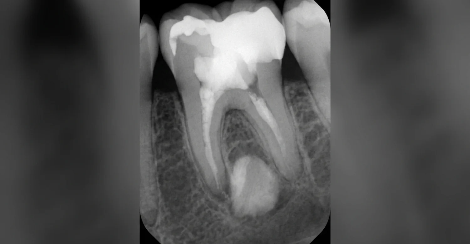
On a condensing osteitis X-ray, the dentist usually sees a localized white, or radiopaque, area. This reflects increased bone density. It commonly appears around the root tip of a tooth, often a lower molar or premolar. That is where irritation from the pulp reaches the bone. Dental X-rays are essential for spotting this finding, since it cannot be seen in the mouth. However, the appearance alone does not establish the cause. Clinical tests must confirm what is happening.
Is Condensing Osteitis the Same as an Infection?
Not exactly. This finding is a bone reaction to inflammation, often from pulpal disease inside the tooth. In other words, the bone is responding to irritation rather than being infected itself. However, the pulp that triggered this response may still be inflamed or infected. That is why the underlying tooth may still need treatment, even when the bone change looks stable. Your dentist will focus on the source of the problem, not just the white area on the image.
What Are the Symptoms of Condensing Osteitis?
Condensing osteitis symptoms vary depending on the condition of the affected tooth and its pulp. Many signs come from the tooth rather than the bone. You may notice:
Tooth tenderness when biting or chewing
A mild or persistent toothache
Sensitivity linked to the affected tooth
Discomfort around the root area
Symptoms related to pulpitis or chronic pulp inflammation
No symptoms at all
Because these signs overlap with other dental problems, a professional examination is the only reliable way to understand what they mean.
Can Condensing Osteitis Cause Tooth Pain?
Yes, pain can occur. However, it usually comes from inflammation inside the tooth rather than from the dense bone. When the pulp is irritated, you may feel aching, throbbing, or soreness when biting. Pain severity does not necessarily reflect the extent of the bone change. A large dense area may cause little discomfort, while a small one may come with significant pain. Persistent or worsening tooth pain should always be assessed, because the pulp condition may be progressing.
Can You Have Condensing Osteitis Without Symptoms?
Yes. Many cases are incidental findings. This means the condition is discovered on an X-ray taken for another reason, such as a routine check-up or orthodontic planning. You may feel completely normal. Still, an asymptomatic finding deserves professional interpretation. The dense area may signal a quietly inflamed pulp. It may also resemble other jaw bone lesions that need a different approach. Your dentist can confirm the diagnosis and decide whether treatment or monitoring is appropriate.
What Causes Condensing Osteitis?
Condensing osteitis causes are usually linked to prolonged, low-grade irritation from an affected tooth. When the pulp stays inflamed for a long time, inflammatory signals can reach the bone around the root tip. Instead of breaking down, the bone in some people responds by becoming thicker and denser. This is why the finding is sometimes described as abnormal bone growth in jaw tissue near a tooth. The most common triggers are explained below.
Pulpitis and Chronic Pulpitis
Pulpitis is inflammation of the dental pulp, the soft tissue containing nerves and blood vessels inside the tooth. Reversible pulpitis is mild, and the pulp can recover once the irritant is removed. Irreversible pulpitis means the pulp cannot heal. When inflammation lasts for months, it is called chronic pulpitis. This long-lasting irritation can spread through the root tip and stimulate a localized bone response. It is one of the most common triggers of condensing osteitis.
Deep Tooth Decay
Untreated decay starts in the enamel and gradually moves into the dentin. Over time, it can reach the pulp. Bacteria and their byproducts then irritate the pulp tissue, causing inflammation. If the decay stays untreated, this irritation can persist for a long time. A chronically irritated tooth may eventually show changes in the surrounding bone on X-rays. Treating decay early is one of the simplest ways to prevent this chain of events. Small cavities are also easier to repair.
Previous Dental Trauma
A blow to the mouth can damage the blood supply to the pulp, even if the tooth looks fine afterward. Pulpal problems may then develop slowly over months or years. However, trauma does not automatically cause this bone change. Many injured teeth heal without any bone reaction. Still, a history of trauma is relevant during diagnosis. It can explain why a tooth without decay shows signs of pulp damage. Always tell your dentist about old injuries.
Long-Standing Root-Related Inflammation
Sometimes irritation around a tooth root continues for a long time. This may come from a leaking filling, a cracked tooth, or an earlier root canal treatment that did not fully resolve the problem. This continuous low-level stimulation may cause the bone to thicken. The key point is that the underlying cause needs to be identified. Treating only the radiographic appearance would miss the real problem. Diagnosis should always focus on the source.
Tips for Patients
A few simple steps can make your dental visit more productive:
Bring previous dental X-rays if you have them, especially when another dentist discovered the lesion. They show whether it has changed.
Tell your dentist about old tooth injuries, even if the trauma happened years ago.
Mention pain when biting, even if the discomfort seems minor or intermittent.
Ask whether the affected tooth needs pulp testing before deciding on treatment.
Ask what the X-ray finding means for your specific tooth, rather than assuming every dense area requires treatment.
What Other Symptoms Can Occur With Condensing Osteitis?
Many signs linked to this finding actually come from the affected tooth rather than the bone itself. Your dentist may look for:
Pain when chewing
Prolonged sensitivity to hot or cold
Tooth discoloration, often grey or dark
Deep decay
Previous trauma
Tenderness around the tooth
A history of unsuccessful dental treatment
Each of these clues points toward the health of the pulp. Together, they help explain why the bone has reacted.
How Can Pulpitis Be Related to Condensing Osteitis?
Pulp inflammation often comes first, and the bone change follows. As pulpitis continues, irritants leave the tooth through the root tip and stimulate the nearby bone. Pulp testing helps determine whether the tooth is still vital, meaning the pulp is alive. Symptoms can also change as pulpal disease progresses. Early pulpitis may cause sharp sensitivity. Later stages may cause lingering pain. If the pulp dies, there may be very little sensation at all, which can be misleading.
How Is Condensing Osteitis Diagnosed?
Condensing osteitis diagnosis combines several steps. Your dentist reviews your history, examines the tooth, performs pulp testing, and studies dental imaging. No single test is enough. The X-ray shows the bone change, while clinical tests reveal the condition of the tooth. Together, they help confirm the diagnosis or point toward another jaw lesion. This combined approach also guides treatment. The right plan depends on whether the pulp is healthy, inflamed, or no longer vital.
Dental X-Rays and Other Imaging
Periapical X-rays are usually the first step. They show the whole tooth and the bone around its root tip. The characteristic finding is increased radiopacity, a white, dense area usually without a dark border. When the picture is unclear, additional imaging such as cone beam computed tomography (CBCT) may be considered. It gives a 3D view of the area. Still, imaging findings must always be interpreted alongside clinical findings, never on their own.
What Does the Dentist Check During an Examination?
During the examination, the dentist assesses several features of the tooth and its surroundings:
Tooth vitality, using cold or electric pulp tests
Pain response and how long it lasts
Sensitivity to percussion, or gentle tapping
Existing decay or restorations
Cracks or signs of trauma
Periodontal condition of the gums and supporting bone
Previous root canal treatment
Changes visible on imaging
Each finding adds to the overall picture and helps narrow down the cause.
When Might an Endodontist Be Involved?
An endodontist is a dentist who specializes in diseases of the dental pulp and the tissues around tooth roots. Referral may be useful when the pulp diagnosis is unclear. It may also help when symptoms do not match test results, or when the tooth has complex anatomy. An endodontist may be involved when root canal treatment or retreatment is needed. Their focused expertise can improve diagnostic accuracy in difficult cases, particularly in molars with curved or narrow canals.
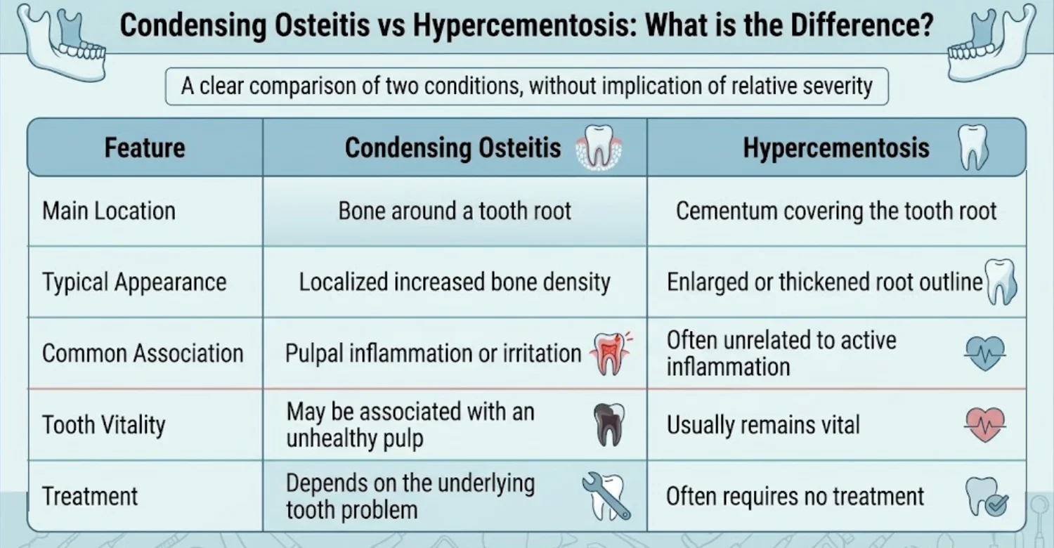
In short, focal sclerosing osteitis involves the bone, while hypercementosis involves the root itself.
Why Can These Conditions Look Similar on an X-Ray?
Both conditions may appear as a white, dense area around the root on dental X-rays. Because they sit so close together, they can be hard to tell apart on a single image. That is why the exact anatomical location matters. If the density is part of the root outline, it suggests hypercementosis. If it lies in the surrounding bone, it suggests condensing osteitis. Clinical testing, especially pulp vitality testing, helps confirm the difference.
Clinical Note
Dr. Rifat Alsaman, Head of the Medical Team and cosmetic dentist at Vitrin Clinic, notes that the most useful approach is to identify the source of the bony response. Focusing only on the X-ray appearance can be misleading. A dense area around a tooth root may reflect a response to chronic irritation. However, other findings can sometimes resemble it. A careful dental history, clinical examination, pulp assessment, and appropriate imaging can help establish the diagnosis accurately.
Can Condensing Osteitis Go Away on Its Own?
The radiographic bone change may not disappear immediately. Sometimes it remains visible for a long time. What matters most is the underlying dental problem. Treating an inflamed or infected pulp is often the main priority. In selected cases, observation may be appropriate after a proper diagnosis, especially when the tooth is healthy and symptom-free. However, unexplained jaw lesions should never simply be ignored. A dentist should confirm what the finding is first.
When Should You See a Dentist About a Jaw Lesion?
It helps to distinguish between an incidental radiographic finding and symptoms that need timely attention. A dense area found on a routine X-ray, with no pain, still deserves professional review. However, it is rarely urgent. On the other hand, swelling, fever, or worsening pain suggest an active problem. These should be assessed promptly. Below are the signs that should prompt an examination, followed by situations that may need further investigation.
Signs That Should Prompt a Dental Examination
Book a dental examination if you notice any of the following:
Persistent tooth pain
Pain when biting
Swelling of the gum or face
Drainage or a bad taste in the mouth
Fever
Tooth discoloration
Increasing sensitivity
A newly discovered, unexplained jaw lesion
Swelling and fever, in particular, may signal an active infection. Do not wait for symptoms to settle on their own. Early assessment often means simpler treatment.
When Could Further Investigation Be Needed?
Sometimes the first examination is not enough. Further investigation may be needed in these situations:
An unusual imaging appearance
Symptoms that do not match the X-ray findings
Cases where another jaw lesion needs to be excluded
Findings that change over time
Lesions with unclear boundaries
In these cases, your dentist may recommend 3D imaging, specialist referral, or, rarely, a biopsy. These steps help rule out other conditions that look similar.
What Is the Treatment for Condensing Osteitis?
Condensing osteitis treatment depends on the condition of the affected tooth and its pulp. The dense bone itself is not the target. Instead, the goal is to remove the source of irritation. Options range from simple fillings to root canal treatment or, in some cases, extraction. A treatment plan is made only after examination, pulp testing, and imaging. This ensures the chosen option matches the real cause rather than the X-ray appearance.
Treating the Underlying Tooth Problem
When decay is the cause and the tooth remains restorable, the dentist removes the decay and places a filling or crown. If pulpitis is still reversible, removing the irritant may allow the pulp to recover. Leaking restorations, cracks, or bite problems may also need correction. The main principle is removing the source of persistent irritation. Once that source is gone, the bone has a chance to remodel gradually over time.
Condensing Osteitis and Root Canal Treatment
Condensing osteitis root canal treatment may be considered when the pulp is irreversibly inflamed or infected. During the procedure, the dentist or endodontist removes the damaged pulp, cleans and shapes the canals, and seals them. The decision depends on clinical and diagnostic findings, not on the bone change alone. After treatment, the radiographic bone response may take months or longer to change. Some dense areas never fully disappear, even when the tooth is healthy.
When Is Tooth Extraction Considered?
Extraction may be considered when a tooth cannot be predictably restored. This can happen with severe structural damage, vertical root fractures, extensive decay below the gum line, or advanced bone loss. Even then, extraction is not automatic. Every case needs individualized treatment planning. If removal is necessary, your dentist will also discuss replacement options, such as a dental implant or bridge. These help protect your bite and nearby teeth.
What Can You Expect After Treatment?
After the underlying cause is addressed, symptoms related to the affected tooth often improve. Biting pain and sensitivity may settle within days or weeks. Radiographic bone changes, however, usually resolve more gradually. Some may shrink partially, and others may stay visible. That is why follow-up dental X-rays may be recommended. Clinically, improvement means comfort and normal function. Radiographically, it means the dense area becomes smaller or stays stable over time.
How Long Does Condensing Osteitis Take to Heal?
Healing is a variable process. Symptoms and radiographic changes often follow different timelines. Pain may ease soon after treatment, while bone remodeling can take many months. In some cases, part of the dense area remains visible indefinitely without causing problems. Follow-up timing depends on the tooth, the treatment performed, and the imaging findings. For this reason, there is no universal healing period. Your dentist will advise you on when to return.
What We Notice Clinically
Dr. Rifat Alsaman, Head of the Medical Team and cosmetic dentist at Vitrin Clinic, emphasizes that an unusual radiographic finding should be interpreted in the context of the tooth's clinical condition. The key question is not simply whether the X-ray shows increased bone density. The clinician also needs to determine whether the tooth has pulpal inflammation, previous trauma, decay, or another source of irritation. Answering this question guides the right treatment.
Why the X-Ray Should Not Be Interpreted Alone
An X-ray shows shapes and densities, but it cannot show how the pulp functions. Clinical examination fills that gap. Pulp testing reveals whether the tooth is vital, and your symptom history adds context. Similar-looking jaw lesions can have very different causes, from harmless bone islands to conditions that need further investigation. Combining imaging with clinical findings supports an individualized diagnosis. This reduces the risk of unnecessary or delayed treatment.
How Can You Reduce the Risk of Problems That May Lead to Condensing Osteitis?
You cannot control every factor, but good habits lower the chance of pulp problems that may trigger condensing osteitis:
Maintain regular dental examinations at the intervals your dentist recommends.
Treat tooth decay before it reaches the pulp.
Seek evaluation after significant dental trauma, even if the tooth feels fine.
Do not ignore persistent tooth sensitivity or pain.
Complete recommended dental treatment and follow-up visits.
Maintain daily brushing and interdental cleaning with floss or interdental brushes.
Dental Care at Vitrin Clinic
At Vitrin Clinic, every case begins with an individualized dental assessment. This includes a clinical examination, dental X-rays, pulp testing, and 3D imaging when indicated. Treatment planning depends on the condition of the affected tooth, not just the image. When a case requires multidisciplinary assessment, the relevant dental disciplines coordinate their input. Dr. Rifat Alsaman, Head of the Medical Team and cosmetic dentist at Vitrin Clinic, oversees individualized treatment planning to keep care consistent.
.jpg)
.jpg)
.jpg)
What Should You Do If an X-Ray Shows Condensing Osteitis?
If an X-ray shows condensing osteitis, try not to panic. It is usually a localized bone response associated with irritation or inflammation involving a tooth. It may cause symptoms, or it may be discovered incidentally. Diagnosis relies on the clinical picture, pulp assessment, and dental imaging rather than the X-ray appearance alone. Your next step is a thorough examination. Once the cause is clear, your dentist can recommend monitoring or treatment.
Ready to transform your smile?
Talk to our team and get a FREE treatment plan.
References:
FAQs
Dr. Rifat Alsaman has more than 5 years of clinical experience in dentistry and currently serves as the Head of the Medical Team at Vitrin Clinic. He is dedicated to providing exceptional patient care, overseeing treatment planning, and ensuring the highest clinical standards across the team. His expertise, attention to detail, and commitment to continuous professional development have helped countless patients achieve healthier, more confident smiles.


