Endodontics
October 20, 2026
Dynamic vs Static Navigation in Endodontic Microsurgery: Which Is Better?
 Dr. Rifat Alsaman

Dynamic vs Static Navigation in Endodontic Microsurgery compares two computer-guided ways of reaching an infected root tip through the jawbone. Both methods use three-dimensional scans to plan the surgery before the first incision. Dynamic navigation tracks instruments live on a screen, while static navigation uses a custom 3D-printed guide. The best choice depends on anatomy, mouth opening, equipment, and the surgeon's experience.
Vitrin Clinic helps patients understand these options during a personalized endodontic assessment. This guide explains accuracy, recovery, side effects, and costs in the US, UK, and Turkey.
Microsurgery success: About 94% in a pooled meta-analysis of 21 studies.
Dynamic navigation accuracy: About 1.2 mm apical deviation and 2.3° angular deviation on average.
Typical procedure time: About 60 to 90 minutes for a single tooth.
Estimated cost per tooth: About $1,100 in the US, $950 in the UK, and $350 in Turkey.
Ready to transform your smile?
Talk to our team and get a personal FREE treatment plan.
What Is Dynamic vs Static Navigation in Endodontic Microsurgery?
Dynamic vs Static Navigation in Endodontic Microsurgery describes two digital methods for guiding root-end surgery with high precision. Both aim to remove disease while protecting healthy bone, neighboring roots, and nearby nerves. Understanding the basics helps patients follow their dentist's recommendations with confidence.
What Is Endodontic Microsurgery and When Is It Needed?
Endodontic microsurgery treats infection that persists at the root tip after a root canal treatment. It is often called apicoectomy, periapical surgery, or root-end surgery. The surgeon opens a small window in the bone, removes about 3 mm of the root tip, and seals it. Operating microscopes, ultrasonic tips, and biocompatible sealing materials make modern surgery far more predictable than older techniques. A 2010 meta-analysis reported 94% success for microsurgery, compared with 59% for traditional root-end surgery. That means microsurgery was about 1.58 times more likely to succeed. Surgery is usually considered when retreatment is impossible, risky, or has already failed. Common reasons include large posts, new crowns, blocked canals, or lesions persisting after good root canal treatment.
What Is Dynamic Navigation in Endodontic Microsurgery?
Dynamic navigation is a computer-guided system that shows the drill's position on a screen in real time. It works much like a GPS for the surgeon's handpiece inside the jawbone. Optical cameras track reference markers attached to the patient's teeth and to the handpiece. The software overlays the live drill position onto the patient's three-dimensional scan. This lets the surgeon follow a planned path to the root apex without any physical guide. The technology was adapted from guided implant surgery and is now studied widely in endodontics.
Real-Time Instrument Tracking and Virtual Surgical Planning
Planning starts with a cone-beam computed tomography scan imported into navigation software. The surgeon marks the apex, entry point, drill angle, and target depth on the 3D model. During surgery, the screen shows live deviation from that plan in millimeters and degrees. A 2025 systematic review of 14 studies and 240 roots reported average apical deviation of 1.21 mm. The same review reported an average angular deviation of about 2.3 degrees. These figures suggest consistently close alignment between the virtual plan and the real procedure. Real-time tracking also helps the surgeon stop exactly at the planned depth.
What Is Static Navigation in Endodontic Microsurgery?
Static navigation uses a custom 3D-printed surgical guide that fits snugly over the patient's teeth. The guide contains a sleeve or window aligned with the planned path to the root tip. The surgeon drills through that sleeve, so direction and depth are controlled mechanically. This approach is also known as targeted endodontic microsurgery or guided apicoectomy. It borrows principles from guided implant surgery, which has used printed guides for many years. Because the guide does the steering, no tracking cameras are needed during surgery.
CBCT Imaging, Digital Planning, and Custom Surgical Guides
The workflow combines a CBCT scan with a digital intraoral scan or stone model. Software merges both datasets so the guide fits the teeth precisely. The surgeon plans the drill or trephine path, depth, and diameter on screen. The final design is then sent for 3D printing and sterilization. An early laboratory study reported guided drill deviation of 0.79 mm, compared with 2.27 mm freehand. That represents roughly a 65% reduction in positional error. Guide fabrication often adds several days between planning and surgery.
How Do Dynamic and Static Navigation Differ From Conventional Freehand Surgery?
Conventional freehand surgery relies on radiographs, anatomical landmarks, and the surgeon's tactile judgment. The surgeon estimates where the apex lies, then removes bone gradually until the root appears. This works well in accessible front teeth with clear anatomy and visible bone loss. However, thick bone or deep roots can lead to larger bone windows and longer searching. With Dynamic vs Static Navigation in Endodontic Microsurgery, both options replace estimation with a measured digital path. That can mean smaller osteotomies and less exploratory drilling inside the bone. Freehand surgery remains valuable, but guidance adds precision where visibility is poor.
What Is the Difference Between Endodontic Microsurgery and Guided Implant Surgery?
Guided implant surgery places a titanium implant where a tooth is already missing. Endodontic microsurgery saves a natural tooth by treating disease at its root tip. Both can use CBCT planning, printed guides, or dynamic navigation systems. However, endodontic targets are smaller and hidden inside bone beside the root. The goal is precise removal of diseased tissue, not preparing a site for an implant. Many navigation systems were first developed for implants, then adapted for endodontic use. Saving a natural tooth often preserves bone, gum contours, and natural chewing sensation.
.jpg)
.jpg)
.jpg)
.jpg)
.jpg)
.jpg)
Dynamic vs Static Navigation in Endodontics: Key Differences
The two techniques share digital planning but differ sharply in how guidance reaches the instrument. Choosing in Dynamic vs Static Navigation in Endodontic Microsurgery starts with understanding these practical differences. Knowing them helps patients ask better questions during consultation.
How Does Dynamic Navigation Work During Root-End Surgery?
First, a tracking array is attached to the patient's teeth, often using a thermoplastic splint. The system is then calibrated so the handpiece and the CBCT scan share coordinates. The surgeon registers reference points to match the real jaw with the virtual one. During drilling, the surgeon watches the monitor as well as the surgical field. Visual cues show whether the drill stays on angle and reaches the planned depth. Cadaver studies report navigation-assisted steps taking about 4 to 9 minutes. A pooled review reported an average of 5.6 minutes for these navigated steps. After the bone window is created, the surgeon continues under the operating microscope.
How Does a Static Surgical Guide Direct Apical Microsurgery?
The printed guide is seated firmly over the teeth, often after the gum flap is raised. A metal sleeve in the guide holds a trephine bur or drill at the planned angle. A depth stop limits how far the instrument can travel into the bone. The trephine cuts a round bone window and can respect the root tip at the same time. The surgeon checks the guide's fit carefully before any drilling begins. Once the guide is removed, the surgeon continues under the microscope as usual. This method keeps the surgeon's eyes on the patient rather than on a screen.
What Are the Main Differences in Planning, Instrument Guidance, and Workflow?
Planning is similar for both, since each depends on a high-quality CBCT scan. Guidance is visual and live with dynamic navigation, but physical and fixed with a static guide. Workflow timing also differs between the two approaches.
Feature | Dynamic navigation | Static navigation |
Guidance | Live screen tracking | 3D-printed guide |
Changes during surgery | Possible | Limited |
Preparation | Chairside calibration | Lab design and printing |
Mouth opening needed | Less | More |
Main cost driver | Equipment and training | Per-case guide fabrication |
Static guides need design and printing before the appointment. Dynamic systems need calibration and registration at the appointment itself.
Which Technique Offers More Flexibility During Surgery?
Dynamic navigation generally offers more flexibility during surgery. If the surgeon sees unexpected anatomy, the angle can be adjusted immediately on screen. Some systems also allow the plan to be modified during the procedure. A static guide fixes the path, so changes require removing the guide and continuing freehand. This flexibility matters when lesion borders are unclear or root positions vary from the scan. It is also helpful if the surgeon needs to extend the bone window slightly. For straightforward paths, however, the fixed guidance of a static guide is often an advantage.
Which Technique Requires More Preoperative Preparation?
Static navigation usually requires more preoperative preparation. The guide must be designed, printed, sterilized, and test-fitted before surgery. This can add several days and a separate laboratory step to treatment. Dynamic navigation needs less work beforehand but more setup in the operating room. Calibration, registration, and accuracy checks add chairside time on the day of surgery. Both techniques still require careful CBCT review and three-dimensional planning first. For international patients, the extra lead time for a static guide should be planned in advance.
Dynamic Navigation vs Static Surgical Guides in Endodontics: Accuracy and Clinical Outcomes
Accuracy is the main reason clinicians adopt computer guidance for root-end surgery. Research on Dynamic vs Static Navigation in Endodontic Microsurgery shows both methods achieve high precision. Yet laboratory precision and long-term healing are related, but not identical, outcomes.
How Accurate Is Dynamic Navigation in Endodontic Microsurgery?
A 2025 systematic review pooled 14 dynamic navigation studies covering 240 roots. It reported mean platform deviation of 1.17 mm and apical deviation of 1.21 mm. Mean angular deflection was 2.29 degrees across the included studies. One cadaver trial found experienced endodontists achieved 0.8 mm platform deviation versus 1.7 mm for novices. Angular deviation was 1.3 degrees for experienced users and 2.5 degrees for novices. Another cadaver trial reported a mean angular deviation of only 1.1 degrees. These results show high accuracy, but operator skill still clearly matters. Most data come from cadavers and small clinical series rather than large trials.
How Accurate Are Static Surgical Guides for Endodontic Microsurgery?
Static guides also show strong accuracy in laboratory and cadaver studies. An in vitro study of 86 guided apicoectomies reported mean global deviation of 1.31 to 1.53 mm. Angular deviation in that study averaged about 3.3 to 3.5 degrees. A cadaver study using trephine guides reported global deviation of about 1.23 mm. Earlier work found guided drills deviated 0.79 mm, compared with 2.27 mm freehand. Adding a depth stop to a trephine reduced deviation from 2.35 mm to 0.92 mm in one study. Overall, static and dynamic accuracy appear broadly comparable in published research.
What Does Research Say About Accuracy in Root-End Resection?
Root-end resection usually removes about 3 mm of the apex at a controlled angle. Classic research shows a 3 mm resection removes about 98% of apical ramifications. The same resection removes about 93% of lateral canals, where bacteria commonly persist. In Dynamic vs Static Navigation in Endodontic Microsurgery, accurate resection length and angle remain key goals. The 2025 review reported an average resection angle deviation of about 7.5 degrees. Incomplete resection occurred in some roots in cadaver studies, reaching about 10% in one trial. This shows that guidance helps, but microscope inspection after resection remains essential.
Do Dynamic and Static Navigation Improve Surgical Outcomes Compared With Freehand Techniques?
Navigation appears to improve precision and efficiency compared with freehand techniques. Studies report smaller, more targeted bone windows and fewer instrument detours. Laboratory data show freehand deviation can be nearly three times greater than guided drilling. A clinical series reported complete healing in 33 of 35 navigated teeth at one year. That equals a healing rate of about 94% on CBCT assessment. However, conventional microsurgery already achieves about 91% to 94% success in strong studies. Within Dynamic vs Static Navigation in Endodontic Microsurgery, the main gain is safer access in difficult cases. Large randomized trials comparing long-term healing between methods are still limited.
Does Greater Navigation Accuracy Guarantee Better Long-Term Healing?
Greater accuracy does not guarantee better long-term healing on its own. Healing depends on removing bacteria, sealing the root end, and the tooth's overall condition. Cracks, gum disease, poor restorations, or missed canals can still cause failure. A meta-analysis of randomized trials reported 91.3% long-term success for endodontic microsurgery. Prospective studies with follow-up of up to 13 years showed about 78.4% success. This gap reminds us that long-term outcomes depend on many factors beyond the operation itself. Navigation supports good surgery, but it cannot replace correct diagnosis and case selection.
What Factors Influence Navigation Accuracy?
Several factors influence how closely the final result matches the digital plan. These include imaging quality, registration, stability, calibration, operator experience, and anatomy. In Dynamic vs Static Navigation in Endodontic Microsurgery, small errors at each stage can add up during surgery. Understanding them explains why experienced teams check accuracy repeatedly.
CBCT Image Quality and Registration Accuracy
Navigation can only be as accurate as the scan it relies on. Patient movement during scanning can blur fine details of the root and bone. Metal crowns, posts, and root fillings can create streak artifacts on CBCT images. These artifacts may hide the true root outline or the lesion border. Registration errors happen when software misaligns the scan with the patient's real anatomy. Careful reference points and verification checks reduce this risk before drilling begins. Using a suitable field of view and resolution also improves planning accuracy.
Patient Movement, Guide Stability, and Instrument Calibration
Dynamic navigation tracks the jaw itself, so stable markers matter more than keeping the patient perfectly still. If a tracking splint loosens, every reading afterward can become inaccurate. Static guides must seat completely, because a small rocking movement changes the drill angle. Even a 1-degree tilt can shift the tip noticeably over a long drilling path. Instrument calibration also matters for both systems. A bent bur or wrongly entered drill length can shift the endpoint by millimeters. Routine checks on known landmarks help catch these problems early.
Operator Experience and Anatomical Complexity
Experience shortens the learning curve and improves accuracy with navigation. In one cadaver trial, novices needed 7.7 minutes compared with 4.3 minutes for experienced endodontists. Novices also showed about twice the platform deviation of experienced users. Anatomy adds another layer of difficulty to every case. Thick cortical bone, molar roots, and nearby nerves make every millimeter more significant. Complex cases benefit most from guidance, but they also demand the greatest surgical skill.
Ready to transform your smile?
Talk to our team and get a personal FREE treatment plan.
What We Notice Clinically
In daily practice, the hardest part of root-end surgery is often finding the target safely. These observations explain where Dynamic vs Static Navigation in Endodontic Microsurgery can make a practical difference. They reflect common clinical patterns rather than guarantees for any individual patient.
Why Can Locating the Root Apex Be Difficult During Microsurgery?
The root tip is often hidden under several millimeters of intact bone. When the lesion has not eroded the outer bone, there is no visible entry point. Lower molar roots can sit beneath thick bone, sometimes more than 5 mm deep. Angled roots and curved anatomy make visual estimation even harder. Searching freehand can enlarge the bone window and lengthen the procedure. A larger window may also increase postoperative swelling and discomfort. Navigation addresses this by showing the target clearly before any bone is removed.
How Do Nearby Nerves, Sinuses, and Other Anatomical Structures Affect Surgical Planning?
Lower premolars sit near the mental nerve, which supplies sensation to the lip and chin. Lower molars may lie close to the inferior alveolar nerve canal. Upper molar and premolar roots often project near or into the maxillary sinus. Palatal roots of upper molars can be difficult to reach from the cheek side. CBCT shows these relationships in three dimensions before surgery begins. Surgeons measure safe distances and plan drill depth with these structures in mind. That information influences whether navigation, freehand surgery, or another treatment is safest.
Why Does Preserving Healthy Bone and Tooth Structure Matter?
Smaller bone windows usually heal faster and support the tooth better over time. Research has linked larger lesions and greater bone loss with slower healing after surgery. Preserving root length also maintains a healthier crown-to-root ratio for chewing. Navigation studies often report osteotomy diameters of around 4 mm. One cadaver study reported an average osteotomy volume of about 82 cubic millimeters. That conservative approach helps protect the outer bone plate and surrounding gum tissue. Less bone removal can also mean a more comfortable early recovery.
Common Misconceptions About Computer-Guided Endodontic Microsurgery
One common misconception is that a computer performs the surgery automatically. In reality, the surgeon still controls every instrument and every decision. Another myth is that guided surgery suits every tooth. Some cases are better treated with retreatment, freehand microsurgery, or extraction. A third misconception is that navigation removes all surgical risk. Infection, swelling, and nerve irritation can still occur, although careful planning reduces them. Finally, more technology does not always mean higher cost-effectiveness for simple cases.
When Can Dynamic or Static Navigation Be Helpful in Complex Cases?
Navigation is especially helpful when the apex is deep, hidden, or close to vital structures. It can assist with lower molars, palatal roots, and teeth surrounded by thick bone. It may also help when there is no bone perforation to show the way. Cases needing minimal bone removal, such as narrow ridges, can benefit too. Teeth with previous failed surgery may also benefit from precise relocation of the apex. In Dynamic vs Static Navigation in Endodontic Microsurgery, the case's anatomy usually points toward one method. In simpler front-tooth cases, conventional microsurgery often remains entirely appropriate.
Clinical Note
Technology should follow diagnosis, never lead it. The first question is always whether the tooth is restorable and worth saving. Only then does the choice between dynamic, static, or freehand surgery become meaningful. Every recommendation should rest on clinical examination and appropriate imaging. Patients benefit most when the simplest safe approach is chosen for their tooth.
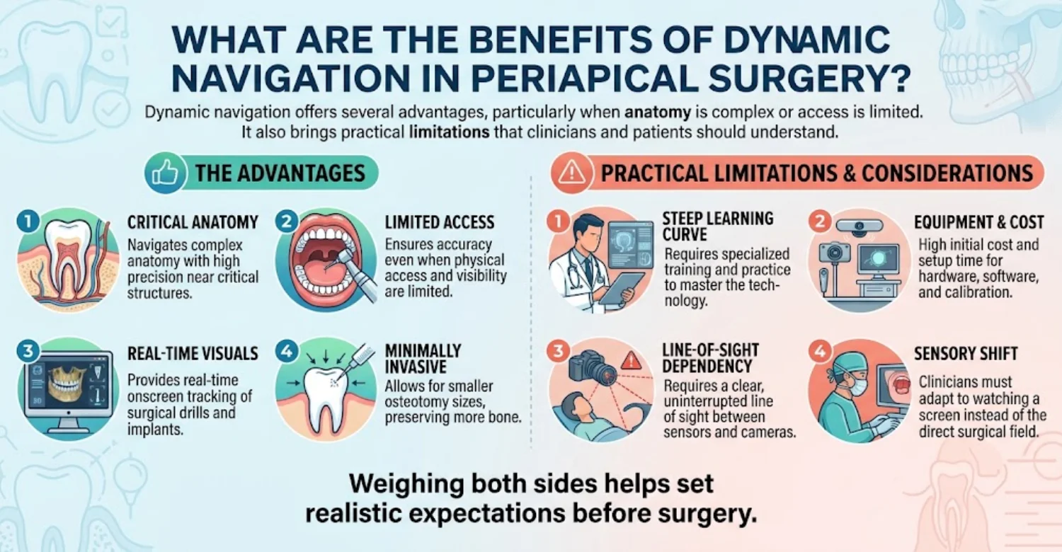
Real-Time Visualization and Instrument Guidance
The surgeon sees the drill's position inside the bone continuously on the monitor. This reduces guesswork when the root tip is not visible directly. Deviation alerts allow correction within fractions of a millimeter before errors grow. The 2025 review reported a mean global apex deviation of about 0.98 mm. Real-time feedback also confirms when the planned depth has been reached. That helps avoid over-drilling toward nerves or the sinus floor. For patients, this often means a more targeted and conservative procedure.
Adaptability During Root-End Resection
Dynamic systems let the surgeon change the angle or depth during surgery if needed. This is useful when the lesion appears larger or positioned differently than expected. The surgeon can extend the bone window without removing any guide. Root-end resection can also be visualized relative to the planned 3 mm level. Some systems allow the trajectory to be replanned within the same session. That adaptability supports a tailored approach for each individual root. It is particularly valuable in multi-rooted teeth with variable anatomy.
Potential Benefits for Accessing Difficult Root Apices
Dynamic navigation may help patients with limited mouth opening because it needs no bulky guide. Posterior teeth are often easier to reach without a guide occupying vertical space. One clinical series on 51 navigated roots reported only two postoperative sinus tracts. That corresponds to a complication rate of about 3.9% in that series. Many studies describe navigation-assisted steps taking around 4 to 5 minutes. These benefits are most noticeable with deep, posterior, or anatomically challenging roots. They may also reduce how much bone must be removed to locate the apex.
Limitations of Dynamic Navigation Systems
Dynamic navigation is not ideal for every clinic or every case. Its main limitations involve equipment, training, and technical reliability. Understanding these helps patients see why some surgeons prefer static guides or freehand surgery.
Equipment, Calibration, and Learning Requirements
Navigation systems require significant investment in hardware, software, and maintenance. Surgeons need dedicated training before achieving consistent accuracy. Studies show novices deviate more than experienced users, especially in angle control. In one trial, novice angular deviation was nearly double that of experienced endodontists. Each procedure also requires calibration of the handpiece and drill. Watching a screen while working in the mouth requires practiced hand-eye coordination. These factors can increase the cost of treatment in some clinics.
Potential Tracking Errors and Technical Interruptions
Tracking can be interrupted if the camera's line of sight is blocked. Blood, hands, or instruments may obscure the markers temporarily. A loose tracking array can produce misleading readings without obvious warning. Software issues or calibration drift may require pausing to recheck accuracy. These interruptions can lengthen the procedure slightly. Experienced teams confirm accuracy on known landmarks before and during drilling. Backup freehand skills remain essential whenever technology does not perform as expected.

Preplanned Instrument Trajectory and Guided Access
The guide physically constrains the drill to the planned angle and depth. The surgeon does not need to look away from the surgical field. This straightforward approach can be easier for teams new to guided surgery. A depth stop helps prevent over-drilling toward nerves or sinus spaces. Because the plan is fixed in advance, the surgical steps are predictable. That predictability can make the procedure smoother and more efficient.
Potential Benefits for Precise Root-Apex Localization
Static guides can locate deep apices accurately even without any bone perforation. Laboratory studies report global deviations of roughly 0.8 to 1.5 mm with guided drilling. That compares favorably with freehand deviations above 2 mm in similar models. Trephine guides can create the bone window and respect the apex in one step. This may shorten the time spent searching for the root tip. Some studies report guided surgical time falling to about one-third of conventional time. Faster, targeted access can also limit bone removal around the root.
Reproducibility of the Planned Surgical Path
Because the path is fixed, results are highly reproducible from case to case. Two surgeons using the same guide should reach a very similar endpoint. This consistency helps when training teams or standardizing complex procedures. Reproducibility also makes it easier to compare postoperative scans with the original plan. Measured deviations can then guide improvements in future guide designs. For patients, it means the surgery follows the plan they discussed beforehand.
Limitations of Static Surgical Guides
Static guides have practical drawbacks that affect suitability for some patients. These relate mainly to fit, access, flexibility, and preparation time. Recognizing them early helps the clinician choose the right approach.
Guide Fit, Mouth Opening, and Restricted Access
A guide adds height between the teeth, so adequate mouth opening is required. Back teeth can be difficult to reach when the guide and drill stack together. Patients with limited openings may not tolerate a bulky guide comfortably. Poor seating from crowns, tooth movement, or printing errors can reduce accuracy. Some guide designs also limit irrigation and cooling around the drill. This requires careful management to protect the bone from heat during drilling.
Reduced Flexibility When Intraoperative Conditions Change
Once printed, a static guide cannot adapt to new findings during surgery. If the lesion extends further than expected, the surgeon must continue freehand. Changes in tooth position since scanning can also make the guide inaccurate. A new crown or filling placed after the scan may prevent proper seating. Surgeons therefore verify fit carefully before starting any osteotomy. Planning a short interval between scanning and surgery helps minimize these issues.
Guide Fabrication Time and Additional Planning Requirements
Designing and printing a guide usually takes several days. Some cases require an intraoral scan in addition to CBCT imaging. Guide design fees, materials, and laboratory work add to the overall cost. A test fitting may be needed before the surgical appointment. Urgent cases may not suit this workflow if delay is undesirable. International patients should allow time for this step in their travel plans.
Dynamic vs Static Navigation in Endodontic Microsurgery: Which Technique Is Best for Different Cases?
No single technique is best for every patient or every tooth. Dynamic vs Static Navigation in Endodontic Microsurgery should always be chosen case by case. The deciding factors are anatomy, access, available equipment, surgeon experience, and cost.
When Might Dynamic Navigation Be Preferred?
Dynamic navigation may be preferred for posterior teeth with limited space for a guide. It can suit patients with restricted mouth opening or a strong gag reflex. It also helps when lesion borders are uncertain and adjustment may be needed. Clinics with trained navigation teams may choose it for complex molar cases. It is useful when there is little time for laboratory guide fabrication. When comparing Dynamic vs Static Navigation in Endodontic Microsurgery, flexibility often favors the dynamic option. Its value increases with anatomical complexity and the surgeon's navigation experience.
When Might Static Navigation Be Preferred?
Static navigation may be preferred when a fixed, reproducible path matters most. It suits accessible teeth where the guide can seat securely and stably. It can also be practical in clinics without dynamic tracking equipment. Many surgeons favor static guides for front teeth and premolars with a good opening. Static guides also work well when the plan is unlikely to change during surgery. For predictable anatomy, static guidance offers precision with a simpler operating-room setup. It can also be cost-effective when the clinic already has digital planning workflows.
Can Conventional Freehand Microsurgery Still Be Appropriate?
Yes, conventional freehand microsurgery remains appropriate for many cases. Front teeth with visible lesions or bone perforation are often straightforward without guidance. Outcomes with modern microsurgery already reach about 94% in pooled data. Freehand surgery avoids guide fabrication fees and navigation technology costs. It also avoids extra setup time in the operating room. The choice in Dynamic vs Static Navigation in Endodontic Microsurgery includes the option of neither. A skilled microsurgeon can achieve excellent results freehand when anatomy is favorable.
How Should Clinicians Balance Accuracy, Accessibility, Cost, and Patient-Specific Needs?
Clinicians should weigh the benefit of extra accuracy against cost and complexity. A deep molar near the nerve may justify guidance more than a simple front tooth. Patient factors include mouth opening, medical history, anxiety, and travel schedules. Cost also matters, since guides and navigation add separate fees. The best plan uses the simplest approach that safely achieves the treatment goal. Discussing Dynamic vs Static Navigation in Endodontic Microsurgery openly helps patients make informed decisions. Shared decision-making leads to treatment plans patients understand and trust.
Diagnosis and Treatment Planning Before Guided Endodontic Microsurgery
Successful surgery begins with an accurate diagnosis of why the tooth has not healed. In Dynamic vs Static Navigation in Endodontic Microsurgery, planning determines whether either method is needed. Diagnosis also confirms whether surgery, retreatment, or another option is most suitable.
How Is Persistent Periapical Disease Diagnosed?
Persistent periapical disease means inflammation or infection remains around the root tip after treatment. Diagnosis combines symptoms, clinical tests, and radiographs. Patients may notice pain, swelling, a gum boil, or tenderness when biting. Some lesions cause no symptoms and are found only on routine X-rays. Comparing current images with older ones shows whether a lesion is healing, stable, or growing. A lesion that has not reduced after about one year usually needs further evaluation. Accurate diagnosis prevents unnecessary surgery on teeth that are slowly healing.
What Clinical Examinations Are Needed Before Root-End Surgery?
A thorough examination checks the tooth, gums, bite, and surrounding soft tissues. These tests help separate endodontic problems from gum disease or cracked teeth. They also confirm which tooth is truly responsible for the symptoms.
Symptoms, Tooth Vitality, Percussion, and Periodontal Assessment
The dentist records pain history, swelling, and any draining sinus tract. Percussion and palpation tests check for tenderness around the root. Vitality testing of nearby teeth helps confirm which tooth is the source. Periodontal probing measures pocket depths around the tooth. A deep, narrow pocket may suggest a vertical root fracture rather than simple infection. Bite tests can also reveal cracks that surgery alone cannot fix.
Assessment of Previous Root Canal Treatment and Restorability
The quality of the existing root filling is reviewed carefully. Missed canals, short fillings, or leaking crowns can explain persistent disease. The dentist also checks whether enough tooth structure remains for a lasting restoration. A tooth that cannot be restored is usually not a good surgical candidate. Mobility, cracks, and bone support all influence the final recommendation. This assessment ensures surgery is offered only when it can genuinely help.
When Is Periapical Radiography Sufficient, and When Is CBCT Appropriate?
Periapical radiographs are the standard first-line image for most root canal problems. They are quick, low-dose, and show many lesions clearly. However, two-dimensional images can hide lesions that overlap with dense bone. Studies consistently show CBCT detects more periapical lesions than conventional radiographs. CBCT is appropriate when planning surgery near nerves, sinuses, or complex roots. Both dynamic and static navigation require CBCT for virtual planning. Professional guidelines recommend the smallest field of view necessary to limit radiation.
How Do Clinicians Assess the Root Apex and Nearby Anatomical Structures?
On CBCT, clinicians measure the distance from the bone surface to the apex. They map the mandibular canal, mental foramen, and maxillary sinus floor. Root curvature, length, and number of canals are recorded carefully. Lesion size and whether the outer bone is perforated are also noted. The thickness of bone over the root helps predict surgical difficulty. These measurements guide the planned entry point, angle, and drilling depth. They also show whether guidance will add meaningful safety for that tooth.
When Should Nonsurgical Root Canal Retreatment Be Considered Before Surgery?
Retreatment is often preferred when the original root canal is clearly deficient. It addresses bacteria along the whole canal, not only at the root tip. A systematic review reported about 77% healing after nonsurgical retreatment. Surgery may be chosen when posts, crowns, or blockages make retreatment risky. It may also be chosen when retreatment has already been performed well but failed. Sometimes both are combined, with retreatment first and surgery later if healing fails. The best sequence depends on the cause of persistent disease.
How Does Digital Planning Determine Whether Dynamic or Static Navigation Is Suitable?
Digital planning reveals apex depth, access angle, and space available for instruments. If mouth opening is limited, dynamic navigation may be more practical. If the path is straightforward and the guide can seat firmly, static guidance may suit. Planning also shows when freehand microsurgery is entirely sufficient. In Dynamic vs Static Navigation in Endodontic Microsurgery, the scan often makes the decision clearer. The surgeon can simulate the trajectory and check distances to nerves and sinuses. This virtual rehearsal helps predict difficulties before the patient is in the chair.
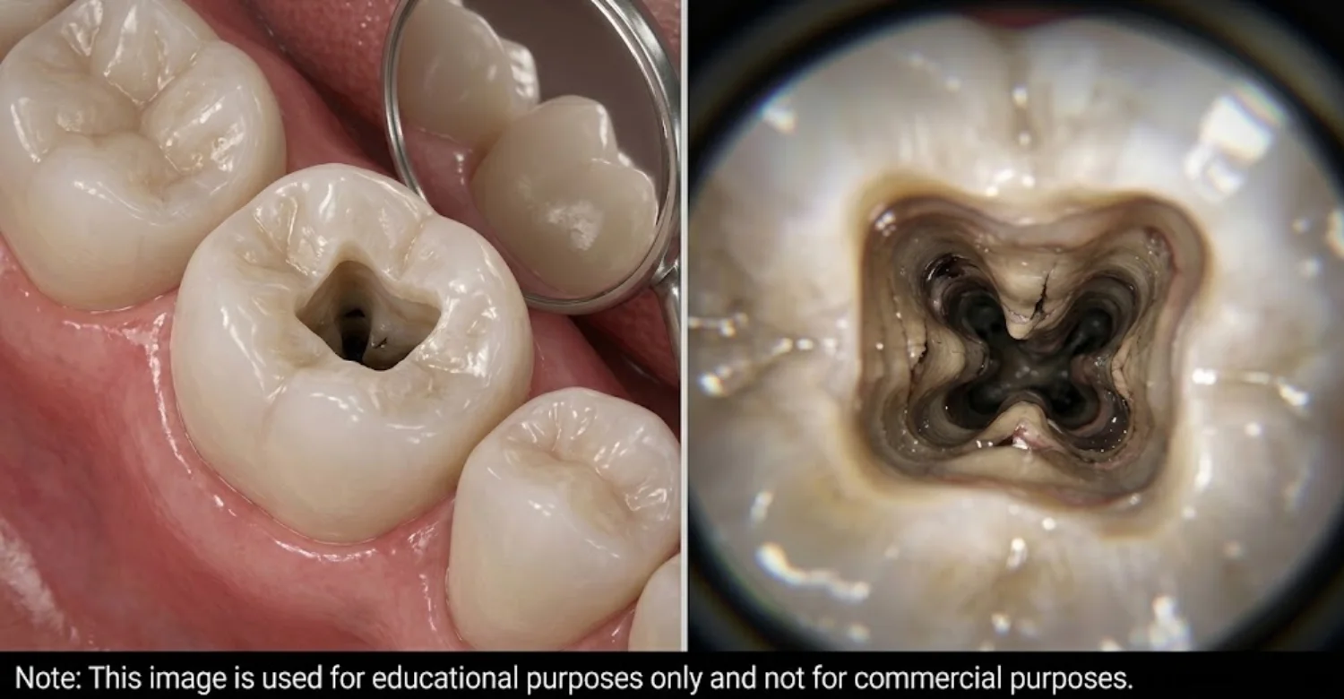
What Happens During Dynamic or Static Guided Endodontic Microsurgery?
Guided microsurgery follows a clear sequence from planning to wound closure. Most patients are treated under local anesthesia in a single appointment. Knowing each step can make the experience feel calmer and more predictable.
Step 1: Clinical Examination and Three-Dimensional Treatment Planning
The dentist examines the tooth and confirms the diagnosis with appropriate imaging. A CBCT scan is taken, often with an intraoral scan for guide planning. The surgeon then plans the apex location, entry point, and drill trajectory. At this stage, Dynamic vs Static Navigation in Endodontic Microsurgery is decided for the specific tooth. Risks, alternatives, and costs are explained before the patient gives consent. Patients can ask questions and review their scan with the dentist.
Step 2: Navigation Setup or Surgical Guide Preparation
For static surgery, the guide is designed, printed, sterilized, and test-fitted. For dynamic surgery, a tracking splint is attached and the system is calibrated. Registration links the patient's jaw to the virtual scan accurately. Accuracy is checked on known teeth or landmarks before any surgical cut. This verification step is one of the most important safety checks in guided surgery.
Step 3: Anesthesia and Surgical Access
Local anesthetic numbs the tooth and surrounding tissues thoroughly. The surgeon makes a small incision and gently lifts a gum flap to expose the bone. Some guided techniques allow smaller flaps than conventional surgery. Patients usually feel pressure and vibration, but not sharp pain. Sedation options may be discussed for anxious patients in suitable cases.
Step 4: Osteotomy and Root-End Resection
The surgeon creates a small bone window following the navigation or guide. Navigation studies typically describe osteotomies of about 4 mm in diameter. About 3 mm of the root tip is removed, along with infected tissue. The cut root surface is inspected under high magnification for cracks or missed canals. Staining dyes may help reveal fine cracks or isthmus areas.
Step 5: Root-End Preparation and Retrograde Filling
Ultrasonic tips prepare a small cavity about 3 mm deep in the root end. This cavity is cleaned and dried carefully. A biocompatible material, such as MTA or bioceramic putty, seals the root end. These materials support healing and have shown favorable results in clinical studies. A good seal prevents bacteria from re-entering the surrounding bone.
Step 6: Wound Closure and Postoperative Instructions
The gum flap is repositioned and closed with fine sutures. Gentle pressure and cold compresses help limit swelling. The patient receives written instructions on medication, diet, and oral hygiene. Sutures are usually removed after about 3 to 7 days. A contact number is provided in case questions arise during recovery.
How Long Does Guided Endodontic Microsurgery Take?
Most guided procedures take about 60 to 90 minutes for a single tooth. Complex molars or multiple roots may take longer. Navigation-assisted steps themselves often take only about 4 to 9 minutes in studies. Setup, flap management, and root-end filling make up most of the session. Planning and guide fabrication happen days before surgery. Patients should also allow time for a review visit about one week later.
Ready to transform your smile?
Talk to our team and get a personal FREE treatment plan.
Relief After Endodontic Microsurgery: What Can Patients Expect?
Most patients experience manageable discomfort that improves within a few days. Understanding the normal recovery pattern helps reduce anxiety after surgery. Recovery is similar whether dynamic, static, or freehand techniques are used.
How Long Does Pain and Swelling Usually Last?
Pain usually peaks within the first 24 to 48 hours after surgery. Swelling often peaks around the second or third day. Most discomfort settles significantly within 3 to 5 days. Mild tenderness when chewing may last about one to two weeks. Bruising, if present, fades gradually over several days. Most patients describe the discomfort as mild to moderate and manageable.
What Can Help Relieve Mild Postoperative Discomfort?
Cold compresses on the cheek during the first day can reduce swelling. Pain relievers should be taken exactly as your dentist advises. Soft, lukewarm foods help avoid irritating the surgical site. Resting with your head slightly raised may also limit swelling. Avoid smoking and alcohol, as both can slow healing. Gentle rest during the first day supports a smoother recovery.
When Should Patients Expect Improvement?
Most patients notice clear improvement by the third or fourth day. Normal daily activities can often resume within one or two days. Bone healing takes longer and continues quietly for months. Radiographic healing is usually assessed at 6 to 12 months. Complete bone fill may take a year or more in larger lesions. Symptoms often resolve long before the bone fully heals.
Does Dynamic or Static Navigation Reduce Postoperative Pain?
Guided techniques may reduce discomfort by creating smaller, more targeted bone windows. Less bone removal and shorter surgical time can mean less tissue trauma. However, strong comparative pain data for Dynamic vs Static Navigation in Endodontic Microsurgery remain limited. Pain also depends on flap size, inflammation, and individual sensitivity. Patients should expect normal post-surgical discomfort with any technique. Following aftercare advice usually matters more than the navigation method itself.
What Should Patients Know About Healing and Follow-Up Appointments?
A review visit about a week after surgery checks the gum and removes sutures. Follow-up radiographs or scans are typically taken at 6 and 12 months. Some clinicians continue monitoring for several years. Healing is judged by symptom relief and bone fill on images. Keeping these appointments helps detect problems early. International patients can often share follow-up images with the clinic remotely.
Side Effects and Potential Complications of Guided Endodontic Microsurgery
Like any surgery, guided endodontic microsurgery carries some risk of side effects. Most effects are mild and temporary, while serious complications are uncommon. Knowing what is normal helps patients recognize when to seek advice.
What Are the Common Temporary Side Effects?
Common side effects reflect the normal healing response of gum and bone. They usually improve steadily over the first week. Most do not require additional treatment beyond routine aftercare.
Mild Pain, Swelling, Bruising, and Tenderness
Mild to moderate pain is expected for a few days. Swelling of the cheek or lip is common and usually peaks around day two or three. Bruising may appear on the face, especially after surgery on lower teeth. Tenderness when touching the area can last about a week. These effects usually respond well to prescribed care and cold compresses.
Temporary Gum Sensitivity and Difficulty Chewing
The gum around the incision may feel tight or sensitive. Chewing on the treated side may be uncomfortable at first. Temporary numbness near the surgical site can occur from swelling. Most patients chew normally again within one to two weeks. Minor gum shrinkage can occasionally occur near the incision line.
What Are the Less Common Surgical Complications?
Less common complications include infection, delayed healing, nerve irritation, and incomplete healing. Careful planning and aftercare reduce these risks considerably. Prompt review helps manage them effectively if they occur.
Infection, Delayed Healing, or Persistent Symptoms
Postoperative infection can cause increasing pain, swelling, or discharge. Smoking, diabetes, and poor oral hygiene can slow healing. In one navigation series, 2 of 51 roots developed a postoperative sinus tract. Persistent symptoms beyond the expected recovery period need prompt review. Most infections respond well to timely treatment.
Injury to Nearby Anatomical Structures
Nerve irritation may cause tingling or numbness of the lip, chin, or gum. This is usually temporary, though rarely it can last longer. Surgery on upper teeth may sometimes create a small opening into the maxillary sinus. CBCT planning and guidance help reduce these risks but cannot eliminate them completely.
Root Damage or Incomplete Resolution of Periapical Disease
Excessive resection can shorten the root and weaken its bone support. Some lesions may not heal fully because of hidden cracks or residual bacteria. Pooled data suggest about 6% to 9% of microsurgery cases do not heal as hoped. Further surgery, retreatment, or extraction may then be discussed.
Can Navigation Errors Affect the Surgical Outcome?
Yes, navigation errors can affect the outcome if they are not detected. A registration or guide-fit error may shift the drill toward the wrong location. Studies report average deviations of about 1 to 1.5 mm, but individual outliers occur. Within Dynamic vs Static Navigation in Endodontic Microsurgery, both methods carry this small risk. Experienced surgeons verify position frequently under the microscope. Guidance is a tool, so clinical confirmation remains essential throughout surgery.
What Warning Signs Require Prompt Dental Assessment?
Some symptoms suggest that healing is not progressing normally. Contact your dentist promptly if any of the following warning signs occur. Early assessment often prevents minor problems from becoming serious.
Increasing Swelling, Fever, or Worsening Pain
Swelling that increases after day three may indicate infection. Fever or feeling generally unwell needs prompt assessment. Pain that worsens instead of improving should also be reviewed quickly.
Persistent Bleeding, Pus, or Difficulty Opening the Mouth
Light oozing is normal on the first day, but persistent bleeding is not. Pus or a bad taste may signal infection at the surgical site. Increasing difficulty opening the mouth can indicate spreading inflammation.
Difficulty Breathing or Swallowing as an Emergency Warning Sign
Difficulty breathing or swallowing is a medical emergency. It may indicate a rapidly spreading infection in the face or neck. Seek emergency care immediately rather than waiting for a dental appointment.
Ready to transform your smile?
Talk to our team and get a personal FREE treatment plan.
Tips for Patients Considering Guided Endodontic Microsurgery
Being prepared helps you make confident decisions and recover more comfortably. These practical tips cover questions, preparation, and aftercare. They apply to dynamic, static, and conventional microsurgery alike.
What Should You Ask Your Dentist Before Choosing a Navigation Technique?
Ask why surgery is recommended over retreatment or extraction. Ask which technique is proposed and why it suits your anatomy. Ask how many guided procedures the surgeon has performed. Ask about expected success rates, risks, and recovery time. When discussing Dynamic vs Static Navigation in Endodontic Microsurgery, request an itemized cost estimate. Also ask what happens if the lesion does not heal as expected.
How Can You Prepare for an Endodontic Microsurgery Appointment?
Share your full medical history and current medications with the clinic. Mention blood thinners, diabetes, or recent heart problems clearly. Eat a light meal beforehand unless instructed otherwise. Arrange soft foods and ice packs at home for recovery. Plan for someone to accompany you if sedation is used. International patients should plan several days locally after surgery.
What Should You Avoid During the Initial Recovery Period?
Avoid smoking for as long as possible, ideally for at least several days. Avoid alcohol while taking medication and during early healing. Do not pull your lip to look at the stitches. Avoid hard, crunchy, or very hot foods for several days. Avoid strenuous exercise for about 48 hours. Avoid vigorous rinsing or spitting during the first day.
How Should You Follow Postoperative Care Instructions?
Read your written instructions carefully and keep them nearby. Take medications at the times and doses prescribed. Use cold compresses as advised during the first day. Rinse gently only when your dentist says it is safe. Keep every scheduled review appointment. Ask the clinic if any instruction is unclear.
When Should You Contact Your Dentist After Surgery?
Contact your dentist if pain or swelling increases after the third day. Call if numbness persists long after the anesthetic should have worn off. Report bleeding that does not stop with gentle pressure. Contact the clinic for any fever, pus, or loose sutures. When in doubt, it is always better to ask.
Medical Tips for Supporting Recovery Safely
Good recovery habits support both bone and soft-tissue healing. Follow these medical tips alongside your dentist's personal advice. They help reduce complications and support comfortable healing.
Follow Prescribed Medication Instructions
Take pain relievers and any antibiotics exactly as prescribed. Do not exceed the recommended dose or combine medicines without advice. Tell your dentist about any side effects or allergic reactions.
Maintain Gentle Oral Hygiene Without Disturbing the Surgical Site
Brush your other teeth normally while avoiding the surgical area initially. Use a soft brush near the site once your dentist advises it. Prescribed mouth rinses can help keep bacteria levels low.
Attend Recommended Follow-Up and Healing Assessments
Follow-up visits confirm that the gum and bone are healing properly. Radiographs at 6 to 12 months show whether the lesion is resolving. Early review allows timely treatment if healing slows.
Dynamic vs Static Navigation in Endodontic Microsurgery: Average Cost in the US, UK, and Turkey
Costs vary by country, clinic, tooth type, and technology used. The figures below are estimates for planning and do not replace a personal quote. Dynamic vs Static Navigation in Endodontic Microsurgery may involve additional fees beyond standard apicoectomy pricing.
How Much Does Endodontic Microsurgery Cost in the US?
In the US, endodontic microsurgery costs an estimated average of $1,100 per tooth. Molars, complex cases, and guided procedures may cost more. CBCT scans, crowns, and follow-up imaging may be billed separately.
Cost component | Estimated cost |
Average endodontic microsurgery | $1,100 per tooth |
Lower planning estimate | $850 per tooth |
Higher planning estimate | $2,000 per tooth |
What Is the Average Cost of Endodontic Microsurgery in the UK?
In the UK, private endodontic microsurgery averages around $950 per tooth. Specialist fees, guided techniques, and additional imaging can increase the total. Confirm whether follow-up appointments are included in the quote.
Cost component | Estimated cost |
Average endodontic microsurgery | $950 per tooth |
Lower planning estimate | Approximately $700 per tooth |
Higher planning estimate | Approximately $1,500 per tooth |
The planning range is expressed in pound sterling equivalents and should not be treated as a verified national price range.
How Much Does Endodontic Microsurgery Cost in Turkey?
In Turkey, endodontic microsurgery averages approximately $350 per tooth. Prices depend on the clinic, specialist experience, imaging, and surgical technique. International patients should also budget for travel and accommodation.
Cost component | Estimated cost |
Average endodontic microsurgery | $350 per tooth |
Lower planning estimate | $300 per tooth |
Higher planning estimate | $900 per tooth |
Average Endodontic Microsurgery Costs by Country
Based on these averages, the estimated cost in Turkey is approximately 68% lower than in the US. These estimates exclude crowns, travel, and accommodation. Actual prices depend on the tooth, technique, and imaging required.
Does Dynamic Navigation Cost More Than Static Surgical Guides?
Dynamic navigation may cost more because clinics invest in tracking hardware and software. Static guides involve design, fabrication, and materials. However, the price difference depends on each clinic's fee structure.
Cost factor | Dynamic navigation | Static surgical guide |
Technology | Tracking hardware and software | Guide design and fabrication |
Planning | Digital surgical planning | Digital planning and guide design |
Equipment or production costs | Navigation system costs | Printing and material costs |
Pricing structure | May include a separate technology fee | May include a separate guide fee |
Main cost consideration | Equipment and system use | Custom guide production |
Some clinics include these services in their standard surgical fee, while others charge separately. Request itemized quotes before comparing Dynamic vs Static Navigation in Endodontic Microsurgery.
What Additional Costs Can Affect the Final Treatment Price?
Additional expenses may increase the overall treatment cost. Ask the clinic which services are included before accepting a quote.
Additional cost | What it may cover | Potential pricing consideration |
CBCT imaging | Three-dimensional assessment of the tooth and surrounding structures | May be charged separately |
Digital treatment planning | Review of scans and surgical planning | May be included or billed separately |
Custom surgical guide | Design, printing, materials, and sterilization | Usually depends on the case |
Navigation equipment | Tracking hardware, software, and system use | May involve a technology fee |
Surgical materials | Root-end filling and other materials | Depends on materials used |
Follow-up appointments | Postoperative reviews and healing assessment | Confirm how many visits are included |
Additional restoration | Crown or other restorative treatment, if needed | May substantially increase total cost |
CBCT Imaging and Digital Treatment Planning
Service | Why it may be needed | Possible additional charge |
CBCT scan | Evaluates root anatomy and surrounding structures when indicated | Depends on the clinic |
Digital planning | Supports assessment and procedure planning | May be included in the surgical fee |
Follow-up imaging | Checks healing when clinically appropriate | May be charged separately |
Custom Surgical Guide Design and Fabrication
Service | What it involves | Cost consideration |
Digital guide design | Planning the guide from diagnostic scans | Case-dependent |
3D printing | Manufacturing the guide | Depends on materials and production |
Sterilization and preparation | Preparing the guide for surgery | May be included in the guide fee |
Intraoral scanning | Capturing details needed for guide design when required | May incur an additional fee |
Navigation Equipment, Surgical Fees, and Root-End Filling Materials
Component | What can affect the price |
Dynamic navigation equipment | System use, tracking technology, and clinic pricing |
Surgical fees | Tooth position, root anatomy, and procedural complexity |
Root-end filling materials | Material type and amount required |
Postoperative care | Follow-up visits and any additional treatment |
Are Guided Endodontic Microsurgery Prices Different From Conventional Apicoectomy Fees?
Guided microsurgery may cost more than conventional apicoectomy because it can require additional imaging, planning, guide fabrication, or navigation technology. Whether this additional expense is worthwhile depends on the individual case.
Treatment approach | Potential cost considerations | When it may be considered |
Conventional apicoectomy | Standard surgical fees and materials | Selected cases suitable for conventional techniques |
Static-guided microsurgery | Surgical fees plus guide design and production | Cases where a custom guide may support access and positioning |
Dynamic-guided microsurgery | Surgical fees plus navigation technology costs | Selected cases where real-time instrument tracking may be beneficial |
Guided techniques are not automatically necessary for every tooth. Ask your endodontist whether navigation is expected to provide a meaningful clinical benefit in your case.
What Should an Itemized Dental Surgery Quote Include?
A clear quote should identify the main treatment costs and any services that may be charged separately.
Quote item | What to confirm |
Consultation | Whether the initial examination is included |
Imaging | Whether CBCT or other diagnostic imaging is included |
Digital planning | Whether planning carries a separate fee |
Navigation or surgical guide | Whether technology use or guide fabrication is included |
Surgical procedure | The total fee for the planned microsurgery |
Materials | Whether root-end filling materials are included |
Follow-up care | Number of included postoperative appointments |
Additional treatment | Whether crowns or other restorations would cost extra |
How Can Patients Compare Treatment Costs Across Countries Safely?
Compare equivalent treatments rather than headline prices alone. Review each clinic's specialist qualifications, experience, technology, materials, and follow-up arrangements. International patients should also account for travel, accommodation, and time away from work.
Comparison factor | Questions to ask |
Clinical expertise | Who performs the surgery, and what is their relevant experience? |
Technology | Is dynamic navigation or a static guide included in the price? |
Diagnostic imaging | Are required scans included? |
Surgical materials | Are root-end filling materials covered? |
Follow-up | How will recovery be monitored after treatment? |
Total cost | Are there additional fees for restoration, travel, or accommodation? |
Request an itemized treatment plan from each clinic and compare the same services. This makes it easier to evaluate the real cost of Dynamic vs Static Navigation in Endodontic Microsurgery across the US, UK, and Turkey.
At Vitrin Clinic: Personalized Assessment for Endodontic Treatment
At Vitrin Clinic, treatment decisions begin with understanding why a tooth has not healed. Our approach prioritizes accurate diagnosis before recommending any technology. Every plan is built around the patient's tooth, health, and goals.
Why Is Identifying the Cause of Persistent Root-End Disease the First Step?
Persistent disease may result from missed canals, leaking restorations, cracks, or cysts. Each cause may require a different solution. Surgery cannot fix a cracked root or an unrestorable tooth. Identifying the cause first helps avoid unnecessary procedures. It also makes the choice in Dynamic vs Static Navigation in Endodontic Microsurgery more meaningful. A clear diagnosis gives patients realistic expectations from the start.
How Can Clinical Examination and Appropriate Digital Imaging Guide Treatment Decisions?
Clinical tests confirm which tooth is causing symptoms and how healthy the gums are. Periapical radiographs provide an initial view of the root and lesion. CBCT is used when three-dimensional detail is needed for planning. Together, these findings show whether retreatment, surgery, or another option is suitable. Imaging is selected carefully to balance diagnostic value with radiation exposure.
When Might Root Canal Retreatment or Endodontic Surgery Be Considered?
Retreatment may be considered when the original root canal shows clear deficiencies. Surgery may be considered when retreatment is risky or has not resolved the disease. Some patients benefit from both treatments in sequence. Extraction and implant options are also discussed when saving the tooth is unlikely. Each option is explained with its expected benefits and limitations.
How Is a Personalized Treatment Plan Developed for International Patients?
International patients can share previous X-rays, scans, and dental records before arrival. This allows an initial review and preliminary planning. On arrival, a full examination confirms or refines the plan. Treatment timing considers healing time and travel dates. Remote follow-up can be arranged after patients return home.
What Should Patients Understand About Treatment Options, Risks, and Expected Costs?
Patients should understand every option, including no treatment and its consequences. Risks such as swelling, nerve irritation, and incomplete healing should be discussed openly. Expected success rates should be presented realistically, not as guarantees. Costs should be itemized so patients know exactly what is included. Informed patients make decisions they feel confident about.






How Vitrin Clinic Supports Your Treatment Journey
Vitrin Clinic aims to make each stage of endodontic care clear and well coordinated. Support continues from the first contact through follow-up. Each step is designed to keep patients informed and comfortable.
Initial Consultation and Review of Previous Dental Records
The journey begins with a consultation and a review of your dental history. Previous root canal records, X-rays, and scans help clarify what has already been tried. This review can begin remotely for international patients.
Assessment of Teeth, Gums, Oral Tissues, and Relevant Anatomical Structures
The examination covers the affected tooth, neighboring teeth, gums, and soft tissues. Relevant anatomy such as nerves and sinuses is assessed when surgery is considered. This complete picture helps ensure treatment is safe and appropriate.
Individualized Diagnostic and Treatment Planning
Each plan reflects your diagnosis, anatomy, and personal priorities. When surgery is indicated, the approach is selected to suit your specific case. Discussions about Dynamic vs Static Navigation in Endodontic Microsurgery are based on your scan findings.
Clear Communication About Treatment Alternatives and Cost Estimates
You receive clear explanations of alternatives, benefits, and risks. Cost estimates are itemized so there are no unexpected charges. Questions are welcome at every stage before you decide.
Appointment Coordination and Guidance for International Patients
The team helps coordinate appointment dates around your travel plans. Guidance on recovery time before flying is provided in advance. Practical information helps international patients plan their stay comfortably.
Follow-Up Planning and Communication After Treatment
Follow-up instructions are provided before you leave the clinic. Healing reviews can be arranged in person or coordinated with your local dentist. Ongoing communication helps address any questions during recovery.
Ready to transform your smile?
Talk to our team and get a personal FREE treatment plan.
Key Takeaways
These points summarize the most important findings from this guide. They can help you prepare for a conversation with your dentist.
Dynamic vs Static Navigation: The Main Differences at a Glance
Dynamic navigation tracks instruments live on a screen and allows adjustments during surgery. Static navigation uses a printed guide to fix the drill's path and depth. Both rely on CBCT planning and show high accuracy in research. Dynamic vs Static Navigation in Endodontic Microsurgery mainly differs in flexibility, setup, and access requirements.
Why Accuracy, Anatomy, and Clinical Experience Matter More Than Technology Alone
Navigation studies report average deviations of about 1 to 1.5 mm. Yet healing depends on diagnosis, sealing quality, and tooth condition. Experienced surgeons achieve better accuracy with either system. In Dynamic vs Static Navigation in Endodontic Microsurgery, technology supports skill but never replaces it.
What Patients Should Know About Relief, Side Effects, and Recovery
Discomfort usually peaks within 48 hours and improves within several days. Swelling and bruising are common and temporary. Serious complications are uncommon but require prompt care. Bone healing is monitored over 6 to 12 months.
Why Treatment Costs Must Be Compared Using Equivalent Services
Average costs range from about $350 in Turkey to $1,100 in the US. Quotes may differ in imaging, guidance, materials, and follow-up. Compare itemized quotes to make fair decisions. When comparing Dynamic vs Static Navigation in Endodontic Microsurgery prices, check exactly what is included.
When to Seek Professional Dental Assessment or Urgent Care
Seek assessment for persistent pain, swelling, or a gum boil after a root canal. Seek urgent care for fever or rapidly increasing swelling. Difficulty breathing or swallowing requires emergency medical attention immediately.
How Does Vitrin Clinic Provide Personalized Endodontic Treatment?
At Vitrin Clinic, our endodontic treatment focuses on diagnosing and treating problems affecting the tooth pulp and root tissues. If you experience persistent pain, swelling, or discomfort after a root canal, our team can assess your condition and discuss suitable treatment options. Contact Vitrin Clinic to book your consultation and receive a personalized endodontic treatment plan.
IMPORTANT :
This article is for general information and does not replace a personal assessment by a qualified dentist. Prices are approximate 2026 averages, with UK and Turkey figures converted to US dollars, and may vary by clinic and case.
Not sure whether a bridge or an implant is right for you? Book your free consultation with Vitrin Clinic and get a personalized assessment and treatment plan from our dental team.
Reference
FAQs
Dr. Rifat Alsaman has more than 5 years of clinical experience in dentistry and currently serves as the Head of the Medical Team at Vitrin Clinic. He is dedicated to providing exceptional patient care, overseeing treatment planning, and ensuring the highest clinical standards across the team. His expertise, attention to detail, and commitment to continuous professional development have helped countless patients achieve healthier, more confident smiles.


