Endodontics
October 18, 2026
Photobiomodulation for Dental Luxation Injuries: Can Light Therapy Support Tooth Healing?
 Dr. Rifat Alsaman

A knock to the mouth can shift a tooth within seconds. The tooth may look slightly out of place, feel loose, or ache when you bite. Many patients now ask whether light therapy can help the injured tissues recover. This guide explains Photobiomodulation for Dental Luxation Injuries in clear, practical terms. You will learn what the evidence shows, where it falls short, and why standard trauma care always comes first. We also cover symptoms, diagnosis, recovery timelines, follow-up care, and typical costs in the US, UK, and Turkey.
Ready to transform your smile?
Talk to our team and get a personal FREE treatment plan.
What Is Photobiomodulation for Dental Luxation Injuries?
Photobiomodulation for Dental Luxation Injuries means using low-intensity light on tissues around a displaced tooth. The aim is to support comfort and natural healing processes. It does not move the tooth or replace emergency treatment. Instead, it may be considered as an adjunct after the dentist has repositioned and stabilized the tooth. Understanding the injury itself helps explain where light therapy might fit.
What Is Photobiomodulation Therapy in Dentistry?
Photobiomodulation therapy, often called PBM, uses red or near-infrared light at low power. It was previously known as low-level laser therapy, or LLLT. Unlike surgical lasers, PBM devices do not cut, burn, or vaporize tissue. Dentists already use PBM for oral ulcers, post-surgical pain, and orthodontic discomfort. The World Association for Photobiomodulation Therapy (WALT) publishes guidance on safe dosing. Most dental PBM devices emit wavelengths between roughly 600 and 1,000 nanometers. These wavelengths penetrate soft tissue and, to some degree, bone. The therapy is typically quick, painless, and delivered chairside.
How Does Low-Level Light Therapy Work at the Cellular Level?
Light energy is absorbed by molecules inside cells called chromophores. The best-studied chromophore is cytochrome c oxidase, found in mitochondria. When this enzyme absorbs light, cellular energy production may increase. Cells can then produce more adenosine triphosphate, or ATP, their main energy source. Laboratory studies also show changes in reactive oxygen species and nitric oxide signaling. These shifts may influence inflammation, cell growth, and blood flow. Researchers describe a "biphasic dose response," meaning too little or too much light reduces benefit. This is why correct dosing matters as much as the device itself. The same dosing principle shapes every protocol for Photobiomodulation for Dental Luxation Injuries.
What Happens to the Tooth and Periodontal Ligament During Luxation?
Each tooth sits in a bony socket, held by the periodontal ligament (PDL). The PDL is a thin layer of fibers, only about 0.15–0.38 millimeters wide. It acts like a shock absorber and contains nerves, blood vessels, and repair cells. During luxation, these fibers stretch, compress, or tear. The blood supply entering the root tip may also be damaged. This can affect the pulp, the living tissue inside the tooth. Bone around the socket may crack or compress in more severe cases. Healing therefore involves several tissues, not just one. Supporting these tissues is the main biological goal of Photobiomodulation for Dental Luxation Injuries. Severe cases may also involve gum lacerations needing sutures.
Which Types of Dental Luxation Injuries May Be Considered for Photobiomodulation?
The International Association of Dental Traumatology (IADT) recognizes several luxation categories. Each type carries a different risk of complications. Research shows pulp necrosis rates range from about 3% after concussion to over 80% after intrusion. Interest in Photobiomodulation for Dental Luxation Injuries spans this whole spectrum. However, its potential role differs with injury severity.
Concussion and Subluxation Injuries
Concussion means the tooth is tender but not loose or displaced. Subluxation means the tooth is loose but still in its normal position. Both are considered milder injuries. Studies report pulp necrosis in roughly 3% of concussions and 6% of subluxations. These patients often mainly need pain control and monitoring. PBM may be discussed for comfort, although direct trial evidence is limited. For these milder cases, Photobiomodulation for Dental Luxation Injuries would focus mainly on pain relief.
Extrusive and Lateral Tooth Luxation
Extrusion means the tooth is partially pushed out of its socket. Lateral luxation means the tooth is displaced sideways, often with a bone fracture. Pulp necrosis occurs in about 26% of extrusions and up to 58% of lateral luxations. These teeth need prompt repositioning and a flexible splint. Photobiomodulation for Dental Luxation Injuries of this kind would only follow those steps.
Intrusive Luxation and More Severe Displacement Injuries
Intrusion means the tooth is driven into the jawbone. It is the most severe type of luxation type. In mature permanent teeth, pulp necrosis rates can exceed 85%. Root resorption and ankylosis are also common long-term risks. These cases often need specialist care, sometimes including surgical or orthodontic repositioning. Any light therapy here remains strictly experimental and supportive. Long-term monitoring for at least five years is usually recommended after intrusion.
.jpg)
.jpg)
.jpg)
.jpg)
.jpg)
.jpg)
Can Photobiomodulation Help a Luxated Tooth Heal?
Patients understandably want anything that might speed healing. The honest answer is that research is promising but incomplete. Laboratory and animal data suggest biological effects on healing tissues. Human trials on Photobiomodulation for Dental Luxation Injuries specifically remain scarce. The sections below explain what is known and what is not.
What Does Current Research Say About Photobiomodulation for Dental Trauma Recovery?
Most research on PBM in dentistry focuses on related conditions. These include orthodontic pain, extraction healing, and oral mucositis. A 2019 systematic review found PBM reduced orthodontic pain in most included trials. Animal studies on replanted teeth have reported improved PDL organization after laser exposure. Some rat studies also showed less inflammatory root resorption. However, high-quality randomized trials in human dental trauma patients are very limited. Current IADT 2020 guidelines do not include PBM as a standard recommendation. Evidence therefore supports cautious interest, not routine use.
Can Light Therapy Support Periodontal Ligament Healing?
PDL fibroblasts are the main repair cells after luxation. In cell culture studies, PBM has increased fibroblast proliferation and collagen production. Some studies also reported higher levels of growth factors involved in tissue repair. In theory, this could help torn PDL fibers reorganize faster. Animal models of tooth replantation support this possibility. Yet a laboratory dish is very different from an injured human mouth. Blood clots, bacteria, and bite forces all affect real healing. Human confirmation is still needed before firm conclusions are drawn.
Can Photobiomodulation Reduce Pain and Inflammation After Tooth Displacement?
Pain relief is the most consistent finding in dental PBM research. Studies in orthodontics and oral surgery show modest but measurable pain reductions. Several meta-analyses report lower pain scores during the first 24 to 72 hours. PBM may reduce inflammatory mediators such as prostaglandin E2 and interleukin-1β. It may also influence nerve signaling and local swelling. These findings make pain management the most plausible benefit for relaxation patients. Still, standard pain relievers remain the first-line approach. Pain relief is therefore the most realistic expectation from Photobiomodulation for Dental Luxation Injuries. Benefits are usually modest rather than dramatic.
Can Low-Level Laser Therapy Improve Tooth Stability or Healing Outcomes?
Tooth stability depends mainly on correct repositioning and splinting. It also depends on how quickly the PDL fibers reattach. Some animal studies suggest PBM may speed bone and ligament repair. However, no strong human trials show that PBM reduces splinting time. Similarly, there is no clear proof that it lowers pulp necrosis rates. Long-term outcomes such as tooth survival have not been adequately studied. For now, any effect on stability should be considered unproven. Patients should be cautious of any clinic promising guaranteed stability results.
What Are the Limitations of Current Evidence for Photobiomodulation After Dental Trauma?
Evidence gaps are important for patients and clinicians alike. Many studies use different wavelengths, doses, and session schedules. This makes results hard to compare or combine. Sample sizes are often small, sometimes fewer than 30 participants. Follow-up periods are frequently too short to detect late complications. These limitations shape how responsible clinics discuss Photobiomodulation for Dental Luxation Injuries.
Evidence From Laboratory Studies Versus Human Clinical Trials
Laboratory studies test cells or animals under controlled conditions. They are useful for understanding mechanisms. However, many promising lab treatments fail when tested in people. Human trauma cases vary in age, injury type, and time to treatment. Randomizing emergency patients is also ethically and practically difficult. As a result, high-level human evidence for PBM after luxation remains thin.
Why Results From Orthodontic Tooth Movement Cannot Automatically Be Applied to Dental Luxation
Orthodontic movement involves slow, controlled pressure on healthy tissues. Luxation involves sudden force, tearing, and sometimes bone fracture. Orthodontic patients rarely have disrupted pulp blood supply. Trauma patients often do. So pain relief seen in braces patients may not translate exactly. Healing biology after a sudden injury follows a different pattern. Extrapolation should therefore be cautious.
What Causes Dental Luxation Injuries?
Traumatic dental injuries are extremely common worldwide. A 2018 global analysis estimated that over one billion people have experienced one. Prevalence reaches about 15% in permanent teeth and 23% in primary teeth. Luxation injuries are among the most frequent types, especially in young children. Knowing the causes helps families reduce risk.
Sports Injuries, Falls, and Accidental Impacts
Falls are the leading cause of dental trauma in young children. Toddlers learning to walk often strike their upper front teeth. In older children and teenagers, sports become a major cause. Contact sports, cycling, skateboarding, and playground activities carry the highest risk. Studies suggest sports cause between 13% and 39% of all dental injuries. Upper central incisors are involved in roughly 80% of cases. Wearing a mouthguard significantly reduces this risk. Children aged 8 to 12 years show a particular peak in sports-related injuries.
Road Traffic Accidents and Direct Facial Trauma
In adults, road traffic accidents are a significant cause of relaxation. Bicycle and motorcycle accidents often cause multiple tooth injuries. Assaults and workplace accidents are other common sources. These injuries often involve greater force than simple falls. As a result, they are more likely to include bone fractures. Patients may also have injuries to the lips, jaw, or face. A full medical assessment may be needed alongside dental care. Head injuries should always be ruled out first.
How Do Dental Luxation Injuries Affect Permanent and Primary Teeth Differently?
Primary teeth sit above developing permanent tooth buds. A displaced baby tooth can damage the permanent tooth underneath. Possible effects include enamel defects, discoloration, or altered eruption. For this reason, primary teeth are rarely forced back into place. Permanent teeth, however, are usually repositioned to save them. Immature permanent teeth with open root tips have better healing potential. Their pulp may even regain blood supply after injury. Peak injury ages are about 2–3 years for primary teeth and 8–10 years for permanent teeth.
Which Factors Increase the Risk of Complications After Tooth Displacement?
Several factors raise complication risks after luxation. Mature teeth with closed root tips heal less predictably than immature teeth. Greater displacement distance increases the chance of pulp necrosis. Delayed treatment also worsens outcomes. Combined injuries, such as crown fractures with luxation, add further risk. Studies show crown fracture plus luxation can raise necrosis risk several times. Smoking, poor oral hygiene, and systemic conditions may slow healing. An overjet greater than 3 millimeters roughly doubles the risk of front-tooth injury.
What Are the Symptoms of a Luxated Tooth?
Symptoms vary with the type and severity of injury. Some signs are obvious immediately, while others develop over weeks. Recognizing them early improves the chances of saving the tooth. Even minor-looking injuries deserve professional assessment.
Tooth Displacement, Looseness, and Changes in Bite
A displaced tooth may look longer, shorter, or tilted. It may feel loose when touched with the tongue. Many patients notice their teeth no longer meet normally. Biting may feel uneven or blocked by the injured tooth. In lateral luxation, the tooth may be locked firmly in its new position. This can make it feel solid despite being displaced. Any noticeable change in your bite after trauma needs a dental check.
Pain, Tenderness, Gum Bleeding, and Swelling
Pain and tenderness to touch are very common after luxation. Biting or tapping on the tooth often increases discomfort. Bleeding from the gum line suggests torn PDL fibers. The gums and lips may swell during the first 48 hours. Bruising of the lip or face is also common. These symptoms usually ease gradually with proper care. Worsening symptoms after three days, however, should be reported promptly.
Tooth Discoloration and Changes in Pulp Sensibility
A tooth may turn pink, grey, or dark after injury. Early pink discoloration can reflect bleeding inside the pulp. Some temporary color changes fade within weeks. Persistent grey or dark color may signal pulp necrosis. The tooth may also stop responding to cold or electric testing. These changes require careful monitoring rather than immediate conclusions. Photographs taken at each visit help track subtle color shifts over time.
Can a Tooth Be Luxated Without Severe Pain?
Yes, some luxated teeth cause surprisingly little pain. Damaged nerves inside the pulp may temporarily stop sending signals. Intruded teeth may also feel firm and painless. This can falsely reassure patients and parents. Children especially may underreport symptoms. Lack of pain does not mean lack of damage, so assessment remains essential.
Which Symptoms Require Immediate Dental Assessment?
Seek urgent dental care if a tooth is visibly displaced or very loose. Bleeding that does not stop after 10–15 minutes of pressure also needs attention. A tooth pushed into the gum requires immediate examination. Facial swelling, fever, or difficulty opening the mouth are warning signs. Any knocked-out tooth is a true emergency. Ideally, displaced teeth should be seen within hours. Early care improves outcomes far more than any later adjunct therapy.
How Are Dental Luxation Injuries Diagnosed Before Photobiomodulation?
Accurate diagnosis must come before any treatment decision. The dentist needs to identify the injury type, severity, and associated damage. This determines whether repositioning, splinting, or specialist referral is needed. Only then can adjunctive options be discussed responsibly. Diagnosis is therefore the first step before considering Photobiomodulation for Dental Luxation Injuries.
Clinical Examination of Tooth Position, Mobility, and Occlusion
The dentist first checks the position of each injured tooth. They compare it with neighboring teeth and the patient's normal bite. Mobility is tested gently in different directions. A metallic, high-pitched sound on tapping may suggest ankylosis or intrusion. The dentist also checks soft tissues for cuts or foreign bodies. Photographs are often taken to document the injury. These records become valuable baselines for later comparison.
Dental X-Rays and Additional Imaging When Indicated
IADT guidelines recommend several periapical radiographs from different angles. An occlusal X-ray is also commonly used. These images show root position, socket changes, and possible fractures. A widened PDL space may indicate extrusion or lateral luxation. In complex cases, cone-beam CT (CBCT) provides three-dimensional detail. CBCT can reveal hidden root or bone fractures missed on standard X-rays.
How Dentists Assess Periodontal Ligament and Supporting Bone Damage
PDL damage is assessed through mobility, tenderness, and X-ray findings. Bleeding from the gum crevice suggests torn ligament fibers. Widening or loss of the PDL space on images indicates displacement. The dentist also feels the bone around the tooth for steps or fractures. Alveolar bone fractures often accompany lateral luxation. These findings guide splint type and duration.
Pulp Sensibility Testing and Monitoring for Pulp Necrosis
Pulp tests check whether the nerve inside the tooth responds. Common tests include cold spray and electric pulp testing. Newer methods, such as laser Doppler flowmetry and pulse oximetry, measure blood flow. Blood flow tests may be more accurate after trauma. Results are recorded at the first visit and compared over time. Trends matter more than a single reading. Immediately after trauma, up to half of injured teeth may show no response temporarily.
How Dentists Distinguish Luxation From Tooth Fracture, Root Fracture, and Avulsion
A crown fracture is usually visible as a chipped or broken tooth. Root fractures may cause mobility similar to luxation. Multiple X-ray angles help reveal horizontal root fracture lines. Avulsion means the tooth is completely out of the socket. Distinguishing these injuries is critical because treatments differ greatly. Sometimes several injury types occur together.
Why Is Follow-Up Testing Necessary Even When the Tooth Initially Appears Healthy?
Many complications appear weeks or months after the injury. Pulp necrosis may develop slowly without obvious symptoms. Root resorption can begin silently and progress quickly in some cases. Studies show most pulp necrosis is detected within the first year. However, some complications appear several years later. Regular follow-up allows early treatment before damage becomes irreversible. This is true whether or not Photobiomodulation for Dental Luxation Injuries is part of the plan.

Note : All images used are for editorial and illustrative purposes only and may not originate from the original news provider or associated company.
What Is the Standard Treatment for a Luxated Tooth?
Standard treatment follows evidence-based IADT guidelines. The goals are to restore position, stabilize the tooth, and protect the pulp and PDL. Treatment varies by injury type and patient age. Any discussion of Photobiomodulation for Dental Luxation Injuries happens within this framework, never instead of it.
Why Prompt Dental Trauma Management Comes Before Adjunctive Light Therapy
Time strongly affects outcomes after luxation. Displaced teeth become harder to reposition as clots form and bone remodels. Delayed repositioning may increase pulp necrosis and resorption risks. Light therapy cannot correct position or stabilize a loose tooth. Therefore, emergency management always takes priority. Adjuncts are only considered once the tooth is correctly placed and secured.
How Dentists Treat Different Types of Tooth Luxation
Treatment is tailored to the specific injury pattern. Milder injuries may need only monitoring. Displaced teeth usually need repositioning and splinting. Some teeth later require root canal treatment. Children with primary teeth follow a separate approach.
Monitoring and Supportive Care for Selected Minor Injuries
Concussion injuries usually need no splint. Subluxated teeth may receive a short flexible splint for comfort, up to 2 weeks. Patients are advised to eat soft foods and keep the area clean. Pain relief and regular check-ups complete the plan. Most of these teeth heal without major complications.
Repositioning and Flexible Splinting for Displaced Teeth
Extruded and laterally luxated teeth are gently repositioned under local anesthesia. A flexible splint then holds the tooth while tissues heal. IADT guidelines recommend about 2 weeks for extrusion. Lateral luxation typically needs about 4 weeks, especially with bone fracture. Intruded teeth may be repositioned surgically, orthodontically, or allowed to re-erupt. The choice depends on root maturity and intrusion depth.
Root Canal Treatment When Pulp Necrosis or Infection Develops
If the pulp dies, bacteria can infect the root canal. This can trigger inflammatory root resorption. Root canal treatment removes the infected tissue and seals the canal. In mature intruded teeth, it is usually started within 2 weeks. Immature teeth may receive regenerative endodontic procedures or apexification instead. Early detection helps preserve the tooth long-term.
Management of Luxation Injuries in Children With Primary Teeth
Primary teeth are treated conservatively to protect developing permanent teeth. Many displaced baby teeth are allowed to reposition spontaneously. Severely displaced or dangerously loose primary teeth may be extracted. Splinting is rarely used in young children. Parents should watch for color change, swelling, or abscess formation. Follow-up continues until the permanent tooth erupts.
Can Photobiomodulation Be Used Alongside Conventional Luxation Treatment?
PBM may be used alongside conventional treatment in selected cases. It does not interfere with splints, medications, or root canal procedures. Some clinicians apply it after repositioning to support comfort. Others use it during early follow-up visits. Patients should understand it remains an adjunct with limited trauma-specific evidence. Photobiomodulation for Dental Luxation Injuries should always be documented as optional and supportive.
When Is Referral to an Endodontist or Dental Trauma Specialist Appropriate?
Referral is advisable for intrusion, complex combined injuries, or multiple displaced teeth. Immature teeth needing regenerative treatment often benefit from specialist input. Signs of root resorption also warrant specialist assessment. Endodontists have additional training in pulp and root canal management. Oral surgeons may help with bone fractures. Collaborative care often improves long-term tooth survival. Specialists can also advise on the suitability of adjunctive therapies.
How Is Photobiomodulation Therapy for Tooth Luxation Performed?
PBM sessions are usually short, simple, and comfortable. The process involves choosing a device, setting parameters, and applying light to target areas. Careful technique matters because dosing affects results. Here is how a typical session may be planned.
How Dentists Select the Light Source and Treatment Parameters
Dentists may use diode lasers or light-emitting diode (LED) devices. Lasers produce a focused, coherent beam. LEDs cover a broader area with non-coherent light. Both can deliver PBM when correctly dosed. The clinician considers tissue depth, injury location, and patient factors. Device calibration is important because actual output can differ from labeled settings. Reliable equipment is a basic requirement for Photobiomodulation for Dental Luxation Injuries.
Wavelength, Energy Density, Power, and Treatment Duration
Red wavelengths, around 630–680 nm, mainly target superficial tissues. Near-infrared wavelengths, around 780–980 nm, penetrate deeper into bone and ligament. Studies commonly report energy densities between about 2 and 10 J/cm² per point. Power output is usually low, often between 50 and 500 milliwatts. Each point may receive light for roughly 10 to 60 seconds. Exact protocols vary widely between studies. WALT recommends that clinicians document every parameter for each session.
Where Is the Light Applied During Dental Trauma Treatment?
Light is usually applied over the root area of the injured tooth. Points may include the cheek or lip side and the tongue or palate side. Some protocols also target the root tip region. The probe is held in contact or very close to the gum. Surrounding soft tissue injuries may also be treated. Coverage aims to reach the PDL and nearby bone. Points are often spaced about one centimeter apart.
How Many Photobiomodulation Sessions May Be Considered?
There is no established session count for luxation injuries. Protocols borrowed from oral surgery often use 3 to 6 sessions. Sessions may be spaced every 24 to 72 hours during the first week or two. More sessions do not automatically mean better results. The biphasic dose response means overtreatment may reduce benefit. Session plans for Photobiomodulation for Dental Luxation Injuries should be individualized and reviewed regularly.
When Might Treatment Begin After Traumatic Tooth Displacement?
PBM is generally considered after repositioning and splinting are complete. Some clinicians start on the same day as emergency treatment. Early sessions may target pain and inflammation during the acute phase. Later sessions may aim to support tissue repair. Timing should never delay essential emergency care. The treating dentist decides based on overall clinical status. Ideally, Photobiomodulation for Dental Luxation Injuries begins within the first few days after injury.
Does Photobiomodulation Hurt, and Is Anesthesia Necessary?
PBM is painless for most patients. Some people feel mild warmth or nothing at all. Because no tissue is cut, anesthesia is not required. Sessions typically last only a few minutes. Children usually tolerate PBM well, which can be helpful after a frightening injury. Patients can return to normal activities immediately afterward.
Can Photobiomodulation Be Used With a Dental Splint?
Yes, PBM can usually be applied while a splint is in place. Light passes through gum tissue around the splint without disturbing it. The clinician avoids pressing on the splinted teeth. Composite and wire splints do not block light at the root level. Patients should not remove or adjust splints themselves. Splint stability always takes priority over light application.
Ready to transform your smile?
Talk to our team and get a personal FREE treatment plan.
What Are the Benefits and Limitations of Photobiomodulation for Luxated Teeth?
Every treatment has potential advantages and real limits. Understanding both helps patients set realistic expectations. PBM may offer comfort and possible support for healing. However, it cannot fix structural problems or guarantee outcomes. Balanced information leads to better decisions about Photobiomodulation for Dental Luxation Injuries.
Potential Effects on Inflammation, Pain, and Tissue Repair
The most supported benefit is short-term pain reduction. Meta-analyses in related dental fields report lower pain scores after PBM. Reduced inflammation may also ease swelling and tenderness. Some studies show faster soft tissue healing after oral surgery. These effects could make early recovery more comfortable. However, the size of benefit after luxation specifically is unknown.
Potential Support for Periodontal Ligament and Surrounding Tissue Recovery
Laboratory studies show PBM can stimulate PDL fibroblasts and bone-forming cells. Animal studies suggest better collagen organization after laser exposure. This could theoretically support ligament reattachment after displacement. Some research also shows improved blood vessel formation. These findings provide a biological rationale for further study. They do not yet prove clinical benefit in human luxation cases.
Why Photobiomodulation Cannot Reposition a Displaced Tooth
A displaced tooth needs mechanical correction. Only manual, surgical, or orthodontic repositioning can return it to place. Light therapy has no physical force capable of moving teeth. Delaying repositioning while trying PBM could worsen outcomes. Unrepositioned teeth may heal in the wrong position or interfere with the bite. This is a firm, non-negotiable limitation.
Can Light Therapy Prevent Root Resorption or Tooth Loss?
Root resorption is a major cause of tooth loss after severe relaxation. After intrusion, resorption rates in some studies exceed 50%. A few animal studies suggest PBM may reduce inflammatory resorption. However, no human trials confirm this protective effect. Timely root canal treatment remains the proven way to stop infection-related resorption. Patients should not rely on light therapy for resorption prevention.
Why Treatment Outcomes Depend on Injury Severity and Timely Conventional Care
Outcomes are driven mainly by injury type, root maturity, and treatment timing. For example, concussion carries a far lower necrosis risk than intrusion. Fast repositioning and appropriate splinting improve prognosis. Regular monitoring catches complications early. Adjunctive therapies may only add marginal benefit on top of these factors. Photobiomodulation for Dental Luxation Injuries cannot overcome a severe injury managed late.
Are There Known Side Effects of Low-Level Laser Therapy in Dentistry?
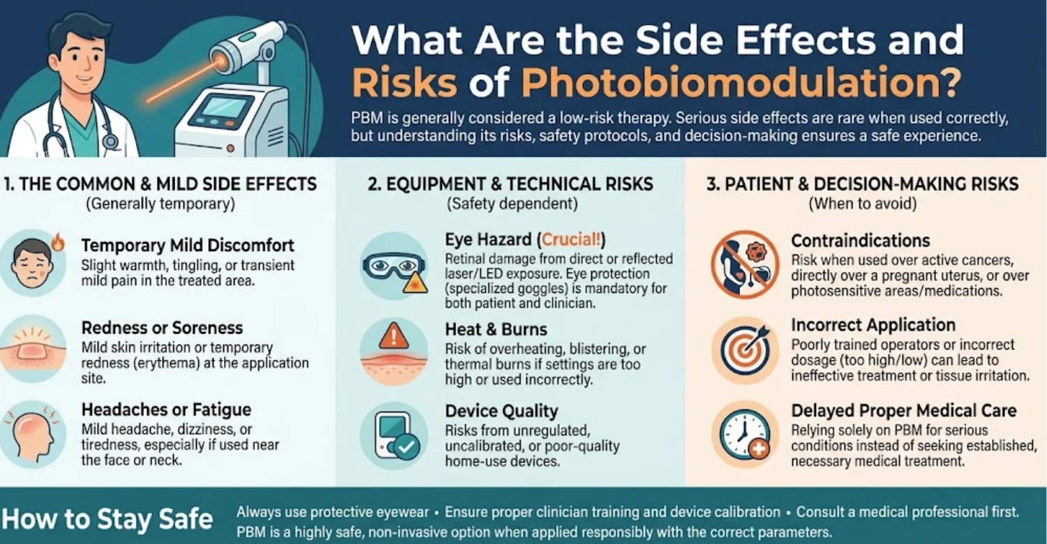
Reported side effects are usually mild and short-lived. Some patients feel slight warmth or tingling during treatment. Temporary sensitivity has occasionally been reported. Systematic reviews across dental applications report very few adverse events. PBM is non-invasive and does not cause bleeding or tissue damage at correct doses. This favorable safety profile explains its growing popularity.
Can Incorrect Laser Parameters Cause Tissue Heating or Injury?
Yes, excessive power or prolonged exposure can heat tissue. Overheating may irritate the gums or the pulp. Dark pigmented tissues absorb more energy and heat faster. High doses may also inhibit rather than stimulate healing. This is why trained clinicians calibrate devices and follow published guidance. Home or unregulated devices are not recommended after dental trauma. Safe Photobiomodulation for Dental Luxation Injuries requires professional-grade, calibrated equipment.
Eye Safety and Protective Measures During Laser Treatment
Laser light can damage the retina if it enters the eye directly. Near-infrared light is especially risky because it is invisible. Patients, staff, and clinicians should wear wavelength-specific protective glasses. The beam should never be directed toward the face or eyes. Clinics should follow local laser safety regulations. LED devices carry lower but not zero eye risks.
What Are the Risks of Relying on Light Therapy Instead of Standard Treatment?
The biggest risk is delaying proven care. A displaced tooth left unrepositioned may heal in a poor position. Missed pulp necrosis can lead to infection and rapid root resorption. Untreated infections may cause abscesses and bone loss. In children, delayed care may also affect developing permanent teeth. Light therapy should never replace emergency treatment or follow-up visits.
What Should Patients Discuss With Their Dentist Before Treatment?
Ask your dentist why PBM is being suggested for your injury. Clarify the expected benefits and the strength of evidence. Ask about the device, settings, and number of sessions. Mention any photosensitive medications, pregnancy, or medical conditions. Discuss total costs and whether PBM is optional. A clear conversation helps you make an informed choice about Photobiomodulation for Dental Luxation Injuries.
How Can Patients Find Relief After a Dental Luxation Injury?
Comfort matters during recovery after any tooth injury. Simple measures often control pain and swelling effectively. Following professional advice also protects the healing tissues. These steps apply whether or not light therapy is used.
What Helps Relieve Pain and Swelling After Tooth Displacement?
Resting the injured area reduces irritation and pain. Soft foods limit pressure on the tooth. Cold compresses help control early swelling. Keeping the head slightly raised while sleeping may reduce facial swelling. Gentle oral hygiene prevents infection that could increase pain. Most discomfort improves noticeably within the first few days. Persistent discomfort beyond one week deserves a professional review.
Which Pain-Relief Medicines May Be Appropriate?
Over-the-counter painkillers such as ibuprofen or paracetamol (acetaminophen) are commonly recommended. Ibuprofen also helps reduce inflammation. Always follow the dose on the package or your dentist's instructions. Children's doses depend on age and weight, so check with a pharmacist. People with stomach, kidney, or bleeding conditions should seek advice first. Antibiotics are not routinely needed for luxation injuries.
When Are Cold Compresses Helpful?
Cold compresses are most useful during the first 24 to 48 hours. They reduce swelling, bruising, and pain. Apply a wrapped ice pack to the cheek or lip for 15–20 minutes. Allow breaks between applications to protect the skin. Never place ice directly on the injured tooth. After 48 hours, swelling usually begins to settle naturally. Some patients then find gentle warmth more soothing.
Should Patients Avoid Biting or Chewing With the Injured Tooth?
Yes, avoiding direct biting helps the PDL heal. IADT guidelines recommend a soft diet for up to 2 weeks. Chew with back teeth on the opposite side when possible. Avoid biting into apples, sandwiches, or hard snacks with the front teeth. Nail biting and pen chewing should also stop. Excess force can disrupt early healing and splint stability.
Why Should Patients Avoid Repositioning a Tooth Themselves?
Pushing a displaced tooth yourself can worsen the injury. Force may further tear the PDL or fracture the root. In primary teeth, it may harm the developing permanent tooth. Self-repositioning also risks introducing bacteria. The exception is an avulsed permanent tooth, where immediate replanting is advised. For relaxation, gently bite on a clean cloth and see a dentist quickly.
When Should Worsening Pain, Swelling, or Fever Be Evaluated Urgently?
Pain should gradually improve, not worsen, after the first few days. Increasing pain may signal infection or pulp necrosis. Swelling that spreads to the face or neck needs urgent care. Fever, pus, or a bad taste suggest an abscess. Difficulty swallowing or breathing is a medical emergency. Contact your dentist or emergency services immediately in these situations.
Ready to transform your smile?
Talk to our team and get a personal FREE treatment plan.
What We Notice Clinically
Clinical experience adds context to research findings. Trauma cases rarely follow textbook patterns exactly. Observing patients over time reveals recurring lessons. These observations help shape cautious, individualized care.
Why the Same Injury Can Produce Different Healing Outcomes
Two patients with similar injuries may heal very differently. Root maturity strongly influences pulp survival. Immature teeth often regain blood supply, while mature teeth frequently do not. Treatment timing, oral hygiene, and general health also matter. Studies show necrosis risk after lateral luxation varies widely by root development. Individual prognosis therefore requires careful assessment. Statistics guide expectations, but each tooth must be judged individually.
Why Tooth Position Alone Does Not Reveal the Extent of Internal Damage
A tooth may look nearly normal after a significant injury. The PDL and pulp blood supply may still be badly damaged. Root fractures can hide beneath a straight-looking crown. Bone fractures may also be invisible without imaging. This is why X-rays and pulp testing are essential. Appearance alone can be misleading. Clinically, this is one of the most important lessons in trauma care.
Why an Initially Negative Pulp Test Does Not Always Confirm Pulp Necrosis
Injured pulp nerves may stop responding temporarily. This is sometimes called "pulp shock." Research shows some teeth regain sensibility after several weeks or months. Transient apical breakdown can also mimic pathology on X-rays. Therefore, a single negative test should not trigger immediate root canal treatment. Dentists look for multiple signs before diagnosing necrosis.
Common Misconceptions About Laser-Assisted Healing for Luxated Permanent Teeth
A common myth is that lasers can "fix" a loose tooth. Another is that light therapy replaces splinting. Some patients believe PBM guarantees the tooth will survive. Others assume all dental lasers work the same way. In reality, PBM devices differ greatly from surgical lasers. Clear explanations help prevent unrealistic expectations about Photobiomodulation for Dental Luxation Injuries.
Clinical Note: Dr. Rifat Alsaman on Individualized Dental Trauma Assessment
At Vitrin Clinic, Dr. Rifat Alsaman's approach to dental trauma centers on careful, individualized assessment. Every injured tooth receives a structured examination before any treatment decision. The focus is on understanding the full extent of the injury. Adjunctive therapies are considered only after essential care is complete.
Why Identifying the Injury Type Must Come Before Selecting Adjunctive Therapies
Each luxation type carries a different risk profile and treatment pathway. Choosing therapies before diagnosis can lead to missed fractures or delayed repositioning. Accurate classification guides splinting, monitoring, and referral decisions. Only then can adjuncts like PBM be weighed sensibly. Diagnosis is the foundation of safe trauma care.
How Clinical Findings and Follow-Up Guide Decisions About Further Treatment
Decisions evolve as the tooth heals. Pulp test trends, color changes, and X-ray findings guide next steps. Some teeth recover fully, while others need root canal treatment. Follow-up visits allow timely intervention when problems appear. This staged approach avoids both overtreatment and undertreatment. It also gives patients a clear picture of progress at every visit.
.jpg)
.jpg)
.jpg)
.jpg)
How Long Does Recovery Take After a Dental Luxation Injury?
Recovery time depends on injury type, age, and treatment. Most early symptoms settle within weeks. However, deeper healing and monitoring continue for months or years. Understanding the timeline helps patients stay patient and attentive.
What to Expect During the First 24–72 Hours
The first three days are usually the most uncomfortable. Pain, swelling, and tenderness typically peak during this period. Gum bleeding often stops within the first day. A splint may feel strange but usually becomes tolerable quickly. Soft foods, cold compresses, and painkillers help most patients manage. If used, PBM sessions often focus on this early phase.
Recovery During the First Two Weeks
Tenderness usually decreases steadily during the first two weeks. Gum tissue begins healing within days. Initial PDL repair starts within about a week. IADT guidelines recommend a check-up around 2 weeks after injury. Splints for extrusion and subluxation are often removed at this point. Patients can slowly return to normal eating as advised. Most patients report minimal discomfort by the end of this period.
Healing and Monitoring Over the Following Months
PDL and bone healing continue for several months. Splints for lateral luxation and intrusion are usually removed around 4 weeks. IADT recommends follow-ups at about 4 weeks, 8 weeks, 12 weeks, 6 months, and 1 year. For severe injuries, yearly checks may continue for at least 5 years. Each visit tracks pulp health and root condition.
How Long Does Periodontal Ligament Healing Usually Take?
Experimental studies show PDL fibers begin reconnecting within about 1 to 2 weeks. By roughly 2 months, ligament strength recovers substantially in uncomplicated cases. Bone around the socket may take several months to remodel fully. Severe injuries with crushed PDL cells heal less predictably. In these cases, ankylosis may develop instead of normal reattachment. Timelines are approximate and vary between patients. Researchers hope future trials will show whether Photobiomodulation for Dental Luxation Injuries shortens these timelines.
What Factors Can Delay Recovery After Tooth Displacement?
Delayed treatment is one of the strongest negative factors. Severe displacement and bone fractures also slow healing. Poor oral hygiene increases infection risk. Smoking reduces blood flow and impairs tissue repair. Conditions such as uncontrolled diabetes may delay healing. Repeated trauma to the same tooth can also disrupt recovery. Ignoring follow-up visits is another common cause of delayed problem detection.
Which Signs May Indicate Complications During Recovery?
Some warning signs appear only after the first weeks. Recognizing them early allows faster treatment. Patients should report any changes between scheduled visits. The following signs deserve particular attention.
Persistent Mobility or Pain
A tooth should gradually firm up after splint removal. Ongoing looseness may suggest poor PDL healing or a root fracture. Pain that persists or returns can signal pulp problems. Pain on biting may indicate infection at the root tip. Early assessment improves management options.
Progressive Tooth Discoloration or Swelling
Gradually darkening grey color often suggests pulp necrosis. Yellow discoloration may indicate pulp canal obliteration, where the canal narrows. Studies report canal obliteration in roughly 15–40% of luxated teeth. Gum swelling or a small pimple near the tooth may signal infection. These signs require a dental visit.
Signs of Infection or Root Resorption
Infection can cause swelling, pus, bad taste, or tenderness. Root resorption often has no symptoms in early stages. It is usually detected on follow-up X-rays. Replacement resorption may make the tooth sound metallic when tapped. In growing children, an ankylosed tooth may appear to sink. Prompt treatment can slow progression.
What Follow-Up Care Is Needed After Photobiomodulation and Luxation Treatment?
Follow-up care is essential regardless of whether PBM was used. Many complications develop silently over months. Scheduled reviews give the best chance of saving the tooth. Here is what ongoing care usually involves.
Recommended Clinical and Radiographic Monitoring
IADT guidelines outline specific follow-up intervals for each luxation type. Typical visits occur at 2 weeks, 4 weeks, 8 weeks, 12 weeks, 6 months, and 1 year. Severe injuries often need yearly reviews for at least 5 years. Each visit includes clinical examination and pulp testing. X-rays are taken when needed to check roots and bone. Missing visits can allow complications to progress unnoticed.
How Dentists Monitor Pulp Health and Root Development
Dentists compare pulp test results with previous readings. Tooth color is recorded and photographed. X-rays reveal root growth in immature teeth. Continued root lengthening and wall thickening suggest a healthy pulp. Arrested development may indicate necrosis. These comparisons guide decisions about root canal or regenerative treatment.
Oral Hygiene and Dietary Adjustments During Recovery
Good hygiene reduces bacterial load around healing tissues. Use a soft toothbrush and gentle strokes around the injured tooth. Chlorhexidine 0.12% mouthwash is often recommended for 1–2 weeks. Choose soft foods such as yogurt, eggs, pasta, and soups. Avoid very hot, cold, or sticky foods early on. Gradually reintroduce firmer foods as your dentist advises.
Returning to Sports and Using a Mouthguard
Avoid contact sports until your dentist confirms adequate healing. This usually means waiting at least until splint removal. A custom-fitted mouthguard offers the best protection. Research suggests non-users face roughly 1.6 to 1.9 times higher risk of orofacial injury. Custom guards fit better and are more comfortable than generic ones. Replace mouthguards as children grow.
Why Long-Term Monitoring Matters Even After Symptoms Resolve
Feeling well does not always mean the tooth is healthy. Pulp necrosis and resorption may progress without pain. Some complications appear two to five years after injury. Early detection allows simpler, more successful treatment. Long-term monitoring protects your investment in initial care. It also supports confident decisions about future restorations. Studies show most complications appear within the first year after injury.
What Is the Average Cost of Photobiomodulation for Dental Luxation Injuries in the US, UK, and Turkey?
The cost of photobiomodulation (PBM) for dental luxation injuries varies by country, clinic, and treatment plan. PBM is generally priced per session, although some clinics offer packages. The estimates below are illustrative planning figures, not verified market averages or guaranteed quotes. Total costs depend on the injury's severity and the conventional dental treatment required.
Average Photobiomodulation Therapy Cost in the United States
In the United States, photobiomodulation sessions may cost approximately 50–200. Specialist fees, clinic location, and the number of sessions influence the final price. Some clinics offer discounted packages for multiple visits.
Dental trauma may also require emergency assessment, X-rays, repositioning, splinting, and follow-up appointments. These services are generally billed separately unless included in a treatment package. Insurance coverage for PBM varies, so patients should confirm benefits with their provider.
Average Photobiomodulation Therapy Cost in the United Kingdom
In the UK, private PBM sessions may cost approximately $40–$150. Fees vary by region, provider, and treatment duration. Some private practices may combine PBM with follow-up appointments or other dental services.
PBM is not a standard NHS treatment for every dental luxation injury. However, eligible patients may access urgent dental assessment through NHS services. Private treatment quotes should distinguish the cost of PBM from essential trauma management.
Average Photobiomodulation Therapy Cost in Turkey
In Turkey, PBM sessions may cost approximately 20–100 at some clinics. Prices depend on the device, clinician, number of sessions, and services included. Some clinics may offer bundled treatment arrangements.
International patients should account for diagnostic appointments, conventional trauma treatment, and follow-up care. Dental luxation injuries may require monitoring after the patient returns home. Patients should arrange continuity of care with a local dentist before travelling.
Estimated Cost Comparison for Photobiomodulation Sessions
The following table compares illustrative session prices and estimated totals for three to six sessions.
Country | Estimated Cost per Session | 3 Sessions | 6 Sessions |
United States | 50–200 | 150–600 | 300–1,200 |
United Kingdom | $40–$150 | $120–$450 | $240–$900 |
Turkey | 20–100 | 60–300 | 120–600 |
These estimates are illustrative, not verified national averages. The number of PBM sessions should be determined by the treating clinician rather than assumed in advance.
Average Additional Costs for Dental Luxation Injury Treatment
PBM is only one possible component of dental trauma management. The following table outlines additional services that may contribute to the overall cost.
Treatment or Service | United States (USD) | United Kingdom (USD) | Turkey (USD) |
Emergency dental examination | 100–300 | $50–$150 | 30–100 |
Dental X-rays | 25–150 | $10–$50 | 10–50 |
Tooth repositioning and splinting | 200–800 | $150–$500 | 100–400 |
Root canal treatment, if required | 700–1,500 | $400–$1,000 | 150–500 |
Follow-up appointments | 50–200 per visit | $30–$100 per visit | 20–80 per visit |
Additional-cost figures are broad illustrative estimates for budgeting only. They are not verified current price surveys, and the actual fees may fall outside these ranges. Services may be bundled, and treatment needs differ by injury type.
What Is Included in the Price of Laser Therapy for Dental Trauma?
PBM session fees may cover light application, protective eyewear, and basic clinical checks. However, initial examinations, imaging, and conventional trauma treatment may be charged separately.
Potentially Included Item | What Patients Should Confirm |
PBM light application | Whether the quoted fee covers the complete session |
Protective eyewear | Whether safety equipment is provided |
Clinical review | Whether symptom assessment is included |
Initial examination | Whether consultation fees are separate |
Dental imaging | Whether X-rays or other imaging are included |
Follow-up care | Whether reassessment visits incur extra fees |
An itemised written quote helps patients compare treatment plans accurately and identify possible additional expenses.
Which Additional Costs May Apply to Luxation Injury Treatment?
The total cost depends on the type and severity of the injury. Lateral luxation, extrusive luxation, and intrusive luxation can require different management approaches. Some injuries need repositioning and splinting, while others require more complex treatment.
Additional Treatment | When It May Be Needed | Cost Implication |
Emergency examination | To diagnose the injury and assess tooth stability | Initial consultation fee |
Periapical X-rays | To assess the tooth and surrounding tissues | Imaging fee |
CBCT imaging | When conventional imaging is insufficient and three-dimensional assessment is justified | Additional imaging expense |
Repositioning and splinting | For luxation injuries requiring stabilisation | Procedure and materials fees |
Root canal treatment | If indicated by the injury and subsequent pulpal assessment | Additional endodontic fee |
Restorative treatment | If the tooth has associated structural damage | Filling or restoration fee |
Long-term monitoring | To assess healing, pulp status, and complications | Possible repeat consultation fees |
Not every patient requires all these services. The treatment plan should be based on the injury's clinical findings and subsequent healing.
Why Do Photobiomodulation Prices Vary Between Clinics and Treatment Plans?
PBM costs vary according to device specifications, treatment parameters, clinician experience, session duration, and the number of visits. Geographic location and clinic overheads also influence fees.
Pricing Factor | How It Can Affect the Cost |
Device and equipment | Different systems may have different operating costs |
Clinician experience | Specialist assessment may increase fees |
Number of sessions | More sessions increase the total expenditure |
Injury complexity | Additional trauma care may be necessary |
Clinic location | Local operating costs affect pricing |
Treatment packages | Bundled services may change the overall price |
Patients should compare the complete treatment scope instead of choosing a clinic solely because it advertises the lowest session fee.
Is Photobiomodulation Included in the Cost of Conventional Dental Trauma Care?
Usually, PBM should be treated as a potentially additional service unless the clinic confirms that it is included. Conventional dental trauma care may involve examination, imaging, repositioning, splinting, and monitoring.
Cost Category | Usually Part of Conventional Trauma Care? | Separate PBM Charge Possible? |
Emergency examination | Yes, when clinically indicated | Yes |
Diagnostic imaging | When indicated | Yes |
Tooth repositioning | When required | Yes |
Splinting | When required | Yes |
Follow-up monitoring | According to the injury and treatment plan | Yes |
Photobiomodulation | Not routinely included | Yes |
PBM is an adjunctive treatment, and evidence for its benefits in dental luxation injuries remains dependent on the specific clinical application. It should not replace established trauma management. Patients should request an itemised quote that separates conventional treatment from optional adjunctive services before making a decision.






Tips for Patients Recovering From Dental Luxation
Small daily habits make a meaningful difference during recovery. These practical tips support healing and reduce complications. They also help you get the most from professional care.
What to Do Immediately After a Tooth Is Displaced
Stay calm and check for other injuries, especially to the head. Rinse the mouth gently with clean water. Apply pressure with gauze to control bleeding. Do not force the tooth back into position. Contact a dentist or emergency service as soon as possible. Bring any broken tooth fragments in milk or saline.
What to Eat and Drink During Early Recovery
Choose soft, nutritious foods that need little chewing. Good options include soups, mashed potatoes, eggs, yogurt, and smoothies. Cut food into small pieces and chew with back teeth. Drink plenty of water to stay hydrated. Avoid very hot drinks that may irritate tender tissues. Limit sugary foods that increase infection risk.
How to Maintain Oral Hygiene Without Disturbing Injured Tissues
Brush gently with a soft or extra-soft toothbrush after each meal. Clean around the splint carefully without pulling on it. Use antiseptic mouthwash if prescribed by your dentist. A water flosser on low settings may help some patients. Avoid aggressive flossing near the injured tooth initially. Clean tissues heal faster and with fewer complications.
What to Avoid While the Tooth and Supporting Tissues Heal
Avoid biting hard foods with the injured tooth. Do not smoke, as smoking slows tissue healing. Avoid contact sports until cleared by your dentist. Do not wiggle the tooth with your tongue or fingers. Avoid using straws vigorously if soft tissues are torn. Skip unregulated home laser or light devices.
Questions to Ask Before Choosing Photobiomodulation Therapy
Ask what evidence supports PBM for your specific injury type. Ask what device and wavelength will be used. Clarify the number of sessions and total cost. Ask how success will be measured. Confirm that PBM will not replace standard follow-up care. These questions help you decide whether Photobiomodulation for Dental Luxation Injuries suits your situation.
How to Prepare for Follow-Up Dental Appointments
Keep a short diary of pain, color changes, and swelling. Note any foods or activities that cause discomfort. Bring a list of current medications. Write down questions before each visit. Arrive on time, as testing may take several steps. Keeping every appointment supports the best long-term outcome. Missed visits are a common reason complications are found late.
At Vitrin Clinic: Personalized Assessment for Dental Trauma and Recovery
At Vitrin Clinic, dental trauma care begins with careful, individualized evaluation. Our team focuses on accurate diagnosis before recommending treatment. We discuss all options transparently, including adjunctive therapies and their limits. Each plan reflects the injury, the tooth's prognosis, and the patient's goals.
Assessing Tooth Position, Gum Health, and Supporting Structures
We examine the injured tooth's position, mobility, and bite relationship. Gum tissues are checked for tears, bleeding, and swelling. Neighboring teeth are also assessed for hidden injury. Pulp sensibility testing establishes a baseline for future comparison. This structured assessment supports safe, informed decisions.
When Digital Dental Imaging May Help Clarify the Injury
Digital X-rays show root position, PDL space, and bone condition. They also use lower radiation than older film systems. When standard images leave questions, three-dimensional imaging may be considered. CBCT can reveal root or bone fractures not visible otherwise. Imaging choices are based on clinical need, not routine. This approach keeps radiation exposure as low as reasonably achievable.
Treatment Planning Based on Injury Severity and Tooth Prognosis
Treatment plans follow established dental trauma guidelines. Minor injuries may need monitoring and supportive care. Displaced teeth usually require repositioning and splinting. Higher-risk injuries receive closer follow-up and earlier endodontic review. Each plan is adjusted as healing progresses. Patients receive a clear explanation of expected timelines and possible complications.
Discussing Evidence, Limitations, and the Suitability of Adjunctive Therapies
We explain what current research supports and where uncertainty remains. Patients interested in Photobiomodulation for Dental Luxation Injuries receive balanced information. We clarify that PBM is supportive and never a replacement for standard care. Suitability depends on injury type, timing, and patient preferences. Decisions are made together after open discussion.
Exploring Relevant Dental Treatments When Additional Care Is Needed
Some injuries need further treatment beyond initial care. Complications may affect the pulp, the tooth structure, or the gums. Coordinated care helps address these needs efficiently.
Endodontic Assessment and Root Canal Treatment
If pulp necrosis or infection develops, root canal treatment may be recommended. Early intervention can prevent inflammatory root resorption. Immature teeth may be considered for regenerative or apexification approaches.
Restorative Treatment for Associated Tooth Damage
Luxation often occurs alongside chipped or fractured crowns. Fragments can sometimes be reattached. Otherwise, composite restorations, veneers, or crowns may restore appearance and function.
Periodontal Evaluation and Ongoing Monitoring
Gum and bone health are reviewed throughout recovery. Probing and X-rays help detect attachment loss or resorption. Ongoing monitoring supports long-term tooth stability.
How Vitrin Clinic Supports Your Treatment Journey
Dental trauma can feel stressful and confusing. Clear guidance at each stage helps patients feel confident. Our team supports you from first consultation through long-term follow-up. Here is how that support is organized.
Coordinating an Initial Consultation and Clinical Assessment
We arrange a prompt consultation after you contact us. The visit includes history-taking, examination, and necessary imaging. We prioritize urgent cases involving visible displacement. Your concerns and questions are addressed from the start. Patients also receive first-aid advice while waiting for their appointment.
Reviewing Diagnostic Findings Before Finalizing a Treatment Plan
After assessment, we review the findings with you clearly. Images are explained in simple terms. We describe the injury type and associated risks. The treatment plan is finalized only after this discussion.
Explaining Available Treatment Options, Benefits, and Limitations
We outline every relevant option, including conventional and adjunctive treatments. Each option is explained with its expected benefits and limitations. We are honest about where evidence is strong or limited. This transparency supports informed consent.
Organizing Follow-Up and Monitoring Recommendations
We provide a written schedule of recommended follow-up visits. Reminders help you stay on track over months and years. Each visit's findings are recorded for comparison. Changes in the plan are discussed promptly. Patients can contact the clinic between visits if new symptoms appear.
Helping International Patients Understand Treatment Stages and Expected Costs
International patients receive clear explanations of each treatment stage. We outline expected visits, timelines, and estimated costs. Guidance is provided for continuing monitoring after returning home. Our team helps coordinate records so your local dentist can follow progress.
Ready to transform your smile?
Talk to our team and get a personal FREE treatment plan.
Key Takeaways
Dental luxation injuries are common and sometimes deceptively serious. Prompt, guideline-based treatment gives the tooth its best chance. Light therapy may offer supportive benefits but has clear limits. These takeaways summarize the most important points.
What Patients Should Know About Photobiomodulation for Dental Luxation Injuries
PBM uses low-level light to support comfort and tissue repair. The strongest evidence relates to pain reduction in related dental procedures. Laboratory and animal studies suggest possible PDL healing benefits. Human trials specific to luxation remain limited. Photobiomodulation for Dental Luxation Injuries is best viewed as an optional adjunct.
Why Early Diagnosis and Appropriate Conventional Treatment Remain Essential
Repositioning, splinting, and monitoring are the proven foundations of care. Pulp necrosis rates range from about 3% to over 80% depending on injury type. Early treatment improves outcomes significantly. Diagnosis must come before any adjunctive therapy.
The Importance of Evidence-Based Decisions About Adjunctive Light Therapy
Patients deserve clear, honest information about benefits and uncertainties. Device settings and protocols must follow published guidance. Expectations should match the available evidence. Shared decision-making leads to better satisfaction and safer care.
When to Seek Urgent Dental Care and Why Follow-Up Should Not Be Missed
Seek urgent care for displacement, severe looseness, or uncontrolled bleeding. Swelling, fever, or worsening pain also need prompt evaluation. Follow-up visits detect silent complications like necrosis and resorption. Keeping every appointment protects the tooth long-term. Prevention through mouthguards remains the simplest protection of all.
At Vitrin Clinic
At Vitrin Clinic, endodontic treatment may be recommended when clinically indicated for dental luxation injuries. Our team carefully assesses each case and discusses whether Photobiomodulation may support healing as an adjunctive option. We provide personalised treatment plans and appropriate follow-up to support recovery.
Reference
FAQs
Dr. Rifat Alsaman has more than 5 years of clinical experience in dentistry and currently serves as the Head of the Medical Team at Vitrin Clinic. He is dedicated to providing exceptional patient care, overseeing treatment planning, and ensuring the highest clinical standards across the team. His expertise, attention to detail, and commitment to continuous professional development have helped countless patients achieve healthier, more confident smiles.


