Dental Implant
October 7, 2026
Dental Implant Placement Accuracy: How Accurate Is Implant Surgery?
 Dr. Rifat Alsaman
Dental Implant Placement Accuracy describes how closely an implant matches its planned three-dimensional position. Computer-assisted workflows can reduce positional deviations compared with conventional freehand approaches. However, no implant technique guarantees mathematical perfection during clinical surgery. Bone anatomy, guide stability, imaging quality, operator experience, and patient movement can all affect outcomes. The goal is therefore clinically appropriate positioning rather than an unrealistic promise of zero deviation. This article explains accuracy measurements, imaging, guided surgery, navigation, costs, recovery, and common patient questions. It also explains how Vitrin Clinic approaches individualized implant planning for international patients.
What Is Dental Implant Placement Accuracy?
Implant placement accuracy refers to agreement between the planned and achieved implant position. Clinicians evaluate this agreement using linear, angular, vertical, and three-dimensional measurements. The reference plan usually comes from CBCT imaging and digital prosthetic planning. In research, Dental Implant Placement Accuracy is reported at the implant platform and apex. Angular deviation describes differences between the planned and actual implant axis. Clinical significance still depends on anatomy, restoration design, and proximity to vital structures.
What Does Dental Implant Positioning Accuracy Mean?
Positioning accuracy means achieving the planned location, direction, and depth within clinically acceptable limits. The desired position is rarely based only on available bone. The final restoration also influences where the implant should ideally sit. This approach is called prosthetically driven planning. A small positional difference may matter greatly in a narrow anterior site. The objective is a biologically and prosthetically appropriate implant position, not a copy of a computer model.
How Is Dental Implant Placement Precision Measured?
Researchers compare planned implant coordinates with postoperative implant coordinates. One large review found computer-assisted workflows produced lower average deviations than noncomputer-assisted approaches. Clinical differences favored computer assistance by 0.65 millimeters coronally and 1.10 millimeters apically. Angular deviation also favored computer-assisted workflows by 3.87 degrees. These figures describe Dental Implant Placement Accuracy across groups of patients. They do not represent guaranteed results for any individual patient.
What Are Platform, Apex, Vertical, and Angular Deviations?
Platform deviation measures displacement near the implant entrance. Apex deviation measures displacement near the implant's deeper endpoint. Vertical deviation describes differences in implant depth or height. Angular deviation measures differences between planned and actual implant axes. A small coronal error can become larger at the apex when angulation changes. Studies therefore report several measurements rather than one single accuracy value.
Why Does the Planned Versus Actual Implant Position Matter?
The planned position sets the expected relationship between implant, bone, restoration, and anatomical structures. A significant deviation can alter restorative emergence or available prosthetic space. It can also reduce clearance from adjacent roots, nerves, or sinus boundaries. Surgical guides are designed to transfer virtual plans into the patient's real anatomy. Understanding Dental Implant Placement Accuracy helps clinicians make informed decisions rather than technology-driven promises.
Why Is Accurate Dental Implant Placement Important?
Appropriate positioning supports biological healing and long-term restorative function. Implants must occupy available bone while keeping safe distances from nearby structures. Guided techniques can improve positional consistency in selected complex cases. Nevertheless, guided surgery does not replace diagnosis or surgical judgment. The best workflow depends on bone anatomy, implant location, restoration type, and operator experience.
Protecting Nerves, Sinuses, and Adjacent Teeth
The inferior alveolar nerve is especially relevant in the lower jaw. Upper posterior implants may approach the maxillary sinus. Adjacent tooth roots can also limit the available space. CBCT can show cross-sectional anatomy when three-dimensional information is clinically justified. The AAOMR identifies CBCT as the preferred cross-sectional modality for implant-site assessment. Safety ultimately depends on interpretation, planning, surgical control, and adequate margins.
Supporting Implant Stability and Prosthetic Function
Implant stability depends on bone quality, implant design, site preparation, and biological healing. An unfavorable angle can complicate prosthetic fabrication and occlusal management. Poor positioning may also limit the choice of abutment or restoration. Good Dental Implant Placement Accuracy therefore connects surgical and restorative goals. Still, positioning is only one part of long-term implant success. Hygiene, periodontal health, smoking, systemic factors, and maintenance also matter.
Achieving Appropriate Implant Angulation and Emergence
Implant angulation affects how the restoration emerges through the gum. It also influences screw-access position for screw-retained restorations. Anterior implants need careful attention to facial and palatal positioning. An ideal emergence profile supports hygiene and natural-looking contours. Computer planning can visualize implant axes before surgery begins. Yet clinical studies still report measurable angular differences between plans and outcomes.
Improving Prosthetically Driven Implant Planning
Prosthetically driven planning starts with the intended restoration rather than the bone alone. The dentist evaluates tooth position, occlusion, emergence profile, and material requirements. Digital planning then positions the implant beneath the planned crown. The final plan must balance prosthetic objectives with anatomical constraints. Bone augmentation may sometimes be needed to achieve that balance. It does not mean every patient needs a fully guided procedure.
Why Accuracy Matters in Single Implants and Full-Arch Rehabilitation
Single implants often sit in narrow spaces between adjacent roots. Full-arch rehabilitation creates different challenges because several implants support one prosthesis. Errors can accumulate when multiple components share one restorative framework. A 2026 review evaluated 1,092 implants placed with different guide supports. It found no significant difference between bone-supported and mucosa-supported guides. Guide design alone does not determine clinical accuracy.
What We Notice Clinically
Clinical decision-making begins before any guide or drill is selected. Bone volume, soft tissue, occlusion, restorability, and access all shape the plan. A highly accurate guide cannot correct an inappropriate treatment plan. At Vitrin Clinic, planning can combine clinical assessment with digital diagnostics when appropriate. The workflow is selected around each patient's anatomy and restorative objectives.
Common Patient Misconceptions About “Perfect” Implant Placement
Some patients believe guided surgery guarantees zero implant deviation. Clinical evidence does not support that expectation. Every transfer from anatomy to imaging introduces small measurement differences. Research on Dental Implant Placement Accuracy consistently reports measurable deviations in guided procedures. The meaningful question is whether deviation stays within an acceptable range. A slightly imperfect position can still deliver excellent long-term treatment.
Why A More Accurate Technique Does Not Replace Clinical Judgment
Computer assistance improves information and procedural control. It does not decide whether an implant should be placed at all. Clinicians must still evaluate periodontal status, bone anatomy, occlusion, and restorative needs. The patient's anatomy may differ from assumptions made during planning. The ADA emphasizes thorough examination and discussion of options before complex dental procedures. Technology is best viewed as decision support, while responsibility stays with the clinician.
How Bone Anatomy and Prosthetic Requirements Change the Ideal Implant Position
The ideal implant position depends on more than bone availability. A facially positioned implant may have adequate bone but poor restorative emergence. A steeply angled implant may avoid one structure while complicating prosthetic access. Bone grafting or sinus augmentation can sometimes create more restorative options. The final position should be individualized rather than standardized for every patient.
Clinical Note: Dr. Rifat Alsaman on Planning Implant Position Around the Patient’s Anatomy and Final Restoration
Dr. Rifat Alsaman’s clinical perspective: implant planning should begin with the patient's anatomy and final restoration. The implant position must support healthy tissues and practical restorative needs. CBCT may help identify bone dimensions and important anatomical boundaries. The clinician compares imaging with clinical findings before choosing a surgical approach. Clinical findings may still require changes to the digital plan. Patient safety and the restorative outcome remain the primary considerations.
What Factors Affect Dental Implant Placement Accuracy?
Dental Implant Placement Accuracy depends on several connected stages rather than one device. Imaging creates the anatomical reference for virtual planning. Planning software translates these datasets into a surgical trajectory. Guide fabrication or navigation then converts the plan into a clinical workflow. Finally, the clinician must transfer the plan during surgery. The strongest results occur when diagnosis, planning, hardware, and execution are well coordinated.
Bone Quantity, Density, and Ridge Anatomy
Bone width can limit implant position and diameter. Vertical bone height can constrain implant length. Irregular ridges make planned trajectories harder to maintain. Very dense bone may need adapted preparation, while soft bone needs attention to primary stability. These biological limits affect Dental Implant Placement Accuracy in ways software cannot fully predict. Radiographic data must therefore be combined with direct clinical examination.
Implant Site Location and Anatomical Limitations
Anterior and posterior regions present different surgical conditions. Upper anterior implants need careful facial and palatal positioning. Posterior maxillary implants may approach the sinus. Posterior mandibular implants may approach the inferior alveolar canal. Soft-tissue thickness may affect guide stability and seating. Accuracy should therefore always be judged within the specific anatomical site.
Implant Angulation, Depth, and Three-Dimensional Position
An implant can sit close to the planned entry point yet follow a different angle. For this reason, accuracy studies measure more than one coordinate. Angulation can shift the apical portion substantially. Longer implants amplify small angular differences at the apex. Computer-assisted methods help constrain drilling directions and implant axes. Positioning must still remain anatomically and prosthetically appropriate.
Clinician Experience and Surgical Technique
Operator experience influences how accurately a plan transfers into surgery. Freehand surgery depends heavily on visual judgment and clinical skill. Guided procedures still require correct guide seating and careful instrument control. Clinicians must understand the limits of each guidance system. Experienced operators recognize anatomy that calls for a modified approach. Accuracy should never be credited to technology alone.
Patient Movement and Surgical Access
Patient movement can affect guide stability and instrument control. Reduced access makes posterior drilling more challenging. The tongue and cheeks can interfere with surgical visibility. A stable, comfortable patient position supports better surgical control. Sedation may be considered for selected patients based on clinical circumstances. However, sedation never replaces careful surgical planning.
Accuracy of Digital Scans, CBCT Data, and Planning Software
Digital accuracy begins with reliable source data. CBCT provides volumetric information about bone and anatomical structures. Intraoral scanners capture surface geometry and restorative relationships. These datasets must be aligned correctly before planning begins. Registration errors in software can shift the virtual implant position. Any error at this stage affects Dental Implant Placement Accuracy later in surgery.
Surgical Guide Fit, Support, and Stability
Guide stability directly affects transfer of the planned implant trajectory. Tooth-supported guides use remaining teeth as stable reference points. Mucosa-supported guides may behave differently because soft tissue can compress. Bone-supported guides may require more surgical exposure. A 2026 meta-analysis found no significant difference between bone-supported and mucosa-supported guides in edentulous patients. The guide should be verified before drilling, and any mobility should prompt reassessment.
Sources of Implant Placement Deviation
Deviation can start at several stages of treatment. Digital registration can create mismatches between datasets. Planning assumptions may produce an unsuitable trajectory. Guide movement and instrument play can alter the planned axis. Soft tissue and patient movement can further complicate transfer. Knowing these sources helps clinicians protect Dental Implant Placement Accuracy and set realistic expectations.
How Does CBCT Improve Implant Placement Planning?
CBCT provides three-dimensional cross-sectional information for implant-site assessment. It also helps visualize important anatomical boundaries before surgery. The AAOMR identifies CBCT as the preferred cross-sectional method for implant sites. Imaging should still be clinically justified for each patient. CBCT is therefore a planning tool rather than a routine requirement for every case.
What Is CBCT Dental Implant Planning?
CBCT implant planning combines volumetric imaging with dedicated implant software. The clinician reviews bone and anatomy in several planes. A virtual implant is then positioned within the digital volume. This supports prosthetically driven positioning and early conflict detection. CBCT does not automatically identify the ideal implant position. Professional interpretation remains essential.
How CBCT Visualizes Bone and Critical Anatomical Structures
CBCT displays bone in axial, sagittal, and coronal views. Clinicians can therefore evaluate ridge width and height accurately. The scan also shows the mandibular canal and maxillary sinus. It may reveal bone deficiencies that need additional treatment. The AAOMR recommends systematic review of the entire scanned volume. Findings must be integrated with the clinical examination. A scan should never be interpreted in isolation.
How CBCT Helps Determine Implant Position, Length, Diameter, and Angulation
The virtual implant can be placed within measured bone boundaries. Angulation can be adjusted to support the planned restoration. Depth can be checked against anatomical structures and prosthetic needs. This virtual rehearsal improves Dental Implant Placement Accuracy before the first drill is used. Still, digital planning cannot predict every intraoperative condition.
Combining CBCT With Digital Intraoral Scans
CBCT and intraoral scanning provide complementary information. CBCT captures internal bone anatomy in three dimensions. Intraoral scanning captures teeth, gums, and dental relationships. The clinician can then design the restoration and implant trajectory together. A recent review found no significant difference between CBCT and scanning for several position outcomes. However, clinical evidence for scanner-based assessment remains limited.
How Digital Planning Supports Precise Dental Implants
Digital planning lets the team evaluate implant position before surgery. The planned axis can be transferred to a surgical guide. Systematic reviews generally link these tools to better Dental Implant Placement Accuracy. Digital planning does not remove biological or surgical uncertainty. Technology should support predictable execution rather than create unrealistic promises.
When CBCT May Be Appropriate and Why Imaging Should Be Clinically Justified
CBCT is appropriate when three-dimensional information changes diagnosis or treatment planning. Examples include limited bone volume or closeness to critical structures. Complex rehabilitation may also need cross-sectional assessment. The AAOMR recommends justified imaging rather than routine scanning. The field of view should remain as small as practical. Exposure settings should match the diagnostic purpose. This approach balances diagnostic quality with responsible radiation management.
How Does Computer-Guided Implant Placement Work?
Computer-guided surgery turns a digital treatment plan into a clinical workflow. A guide may then be manufactured to control drilling and placement. Alternatively, dynamic navigation offers real-time guidance without a fixed guide. Both approaches aim to improve Dental Implant Placement Accuracy compared with freehand surgery. Technology must still match individual anatomy and treatment complexity.
From Digital Diagnosis to Virtual Implant Planning
Digital diagnosis combines clinical examination with imaging and surface scans. The dentist identifies missing teeth, bone dimensions, soft tissue, and restorative needs. The intended crown or prosthesis is designed digitally first. The implant is then positioned beneath that planned restoration. The final plan should be reviewed clinically before manufacturing or surgery.
How a Digital Treatment Plan Is Transferred to Surgery
A digital plan is transferred using static guides or dynamic navigation. Static guides physically constrain drill position and angulation. Dynamic navigation displays the instrument against the virtual plan in real time. Transfer can still be affected by guide fit, registration, and hardware tolerances. The planned position is a target, not an absolute guarantee.
What Is a Dental Implant Surgical Guide?
A surgical guide is a patient-specific device used during implant surgery. It provides physical guidance for drills and sometimes for implant insertion. The guide is produced from digital planning data. It may rest on teeth, mucosa, bone, or several structures. Systems use sleeves, metal inserts, or other guiding mechanisms. A guide cannot compensate for incorrect source data. Its value lies in improving procedural control.
How Surgical Guides Improve Implant Positioning
Surgical guides reduce the amount of freehand directional judgment required. Some systems also control drilling depth and implant insertion. Systematic reviews generally show better Dental Implant Placement Accuracy with guided techniques. Results still vary between guide designs and clinical situations. A stable, verified guide supports consistency, but the whole workflow matters most.
Static Guided Implant Surgery and Guide-Supported Placement
Static guided surgery uses a physical template positioned during the procedure. Fully guided workflows may control both drilling and implant insertion. Pilot-guided systems control only the first drilling steps. A 2025 meta-analysis found fully guided surgery more accurate than freehand placement. The guide must remain securely seated throughout the procedure.
3D Guided Dental Implant Surgery: What It Means in Practice
Three-dimensional guided surgery integrates digital anatomy, implant planning, and guided execution. CBCT provides the internal anatomical model. Digital scans add surface and restorative information. Software combines these datasets into one virtual treatment plan. A surgical guide then transfers the planned trajectory. This reduces repeated estimation during surgery. Transfer errors can still occur, so clinical verification remains essential.
How Accurate Is Guided Dental Implant Surgery?
Guided surgery is generally more accurate than conventional freehand placement. A 2025 meta-analysis of 45 studies found significantly lower deviations with computer assistance. Another review found fully guided surgery improved several accuracy measures. Even so, Dental Implant Placement Accuracy varies with anatomy, guide type, operator, and protocol. No technique eliminates all deviations.
What Research Shows About Guided Dental Implant Surgery Accuracy
A major meta-analysis compared computer-assisted and noncomputer-assisted workflows across 45 studies. Clinical results favored computer assistance for coronal, apical, and angular deviation. Another review of 41 articles reported most deviations below two millimeters. Most angular deviations in that review stayed below five degrees. Accuracy evidence supports predictability, not perfection.
Implant Surgical Guide Accuracy and Transfer Errors
Guide accuracy depends on every stage of the digital workflow. Imaging errors can affect the original virtual plan. Dataset alignment can create registration discrepancies. Manufacturing tolerances can alter guide geometry slightly. Guide seating may differ from the planning model. Drilling components introduce small mechanical tolerances. Patient movement can also affect transfer. These errors may accumulate, so the guide must be judged within the whole workflow.
Static Guided Surgery Accuracy
Static guided surgery can achieve small positional deviations under controlled conditions. One review of digital guides reported mean distance deviations generally below two millimeters. Another recent review reported pooled apical deviation of 0.89 millimeters. It also reported depth deviation of 0.43 millimeters and angular deviation of 2.47 degrees. These findings show strong Dental Implant Placement Accuracy in research settings.
Implant Placement Deviation and Accuracy: What the Measurements Mean
A deviation is simply a measurable difference between planned and achieved positions. A small deviation can remain safe within adequate anatomical margins. Apical deviation matters more near nerves or sinus boundaries. Depth deviation can affect crestal and prosthetic relationships. The goal is safe, functional treatment rather than the smallest number alone.
Why Clinical Results Can Differ From Laboratory Accuracy
Laboratory studies operate under highly controlled conditions. Rigid models do not reproduce living soft tissues. Real patients move, open differently, and offer varied access. Mucosa can compress during guide seating. For these reasons, Dental Implant Placement Accuracy in vitro often exceeds clinical accuracy. Clinical trials therefore offer more relevant evidence for patient counseling. Accuracy figures should never be transferred blindly between settings.
Limitations of Surgical Guides and Computer-Assisted Techniques
Computer-assisted surgery adds equipment and planning requirements. Digital datasets must be accurate and correctly aligned. Guides must fit the patient's anatomy precisely. Navigation needs reliable registration before surgery. Technical failures can interrupt the planned workflow. Costs may also rise with custom guides and digital technology. Not every straightforward implant needs the same level of assistance.
Which Is More Accurate?
Freehand
Guided Implant Surgery
Freehand surgery relies on clinical judgment and conventional references. Guided surgery uses a physical or digital system to transfer a predefined plan. Research generally shows better positional accuracy with guided techniques. The difference matters most when anatomy is limited. Straightforward cases may still be managed well with freehand techniques. The right choice depends on risk, restorative demands, access, and clinician expertise.
How Freehand Implant Placement Is Performed
Freehand placement uses clinical landmarks, radiographs, measurements, and operator experience. The surgeon determines entry point and trajectory during the procedure. Drilling sequence and depth are controlled clinically. This allows real-time adaptation to surgical findings. However, the absence of a guide can increase positional variability. Experienced clinicians can still achieve predictable results in appropriate cases.
Guided Implant Surgery vs Freehand Placement
Guided surgery provides predefined positional references during implantation. Freehand surgery relies more on the clinician's intraoperative control. Systematic reviews show guided workflows generally produce lower deviations. Computer assistance may offer greater consistency across multiple implants. Freehand surgery offers flexibility when unexpected conditions appear. Neither technique removes the need for experienced judgment and careful case selection.
What Systematic Reviews Show About Positional Accuracy
Recent evidence favors computer-assisted placement for positional accuracy. A 2025 review found lower coronal, apical, and angular deviations with computer assistance. Another 2025 meta-analysis found fully guided surgery superior to freehand placement. A network meta-analysis of seven randomized trials included 338 immediate implants. All computer-assisted approaches were significantly more accurate than freehand surgery. Study heterogeneity means results should guide expectations, not promise identical outcomes.
When Freehand Placement May Still Be Appropriate
Freehand surgery may suit straightforward anatomy with sufficient bone volume. The restorative plan may also allow some positional flexibility. A single posterior implant can sometimes be placed without sophisticated guidance. The approach should still use appropriate imaging and planning. An absence of a guide does not mean an absence of planning. Patient-specific risk decides whether extra guidance adds real value.
When Guided Surgery May Offer Greater Predictability
Guided surgery is valuable when positioning has narrow tolerances. This includes limited bone width or closeness to critical anatomy. Full-arch prostheses need careful spatial relationships between implants. In these cases, Dental Implant Placement Accuracy directly affects the final restoration. The method should be chosen because it solves a specific planning problem.
Why Treatment Complexity Should Influence the Surgical Approach
Treatment complexity changes the consequences of positional error. Simple cases may tolerate greater positional flexibility. Complex full-arch cases often require coordinated implant positions. Narrow anatomical spaces may demand tighter surgical control. Static guidance provides strong positional constraints. Dynamic navigation helps when real-time adaptation is useful. The treatment plan should dictate the surgical workflow.
for Dental Implants
Static Guides
Dynamic Navigation
Static guides and dynamic navigation both rely on digital implant planning. They differ in how the plan reaches the surgical field. Static guides physically constrain the drill trajectory. Dynamic navigation tracks instruments in real time on a screen. Both can improve Dental Implant Placement Accuracy compared with freehand surgery. The choice depends on complexity, anatomy, access, and operator preference.
What Is Dynamic Navigation?
Dynamic navigation provides real-time visual guidance during implant surgery. The system tracks the patient's anatomy and the surgical instruments. The operator sees the planned trajectory on a display while drilling. Unlike a static guide, it does not physically constrain the drill. The clinician can therefore adjust the trajectory during surgery. Accurate registration, calibration, and training remain essential.
How Dynamic Navigation Tracks Implant Position During Surgery
The system first links the patient's anatomy to the virtual plan. Tracking technology then follows the handpiece or drill. Software displays the instrument relative to the planned implant. The surgeon uses this feedback to maintain the desired path. Registration accuracy determines how reliable the display is. The clinician must verify the system before beginning.
Static Guided Surgery vs Dynamic Navigation
Static guidance constrains preparation through a custom physical template. Dynamic navigation gives visual feedback while the instrument stays free. Static systems are easy to follow once the guide is seated. Dynamic systems allow adaptation when conditions change. Static guides need manufacturing and accurate seating. Dynamic navigation needs calibration, registration, and familiarity. Current evidence does not show one system as universally superior.
What Research Shows About Dynamic Navigation Accuracy
Recent reviews support dynamic navigation as an accurate placement method. One 2025 comparison reported lower deviations with dynamic guidance than static or freehand methods. Other reviews show computer-assisted techniques generally improve positional accuracy. Evidence quality still varies between studies. Operators, tracking systems, anatomy, and measurement methods all affect results. Patients should ask how navigation helps their specific treatment.
When Dynamic Navigation May Be Considered
Navigation may help when flexibility during surgery is valuable. It allows trajectory changes without making a new physical guide. This may benefit selected complex or anatomically variable cases. It can also support minimally invasive approaches in suitable situations. Equipment availability may affect cost and workflow. The clinical team should explain why navigation adds value.
Robotic-Assisted Implant Placement and Emerging Accuracy Data
Robotic implant surgery is an emerging computer-assisted approach. A 2025 systematic review analyzed 16 clinical studies involving 908 implants. Pooled platform deviation was 0.69 millimeters, and apical deviation was 0.72 millimeters. Pooled angular deviation was 1.62 degrees. These results suggest promising Dental Implant Placement Accuracy with robotic systems. However, heterogeneity was substantial and long-term evidence is still limited.
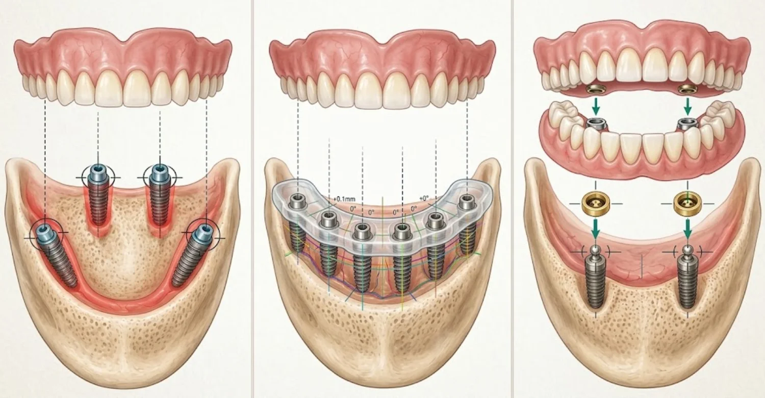
Note : All images used are for editorial and illustrative purposes only and may not originate from the original news provider or associated company.
How Is Dental Implant Placement Accuracy Assessed?
Assessment compares the digital plan with the postoperative implant position. This comparison can use CBCT, digital scanning, or both. Researchers calculate linear and angular differences between the datasets. Common parameters include platform, apex, depth, and angular deviation. Three-dimensional superimposition provides a detailed picture. The measurement method itself can influence reported Dental Implant Placement Accuracy.
Comparing the Planned and Actual Implant Position
The virtual plan shows where the implant should be. Postoperative imaging shows where it was actually placed. Software overlays both datasets for quantitative comparison. This can reveal systematic deviations within a workflow. Routine clinical practice does not always require this measurement. Numbers should always be interpreted alongside clinical examination.
Measuring Coronal and Apical Deviation
Coronal deviation measures displacement near the implant platform. Apical deviation measures displacement near the implant tip. These values can differ when angulation changes. A small platform shift may create larger apical displacement. This relationship matters most with long implants. A small coronal difference may be significant when the apex approaches a nerve.
Measuring Dental Implant Angulation Accuracy
Angular accuracy compares the planned and actual implant axes in degrees. Higher values mean greater divergence from the planned trajectory. Angular errors can increase apical displacement. They can also affect restorative component relationships. One digital-guide analysis reported pooled angular deviation of 2.47 degrees. Clinical significance depends on anatomy and prosthetic needs.
Evaluating Three-Dimensional Implant Position
Three-dimensional evaluation considers displacement in several spatial directions. It combines horizontal, vertical, and depth-related differences. This gives a fuller picture than one coordinate alone. Software compares actual and planned axes for translation and rotation. Computer-assisted techniques generally perform better on these measures. Any numerical deviation must still be related to the patient's anatomy.
Why Small Deviations Do Not Always Mean Treatment Failure
No surgical workflow produces absolute geometric identity. Small deviations commonly occur during clinical treatment. A minor difference can remain within planned safety margins. Implant survival also depends on biological and prosthetic factors. Research measures deviation to compare techniques, not to define universal failure. Safety and function remain the primary clinical objectives.
How Anatomical Site, Guide Support, and Operator Factors Influence Accuracy
Accuracy varies with anatomy and workflow. Anterior and posterior sites present different access challenges. Edentulous arches offer fewer rigid support points for guides. Operator training influences guide handling and navigation. A 2026 review found no significant difference between bone-supported and mucosa-supported guides. This shows that Dental Implant Placement Accuracy rarely depends on one factor alone.
What Happens When an Implant Is Not Placed as Planned?
A difference between planned and actual position does not automatically cause a complication. Its significance depends on the size and direction of deviation. An implant can remain fully functional despite a small positional difference. Larger deviations may affect anatomy or restorative design. Additional imaging can sometimes clarify the situation. Prompt assessment is important when neurological symptoms or major complications occur.
Mild Versus Clinically Significant Positional Deviation
Mild deviation may remain within the intended safety envelope. Clinically significant deviation can reduce anatomical clearance or compromise the restoration. The difference depends on implant dimensions and surrounding structures. A small angular change matters more with a long implant. No single deviation value defines failure in every case. The dentist should explain findings and available options clearly.
Potential Effects of Incorrect Implant Angulation or Depth
Incorrect angulation can complicate crown emergence and restorative access. It may also change load distribution across implant components. Incorrect depth can affect crestal bone and soft-tissue contours. Excessive depth may bring the implant closer to vulnerable anatomy. Some deviations can be accommodated prosthetically. Others may need further assessment or intervention.
Effects on Adjacent Teeth, Nerves, Sinuses, and Prosthetic Components
Implants placed near roots can create biological risks. Implants near the mandibular canal require attention to nerve anatomy. Posterior maxillary implants may approach or enter the sinus. Unfavorable angulation can make restorative components difficult to position. Altered sensation may indicate nerve involvement and needs prompt assessment. Early professional review is better than self-diagnosis.
When an Implant May Need Additional Assessment or Correction
Additional assessment is needed when symptoms or imaging raise concerns. The clinician may review the original plan and postoperative images. The restoration may also be checked for functional problems. Some deviations can be managed without relocating the implant. Others may require surgical or prosthetic correction. Second opinions can be reasonable for complex cases.
Symptoms That Should Be Reported After Implant Surgery
Contact your dental team when symptoms become unusual or persistent. A loose implant needs professional assessment. Significant swelling, fever, or pus may indicate infection. Persistent bleeding also warrants review. Altered sensation in the lip, chin, or tongue should be reported promptly. Urgent symptoms should never be managed through online information alone.
Diagnosis and Clinical Assessment Before Implant Placement
Implant planning begins with diagnosis rather than surgery. The dentist reviews medical history, oral health, and the missing-tooth site. The examination evaluates teeth, gums, bone, and restorative conditions. Imaging is chosen according to the information required. A thorough diagnosis protects Dental Implant Placement Accuracy before any drilling starts. The final workflow should reflect the patient's individual anatomy.
Medical and Dental History Review
Medical history identifies factors that may influence surgery or healing. The dentist reviews medications, allergies, previous procedures, and systemic conditions. Dental history reveals periodontal status and earlier treatments. Smoking status may also affect implant risk. Previous grafting or sinus procedures should be noted. Patients should provide complete and accurate information.
Clinical Examination of Teeth, Gums, and Implant Site
The dentist examines remaining teeth and periodontal tissues. Active disease may need treatment before implant surgery. The proposed site is evaluated for soft-tissue thickness and shape. Occlusion and restorative space are also assessed. Adjacent roots can influence the planned trajectory. The surgical plan should never rely on a digital scan alone.
Evaluating Bone Volume and Anatomical Structures
Bone is assessed in three dimensions when necessary. The clinician evaluates height, width, contour, and the available implant envelope. Anatomical structures guide the choice of implant length and angulation. CBCT provides cross-sectional information when clinically justified. Bone deficiencies may point toward grafting or alternative treatment. The plan balances biological support with restoration design.
CBCT and Other Imaging When Clinically Indicated
Imaging selection should reflect the diagnostic question. Periapical or panoramic images may provide initial information. Cross-sectional imaging helps when three-dimensional anatomy needs evaluation. The AAOMR recommends CBCT for implant sites when clinically justified. The smallest appropriate field of view should be used. More imaging does not automatically create better treatment.
Prosthetic Assessment Before Surgical Planning
The intended crown or prosthesis influences implant position. The dentist evaluates tooth width, emergence profile, occlusion, and restorative space. Implant axes are then planned beneath the final restoration. This reduces compromises caused by purely bone-driven placement. Insufficient bone may require augmentation first. Digital software makes these relationships visible before treatment.
Creating an Individualized Implant Treatment Plan
An individualized plan integrates diagnosis, anatomy, restoration, and patient preferences. It identifies the implant type and surgical approach. It may include grafting, sinus augmentation, or staged procedures. Temporary and final restorations should also be addressed. Guidance technology is selected after these decisions are made. Costs should be explained according to the final plan.
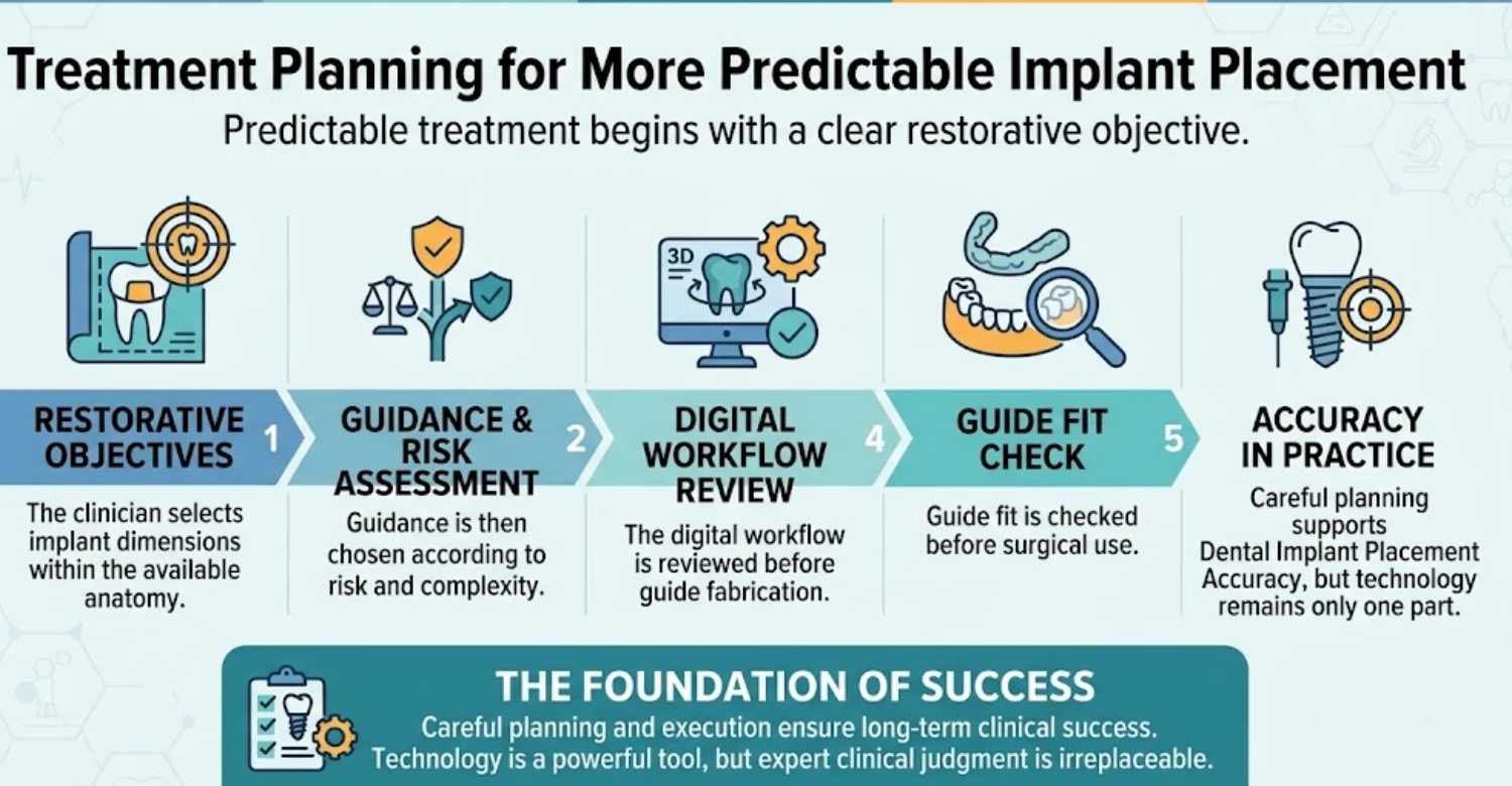
Choosing Implant Size, Position, Depth, and Angulation
Implant selection must match available anatomy and the planned restoration. Diameter depends on ridge width and restorative needs. Length depends on available bone and safety margins. Depth influences crestal and prosthetic relationships. Angulation must support the restoration while remaining anatomically safe. These variables can be tested digitally before treatment.
Matching Implant Position to the Planned Crown or Prosthesis
The crown location sets important restorative targets. The implant should support the restoration from directly beneath. This influences emergence profile and occlusal loading. It can also determine screw-access location. Software shows the implant and crown together for review. Bone limitations may require augmentation or a different restoration.
Deciding Between Freehand, Static, and Dynamic Guidance
Freehand treatment may suit straightforward cases with favorable anatomy. Static guides help when a fixed trajectory is advantageous. Dynamic navigation provides real-time guidance in complex procedures. The decision should reflect anatomical risk and restorative needs. Cost, training, access, and workflow also influence the choice. The clinician should explain why a particular approach was selected.
Planning for Bone Grafting or Sinus Augmentation When Needed
Insufficient bone can restrict the intended implant position. Bone grafting may increase support in selected sites. Upper posterior bone loss may require sinus augmentation. These procedures add surgical steps and healing time. The implant trajectory may change after graft healing. Staged treatment can sometimes provide greater predictability.
Verifying the Surgical Guide and Digital Plan Before Treatment
Guide verification is a key final planning step. The clinician confirms the guide matches the intended arch and support. The fit should be stable and reproducible. Guide components must match the planned implant system and drills. Unexpected instability should trigger reassessment. Verification protects Dental Implant Placement Accuracy at the moment of surgery.
Dental Implant Treatment Cost in
US
UK
Turkey
Implant prices vary by country, clinic, materials, complexity, and included services. Current international listings show substantial price variation. The figures below are indicative planning references, not fixed quotes. Additional procedures can change the final price significantly.
Average Dental Implant Cost Comparison
Country | Indicative Implant + Crown Cost | Cost Context |
United States | About $3,600+ | Market reference for implant placement with crown |
United Kingdom | From about $1,400+ | Private clinic pricing varies by treatment and materials |
Turkey | About 450–610 | Varies by implant brand, crown material, clinic, and city |
Prices are market references verified by Bookimed in October 2026 and may change. They are not guaranteed treatment quotes.
United States: About $3,600+ for a Complete Single Implant
This is an indicative market reference, not an official national average. U.S. fees vary by location, clinician, materials, and complexity. Imaging, grafting, or additional procedures can raise the total cost.
United Kingdom: From About $1,400 for an Implant With Crown
Private implant fees vary widely between clinics. The NHS Band 3 charge in England is $332.10 from April 2026, but this is an NHS dental charge and should not be presented as a private implant price.
Turkey: About 450–610 for an Implant With Crown
Current 2026 listings show approximately 450–610 for implant placement with a crown in Turkey. Prices vary according to implant brand, crown material, clinic, and location.
What the Implant Cost May or May Not Include
Cost Item | May Be Included | May Be Charged Separately |
Implant fixture | ✓ | ✓ |
Abutment | ✓ | ✓ |
Final crown | ✓ | ✓ |
X-rays / CBCT | ✓ | ✓ |
Temporary restoration | ✓ | ✓ |
Bone grafting | ✓ | |
Sinus augmentation | ✓ | |
Sedation | ✓ | ✓ |
Transport / accommodation | ✓ | ✓ |
How Additional Procedures Can Change the Price
Treatment Factor | Possible Effect on Cost |
CBCT imaging | Additional diagnostic fee |
Surgical guide | Planning and laboratory cost |
Bone grafting | Additional surgical and material fees |
Sinus augmentation | Additional surgical cost |
Premium implant brand | Higher implant component cost |
Customized abutment | Additional laboratory cost |
Premium crown material | Higher restoration cost |
Why Published Implant Prices Should Be Treated as Planning Ranges, Not Fixed Quotes
An online price cannot account for anatomy that has not been examined. Patients may require grafting, additional imaging, or restorative procedures. Published prices are therefore useful for initial budgeting, while a personalized examination and itemized treatment plan determine the actual cost. The ADA also emphasizes that implant treatment should be planned according to the patient's individual needs.
Pain Relief, Side Effects, and Recovery After Implant Placement
Implant surgery commonly causes temporary soreness and swelling. Recovery varies with surgical complexity and individual healing. Cleveland Clinic reports that early soft-tissue healing often takes about one week. Bone integration continues for several months in many cases. Pain medication can help control early discomfort. Unexpected or worsening symptoms require professional assessment.
What Pain and Swelling Can Be Expected After Implant Surgery?
Mild soreness is common after implant surgery. Swelling and bruising may also appear during early recovery. NHS guidance notes that some discomfort and swelling are expected. Swelling is often most noticeable during the first few days. Simple painkillers are usually enough for routine discomfort. Severe or worsening symptoms are not automatically normal.
Commonly Recommended Pain-Relief and Medical Tips
Pain relief should follow your clinician's instructions. Paracetamol or ibuprofen may be suitable when medically appropriate. Check existing medications and contraindications first. Cold compresses may help reduce early swelling. Keeping the head elevated can improve comfort. Avoid smoking, because it can increase implant-related risks.
What Can Help Reduce Irritation During Early Healing?
Keeping the surgical area clean supports recovery. Your team may recommend modified brushing around the site. Soft foods may be advised for a short period. Avoid touching the wound with fingers or objects. Some patients receive antimicrobial rinses based on clinical judgment. Individual instructions matter more than generic online routines.
Which Side Effects Usually Improve During Recovery?
Soreness generally decreases as tissues heal. Swelling and minor bruising usually fade gradually. Temporary sensitivity may occur around the treated site. NHS guidance suggests discomfort usually lasts up to about one week. The implant still needs time to integrate with bone. Persistent or worsening symptoms should be evaluated professionally.
Which Symptoms Should Prompt Contact With Your Dental Team?
Contact the team when symptoms become severe or progressively worse. Persistent bleeding should be assessed. Fever, pus, or drainage can suggest infection. Increasing swelling also warrants evaluation. A loose implant or new altered sensation needs prompt review. Follow the emergency instructions you received after surgery.
Persistent Pain, Increasing Swelling, Fever, Drainage, Mobility, or Altered Sensation as Reasons for Clinical Review
Persistent symptoms need evaluation because several causes are possible. Pain may arise from inflammation, infection, bite problems, or another tooth. Mobility can indicate an implant stability problem. Altered sensation may require neurological assessment. The clinician may use examination and imaging to investigate. Never try to adjust or manipulate an implant yourself.
.webp)
.webp)
.webp)
.webp)
Tips for Patients
Patients can improve communication by asking specific questions. Understanding the planned position helps set realistic expectations. Ask your dentist to explain imaging, guidance, and restorative goals. Learn what Dental Implant Placement Accuracy means for your own case. Check what the quoted price includes. Judge technology by its clinical value rather than marketing language.
What to Ask Your Dentist About Implant Placement Accuracy
Ask how the implant position was planned before surgery. Ask whether the restoration was considered during planning. Ask which imaging was used and why. Ask whether guided surgery suits your case. Ask what happens if the planned position cannot be achieved. Clear answers support informed, diagnosis-based decisions.
Ask How the Implant Position Was Planned and Verified
Ask whether planning included the final crown or prosthesis. Ask whether digital scans were combined with radiographic data. Ask how angulation and depth were selected. Ask whether anatomical safety margins were considered. You can also ask how guide fit will be checked. Complex implant treatment deserves clear communication and informed consent.
Ask Whether CBCT or Digital Scanning Is Clinically Appropriate
CBCT should be used when three-dimensional information offers real clinical benefit. Ask what diagnostic question the scan will answer. Ask whether another imaging method could be sufficient. Digital scans help capture restorative and surface anatomy. CBCT and scanning often provide complementary information. The best imaging protocol depends on your treatment.
Ask Whether Freehand, Static, or Dynamic Guidance Is Recommended and Why
Ask why your clinician chose the proposed surgical method. Freehand placement can suit selected straightforward cases. Static guidance can improve transfer control. Dynamic navigation offers real-time feedback during surgery. Research generally favors computer assistance for Dental Implant Placement Accuracy. Still, the most advanced option is not automatically the best one.
Confirm What Is Included in the Treatment Plan and Cost
Ask whether the quote includes the implant fixture, abutment, and crown. Ask about CBCT, digital scans, guides, and temporary restorations. Confirm whether bone grafting is charged separately. Ask how follow-up visits are handled. International patients should clarify transport and accommodation inclusions. A detailed quote makes comparison much easier.
Follow Preoperative and Postoperative Instructions Carefully
Following instructions reduces avoidable recovery problems. Take prescribed medications exactly as directed. Follow guidance on eating, brushing, and wound care. Avoid smoking as your team recommends. Attend scheduled follow-up appointments. Your own clinical team's instructions always take priority over general information.
Avoid Choosing a Treatment Solely on Price or Technology Claims
The lowest price may exclude important treatment components. The newest technology may not be necessary for every case. Compare diagnosis, clinician expertise, materials, and follow-up. Research supports guided accuracy, but technology does not guarantee outcomes. Look for individualized diagnosis and realistic expectations. Price and technology are only two parts of treatment value.
How Vitrin Clinic Plans for Precise Dental Implant Placement
Vitrin Clinic approaches implant planning through individualized assessment and digital diagnostics. The workflow is selected according to anatomy, restorative needs, and complexity. CBCT and digital scans may be used when clinically appropriate. Planning evaluates position, angulation, depth, and prosthetic relationships. The aim is strong Dental Implant Placement Accuracy without promising zero deviation. International patients can receive coordinated planning before visiting Istanbul.
Clinical Assessment Before Implant Placement
The process begins with evaluating teeth, gums, the implant site, and overall oral health. Bone availability and restorative space are considered before planning. Existing dental problems may need attention first. Functional and esthetic requirements are also reviewed. The treatment approach is then matched to the individual case. A standardized protocol is not appropriate for every patient.
CBCT and Digital Diagnostics When Clinically Appropriate
CBCT provides three-dimensional anatomical information when clinically necessary. Digital scanning captures dental surfaces and restorative relationships. Together they support computer-assisted planning in suitable cases. Vitrin Clinic can include these diagnostics when clinically appropriate. The purpose is better visualization, not technology for its own sake. Imaging findings remain subject to professional interpretation.
Digital Planning for Implant Position, Angulation, and Prosthetic Requirements
Digital planning assesses the implant and future restoration together. The clinician evaluates diameter, length, depth, and angulation. Restorative emergence and available bone are also considered. A surgical guide may follow when clinically appropriate. This coordinated process supports Dental Implant Placement Accuracy from planning to surgery. The goal is predictable treatment rather than a technology-based guarantee.
Using a Patient-Specific Approach to Determine the Appropriate Implant Workflow
Not every implant patient needs the same technology. A straightforward case may suit conventional techniques. A complex case may benefit from static guidance or dynamic navigation. The decision depends on anatomy, restoration, access, and risk. Healing needs and adjunctive procedures are also considered. Technology is chosen only after the clinical problem is understood.
Treatment Planning for Single and More Complex Implant Cases
Single implants require attention to adjacent structures and restorative emergence. Multiple implants require coordination between several planned positions. Full-arch rehabilitation adds further prosthetic and surgical relationships. Digital tools are especially useful when several implants support one restoration. The final workflow follows evaluation of the complete clinical situation. Safe, functional treatment remains the objective.
How Vitrin Clinic Supports Your Treatment Journey
For international patients, treatment involves more than surgery itself. Clear coordination simplifies diagnostics, appointments, treatment stages, and follow-up. Vitrin Clinic can coordinate both clinical and logistical stages. The proposed treatment can be explained before arrival when records allow. Online planning cannot replace an in-person examination. The aim is an organized and transparent treatment journey.
Consultation and Individualized Assessment
Consultation reviews symptoms, dental history, and treatment goals. Clinical findings then guide the diagnostic pathway. Patients can ask about alternatives and expected stages. The team explains whether further assessment is needed. Vitrin Clinic focuses on individualized evaluation rather than generic packages. Final recommendations depend on direct professional examination.
Coordinating Digital Diagnostics and Treatment Planning
Digital diagnostics streamline communication between clinical and restorative stages. CBCT and intraoral scans support planning when appropriate. The plan can be reviewed before surgery to catch conflicts early. International patients may share existing records when useful. Digital communication reduces unnecessary duplication of information. An original clinical assessment remains essential before treatment.
Explaining the Proposed Implant Procedure and Alternatives
Patients should understand why an implant is recommended. Alternatives may include bridges, dentures, other restorative care, or no treatment. The choice depends on diagnosis, anatomy, preferences, and prognosis. Guidance methods should be explained in plain terms. Patients deserve both benefits and limitations of each approach. Informed decisions require balanced information rather than promotional claims.
Supporting Treatment Coordination for International Patients
International patients often work within limited travel schedules. The sequence must account for surgery, healing, and restorative stages. Travel plans should never override biological healing needs. Some cases require several visits or staged treatment. The coordination team can organize appointment logistics. Package inclusions such as accommodation should be confirmed before travel.
Follow-Up and Aftercare Planning
Implant care continues after surgery and restoration. Follow-up lets the team monitor healing and function. Good oral hygiene around implants is essential. Regular professional maintenance may also be recommended. International patients should know where follow-up will happen at home. The clinic can provide records and postoperative guidance when appropriate.
Evidence Behind Dental Implant Placement Accuracy
The evidence base supports better positional control with computer-assisted implant workflows. Recent systematic reviews consistently report lower deviations than freehand approaches. Operator experience, anatomy, guide support, and workflow all influence results. Data on Dental Implant Placement Accuracy should therefore be read as ranges and averages. Research supports guidance as a precision tool, not a guarantee of perfect positioning.
What Systematic Reviews and Meta-Analyses Show
A 2025 meta-analysis included 45 studies comparing computer-assisted and noncomputer-assisted workflows. Clinical studies favored computer assistance by 0.65 millimeters for coronal deviation. Apical deviation favored computer assistance by 1.10 millimeters. Angular deviation favored computer assistance by 3.87 degrees. A separate review of 67 studies reported 1.11 millimeters entry and 1.40 millimeters apical deviation. Its overall angular deviation was 3.51 degrees.
Evidence on Static Guided Surgery, Dynamic Navigation, and Freehand Techniques
Comparative evidence favors both static and dynamic approaches over freehand surgery. A 2025 review found greater accuracy with both systems in randomized trials. Another meta-analysis found fully guided placement superior to freehand treatment. Evidence does not show static guidance always outperforming dynamic navigation. Operator training and workflow quality influence real-world performance. The best approach addresses the patient's specific risk.
Evidence on CBCT-Based Implant Planning
CBCT provides cross-sectional information that supports implant-site evaluation. The AAOMR recommends CBCT when three-dimensional information is clinically justified. Research also explores intraoral scanning for implant-position assessment. A 2026 meta-analysis found no significant differences between CBCT and scanning for several measures. Clinical evidence for scanner-based assessment remains limited. CBCT stays important when bone and critical structures need cross-sectional review.
Why Study Design, Operator Experience, and Clinical Setting Matter
Laboratory studies do not fully reproduce clinical conditions. Patients move, soft tissues deform, and access varies. Operator experience differs between research studies. Guide designs and navigation systems are not identical. Recent reviews report substantial heterogeneity across technologies and protocols. Evidence supports trends rather than identical outcomes for every patient.
Recommended Evidence Sources: ADA, AAOMR, NIH, NIDCR, PubMed, and Peer-Reviewed Implant Literature
Patients and content teams should prioritize authoritative sources. PubMed provides access to peer-reviewed biomedical research. The AAOMR publishes guidance on dental imaging and CBCT selection. The ADA, NIH, and NIDCR provide evidence-based oral-health information. Systematic reviews summarize results across many clinical studies. Check every source for publication date and study quality.
Ready to transform your smile?
Talk to our team and get a personal FREE treatment plan.
Related Vitrin Clinic Treatments and Patient Guides
Implant treatment may form part of a broader rehabilitation plan. Some patients need bone augmentation before implant placement. Others may need full-arch rehabilitation after extensive tooth loss. Educational resources help patients understand each treatment stage. Vitrin Clinic provides information across implant and restorative pathways. Individualized recommendations still require a clinical examination.
Dental Implant Treatment Information
Dental implants replace missing tooth roots and support restorations. They can support single crowns, bridges, or implant-supported dentures. Full-arch treatments use several implants to support one prosthesis. Cleveland Clinic notes that implants may need several procedures and months of healing. Suitability depends on bone health, oral conditions, and restorative needs. Planning should always begin with professional diagnosis.
Sinus Lift Surgery Before Dental Implants Explained
A sinus lift may be considered when upper posterior bone height is insufficient. The procedure creates space for bone augmentation beneath the sinus. CBCT helps evaluate the sinus and available bone beforehand. Not every posterior implant needs this procedure. Treatment timing depends on the chosen surgical strategy. Discuss healing periods and alternatives with your clinician.
Replacing All Teeth With Implants Cost
Full-arch treatment involves several implants and a prosthetic restoration. Costs therefore differ greatly from single-implant treatment. The price depends on implant number, materials, surgery, and prosthesis design. Grafting and temporary prostheses may add further costs. Travel and accommodation also affect international budgets. A personalized assessment is needed for a reliable quotation.
Related Implant and Full-Arch Treatment Education
Patients benefit from understanding implant types, grafting, restorations, and aftercare. Full-arch cases need extra prosthetic and surgical planning. Single implants have different anatomical and restorative considerations. Digital planning can support both treatment categories. Good educational content explains both advantages and limitations. Patients should seek professional diagnosis before choosing treatment.
Key Takeaways
Successful implant treatment depends on accurate diagnosis and appropriate planning. Digital tools improve visualization and positional control. Guided workflows generally achieve better Dental Implant Placement Accuracy than freehand techniques. No technique eliminates every source of deviation. Cost should be compared through complete treatment inclusions. An individualized treatment plan remains the foundation of safe care.
Dental Implant Placement Accuracy Depends on Planning, Anatomy, Technique, and Clinical Execution
Accurate implant treatment begins well before surgery. The clinician evaluates anatomy, restoration, imaging, and surgical access. The selected technique should match the complexity of the case. Guidance technology can improve positional control. Still, Dental Implant Placement Accuracy also depends on clinical execution. No single device guarantees the same result for every patient.
CBCT and Digital Planning Can Improve Visualization and Treatment Planning When Clinically Indicated
CBCT provides three-dimensional information about bone and anatomical structures. Digital planning links this information with the planned restoration. Implant position can then be evaluated before surgery. The AAOMR supports clinically justified CBCT for implant-site assessment. Digital plans can also feed surgical guides and navigation. More imaging or technology does not automatically mean better care.
Guided Techniques Generally Show Greater Positional Accuracy Than Freehand Approaches, While No Method Eliminates All Deviation
Recent systematic reviews consistently favor computer-assisted methods for positional accuracy. Fully guided surgery has outperformed freehand placement in several analyses. However, deviation remains measurable even with guidance. Guide stability, imaging, registration, and technique influence results. Patients should expect improved predictability rather than mathematical perfection.
Small Planned-to-Actual Differences Must Be Interpreted in Their Clinical Context
Deviation statistics do not automatically indicate treatment failure. A small difference may stay safely within anatomical margins. A similar difference can matter more near critical structures. Angular changes can also enlarge apical displacement. Numbers should be viewed alongside symptoms, anatomy, and restorative needs. Your clinician should explain whether a measured difference is important.
Pain, Swelling, Numbness, Mobility, or Other Concerning Symptoms May Require Professional Assessment
Mild soreness and swelling can occur after implant surgery. Persistent or worsening symptoms should not be ignored. Fever, drainage, pus, and implant mobility warrant dental review. Altered sensation also deserves prompt professional assessment. Follow your postoperative instructions carefully. Serious symptoms always need direct clinical attention.
The Best Implant Approach Is Individualized Rather Than Based on Technology or Price Alone
Technology can improve surgical control, but it does not make the diagnosis. Price does not automatically indicate quality or suitability. The right approach depends on anatomy, restoration, risk, and expertise. Freehand treatment remains suitable for selected straightforward cases. Guided or navigated surgery may add predictability in complex cases. Compare complete treatment plans rather than isolated prices.
A Personalized Clinical Evaluation Is the Starting Point for Safe and Predictable Implant Treatment
Every implant case starts from a different anatomical and restorative situation. The clinician first decides whether implant treatment is appropriate. Imaging and digital planning are chosen according to clinical need. The surgical approach is then adapted to the patient's anatomy. Clinical judgment remains essential through diagnosis, surgery, restoration, and maintenance.
At Vitrin Clinic
At Vitrin Clinic, dental implant care starts with an individualized clinical assessment rather than a fixed technology choice. The team evaluates teeth, gums, bone volume, anatomical structures, and restorative space before planning. CBCT and digital scanning can support three-dimensional planning when clinically appropriate. Plans consider implant position, angulation, depth, bone anatomy, and the final crown or prosthesis. The surgical approach, whether freehand, static guidance, or dynamic navigation, is matched to each case. International patients receive support with appointment scheduling, treatment stages, and follow-up planning. The aim is safe, functional, and predictable implant treatment without promises of zero deviation. Every final recommendation depends on a direct professional clinical examination.
Reference
FAQs
Dr. Rifat Alsaman has more than 5 years of clinical experience in dentistry and currently serves as the Head of the Medical Team at Vitrin Clinic. He is dedicated to providing exceptional patient care, overseeing treatment planning, and ensuring the highest clinical standards across the team. His expertise, attention to detail, and commitment to continuous professional development have helped countless patients achieve healthier, more confident smiles.


