Dental Implant
October 4, 2026
When Are Subperiosteal Implants for Maxillary Reconstruction Used?
 Dr. Rifat Alsaman
Severe upper jaw bone loss can leave too little support for conventional dental implants. Subperiosteal Implants for Maxillary Reconstruction offer a patient-specific framework that rests over the remaining jawbone. Modern designs rely on three-dimensional imaging and computer-aided manufacturing to match each patient's anatomy. This approach may help selected patients avoid some extensive bone augmentation procedures. Suitability still depends on bone anatomy, soft-tissue health, prosthetic needs, medical factors, and surgical assessment. Current studies show promising short-term outcomes, yet long-term evidence remains limited. Complications can include soft-tissue dehiscence and framework exposure.
What Are Subperiosteal Implants for Maxillary Reconstruction?
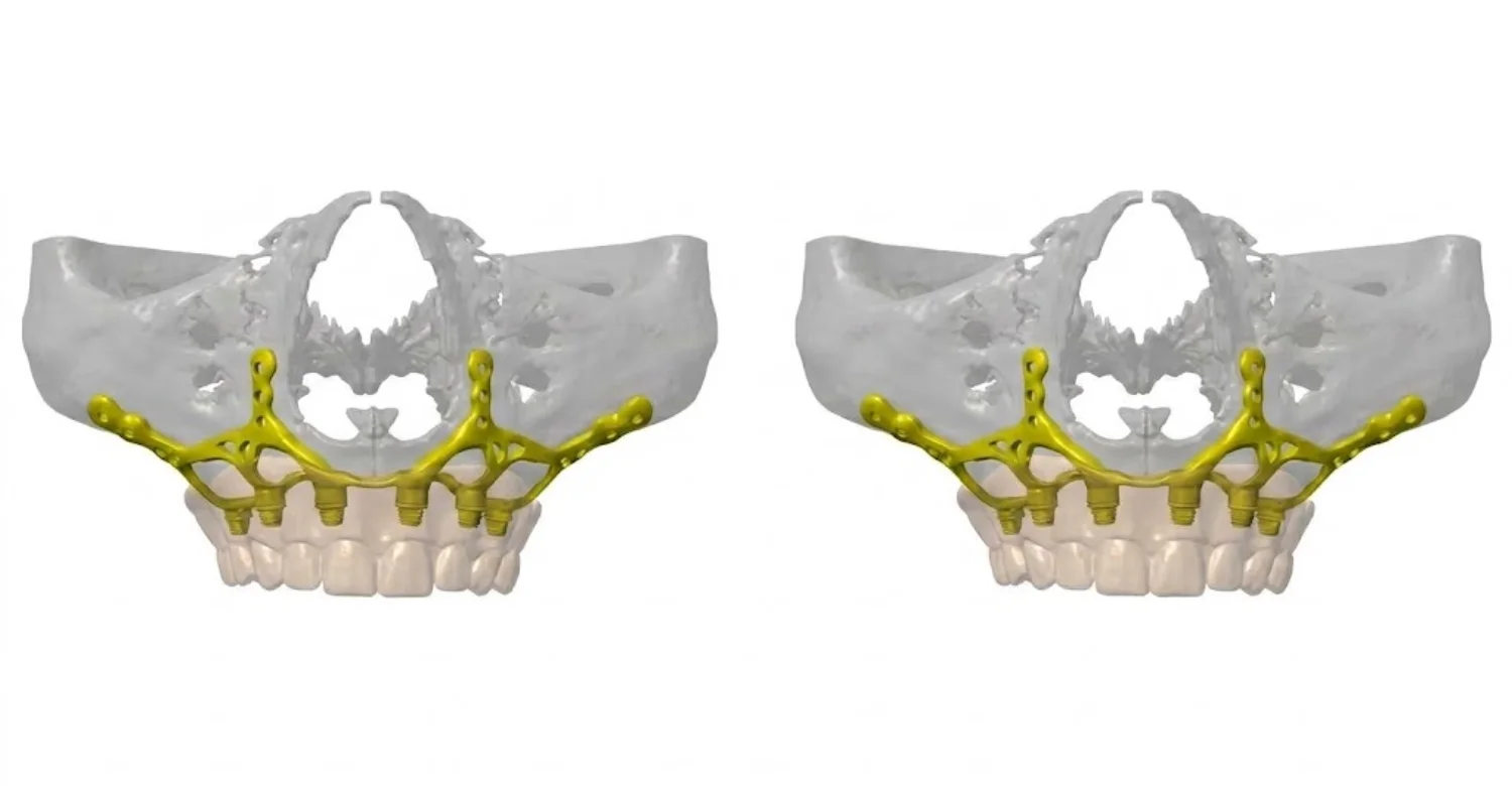
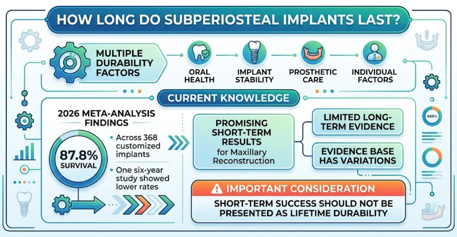
Subperiosteal Implants for Maxillary Reconstruction are custom frameworks that support a prosthesis when conventional implant anchorage is limited. The framework sits on top of the remaining maxillary bone rather than inside it. Contemporary systems are designed from CBCT data and produced through digital workflows. Published reviews describe them as an option for selected patients with severe jaw atrophy. A 2026 meta-analysis reported 87.8% overall survival across 368 implants. The same analysis found soft-tissue dehiscence in 28.6% of cases.
How subperiosteal implants differ from conventional endosteal implants
The main difference lies in how each option obtains support. Endosteal implants are inserted into the jawbone and need adequate bone quantity and quality. A subperiosteal framework rests on the bone surface instead. Posts pass through the gum to connect a fixed or removable restoration. This matters when severe resorption limits conventional implant positioning. However, the framework does not remove surgical risks or biological requirements.
How a subperiosteal framework supports a dental prosthesis
The custom framework spreads functional forces across the available maxillary anatomy. Planned attachment points connect it to a partial or full-arch prosthesis. Digital planning links the framework design with the intended tooth position before surgery. This prosthetically driven approach coordinates fixation points, restorative space, and hygiene access. A 2025 review found 97.6% of implants functional after a mean 17-month follow-up.
Why they may be considered for an atrophic maxilla
Long-term tooth loss can reduce both the height and width of the upper ridge. Advanced resorption may also limit surgical options in the posterior maxilla. Subperiosteal Implants for Maxillary Reconstruction may be considered when remaining bone cannot support conventional implants. Grafting may be unsuitable because of anatomy, health factors, earlier treatment, or patient preference. A custom framework uses the remaining bone surface instead.
How CBCT, CAD/CAM, and 3D manufacturing have modernized subperiosteal implants
CBCT imaging shows the residual maxilla and nearby structures in three dimensions. CAD software then allows virtual framework design matched to that anatomy. Computer-aided manufacturing can mill or print the framework in titanium. Virtual planning also aligns fixation points with restorative goals. A 2025 systematic review examined 24 publications involving 246 implants. It reported 97.6% functional implants after a mean 17-month follow-up.
Are subperiosteal implants the same as zygomatic implants?
No, these are two different treatments. Zygomatic implants are long implants anchored into the cheekbone. They bypass resorbed areas by using stronger facial bones. A subperiosteal framework instead rests over the residual maxillary bone. Both can address severe atrophy, but their surgical pathways and anatomical requirements differ. Comparative evidence does not show one approach is best for every patient.
Why Does Severe Maxillary Bone Loss Develop?
Severe maxillary bone loss can result from biological, infectious, surgical, and structural causes. Subperiosteal Implants for Maxillary Reconstruction may become relevant when remaining bone cannot support conventional implants. Tooth loss gradually reduces ridge dimensions through remodeling. Periodontal disease and chronic infection can destroy supporting tissues. Trauma, previous surgery, and tumor treatment may also remove important areas of bone.
Tooth loss and long-term alveolar ridge resorption
Tooth loss changes the mechanical environment around the alveolar ridge. After extraction, the ridge can gradually become narrower and shorter. These changes may continue for years, although the rate varies between patients. The anterior and posterior maxilla often remodel in different patterns. Dentures restore function but do not recreate the original tooth attachment.
Periodontal disease and chronic dental infection
Periodontal disease progressively damages the tissues that support natural teeth. Advanced destruction can leave localized or generalized bone defects. Chronic dental infection may also cause inflammatory changes in surrounding tissues. Clinicians generally treat active infection before definitive implant reconstruction. This allows a more accurate evaluation of the remaining anatomy.
Trauma and previous jaw surgery
Previous trauma can permanently change the shape, volume, and quality of maxillary bone. Earlier jaw surgery may leave scar tissue or limited surgical access. Existing fixation plates, grafts, or reconstructive materials can also affect planning. CBCT imaging helps identify important structures before surgery. Vitrin Clinic reviews previous treatment records whenever they are available.
Maxillofacial tumor or tissue resection
Tumor treatment can significantly alter the maxilla and surrounding soft tissues. Surgical resection may change the ridge, palate, sinus region, or facial contours. Radiation therapy can introduce further concerns about tissue healing. Treatment therefore requires coordination between surgical, restorative, and medical teams. Timing depends on healing, disease status, tissue condition, and prosthetic goals.
Sinus pneumatization and reduced posterior maxillary bone
The posterior maxilla often becomes challenging after upper molar and premolar loss. The maxillary sinus can expand into areas once occupied by tooth roots. This reduces the vertical bone height available beneath the sinus. Subperiosteal Implants for Maxillary Reconstruction may be considered when this atrophy limits conventional anchorage. Clinicians may also consider sinus augmentation or alternative implant positions.
Risk factors that may complicate maxillary implant reconstruction
Several factors can increase the complexity of implant reconstruction. Smoking can negatively affect oral healing and should be discussed before surgery. Poor periodontal control can increase inflammation around implant-supported prostheses. Certain medications and medical conditions may influence surgical planning. Previous radiation or compromised soft tissues can add further difficulty.
Learn about managing Amelogenesis Imperfecta
What Are the Signs of an Atrophic Maxilla?
An atrophic maxilla does not always produce obvious symptoms in daily life. Patients may notice dentures becoming less stable during speaking or eating. Facial support can change gradually after prolonged tooth loss. Subperiosteal Implants for Maxillary Reconstruction are considered only after examination and imaging confirm substantial bone loss. Some patients feel no pain despite significant anatomical change.
Early signs patients may notice
Early changes are often subtle and develop slowly after tooth loss. A denture may move more during chewing or speech. Food may collect beneath an unstable denture more often. Lip or cheek support may also change over time. These signs do not prove severe bone loss by themselves.
Advanced signs of severe upper jaw bone loss
Advanced maxillary atrophy can severely limit conventional implant positioning. Dentures may stay unstable despite repeated adjustments or relining. Facial support can change noticeably when substantial ridge volume is lost. Severe posterior resorption can complicate implant placement beneath the sinus. However, symptom severity does not accurately measure anatomical severity.
Loose dentures and reduced prosthetic stability
A loose upper denture can result from ridge changes, poor fit, or prosthetic wear. Instability may interfere with chewing, speaking, and social comfort. Repeated adjustments can help temporarily without addressing major anatomical change. A clinician can identify whether the problem involves the prosthesis, tissues, bone, or several factors. Unstable dentures should prompt assessment rather than an automatic implant recommendation.
Why significant maxillary bone loss may have few obvious symptoms
Significant bone loss can progress without sharp pain or visible swelling. Bone remodeling differs from acute infection, which usually causes inflammatory symptoms. Patients often adapt gradually to changes in chewing and denture stability. This adaptation can make functional decline harder to notice. Regular dental examinations can detect changes before major problems develop.
Which warning signs require urgent dental or medical evaluation?
Certain symptoms need prompt assessment rather than a routine implant consultation. Severe facial swelling can indicate an infection requiring urgent treatment. Difficulty breathing or swallowing requires immediate medical attention. Uncontrolled bleeding after dental surgery also needs urgent professional care. Fever with swelling or significant tenderness may require prompt evaluation.
Discover when pocket reduction is needed in Osseous Surgery
Who May Be a Candidate for Subperiosteal Implants?
Candidacy depends on anatomy, oral health, restorative goals, and available alternatives. Subperiosteal Implants for Maxillary Reconstruction may suit selected patients with severe atrophy and inadequate conventional support. Low bone volume alone does not automatically justify this treatment. Clinicians must confirm the framework can achieve stable positioning and prosthetic function. Soft-tissue quality matters because framework components interact closely with the gums.
Patients with severe maxillary bone loss and insufficient implant support
Patients with advanced atrophy may have few sites for conventional implants. The degree of atrophy should be confirmed through examination and three-dimensional imaging. Planning also considers the sinuses, nasal floor, and remaining skeletal structures. A custom framework can be designed around these anatomical limitations. Adequate fixation and healthy soft tissues remain important requirements.
When conventional dental implants may not provide enough available bone
Conventional implants need suitable bone for placement and mechanical stability. Posterior limitations are especially common because of sinus expansion. Anterior defects can also reduce the number of feasible implant positions. Bone quality and quantity should be assessed together. Some patients can still receive conventional implants after augmentation procedures.
When bone grafting or sinus augmentation may be unsuitable or undesirable
Bone augmentation remains an established strategy for many patients. Subperiosteal Implants for Maxillary Reconstruction may be considered when extensive grafting is unsuitable or unwanted. Some patients prefer to avoid additional augmentation stages. Anatomy, medical factors, or earlier surgery can also limit grafting predictability. A 2026 meta-analysis of 22 studies included 6,860 implants after sinus augmentation. Pooled survival was about 97%, though evidence certainty was low.
Partial-arch versus full-arch maxillary reconstruction
The extent of tooth loss shapes the reconstruction design. Subperiosteal Implants for Maxillary Reconstruction can potentially support partial or full-arch restorations. Partial-arch treatment replaces missing teeth within a defined region. Full-arch treatment addresses broader tooth loss and needs more extensive prosthetic planning. Occlusion must be evaluated so restored teeth meet with balanced contacts.
Factors that influence candidacy and surgical planning
Candidacy depends on more than the amount of remaining bone. Clinicians evaluate bone contours, soft tissues, prosthetic space, occlusion, and surgical access. Periodontal health and any active infection also require review. Earlier trauma, surgery, or radiation can influence tissue quality. Medical history may affect anesthesia, healing, and postoperative monitoring.
When might another reconstructive option be more appropriate?
Another treatment may be better when anatomy or biology does not favor a custom framework. Conventional implants may remain appropriate after ridge augmentation or sinus elevation. Zygomatic implants can use cheekbone support when the maxilla is severely resorbed. Pterygoid implants may help in selected posterior situations. Removable prostheses suit patients for whom surgery carries unacceptable risk. Zygomatic implant reviews commonly report survival above 95%.
Understand the role of Gutta Percha Solvents in Root Canal Retreatment
How Is Severe Maxillary Bone Loss Diagnosed and Planned for Implant Treatment?
Accurate diagnosis must come before any complex implant reconstruction is chosen. Subperiosteal Implants for Maxillary Reconstruction need detailed assessment of the residual maxilla and nearby anatomy. Clinicians examine the teeth, gums, mucosa, occlusion, jaw function, and prosthetic space. CBCT provides three-dimensional information about bone contours. Digital scans help connect existing anatomy with the planned restoration.
Clinical examination of teeth, gums, soft tissues, occlusion, and prosthetic space
A clinical examination reveals information that imaging alone cannot provide. Dentists check remaining teeth for decay, periodontal disease, infection, or mobility. Soft tissues are assessed for thickness, inflammation, scarring, and keratinized tissue. Occlusion shows how the upper and lower arches interact during function. Prosthetic space determines whether the restoration can achieve adequate thickness and tooth position.
Why CBCT is important for evaluating the atrophic maxilla
CBCT shows details that two-dimensional dental images cannot fully capture. It helps identify ridge contours, sinus boundaries, nasal anatomy, and fixation surfaces. It may also reveal defects caused by trauma, infection, or earlier surgery. This information guides virtual framework design and surgical positioning. CBCT should be prescribed when its diagnostic value justifies the radiation exposure.
3D scanning and digital impressions for treatment planning
Digital impressions capture teeth, soft tissues, and prosthetic relationships. Combining intraoral scans with CBCT data creates a fuller planning model. This model helps clinicians evaluate tooth position and restorative space. Digital records can also support virtual articulation and framework design. They improve communication between surgical and restorative teams.
Virtual surgical planning and custom framework design
Virtual planning lets the team study anatomy before creating the final framework. Subperiosteal Implants for Maxillary Reconstruction can be digitally designed around available bone and prosthetic requirements. Clinicians assess framework contours, fixation areas, and post locations on screen. Planned tooth positions are considered before the surgical design is finalized. A 2024 systematic review analyzed 26 publications involving 302 digitally planned cases. It reported 95.3% survival and 87.7% success across the included literature.
Assessing the sinus, nasal floor, adjacent anatomy, and surgical access
Complex maxillary anatomy strongly influences reconstruction planning. The sinus can limit posterior implant positioning after ridge resorption. The nasal floor becomes relevant when anterior bone is severely reduced. Earlier surgery may create additional restrictions or altered access. Clinicians also consider the thickness and condition of overlying soft tissues.
What should patients bring to a maxillary implant consultation?
Bringing previous records can make the consultation more efficient. Useful items include earlier CBCT scans, panoramic images, and dental records. Patients should also bring medication lists and details of medical conditions. Information about prior grafts, sinus procedures, or failed implants is valuable. Existing dentures help clinicians evaluate tooth position and expectations.
What questions should a patient ask about the planned implant design?
Patients should understand why a custom framework is recommended over alternatives. Ask which anatomical findings make the proposed design appropriate. Ask where the framework will sit and how fixation will be achieved. Ask which prosthesis will be attached and what maintenance it needs. Patients should also ask about likely complications and possible revisions.
Learn how to prevent enamel loss from Sports Drinks and Dental Erosion
What Treatment Options Exist for Severe Upper Jaw Bone Loss?
Severe maxillary atrophy can be managed through several reconstructive strategies. Subperiosteal Implants for Maxillary Reconstruction represent one customized option among them. Conventional implants may still work after ridge augmentation or sinus elevation. Zygomatic and pterygoid implants offer alternative anchorage in selected anatomy. Removable dentures remain appropriate when surgery is unsuitable or unwanted.
Conventional dental implants after bone augmentation
Bone augmentation can increase the volume available for conventional implants. Ridge grafting addresses localized deficiencies affecting implant positions. Sinus augmentation can add vertical bone in selected posterior sites. Treatment may involve separate surgical and healing stages. This can mean more appointments and a longer overall timeline.
Sinus lift and other graft-based approaches
Sinus augmentation creates additional vertical bone beneath the maxillary sinus. It may use a crestal or lateral approach depending on anatomy. Grafting materials and implant timing vary with each clinical situation. Augmentation remains a surgical procedure with possible complications. These include membrane perforation, infection, swelling, and implant-related problems. Recent evidence from 6,860 implants showed about 97% survival after sinus augmentation.
Zygomatic implants for severe maxillary atrophy
Zygomatic implants use the cheekbone instead of the resorbed maxillary ridge. They can provide fixed support without conventional ridge augmentation in selected cases. The technique requires advanced surgical skill and detailed three-dimensional planning. Possible complications include sinus problems, infection, and soft-tissue issues. A review of 196 publications reported 98.5% success below one year. The reported rate remained 96.1% beyond five years.
Pterygoid or other alternative implant strategies
Alternative strategies can provide support when standard maxillary sites are limited. Pterygoid implants use posterior anatomical structures for anchorage. Their use requires careful evaluation of anatomy, access, angulation, and operator experience. These techniques are not interchangeable because each uses different support. The prosthetic design should guide implant number and distribution.
Custom subperiosteal implants for the maxilla
Custom frameworks are designed for severe atrophy with limited conventional implant sites. Subperiosteal Implants for Maxillary Reconstruction use residual skeletal anatomy as the foundation. Digital planning combines CBCT data with the desired prosthetic position. The framework is manufactured to fit the patient's specific contours. It may reduce the need for extensive augmentation in selected cases. A 2024 review reported 95.3% survival among 302 analyzed cases.
Removable dentures when implant reconstruction is not appropriate
Removable dentures remain an important option for severe tooth loss. Some patients have medical, financial, or personal reasons to avoid complex surgery. A well-designed denture can restore basic chewing and speech. Relining or replacement may improve fit when ridge changes reduce retention. Implant-assisted dentures may be possible when enough support exists.
How do subperiosteal and zygomatic implants compare?
Subperiosteal Implants for Maxillary Reconstruction and zygomatic implants use different anatomical strategies. Zygomatic implants anchor directly into facial bone through the resorbed maxilla. Subperiosteal frameworks rest over available bone with customized components. Both require advanced imaging and specialized surgical planning. A 2023 review found zygomatic implant survival between 90.3% and 100%.
When may bone grafting be preferred over a graftless approach?
Grafting may be preferable when it can create predictable conventional implant sites. Sinus augmentation can also restore posterior bone height when anatomy permits. The choice depends on bone, sinus anatomy, healing potential, and treatment duration. Some patients accept extra surgery for implants with extensive supporting evidence. Others prioritize fewer reconstructive stages when clinically appropriate.
How Does a Subperiosteal Implant Procedure Work?
Subperiosteal Implants for Maxillary Reconstruction involve several coordinated clinical and digital stages. Treatment begins with examination, imaging, and restorative planning. CBCT data and digital scans are combined for virtual design. The custom framework is then manufactured to match the plan. During surgery, it is positioned over the bone and secured.
Initial assessment and digital imaging
The first stage confirms whether the anatomy can support a customized approach. The clinician evaluates teeth, gums, mucosa, occlusion, and prosthetic space. CBCT identifies residual bone contours and nearby structures. Intraoral scanning adds information about teeth and soft tissues. The team then checks whether simpler or alternative options remain feasible. A 2025 review highlighted patient-specific design as a central feature of modern workflows.
Designing the patient-specific implant framework
Framework design converts three-dimensional data into a customized surgical component. The design may include fixation points, transmucosal posts, and prosthetic supports. Planned tooth position should guide the framework rather than follow it. The final design balances stability, adaptation, surgical access, and soft-tissue needs. Manufacturing may use milling or three-dimensional printing in titanium.
Surgical placement over the maxillary bone
The surgeon exposes the planned area and positions the custom framework. The framework sits over residual bone rather than being inserted through it. Its fit is checked against the planned anatomical surfaces. Fixation is then secured according to the design and available bone. Prosthetic connection points must align with the intended restoration.
Managing the soft tissue around the framework
Soft-tissue management is one of the most important steps after placement. Subperiosteal Implants for Maxillary Reconstruction include posts that pass through the surrounding mucosa. Excess tension or poor tissue adaptation can cause inflammation or exposure. Careful flap handling and positioning are therefore essential. A 2026 review found soft-tissue dehiscence in about 28.6% of customized implants.
Connecting the framework to the planned prosthesis
The prosthetic connection must match the previously planned restorative design. Customized posts can support either a fixed or removable prosthesis. The restoration needs good function, appearance, hygiene access, and strength. Bite records and digital data help guide tooth positioning. Temporary components may be used while tissues stabilize.
Temporary and definitive prosthetic stages
Some patients receive a temporary restoration before the definitive prosthesis. Temporary teeth help establish function while soft tissues adapt. Tooth position, occlusion, and contours can then be refined. The definitive restoration is made after the required clinical checks. The exact sequence depends on surgical stability and clinician protocol.
How many treatment stages may be needed?
The number of stages depends on anatomy, planning, surgery, and prosthetic design. Assessment usually comes first, followed by imaging, planning, and manufacturing. Surgery is followed by healing reviews and prosthetic management. Extra visits may address occlusion, hygiene, or tissue healing. Complex cases often need more appointments.
What factors can change the treatment timeline?
Timelines can change when anatomy or healing differs from the original plan. Active infection may need treatment before definitive reconstruction. Previous surgery can create scar tissue or anatomical limitations. Framework manufacturing adds time because each component is made individually. Soft-tissue complications may require extra management before final restoration.
Understand the connection between Circulatory System Diseases and Periodontal Disease
What We Notice Clinically
Severe maxillary atrophy requires evaluation beyond measuring available bone. Subperiosteal Implants for Maxillary Reconstruction may suit selected cases, but bone quantity alone cannot decide treatment. Clinicians also assess soft tissues, prosthetic space, occlusion, hygiene access, and expectations. The same bone loss can lead to different plans in different patients. Digital technology helps visualize anatomy but does not remove uncertainty.
Why “not enough bone” does not automatically mean one treatment is required
Insufficient bone does not dictate one single reconstructive procedure. Conventional implants may become possible after augmentation or sinus elevation. Zygomatic implants may provide support in appropriate cases. Removable prostheses remain reasonable when complex surgery is unsuitable. Options should be compared by anatomy, health, function, cost, and maintenance.
Common patient misconceptions about severe maxillary bone loss
Some patients assume severe bone loss makes fixed teeth impossible. Others believe digital planning guarantees complication-free treatment. Technology improves customization but cannot prevent biological healing problems. Some assume the newest technique must automatically be the safest. Cost alone can also create misleading comparisons between procedures.
Why bone volume is only one part of treatment planning
Bone dimensions are essential, but reconstruction involves many other variables. Adequate bone surface does not guarantee predictable tissue healing. A framework may be mechanically sound yet difficult to clean. Poor prosthetic positioning can affect speech, chewing, and facial support. Existing periodontal disease adds further biological concerns.
The importance of soft-tissue health, prosthetic design, and oral hygiene
Long-term success depends on more than successful framework placement. Inflamed or fragile tissues may raise the risk of dehiscence or exposure. Prosthetic contours should give patients access to key cleaning areas. Regular professional maintenance helps detect inflammation or mechanical problems early. The 2026 meta-analysis reported soft-tissue dehiscence in 28.6% of analyzed implants.
Clinical Note: Dr. Rifat Alsaman
Dr. Rifat Alsaman emphasizes that complex implant decisions require individualized clinical assessment. Subperiosteal Implants for Maxillary Reconstruction should be considered only after evaluating anatomy and restorative goals. Bone loss must be interpreted alongside soft tissues, occlusion, and prosthetic needs. Digital imaging improves planning, but it does not replace clinical judgment. Patients should understand benefits, limitations, risks, and alternatives before treatment.
What Are the Potential Benefits and Limitations of Subperiosteal Implants?
Subperiosteal Implants for Maxillary Reconstruction may offer a graftless alternative for selected severe atrophy cases. Custom frameworks adapt to irregular anatomy and planned prosthetic needs. Digital workflows improve customization and teamwork between surgical and restorative clinicians. However, soft-tissue exposure, inflammation, and mechanical problems can occur. Long-term evidence remains less extensive than for conventional implants. A 2026 meta-analysis reported 87.8% survival and 28.6% soft-tissue dehiscence.
Potential role in severely atrophic maxillae
Severe atrophy can leave limited bone for conventional implant placement. A custom framework can provide support without relying fully on ridge volume. This is relevant when extensive augmentation is undesirable or technically difficult. The approach may also reduce some stages linked with major grafting. One review found 97.6% of implants functional after a mean 17-month follow-up.
Customization around the patient’s existing anatomy
Customization is a defining advantage of modern subperiosteal frameworks. CBCT data shows the available skeletal surfaces and surrounding structures. Digital impressions add detail about teeth, soft tissues, and planned contours. Computer-aided design then tailors the framework to these combined records. Design accuracy depends on diagnostic quality and manufacturing precision. One review of 302 cases reported 95.3% survival and 87.7% success.
Potential use when conventional implant placement is limited
Conventional implants depend on suitable bone, anatomy, and surgical access. Severe horizontal and vertical resorption can restrict their positioning. Posterior sinus expansion adds further limitations. Customized frameworks can use broader residual surfaces for support. This does not make every severely atrophic patient a suitable candidate.
Surgical and biological limitations
Modern technology does not remove the biological challenges of framework placement. Dehiscence or exposure can develop when tissues fail to stay adapted. Inflammation may occur around exposed framework areas. Infection can compromise healing and stability. A 2026 meta-analysis reported 28.6% overall soft-tissue dehiscence. The maxillary rate was 31.9%, though this cannot predict individual outcomes.
Why modern evidence is promising but still developing
Digital workflows have renewed clinical interest in customized subperiosteal treatment. Recent reviews generally report favorable short-term survival and function. However, most studies have small samples and varied methods. Many lack randomized comparisons with other reconstructive techniques. A 2024 review of 227 patients reported 97.8% implants functioning after 21.4 months. It also reported partial exposure in 25.6% of implants.
What does current research say about implant survival and complications?
Research suggests high short-term survival alongside meaningful soft-tissue complication rates. Subperiosteal Implants for Maxillary Reconstruction showed 97.6% function after 17 months in one 2025 review. Partial exposure occurred in 37% of those implants. Another review reported 97.8% functioning implants and 25.6% exposure. A 2026 meta-analysis found 87.8% survival and 28.6% dehiscence.
Learn about the causes and solutions for Habitual Jaw Clenching and Tooth Wear
What Are the Side Effects and Complications of Subperiosteal Implants?
Subperiosteal Implants for Maxillary Reconstruction can involve normal postoperative effects and more serious complications. Pain, swelling, bruising, and tenderness are common after oral surgery. Biological complications include inflammation, wound breakdown, dehiscence, and framework exposure. Mechanical problems can affect framework stability or prosthetic components. A 2024 review reported biological complications at 11.5% and prosthetic problems at 5.9%.
Expected short-term effects such as pain, swelling, and bruising
Postoperative discomfort is common after complex oral surgery. Patients may experience tenderness, swelling, bruising, or temporary difficulty chewing. These effects usually reflect a normal healing response. Pain levels vary with surgical extent and individual sensitivity. A 2025 review reported pain in 25% and swelling in 24% of cases.
Soft-tissue inflammation, dehiscence, and framework exposure
Soft-tissue complications are a key concern with customized subperiosteal treatment. Subperiosteal Implants for Maxillary Reconstruction can become exposed when gum tissues separate or recede. Dehiscence may appear early or later during follow-up. Local inflammation can accompany exposure and complicate hygiene. A 2026 meta-analysis reported 28.6% overall dehiscence, rising to 31.9% in the maxilla.
Infection and wound-healing problems
Infection can disrupt healing and threaten the stability of complex reconstruction. Active dental infections should be identified during preoperative assessment. After surgery, swelling, drainage, fever, or worsening pain need professional evaluation. Wound breakdown can expose parts of the framework to the mouth. One review of printed frameworks reported soft-tissue or persistent infection in 5.3% of patients.
Prosthetic misfit, fracture, or mechanical complications
Mechanical problems can affect both the framework and the attached prosthesis. A framework may fit anatomically yet still create restorative challenges. Provisional restorations can fracture under heavy chewing or grinding forces. Screws, connectors, or attachments may also need adjustment. Balanced occlusion helps reduce excessive stress on the restoration. One review reported 5.9% prosthetic complications across 302 digitally planned cases.
Implant failure and possible framework removal
Failure can occur despite careful planning and modern manufacturing. Severe biological or mechanical problems may require revision or removal. Framework exposure alone does not always mean the treatment has failed. Decisions depend on tissue condition, infection, stability, and overall prognosis. One 2025 review identified five failures among 246 implants.
Which post-treatment symptoms should be treated as warning signs?
Expected discomfort should gradually ease rather than keep worsening. Rapidly increasing facial swelling may signal infection or another acute problem. Persistent bleeding needs assessment, especially when pressure fails to control it. Fever with swelling or drainage may indicate infection. Visible framework exposure should be reported even without pain.
Find out how thick saliva impacts recovery in Salivary Viscosity and Root Canal Retreatment Healing
Relief and Recovery After Maxillary Subperiosteal Implant Surgery
Recovery focuses on protecting surgical tissues while monitoring healing. Subperiosteal Implants for Maxillary Reconstruction need careful aftercare because soft tissues surround framework components. Temporary swelling and tenderness are common early on. Patients receive individual instructions for food, hygiene, medication, and activity. Follow-up visits help detect wound problems or exposure early.
What relief measures may help with expected discomfort
Early discomfort can often be eased with clinician-directed measures. Pain relief should follow the treating clinician's instructions exactly. Cold applications may help reduce swelling during the early period. Patients should avoid pressing directly on the surgical area. Rest supports recovery during the first days after surgery.
Managing swelling and tenderness after surgery
Swelling is a common response to surgical tissue handling. It may increase briefly before beginning to settle. Cold therapy and keeping the head elevated are often advised. Patients should avoid strenuous activity until cleared by their clinician. Swelling that returns after improving needs professional assessment.
Eating and drinking while the tissues heal
Diet helps protect healing tissues in the early postoperative phase. Soft foods reduce pressure on sensitive areas. Very hot foods and drinks may increase discomfort initially. Hard or sharp foods can irritate surgical tissues. Patients should stay well hydrated unless medically restricted.
Oral hygiene around the surgical area
Effective hygiene matters because plaque can trigger inflammation. Patients should follow their clinician's cleaning instructions exactly. Aggressive brushing over fresh wounds may damage healing tissues. Special brushes or cleaning aids may be advised after initial healing. The prosthesis may need extra techniques for hard-to-reach areas.
Medication guidance and questions to discuss with the treating clinician
Medication instructions should always come from the treating clinical team. The right choice depends on history, allergies, current prescriptions, and surgical findings. Patients should share a complete medication list before treatment. They should never change prescribed doses without professional advice. Useful questions include when each medicine starts and stops.
When should normal post-operative discomfort begin to improve?
Discomfort should become more manageable as healing progresses. The exact pattern differs between patients and procedures. Gradual improvement is more reassuring than steady worsening. Persistent or increasing symptoms need contact with the treating clinician. Exposure, drainage, fever, or severe pain can signal a complication.
Note : All images used are for editorial and illustrative purposes only and may not originate from the original news provider or associated company.
Explore how liver health influences gum disease in Cirrhosis and Periodontics
Medical Tips for Patients Preparing for Treatment
Good preparation reduces delays and improves communication before complex reconstruction. Subperiosteal Implants for Maxillary Reconstruction require a full review of dental and medical history. Patients should discuss earlier surgeries, infections, implants, grafts, and anesthesia experiences. Medication lists should include supplements and over-the-counter products. Smoking and vaping should also be disclosed. One meta-analysis found smokers had 2.59 times higher odds of early implant failure.
Review your dental and medical history before surgery
A detailed history helps the team identify factors that may affect planning. Patients should mention extractions, periodontal treatment, sinus procedures, and grafts. Previous implant failures are especially important to report. Relevant medical conditions and earlier surgeries should also be disclosed. Existing imaging should be shared when it remains diagnostically useful.
Tell your clinician about medications and previous jaw procedures
Medication and surgical history can strongly affect complex dental treatment. Patients should list prescriptions, supplements, and any medication allergies. Previous jaw surgery may alter anatomy, soft tissues, and surgical access. Earlier sinus procedures can influence posterior planning. Operative reports and older imaging may help explain these changes.
Discuss smoking or vaping before and after treatment
Smoking and vaping should be discussed openly before implant treatment. A 2026 systematic review found reduced implant survival among cigarette smokers. It also reported greater crestal bone loss in smokers. Evidence about vaping is still developing but appears unfavorable. Smoking cessation should be discussed before surgery.
Ask how existing periodontal or dental infections will be managed
Existing infection should be treated before complex reconstruction whenever necessary. Clinicians may evaluate decayed teeth, gum pockets, abscesses, or ongoing inflammation. Treatment may include cleaning, periodontal therapy, root canal treatment, or extraction. The sequence depends on individual clinical findings. Active infection should never simply be covered by a new prosthesis.
Confirm the proposed prosthesis, materials, and treatment stages
Patients should know exactly what restoration the framework will support. The plan should state whether it is temporary, definitive, fixed, or removable. Materials should be explained in clear, understandable terms. Patients can ask about attachments, expected repairs, and maintenance. The number of appointments should be confirmed before travel.
Questions to ask before agreeing to a custom implant
Ask why a customized framework suits your specific anatomy. Ask how it compares with conventional implants and augmentation. Ask about the prosthesis, materials, fixation method, and maintenance needs. Patients should also ask which complications are most relevant to them. The team should explain what happens if a problem develops.
Compare procedure steps in Simple vs Surgical Tooth Extraction
Can Maxillary Bone Loss Be Prevented or Reduced?
Prevention is easier before major structural loss becomes established. Subperiosteal Implants for Maxillary Reconstruction may help selected patients later, but prevention remains preferable. Healthy gums reduce preventable tooth loss and bone destruction. Prompt treatment of infection can limit local tissue damage. Stopping tobacco use supports healthier implant-related outcomes.
Maintaining periodontal and oral health
Healthy gums are an important foundation for long-term oral rehabilitation. Daily plaque control reduces inflammation around teeth and implant restorations. Professional periodontal maintenance may be needed according to individual risk. Existing gum disease should be treated before complex reconstruction. A 2025 review linked adherence to supportive care with better outcomes.
Replacing missing teeth and monitoring changes in the ridge
Replacing missing teeth restores function and allows early detection of changes. The ridge continues remodeling after extraction over time. The extent and speed of this process vary between individuals. Dentures restore function but do not prevent every ridge change. Regular reviews help clinicians track prosthetic fit and anatomy.
Addressing dental infection and periodontal disease promptly
Untreated dental disease can damage supporting tissues and complicate future treatment. Patients should seek care for swelling, drainage, loose teeth, or unexplained bleeding. Periodontal treatment can lower the inflammatory burden around remaining teeth. Root canal treatment may save suitable infected teeth. Extraction may be needed when a tooth cannot be predictably kept.
Reducing modifiable factors that may affect oral healing
Several lifestyle factors can be addressed before complex dental surgery. Smoking is one of the clearest risks linked with poorer implant outcomes. Vaping should also be discussed because emerging evidence looks unfavorable. Patients should follow guidance on hygiene, nutrition, medication, and activity. A 2024 meta-analysis linked smoking with roughly doubled early implant failure risk.
Why prevention cannot always reverse established jawbone loss
Prevention cannot recreate anatomy that has already been lost. Bone resorption after tooth loss can continue despite good hygiene. Treating gum disease controls inflammation but cannot restore all lost structure. Grafting may be needed when conventional implants remain the goal. Other patients may need a customized or alternative approach.
What current clinical evidence can and cannot tell us
Current evidence offers encouraging information about early survival and function. Most data come from observational studies, case series, and systematic reviews. A 2026 meta-analysis included 11 studies, 268 patients, and 369 implants. It showed high short-term survival but less certainty about long-term outcomes. Studies differ in anatomy, technique, framework design, and prosthetic protocols.
Factors that may influence long-term implant performance
Long-term performance depends on biological and mechanical conditions. Soft-tissue health, hygiene, smoking, loading, and maintenance all play roles. Smoking is linked with lower implant survival and greater crestal bone loss. Poor maintenance allows inflammation to develop around implant surfaces. Excessive biting forces can damage prosthetic components.
The role of prosthetic maintenance and regular follow-up
Professional maintenance is essential for full-arch implant restorations. Framework and prosthetic components can create cleaning challenges. Regular reviews check soft tissues, fit, occlusion, and hygiene. A 2024 review suggested professional hygiene about every six months. It also suggested removing the prosthesis at least annually when appropriate.
Why long-term monitoring is particularly important in severe maxillary atrophy
Severe atrophy leaves less bone support and creates complex prosthetic relationships. Soft-tissue changes can expose framework parts or hinder cleaning. Prosthetic wear may change how forces are distributed over time. Early reviews can catch inflammation, exposure, or loose components. International patients should arrange a local maintenance pathway after returning home.
See what fits your needs in Dental Implants vs Implant Supported Dentures
How Much Do Subperiosteal Implants for the Maxilla Cost?
Costs vary considerably between patients and treatment systems. Subperiosteal Implants for Maxillary Reconstruction pricing depends on design, surgery, diagnostics, prosthetic materials, and follow-up. The provided framework lists about 25,000–50,000 per arch in the United States. It lists about 25,400–57,150 per arch in the United Kingdom. Turkey starts from about $6,000 per arch.
Average Cost of Subperiosteal Implant Reconstruction in the US, UK, and Turkey
The cost framework shows wide geographic variation for custom reconstruction. The United States reference range is 25,000–50,000 per arch. The United Kingdom range is about 25,400–57,150 per arch. The Turkey reference starts from about $6,000 per arch. Design, complexity, materials, diagnostics, and aftercare can change the final amount.
Country | Indicative full-arch subperiosteal implant cost | Cost context |
US | 25,000–50,000 per arch | Custom framework, surgery, and prosthetic reconstruction can vary substantially |
UK | 25,400–57,150 per arch | Specialist treatment and additional diagnostics/procedures may affect the final price |
Turkey | From $6,000 per arch | Custom design, surgical complexity, and prosthetic material can change the final cost |
What Is Usually Included in a Subperiosteal Implant Quote?
A quote should clearly list every major surgical and prosthetic component. Items may include consultation, CBCT, digital scans, design, manufacturing, and surgery. Temporary and definitive restorations are sometimes priced separately. Follow-up appointments may or may not be included. Travel services in international packages should be confirmed in writing.
Which additional procedures can increase the total treatment cost?
Additional procedures can significantly change the final price. Some patients need treatment for infection, gum disease, or failing teeth. Others may need extractions, grafting, or temporary prosthetic care. Earlier surgical complications can add planning complexity. Premium prosthetic materials or designs may raise costs further.
Why custom framework design affects the final price
Customized frameworks need individual digital planning, engineering, manufacturing, and quality control. Subperiosteal Implants for Maxillary Reconstruction are therefore more individualized than standard implant components. CBCT data must be processed into a three-dimensional anatomical model. The framework is then designed around the bone and planned prosthesis. Manufacturing may involve specialized titanium processing and verification.
Are low-cost implant quotes always directly comparable?
Low-cost quotes are not automatically comparable with higher ones. One quote may include imaging, planning, manufacturing, surgery, and final prosthetics. Another may cover only the framework or the surgery itself. Follow-up, laboratory charges, and revisions may also differ. Patients should get written clarification before comparing prices across countries.
What Should Patients Consider Before Choosing Treatment Abroad?
Choosing treatment abroad requires looking beyond the advertised procedure and price. Subperiosteal Implants for Maxillary Reconstruction involve complex surgical and prosthetic planning. Patients should verify the clinician's experience with severe atrophy and custom frameworks. They should know where planning and manufacturing take place. The CDC recommends keeping copies of overseas medical records for later providers.
Verify the clinician’s experience with severe maxillary atrophy
Complex reconstruction requires experience with advanced implant and prosthetic planning. Patients can ask how often the clinician performs this treatment. They can also ask which cases the clinician considers appropriate. Questions should cover surgical protocols and complication management. A 2025 review stressed meticulous patient selection and surgical precision.
Ask how custom implant design and manufacturing are handled
Patients should understand how the framework moves from imaging to the final product. Ask whether CBCT and digital scans are combined in one planning workflow. Ask which software and manufacturing process are used. Patients should also ask how accuracy is checked before surgery. Both milled and printed titanium frameworks appear in current literature.
Confirm what diagnostics and prosthetic stages are included
A complete plan should list every diagnostic and restorative stage. Patients should confirm whether CBCT, scans, design, and surgical planning are included. They should also check whether temporary and definitive prostheses are covered. Complex anatomy may require additional consultations. International patients should confirm appointment numbers before booking flights.
Understand the complication and revision plan
Every surgical reconstruction needs a plan for possible complications. Patients should ask what happens if dehiscence or exposure occurs. They should check whether revision surgery is included in the agreement. Prosthetic fractures may require repairs or replacement. A 2026 meta-analysis reported 28.6% soft-tissue dehiscence across 368 implants.
Plan follow-up care before returning home
Follow-up is part of treatment, not an optional extra. Patients should know which reviews happen before they fly home. They should also identify a qualified local dentist for ongoing maintenance. Copies of imaging, surgical reports, and prosthetic details should be requested. Medication instructions should be included in these records.
.jpg)
.jpg)
.jpg)
.jpg)
Tips for Patients Choosing a Maxillary Implant Solution
Patients should compare plans by clinical suitability rather than convenience. Subperiosteal Implants for Maxillary Reconstruction may suit selected severe atrophy cases. Augmentation, conventional implants, zygomatic implants, or dentures may also be appropriate. Patients should ask clinicians to explain the reasoning behind each recommendation. Discussions should cover benefits, limitations, complications, recovery, and maintenance.
Compare treatment plans rather than headline prices alone
Headline prices can hide important differences between treatment plans. Subperiosteal Implants for Maxillary Reconstruction may involve many stages missing from basic advertisements. Patients should compare diagnostics, surgery, framework production, prosthetics, and aftercare. Travel services may also differ between international packages. A cheap quote can grow when extra stages are added later. Itemized plans give a more reliable comparison than one advertised figure.
Ask why a specific implant option has been recommended
Patients deserve a clear link between their anatomy and the recommendation. Ask which limitations prevent simpler implant placement. Ask whether augmentation could create suitable conventional implant sites. Ask whether a removable prosthesis remains a reasonable option. A 2025 review described these systems as relevant when traditional implants are contraindicated.
Request a clear explanation of benefits, limitations, and alternatives
A balanced consultation presents advantages and disadvantages together. Custom frameworks can avoid some grafting in carefully selected cases. Dehiscence and framework exposure remain possible complications. Patients should ask how the plan compares with augmentation and zygomatic implants. One review found 95.3% survival with 11.5% biological complications. It also reported 5.9% prosthetic problems.
Confirm the expected recovery and aftercare requirements
Recovery needs vary with surgical complexity and prosthetic protocol. Patients should ask when normal chewing can resume. They should also ask when the definitive prosthesis will be delivered. Follow-up visits should be scheduled before travel where possible. Patients should understand what happens if healing takes longer than expected.
What should international patients ask about follow-up after returning home?
International patients should set up their aftercare pathway before leaving. They should ask when the next review should happen. They should request copies of surgical records, imaging, prescriptions, and prosthetic specifications. Patients should also ask whom to contact during evenings or weekends. Clear communication between the clinic and local dentist supports continuity.
How Vitrin Clinic Evaluates Severe Maxillary Bone Loss
Vitrin Clinic evaluates severe bone loss through individualized assessment and planning. Subperiosteal Implants for Maxillary Reconstruction may be discussed when findings show limited conventional implant support. The assessment covers teeth, gums, oral tissues, chewing function, joints, and prosthetic space. Digital X-rays, CBCT, and 3D scanning are used when clinically appropriate. Patients with similar bone loss may need very different strategies.
Individualized assessment for severe maxillary bone loss
Every severe atrophy case needs evaluation according to its own pattern. The clinician reviews bone contours, soft tissues, prosthetic needs, and functional goals. Earlier dental treatment may change the available options. Vitrin Clinic decides whether further diagnostic records are necessary. No single bone measurement can establish candidacy by itself.
Clinical evaluation of teeth, gums, oral tissues, chewing function, and joints
A complex implant assessment should include the whole oral environment. Teeth, gums, mucosa, occlusion, and jaw function are all reviewed. Existing infections are identified and managed when necessary. Chewing muscles and jaw joints can influence functional planning. Consensus guidance highlights periodontitis, smoking, and poor biofilm control as risks.
Digital X-rays, CBCT, and 3D scanning when clinically appropriate
Three-dimensional diagnostics improve understanding of complex maxillary anatomy. CBCT helps evaluate residual bone, sinuses, nasal structures, and framework surfaces. Digital scans add detail about teeth and soft-tissue relationships. These records support prosthetic design and communication between stages. Imaging should be prescribed by clinical need, not routine habit.
Personalized evaluation of whether custom subperiosteal reconstruction is appropriate
A custom framework should be advised only when benefits match the clinical situation. The assessment considers bone, soft tissues, prosthetic goals, hygiene access, and health. Vitrin Clinic compares these findings with other reconstructive approaches. A 2026 review described these systems as promising but selective. Soft-tissue events remain the dominant complication category.
Topic-specific treatment planning based on anatomy and restorative goals
Planning should start with the patient's actual anatomy and desired restoration. The plan may cover fixation, tooth position, soft-tissue contours, and materials. Hygiene access is considered from the earliest planning stage. Recent reviews describe prosthetically driven digital planning as a key modern feature. Patients can discuss how each component supports their final function.
Internal Link Opportunity: Vitrin Clinic Treatments
Patients researching complex reconstruction may want information on related treatments. A link to Vitrin Clinic's treatment hub can provide that context. Relevant topics include conventional implants, full-arch rehabilitation, crowns, and periodontal care. The linked page supports learning but cannot replace a clinical consultation. Descriptive anchor text should clearly explain the destination.
How Vitrin Clinic Supports Your Treatment Journey
International dental care requires coordination beyond the procedure itself. Subperiosteal Implants for Maxillary Reconstruction can involve multiple appointments, diagnostics, surgery, and prosthetic stages. Vitrin Clinic coordinates these stages according to individual needs. Patients can receive help with scheduling, transport, and accommodation. Follow-up planning remains important after patients return home.
Consultation and review of individual treatment needs
The consultation establishes the patient's main concerns and treatment goals. Vitrin Clinic reviews available records and identifies any further diagnostics needed. Discussion can cover function, prosthetic expectations, earlier treatment, and alternatives. Patients can ask about recovery, maintenance, and possible complications. Recent reviews emphasize selective treatment based on anatomy and restorative needs.
Digital treatment planning and coordination between clinical stages
Digital workflows connect diagnostic data with surgical and prosthetic design. Vitrin Clinic can coordinate records across diagnostic, surgical, and restorative stages. This helps ensure the framework reflects the planned prosthesis. One review found 97.6% functional implants, yet 37% showed partial exposure. Digital accuracy supports planning but cannot remove biological risk.
Treatment coordination for international patients
International treatment requires careful scheduling around clinical and travel plans. The number of visits depends on anatomy and treatment protocol. Coordinators help patients understand scheduling requirements before arrival. Timing may shift if healing or findings require further evaluation. Patients should allow reasonable flexibility in their travel arrangements.
Appointment, transport, accommodation, and follow-up support
International patients often need logistical help alongside dental treatment. Vitrin Clinic can assist with appointments, transport, accommodation, and follow-up coordination. Exact services should be listed in written treatment information. Patients should keep copies of clinical records throughout their journey. The CDC recommends carrying overseas medical records for later healthcare providers.
Internal Link Opportunity: Vitrin Clinic Contact Page
Readers needing individual assessment can be directed to the clinic's contact page. This page offers a direct route for consultation requests. It can also explain how international patients begin the assessment process. Online information cannot replace clinical examination and diagnostic imaging. Descriptive anchor text should connect clearly to consultation intent.
When Should You Seek Professional Care?
Persistent oral changes deserve assessment when they affect function or suggest disease. Subperiosteal Implants for Maxillary Reconstruction should never be chosen from symptoms alone. Loose dentures, chewing difficulty, swelling, or repeated infections justify evaluation. Unexplained changes after earlier surgery also warrant review. Severe atrophy can exist without pain or visible swelling.
When severe bone loss or unstable dentures should be professionally assessed
Increasingly unstable dentures may reflect changes in the supporting ridge. Patients should seek review when adjustments no longer restore stability. Difficulty chewing or speaking also justifies professional assessment. Vitrin Clinic can evaluate the denture alongside the underlying anatomy. Advanced resorption often progresses without significant pain.
When persistent pain, swelling, infection, or functional problems need evaluation
Persistent symptoms should be assessed rather than managed indefinitely at home. Ongoing swelling may indicate infection or another postoperative issue. Drainage, fever, worsening pain, or tenderness need professional review. Lasting changes in chewing or prosthetic stability also deserve attention. Infection and wound problems are recognized complications in published literature.
Warning signs after implant surgery that should not be ignored
Some postoperative symptoms need urgent clinical attention. Rapidly increasing swelling requires prompt assessment. Difficulty breathing or swallowing needs immediate medical care. Uncontrolled bleeding also requires urgent evaluation. Severe pain, fever, or heavy drainage should prompt contact with the treating team.
Seek urgent medical care for difficulty breathing or swallowing, uncontrolled bleeding, rapidly increasing facial swelling, severe worsening pain, or other acute concerning symptoms
Emergency symptoms need immediate medical evaluation, not routine dental follow-up. Difficulty breathing or swallowing may signal significant swelling or another acute problem. Uncontrolled bleeding requires prompt professional management. Rapidly spreading facial swelling can indicate a serious infection. Patients should call emergency services when breathing or safety is threatened.
Ready to transform your smile?
Talk to our team and get a personal FREE treatment plan.
Evidence and EEAT Sources to Support the Article
Responsible medical content separates established evidence from emerging evidence. Subperiosteal Implants for Maxillary Reconstruction are supported by recent systematic reviews and clinical reports. These show encouraging outcomes from digitally manufactured systems. However, studies are varied and often have limited follow-up. A 2026 meta-analysis of 368 implants reported 87.8% survival and 28.6% dehiscence.
Peer-reviewed systematic reviews on modern digitally designed subperiosteal implants
Systematic reviews summarize the developing evidence base effectively. A 2025 review analyzed 24 publications and 246 implants. It reported 97.6% functional implants after a mean 17 months. A 2026 meta-analysis covered 368 implants across 14 studies. It reported 87.8% overall survival.
PubMed literature on severe maxillary atrophy and customized subperiosteal reconstruction
PubMed provides access to peer-reviewed research on severely atrophic jaws. A 2025 literature review found modern custom designs may benefit selected patients. A separate 2026 review mapped wider applications and soft-tissue complications. A 2026 meta-analysis supplied pooled survival and dehiscence estimates. Statistics should state the population and follow-up period when available.
NIH and NIDCR resources for oral health, tooth loss, and dental implant information
Government and academic resources provide useful background on general oral health. NIH and NIDCR materials can support content about tooth loss, hygiene, and disease. They should not support unverified claims about specialized implant survival. Content should separate general guidance from procedure-specific evidence. Specialized peer-reviewed reviews remain the strongest quantitative source.
Relevant professional guidance from recognized dental and oral-maxillofacial organizations
Professional consensus helps frame risk factors and maintenance principles. The 2025 AO/AAP consensus lists periodontitis, smoking, and poor biofilm control as risks. It also highlights prosthetic factors and soft-tissue characteristics. Supportive peri-implant maintenance is emphasized for long-term tissue stability. These principles complement procedure-specific studies on custom frameworks.
Cite current clinical literature when discussing survival, complications, candidacy, and treatment comparisons
Clinical claims should rest on current studies whenever evidence exists. Statistics should identify source, sample size, follow-up, and outcome definition. Older historical complication rates should not predict outcomes for modern systems. Historical literature still explains why newer designs were developed. Current reviews show promise without definitive long-term comparative evidence.
Key Takeaways
Severe maxillary atrophy can make conventional implant placement very difficult. Subperiosteal Implants for Maxillary Reconstruction may offer a customized alternative for selected patients. Modern workflows combine CBCT, digital scanning, CAD design, and custom manufacturing. Decisions should weigh bone, soft tissues, prosthetic needs, health, and alternatives. Reported survival or function ranges from about 87.8% to 97.6%.
Subperiosteal implants are a specialized option for selected cases of severe maxillary bone loss
This treatment suits carefully selected patients with advanced maxillary atrophy. It should not be recommended simply because conventional implants seem difficult. Clinicians must evaluate anatomy, soft tissues, prosthetic needs, and alternatives. Modern digital frameworks have broadened possible applications in severe atrophy. A 2025 review reported favorable outcomes while stressing careful patient selection.
CBCT and individualized digital planning are central to assessing treatment suitability
Three-dimensional imaging helps clinicians understand complex anatomy before framework design. CBCT data forms the basis of customized planning. Digital scans complement this data during prosthetic design. Reviews identify virtual planning as a defining feature of modern workflows. Digital planning improves customization but cannot remove biological uncertainty.
Bone grafting, conventional implants, zygomatic implants, and other approaches should be considered where appropriate
Severe bone loss does not lead to one universal treatment pathway. Options include augmentation, conventional implants, zygomatic implants, and removable prostheses. The preferred choice depends on anatomy, health, surgical needs, and preferences. Reviews report high survival across several strategies. Direct comparisons are difficult because study populations and follow-up differ.
Relief, recovery, oral hygiene, and follow-up are important after surgery
Postoperative care contributes directly to the health of the reconstruction. Careful hygiene and temporary diet changes are usually required. Patients should follow individual instructions for pain control, cleaning, and activity. Soft-tissue monitoring matters because exposure and inflammation can occur. A 2025 review reported pain in 25% and swelling in 24% of cases.
Side effects can include expected post-operative discomfort as well as biological and prosthetic complications
Not every postoperative symptom signals treatment failure. Swelling, tenderness, and pain are expected during early healing. More serious problems include dehiscence, exposure, infection, and prosthetic issues. A 2026 meta-analysis reported 28.6% overall soft-tissue dehiscence. Another review reported 5.9% prosthetic complications.
Professional evaluation is appropriate when bone loss, implant suitability, or persistent oral symptoms are uncertain
Symptoms alone cannot reveal how severe maxillary bone loss has become. Clinical examination and imaging are needed before choosing treatment. Loose dentures, infection, or functional limits justify assessment. Severe atrophy can exist with very few symptoms. Evaluation may reveal conventional or alternative options that remain suitable.
Cost should be compared by reviewing the complete treatment plan rather than the headline implant price
Price alone cannot show whether two plans are equivalent. Quotes may include diagnostics, surgery, manufacturing, and prosthetics in different combinations. Reference figures range from about $6,000 per arch in Turkey to much higher US and UK levels. Custom design and manufacturing add to treatment complexity and cost. Patients should confirm inclusions, exclusions, follow-up, and revision policies.
At Vitrin Clinic
At Vitrin Clinic, treatment begins with an individualized assessment of severe maxillary bone loss. Subperiosteal Implants for Maxillary Reconstruction may be considered when conventional implant support is limited. Our team evaluates teeth, gums, oral tissues, bone structure, chewing function, and restorative goals. Digital X-rays, CBCT, and 3D scanning are used when clinically appropriate. International patients receive support with appointments, transport, accommodation, and follow-up.
Reference
FAQs
Dr. Rifat Alsaman has more than 5 years of clinical experience in dentistry and currently serves as the Head of the Medical Team at Vitrin Clinic. He is dedicated to providing exceptional patient care, overseeing treatment planning, and ensuring the highest clinical standards across the team. His expertise, attention to detail, and commitment to continuous professional development have helped countless patients achieve healthier, more confident smiles.


