Dental Implant
October 1, 2026
Are Subperiosteal Implants for Severe Jawbone Atrophy Right for You?
 Dr. Rifat Alsaman
Subperiosteal Implants for Severe Jawbone Atrophy may help when the jaw has become too thin for conventional implants. Whether they suit you depends on anatomy, oral health, general health, and the teeth you hope to receive. A single scan measurement cannot settle the question. A full examination, three-dimensional imaging, and an honest comparison of alternatives should come first. This guide explains how the treatment works, who may benefit, what the evidence shows, and what it may cost.
What Are Subperiosteal Implants for Severe Jawbone Atrophy?
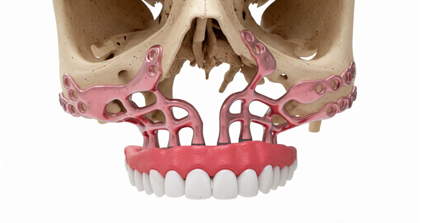
Subperiosteal Implants for Severe Jawbone Atrophy are custom titanium frameworks that rest on the jawbone surface beneath the gum. They do not need to be screwed deep into the bone like standard implants. Small fixation screws hold the framework in place. Posts pass through the gum to support fixed teeth or an overdenture. Modern versions are designed digitally from each patient's scan, which makes them very different from older handmade designs.
How Do Subperiosteal Implants Differ From Conventional Dental Implants?
A conventional implant is a screw-shaped post placed inside the bone. It needs enough bone height and width to surround it securely. A subperiosteal implant sits on top of the bone instead of inside it. It spreads support across a wider surface area. This difference is why it may be considered when bone volume is too limited for conventional placement.
Where Is a Subperiosteal Implant Positioned in the Jaw?
The framework lies between the bone surface and the periosteum, the thin membrane covering bone. The gum and periosteum are lifted, the framework is seated, and the tissues are closed over it. Only the prosthetic posts emerge into the mouth. Exact positioning follows the digital plan and avoids nerves, sinuses, and other sensitive structures.
How Do Modern Patient-Specific Subperiosteal Implants Work?
Modern frameworks begin with a CBCT or CT scan of the jaw. Software converts the scan into a three-dimensional model of the bone surface. Engineers and clinicians then design a framework that matches this surface closely. The framework is manufactured in titanium and fixed with screws during surgery. A provisional or final prosthesis then attaches to the emerging posts.
Framework Design and Bone-Surface Adaptation
The framework is shaped to follow the patient's own bone contours rather than a standard size. Close, passive adaptation helps the framework sit firmly without rocking. Designers place arms and plates where bone is thick and dependable. They avoid thin areas, nerve exits, and sharp transitions that could irritate the gum. Digital checks can reveal seating conflicts before surgery begins.
Fixation Screws and Prosthetic Connections
Fixation screws anchor the framework to areas of stronger bone. Their number, length, and position are planned in advance to share chewing forces. Prosthetic connections are the posts that pass through the gum. They are angled and positioned to suit the planned teeth, bite, and cleaning access. Good connection design makes later repair and maintenance easier.
Why Does Severe Jawbone Atrophy Develop?
Jawbone needs stimulation from teeth to keep its shape. When teeth are lost, the ridge that once held them gradually shrinks. This remodeling is natural, but it can become severe over many years. Gum disease, infection, injury, and long denture use can speed the process. The result may be a ridge too thin or too low for routine implants.
What Causes Severe Jawbone Loss After Tooth Loss?
After extraction, the tooth socket heals and the surrounding bone begins to remodel. Much of the width change can happen within the first months. Without roots to transmit chewing forces, the ridge continues to resorb slowly. Several missing teeth in one area often lead to wider defects. Over decades, the ridge may lose most of its original volume.
How Do Periodontal Disease and Long-Term Edentulism Affect Jawbone Volume?
Periodontal disease destroys the bone supporting teeth even before they are lost. Teeth extracted after severe gum disease often leave already damaged ridges. Long-term edentulism, meaning years without teeth, then adds further resorption. Pressure from a loose denture can concentrate force on small areas of bone. Together, these factors can produce advanced atrophy.
Can Trauma, Infection, Tumor Surgery, or Other Conditions Cause Severe Jaw Atrophy?
Yes, bone loss is not always caused by tooth loss alone. Facial trauma can fracture or remove parts of the jaw. Chronic infection, failed implants, and cyst removal can leave large defects. Tumor surgery may remove substantial sections of bone. Certain medical conditions and medicines can also affect bone healing and should be reviewed individually.
Does Jawbone Atrophy Affect the Maxilla and Mandible Differently?
The upper jaw, or maxilla, usually resorbs upward and inward. It also has expanding sinuses that reduce available bone height over time. The lower jaw, or mandible, often resorbs downward and outward. Its main concern is the inferior alveolar nerve running through the bone. These patterns shape how any implant solution must be designed.
Explore how liver health influences gum disease in Cirrhosis and Periodontics
What Does Severe Jawbone Atrophy Look or Feel Like?
Many patients notice changes gradually rather than suddenly. Dentures may feel loose, painful, or impossible to keep in place. The face may look shorter, with lips appearing thinner and wrinkles deeper. Chewing firm foods can become difficult. These signs suggest change, but only imaging can confirm how much bone remains.
What Signs May Suggest Advanced Jawbone Resorption?
Repeated denture relines that never seem to last are a common sign. A flat or knife-edge ridge felt with the tongue may also indicate loss. Some patients develop sore spots where the denture presses on nerves. Speech changes and food trapping under the denture are also frequent complaints. A clinical examination can clarify these findings.
Can Loose Dentures Be a Sign of Significant Bone Atrophy?
Yes, loose dentures often reflect a shrinking ridge beneath them. A denture fitted years ago no longer matches the changed bone shape. Adhesives may help temporarily but do not stop resorption. Persistent looseness is a good reason to request imaging. Early assessment can sometimes keep more treatment options available.
Is Severe Jawbone Atrophy Always Associated With Pain?
No, bone loss itself is usually painless. Discomfort more often comes from a denture pressing on thin tissue or exposed nerves. Some patients have severe atrophy with little pain, while others feel constant soreness. Pain levels therefore do not indicate how much bone remains. Diagnosis relies on examination and imaging rather than symptoms alone.
Can Facial or Bite Changes Occur After Major Bone Loss?
Yes, losing bone can reduce the vertical height of the lower face. The chin may move closer to the nose, and lip support may decrease. The bite relationship between upper and lower jaws can also shift. Restoring teeth in the correct position can help improve support and function. Planning must consider these changes from the start.
Compare procedure steps in Simple vs Surgical Tooth Extraction
Who May Be a Candidate for Subperiosteal Implants?
Candidates usually have severe bone loss that limits conventional implant placement. They should be in reasonably stable general health and able to undergo surgery. Healthy gums, controlled infection, and good hygiene habits are important. Candidates for Subperiosteal Implants for Severe Jawbone Atrophy must also attend long-term follow-up. Each case is decided after an individual assessment.
Who May Consider Subperiosteal Implants for Severe Bone Loss?
Patients who cannot wear dentures comfortably may consider this option. So may people who wish to avoid large bone grafts or have had grafts fail. Those with limited bone after implant failure or surgery may also qualify. Subperiosteal Implants for Severe Jawbone Atrophy are one option among several graft-free and graft-based approaches.
Are Subperiosteal Implants Suitable for an Atrophic Jaw?
They can be suitable when the remaining bone surface offers enough stable fixation points. The jaw does not need a tall bone, but it needs dependable cortical areas. Soft tissue must be healthy enough to cover the framework reliably. Suitability also depends on whether alternatives offer better long-term evidence. Imaging and examination answer this question for each patient.
Can Patients With Severe Maxillary Atrophy Be Treated?
Yes, many published cases involve severely atrophic upper jaws. The framework can rest on the remaining palatal, ridge, and buttress areas. Design must respect the sinus, nasal floor, and surrounding structures. Subperiosteal Implants for Severe Jawbone Atrophy in the maxilla may avoid sinus grafting in selected patients. Zygomatic implants remain an important alternative to compare.
Can Patients With Severe Mandibular Atrophy Be Treated?
Yes, clinical studies have treated severely atrophic lower jaws with custom frameworks. The design spans the available cortical bone while protecting the nerve. Fixation is placed where bone is thick enough to hold screws securely. Short implants or grafting may also be possible in some cases. The best choice depends on the measured anatomy.
When Might Conventional Implants Not Provide Enough Bone Support?
Conventional implants need minimum bone height and width around them. When the ridge is narrower or shorter than this, stability may be compromised. Nerves and sinuses can further limit safe implant length. In such cases, grafting or an alternative design becomes necessary. Subperiosteal Implants for Severe Jawbone Atrophy are considered when these limits apply.
When May Bone Grafting Still Be Preferred?
Grafting may be preferred when it can predictably rebuild the ridge. Conventional implants placed in grafted bone have a long research history. Younger patients with good healing capacity may benefit from rebuilding bone. Grafting may also suit cases where natural tooth position matters greatly. The longer timeline is often accepted for stronger long-term evidence.
What Medical or Oral Health Factors Can Affect Candidacy?
Uncontrolled diabetes can slow healing and raise infection risk. Smoking reduces blood flow to the gums and bone. Some osteoporosis and cancer medicines affect bone healing and need careful review. Previous radiation to the jaw is a major consideration. Active gum disease and untreated infection must be controlled before surgery.
See what fits your needs in Dental Implants vs Implant Supported Dentures
What We Notice Clinically
In clinical practice, patients often arrive believing only one solution exists. Many have heard that severe bone loss automatically means a custom framework. In reality, several approaches may suit the same jaw. Expectations about speed, cost, and appearance also differ widely. Careful discussion helps align the plan with each patient's priorities.
Why Severe Bone Loss Does Not Automatically Mean Every Patient Needs the Same Implant Solution
Two patients with similar scans may need different plans. One may have healthy gums and good bone density at fixation sites. Another may have thin tissue or medical risks that change the balance. Prosthetic goals, budget, and willingness to accept grafting also vary. Subperiosteal Implants for Severe Jawbone Atrophy are therefore one option, not a default.
Common Misconceptions About Graft-Free Implant Treatment
Some believe graft-free means no surgery. In fact, flaps, fixation, and careful soft-tissue closure are still required. Others assume graft-free always means faster or cheaper. Custom design and manufacturing add their own time and cost. Another misconception is that newer always means better proven. Long-term evidence for custom frameworks is still developing.
The Difference Between Severe Bone Atrophy and Active Oral Disease
Atrophy describes reduced bone volume, which is a structural problem. Active disease means ongoing infection or inflammation that is still progressing. Atrophy can be planned around with suitable designs. Active disease must be treated before any implant is placed. Placing a framework into infected tissue can seriously threaten the result.
Why Prosthetic Planning Matters as Much as Available Bone
The final teeth determine where support is truly needed. A framework that fits bone perfectly can still fail functionally if posts emerge poorly. Tooth position affects chewing, speech, lip support, and appearance. Cleaning access determines how well tissues stay healthy. Planning should therefore start with the teeth and work backward to the framework.
How Smoking, Soft-Tissue Condition, and Oral Hygiene Can Influence Risk
Smoking impairs healing and is linked with higher implant failure rates. A 2024 meta-analysis reported an odds ratio of 2.59 for early implant failure among smokers. Thin or scarred gums are more prone to exposure over the framework. Poor plaque control encourages inflammation around posts. Improving these factors before surgery can meaningfully reduce risk.
Clinical Note: Dr. Rifat Alsaman on Patient Selection and Individualized Planning
Patient selection is treated as the most important step in this treatment. Each case is reviewed through clinical findings, imaging, and restorative goals together. Alternatives are discussed openly before a custom framework is recommended. The aim is a plan that suits the patient's anatomy, health, and maintenance ability. Treatment is scheduled only after these factors are clearly documented.
Evaluate your choices in Implant Supported Bridge vs Traditional Bridge

What Does the Initial Oral Examination Assess?
The examination reviews remaining teeth, gums, and oral mucosa. It checks existing dentures, bite relationships, and jaw movements. The clinician looks for infection, inflammation, and lesions needing treatment. Lip support, smile line, and speech are also observed. Medical history and medicines are reviewed at the same visit.
Why Is Three-Dimensional Imaging Important?
Two-dimensional X-rays flatten the jaw into a single image. They cannot show ridge width, surface contours, or exact nerve paths. Three-dimensional imaging reveals bone shape from every direction. It allows precise measurement of fixation areas and anatomical boundaries. Custom framework design is impossible without this data.
How Are CBCT or CT Data Used in Implant Planning?
CBCT or CT data are exported as digital files to planning software. The software segments bone from soft tissue to create a surface model. Clinicians mark nerves, sinuses, and other structures to avoid. Bone thickness at possible screw sites is measured. The model then becomes the base for framework design.
How Are Intraoral Scans and Digital Models Used?
Intraoral scans capture the gum surface and existing teeth or dentures. They are merged with the CBCT data for a complete picture. This shows how thick the soft tissue is over the bone. Digital models also help design the provisional and final prosthesis. Accurate merging improves both fit and tooth position.
How Is the Bone Surface Mapped for a Custom Implant?
The software builds a detailed surface map of the remaining bone. Designers identify areas of thick cortical bone suitable for support. They trace the framework outline to avoid nerve exits and thin regions. Contact areas are adjusted for broad, even seating. The map guides both framework shape and screw placement.
How Is Occlusion and the Final Prosthesis Considered Before Surgery?
The team first sets the ideal tooth position in a virtual setup. Bite height, jaw relationship, and lip support are checked at this stage. Post positions are then chosen to support those teeth. Forces from chewing are directed toward stronger supporting areas. This prosthesis-first approach guides the whole surgical design.
Bone Anatomy
Bone anatomy determines where the framework can rest and be fixed. Planners evaluate cortical thickness, ridge shape, and buttress areas. They map the positions of nerves, sinuses, and nasal structures. Bone density at screw sites influences fixation choices. Each jaw presents a different combination of these features.
Soft-Tissue Thickness
The gum covering the framework must be thick and healthy enough. Thin tissue is more likely to break down and expose metal. Scarring from previous surgery can reduce blood supply. Planners measure tissue thickness from merged scans. Soft-tissue procedures may sometimes be recommended before or during surgery.
Prosthetic Emergence
Emergence describes where and how posts exit the gum. Ideal emergence supports teeth in the correct position without bulky prosthetics. Posts placed too far forward or back complicate cleaning and appearance. Emergence also influences how tightly gum seals around each post. Good emergence protects both function and tissue health.
Fixation and Load Distribution
Fixation screws hold the framework against the bone surface. Their placement spreads chewing load across several stable areas. Overloading one region can cause screw loosening or bone changes. Planners balance the number of screws with surgical access. A well-distributed design improves long-term mechanical stability.
How Is Severe Atrophy Classified for Treatment Planning?
Classification systems describe how far ridge resorption has progressed. They give clinicians a shared language for discussing severity. Classification helps indicate which treatment groups may be realistic. However, it cannot replace three-dimensional imaging. Individual planning still decides the final design.
Cawood-Howell Classification and Clinical Relevance
The Cawood-Howell classification describes edentulous ridges from Class I to Class VI. Early classes show mild change, while later classes show severe resorption. Class V and VI ridges often limit conventional implant options. The system offers useful context during consultations. Custom frameworks, however, are planned from each patient's actual bone surface.
Learn which procedure you need between Bone Graft vs Sinus Lift
How Are Custom Subperiosteal Dental Implants Designed?
Design starts with imaging and a virtual tooth setup. Designers then shape the framework over the mapped bone surface. Screw holes, posts, and connecting bars are added step by step. Clinicians review and approve each design before manufacturing. Subperiosteal Implants for Severe Jawbone Atrophy rely on this close collaboration between surgeon, prosthodontist, and engineer.
What Are Patient-Specific Subperiosteal Implants?
Patient-specific implants are made for one person's anatomy only. They cannot be swapped between patients or bought in standard sizes. Their shape, screw positions, and posts all follow that patient's plan. This customization allows support on bone that standard implants cannot use. Subperiosteal Implants for Severe Jawbone Atrophy are always patient-specific in modern practice.
How Does CAD/CAM Technology Shape the Implant Framework?
CAD stands for computer-aided design, and CAM for computer-aided manufacturing. CAD software lets designers draw the framework directly onto the bone model. Thickness, curves, and screw angles are adjusted digitally. CAM then converts the design into machine instructions. This workflow improves accuracy compared with older impression-based techniques.
How Does 3D Printing or Additive Manufacturing Work?
Additive manufacturing builds the framework layer by layer from titanium powder. A laser or electron beam melts each thin layer according to the design. Complex shapes can be produced without cutting from a solid block. After printing, the framework is cleaned, finished, and polished in selected areas. Some frameworks are milled instead, depending on the design.
What Materials Are Used for Modern Custom Subperiosteal Implants?
Titanium and titanium alloys are the most common framework materials. They are strong, lightweight, and well tolerated by body tissues. Fixation screws are usually titanium as well. Prosthetic teeth may be acrylic, composite, zirconia, or ceramic. Material choice depends on strength needs, aesthetics, and maintenance plans.
How Is Implant Fit Checked Before Placement?
Designers check fit virtually against the bone model before manufacturing. A printed resin model of the jaw is often produced. The framework or a replica can be seated on this model. Any rocking, gaps, or interference can then be corrected. These checks reduce the need for adjustments during surgery.
What Is the Role of Digital Surgical Guides?
Surgical guides transfer the digital plan into the mouth. They may guide bone reshaping or screw drilling in precise positions. Some frameworks are self-guiding because of their close fit. Guides can shorten surgical time and improve accuracy. Their use depends on the case and the surgeon's protocol.
Check out the differences in Root Canal vs Crown
Can Subperiosteal Implants Be Placed Without Bone Grafting?
Yes, that is one of their main purposes. The framework uses existing bone surface instead of rebuilding lost volume. This avoids waiting months for a graft to heal. Subperiosteal Implants for Severe Jawbone Atrophy may therefore shorten the overall treatment timeline. Graft-free placement still requires careful selection and planning.
When Can Subperiosteal Implants Avoid Traditional Bone Augmentation?
They may avoid augmentation when remaining bone offers stable fixation areas. The soft tissue must be healthy and thick enough for coverage. The planned prosthesis must be achievable from the available post positions. If these conditions are met, grafting may be unnecessary. If not, augmentation or another approach may be safer.
Does “Graft-Free” Mean No Other Surgical Procedure Is Needed?
No, graft-free does not mean surgery-free. The gum must be opened to expose the bone surface. Bone may need minor smoothing for accurate seating. The framework is fixed with screws and the tissues are stitched closed. Extractions or soft-tissue procedures may also be needed in the same session.
How Do Subperiosteal Implants Compare With Bone Grafting?
Grafting rebuilds bone so conventional implants can be placed later. Custom frameworks use the bone that remains without rebuilding it. Grafting usually takes longer but rests on extensive long-term research. Subperiosteal Implants for Severe Jawbone Atrophy may be faster but have less long-term data. The comparison involves time, risk, evidence, and cost.
Treatment Time
Grafting often adds four to nine months before implants can be placed. Further healing may be needed before final teeth are fitted. Custom frameworks need design and manufacturing time instead. Placement and provisional teeth may then occur in a single surgery. Total time still varies with healing and prosthetic steps.
Surgical Complexity
Large grafts may need donor sites, such as the hip or skull. Sinus lifts and block grafts add separate procedures. Custom frameworks require wide flap access and precise fixation. Both approaches are complex and need experienced surgeons. The complexity simply lies in different places.
Bone Requirements
Grafting needs a healthy recipient site that can accept new bones. Conventional implants then need enough grafted volume to surround them. Custom frameworks need stable cortical surfaces for fixation. They do not need tall or wide ridges. Bone quality at screw sites remains important for both approaches.
Healing and Recovery
Graft surgery may involve swelling at both donor and recipient sites. Healing must be complete before implants can be loaded. Framework surgery involves one main healing period for the gums. Early discomfort and swelling usually settle within one to two weeks. Soft-tissue maturation continues for several weeks afterward.
Long-Term Evidence
Conventional implants in grafted bone have decades of follow-up data. Modern custom frameworks have mostly short- to medium-term data. A 2026 systematic review found 97.8 percent pooled survival up to three years. Survival fell to 54.1 percent at six years in the longest follow-up study. These differences should be discussed before choosing. Evidence quality should therefore carry real weight in the final treatment decision.
What Are the Alternatives When Grafting Is Possible?
When grafting is feasible, several routes become available. Conventional implants can be placed after augmentation. Short or narrow implants may fit selected anatomy without major grafting. Zygomatic and pterygoid implants may suit some upper jaws. Comparing these options helps patients choose with full information.
Conventional Endosseous Implants With Bone Augmentation
Endosseous implants are placed inside bone after the ridge is rebuilt. Augmentation may use autogenous bone, substitutes, or membranes. Sinus lifts increase height in the upper back jaw. This approach has the strongest long-term evidence base. Its main drawbacks are longer treatment time and extra surgery.
Short or Narrow Implants in Selected Anatomy
Short implants may suit ridges with limited height but adequate width. Narrow implants may fit thin ridges with adequate height. Both can sometimes avoid major grafting. They have their own limits regarding load and prosthetic design. Imaging decides whether the remaining anatomy can support them.
Zygomatic Implants for Selected Severe Maxillary Cases
Zygomatic implants are long implants anchored in the cheekbone. They can support upper teeth when the maxilla has little bone. They are well researched for severe upper-jaw atrophy. Placement is technically demanding and involves specific risks, including sinus problems. They are not used in the lower jaw. Their prosthetic posts may emerge toward the palate, which can affect bulk and cleaning.
Other Advanced Implant Strategies
Pterygoid implants anchor in bone behind the upper jaw. Tilted implants can avoid the sinus or nerve while using available bone. Nasal and transnasal implants are used in specialized cases. Each strategy has its own indications and evidence base. The treating team should explain why one option is preferred. Comparing them side by side helps patients see trade-offs in evidence, risk, and prosthetic access.
How Are Subperiosteal Implants Used for Severe Maxillary Atrophy?
In the upper jaw, the framework may rest on the ridge, palate, and buttress areas. Subperiosteal Implants for Severe Jawbone Atrophy can avoid sinus grafting in selected cases. Fixation screws are placed in denser bone, such as the zygomatic buttress. Posts are positioned to support a fixed bridge or overdenture. Planning focuses on stability, appearance, and hygiene access.
Why Is Severe Maxillary Atrophy Challenging?
The upper jaw often has softer bones than the lower jaw. Expanding sinuses reduce height in the back regions. The nasal floor limits options at the front. Resorption also moves the ridge inward, complicating tooth position. These factors make conventional implant placement difficult without grafting.
How Is the Custom Framework Adapted to the Atrophic Maxilla?
Designers follow the remaining palatal and buttress contours closely. Arms may extend to stronger areas while avoiding thin sinus walls. The framework avoids the incisive canal and nasal openings. Smooth edges reduce irritation of thin palatal tissue. Post positions are set to restore lip support and a natural smile.
What Anatomical Structures Must Be Considered in the Upper Jaw?
The maxillary sinuses lie above the back teeth area. The nasal floor and piriform aperture lie above the front teeth. The incisive canal carries nerves and vessels behind the front teeth. The greater palatine vessels run along the palate. All these structures must be mapped and protected.
What Fixed Prosthetic Options May Be Supported?
A screw-retained full-arch bridge is a common fixed option. It may be acrylic with a metal bar or fully zirconia. Hybrid designs combine durability with easier repair. The design must allow professional removal for cleaning and inspection. The choice depends on bite forces, aesthetics, and budget. Removable overdentures remain an alternative when cleaning access or budget is a concern.
How Are Subperiosteal Implants Used for Severe Mandibular Atrophy?
In the lower jaw, the framework often spans the front and side regions. Subperiosteal Implants for Severe Jawbone Atrophy in the mandible must protect the inferior alveolar nerve. Fixation is placed in thick cortical bone along the chin and body. Posts support a fixed bridge or removable overdenture. Clinical trials have tested both milled and printed designs.
Why Is Severe Mandibular Atrophy Different From Maxillary Atrophy?
The mandible usually has denser cortical bone than the maxilla. It has no sinus, but the nerve path is critical. Severe resorption can bring the nerve close to the ridge surface. The lower jaw also flexes slightly during opening and chewing. Framework design must account for these different mechanics.
Which Anatomical Structures Must Be Protected in the Lower Jaw?
The inferior alveolar nerve runs through the mandible. It exists at the mental foramen, which may lie on top of a resorbed ridge. The lingual nerve lies close to the inner surface. Floor-of-mouth vessels require care during deep fixation. Precise mapping protects sensation and avoids bleeding.
How Does Fixation Differ in an Atrophic Mandible?
Lower-jaw bone is often denser, allowing strong screw engagement. However, very thin mandibles carry a risk of fracture. Screws must avoid the nerve canal and mental foramen. Fixation is often concentrated in the chin region and basal bone. Load distribution helps protect a fragile jaw.
What Prosthetic Options Can Be Used After Placement?
Fixed full-arch bridges are often chosen for comfort and confidence. Removable overdentures attached to posts or bars are another option. Overdentures can be easier to clean and less costly. Fixed options feel more like natural teeth. The choice depends on anatomy, hygiene ability, and preference.
Read about managing a sharp fragment in your gum with Bone Spicule
What Happens During Subperiosteal Implant Surgery?
Surgery is usually performed under local anesthesia with sedation or general anesthesia. The surgeon opens the gum and exposes the bone surface. The framework is seated, checked, and fixed with screws. The gum is closed around the emerging posts. A provisional prosthesis may then be attached.
What Happens Before the Procedure?
Patients receive written instructions about eating, medicines, and transport. Medical conditions and current medications are reviewed again. Any remaining infection should already be treated. The framework, screws, and prosthetic parts are checked before surgery. Consent forms explain risks, alternatives, and follow-up needs.
How Is the Custom Implant Positioned on the Jaw?
After exposing the bone, the surgeon removes soft tissue from the surface. The framework is placed according to the digital plan. Its close fit helps confirm the correct seating position. The surgeon checks for gaps or rocking at every contact area. Minor bone smoothing may improve seating if needed.
How Is the Framework Stabilized?
Fixation screws are placed through planned holes into stable bone. They are tightened in a sequence that avoids distorting the framework. The surgeon confirms stability after all screws are placed. Correct screw length avoids nerves and sinus walls. A stable framework is essential before any loading.
How Are the Prosthetic Connections Positioned?
Posts are already built into the framework at planned positions. The gum is shaped and stitched around each post. Abutments or multi-unit connectors may be attached to the posts. Their angles match the planned prosthesis. Accurate positioning simplifies provisional and final tooth delivery.
Can a Temporary Fixed Prosthesis Be Provided During the Same Treatment Stage?
In selected cases, yes. Some protocols attach a provisional bridge on the day of surgery or shortly after. This depends on framework stability and the planned bite. The provisional restores appearance and function during healing. A softer diet is usually advised until tissues mature.
What Determines Whether Immediate Loading Is Appropriate?
Immediate loading depends on firm framework stability at surgery. Good bone at fixation sites and balanced bite forces are also required. Heavy clenching or grinding may make delayed loading safer. Soft-tissue condition affects how well tissue heals around posts. The surgeon decides after assessing stability during placement.
Note : All images used are for editorial and illustrative purposes only and may not originate from the original news provider or associated company.
What Are the Benefits and Limitations of Subperiosteal Implants?
Custom frameworks offer real benefits for carefully selected patients. They also carry limitations that patients should understand clearly. Benefits include possible graft avoidance and shorter treatment timelines. Limitations include soft-tissue exposure risk and limited long-term data. Subperiosteal Implants for Severe Jawbone Atrophy should be judged on both sides.
Potential Benefits for Severely Atrophic Jaws
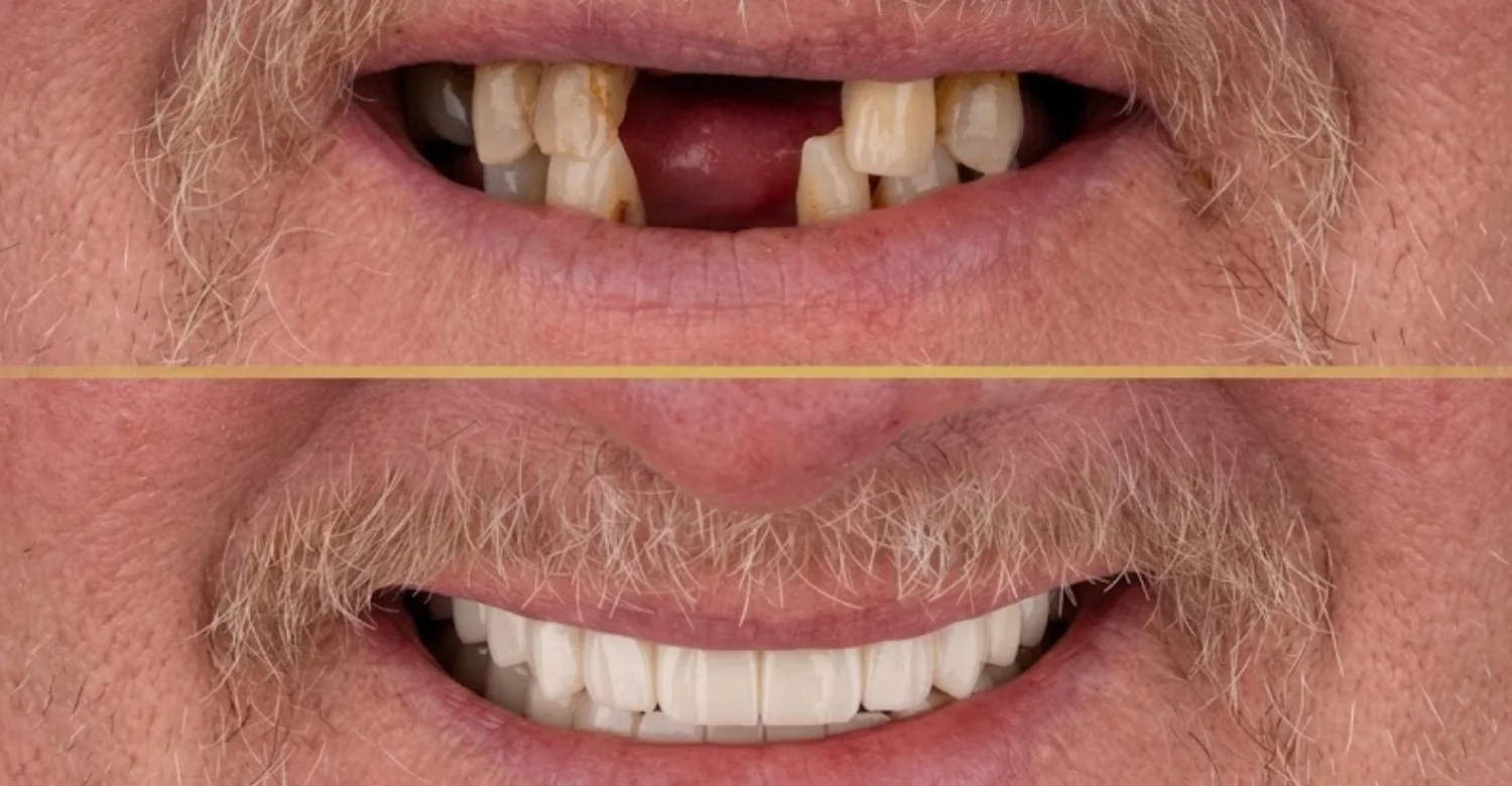
The framework can use bone that conventional implants cannot reach. It may restore fixed teeth to patients who struggled with dentures for years. Many patients report better chewing and greater confidence. Subperiosteal Implants for Severe Jawbone Atrophy may also avoid donor-site surgery. These benefits depend on careful case selection.
Potential Treatment-Time Advantages in Selected Cases
Avoiding grafts can remove several months of healing time. Placement and provisional teeth may occur in one surgical stage. Fewer surgeries can mean fewer recovery periods. Design and manufacturing still require several weeks before surgery. Time savings vary with each case and are not guaranteed.
Why Customization Can Improve Anatomical Adaptation
A custom framework matches the patient's unique bone contours. Close adaptation helps distribute load across a broad surface. Screw positions can target the best available bone. Designers can avoid nerves, sinuses, and thin areas precisely. Standard parts cannot achieve this level of individual fit. Subperiosteal Implants for Severe Jawbone Atrophy use this close fit to spread chewing load safely.
Important Limitations and Uncertainties
Soft-tissue dehiscence and framework exposure remain important concerns. Long-term studies are still fewer than for conventional implants. Removing or revising a framework can be surgically demanding. Results depend heavily on design quality and surgical skill. Subperiosteal Implants for Severe Jawbone Atrophy are promising but not risk-free. Patients should compare these limits with zygomatic, pterygoid, and grafting options before deciding.
Why Modern Results Should Not Be Compared Directly With Historical Subperiosteal Designs
Older subperiosteal implants were made from impressions taken during surgery. They often fitted poorly and had higher complication rates. Modern frameworks use CBCT data, digital design, and precise manufacturing. Materials and fixation methods have also improved. Historical results therefore do not reflect modern outcomes accurately. Modern studies should be judged on their own size, design, and follow-up length.
Find out what causes it when All My Teeth Hurt Suddenly
What Are the Side Effects and Complications of Subperiosteal Implants?
Common side effects include swelling, bruising, and discomfort after surgery. More serious complications include soft-tissue dehiscence and framework exposure. Infection, screw loosening, and prosthetic fractures can also occur. Subperiosteal Implants for Severe Jawbone Atrophy require ongoing monitoring to detect problems early. Most early side effects settle with proper care.
What Side Effects Are Common During Early Healing?
Swelling usually peaks within two to three days after surgery. Bruising may appear on the face or neck. Mild bleeding from the stitches is common at first. Patients may notice tightness when opening the mouth. These effects usually improve steadily during the first one to two weeks.
Is Pain, Swelling, or Bruising Normal After Surgery?
Yes, these are expected responses to flap surgery and fixation. Pain is usually manageable with recommended medicines. Swelling and bruising gradually fade over several days. Symptoms should improve, not worsen, after the first few days. Worsening symptoms should be reported to the clinical team.
Can Soft-Tissue Dehiscence or Framework Exposure Occur?
Yes, this is the most commonly reported complication in recent studies. Dehiscence means the gum opens over part of the framework. Exposed metal can collect plaque and bacteria. Small exposures may be managed with cleaning and monitoring. Larger exposures may need surgical repair or partial framework revision.
Can Infection Develop Around a Subperiosteal Implant?
Yes, infection can develop early or years after placement. Exposure and poor plaque control raise this risk. Signs include swelling, pus, bad taste, and increasing pain. Early treatment may include cleaning, antibiotics, or surgical access. Persistent infection can threaten the framework's survival.
What Can Cause Implant Mobility or Prosthetic Problems?
Mobility may result from screw loosening or bone changes at fixation sites. Excessive bite forces or grinding can overload the framework. Prosthetic teeth may chip, crack, or loosen over time. Poor fit between the bridge and posts adds stress. Regular reviews help detect these problems early.
What Complications May Occur Specifically in the Maxilla?
Screws placed near the sinus may cause sinus irritation if misplaced. Thin palatal tissue may be prone to exposure. Nasal or sinus communication is rare but possible. Upper-jaw bone is softer, which may affect screw stability. Careful planning reduces these maxilla-specific risks.
What Complications May Occur Specifically in the Mandible?
Nerve irritation may cause numbness or tingling in the lip or chin. This is usually temporary but can occasionally persist. Very thin mandibles carry a small fracture risk. Floor-of-mouth bleeding is rare but important. Precise imaging and design help prevent these problems.
Which Complications Require Prompt Clinical Review?
Increasing pain after initial improvement needs prompt review. So do pus, spreading swelling, or persistent bleeding. Visible metal under the gum should be checked early. Loose teeth or a moving bridge require assessment. Numbness that does not improve should also be reported.
Learn why you might see Saliva Blood When Spitting
Pain Relief After Subperiosteal Implant Surgery
Pain is usually strongest during the first two to three days. Most patients manage well with medicines recommended by the team. Cold compresses, rest, and soft foods also help. Pain should steadily decrease as healing progresses. Unexpected pain changes should prompt contact with the clinic.
What Can Help Relieve Normal Postoperative Discomfort?
Cold packs on the face during the first day can reduce swelling. Keeping the head raised while resting may also help. Soft, cool foods are easier to eat during early healing. Taking pain relief on schedule prevents discomfort from building up. Gentle rinsing as instructed keeps the area clean.
Which Pain-Relief Instructions Should Come From the Treating Dental Team?
Medicine type, dose, and timing should come from your treating team. They will consider your medical history and other prescriptions. Some patients cannot take certain anti-inflammatory medicines. Antibiotics, if prescribed, must be completed as directed. Written instructions help avoid confusion during recovery.
Why Should Patients Avoid Self-Medicating Beyond Recommended Instructions?
Extra doses can cause stomach, kidney, or liver problems. Some pain medicines interact with blood thinners and other prescriptions. Strong pain relief may mask signs of infection or complications. Mixing medicines without guidance can be unsafe. Contact the team if recommended medicines do not control pain.
Understand the potential effects of Prion Diseases and Oral Health
What Is Recovery Like After Subperiosteal Implant Surgery?
Recovery happens in stages over several weeks. The first days focus on rest, swelling control, and gentle hygiene. Stitches are usually reviewed or removed within about two weeks. Soft-tissue healing continues while the provisional prosthesis is worn. The final prosthesis follows once tissues stabilize.
What Should Patients Expect During the First Few Days?
Expect swelling, mild bleeding, and tenderness at first. Eating may be limited to soft or liquid foods. Speech may feel different with new provisional teeth. Rest and avoid strenuous activity for the first few days. Follow all cleaning and medicine instructions carefully.
How Long Does Soft-Tissue Healing Usually Take?
Initial gum closure usually occurs within one to two weeks. Tissue maturation around the posts can take several more weeks. Full stabilization may take two to three months. Healing varies with tissue thickness, smoking, and health. Follow-up visits confirm healing progress.
When Can Normal Eating and Oral Hygiene Resume?
Soft foods are usually advised for several weeks after surgery. Firmer foods are reintroduced gradually as healing allows. Gentle brushing around posts often begins after the first few days. Water flossers or special brushes may be added later. Your team will confirm when normal habits can resume.
When Is the Final Prosthesis Usually Delivered?
The final prosthesis is usually delivered after soft tissues stabilize. This often occurs a few months after surgery. Timing depends on healing, bite stability, and prosthetic design. New scans or impressions are taken for the final teeth. Try-in visits check fit, appearance, and bite before completion.
What Follow-Up Visits Are Needed After Treatment?
Early visits check healing, stitches, and provisional fit. Later visits assess tissue health and framework stability. Imaging may be repeated at planned intervals. Professional cleaning and prosthesis removal may be scheduled regularly. Lifelong follow-up is recommended for all implant-supported teeth.
Warning Signs After Subperiosteal Implant Surgery
Some symptoms suggest healing is not progressing normally. These include worsening pain, spreading swelling, or persistent bleeding. Pus, foul taste, or fever may indicate infection. Visible metal or loose components need early review. Recognizing these signs early protects the reconstruction.
When Does Increasing Pain Require Dental Assessment?
Pain should ease gradually after the first few days. If pain increases after improving, contact the clinic promptly. Throbbing pain with swelling may suggest infection. Pain when biting may indicate a bite or prosthesis problem. Early assessment usually allows simpler management.
When Could Swelling or Bleeding Indicate a Problem?
Swelling that keeps growing after three days needs review. Swelling spreading toward the eye or neck needs urgent attention. Bleeding that soaks gauze persistently should be checked promptly. Light oozing on the first day is usually normal. When unsure, contact the clinical team.
What Does Pus, Persistent Bad Taste, or Drainage Mean?
These signs often indicate infection around the framework or wound. They may also reflect food or plaque trapped near the posts. Infection should not be ignored, even if pain is mild. The team may clean the area or prescribe treatment. Early care can prevent spread to deeper tissues.
What Should Patients Do If the Framework or Implant Becomes Exposed?
Contact the clinic and arrange an examination. Do not touch, pick, or try to cover the exposed area. Keep the area clean with the rinses or methods recommended. The team will assess the size and cause of exposure. Treatment may range from monitoring to minor surgical repair.
When Is Urgent Medical or Emergency Care Appropriate?
Seek emergency care for breathing or swallowing difficulty. Rapid facial swelling with fever also needs urgent attention. Uncontrolled bleeding that does not stop with pressure is an emergency. Severe allergic reactions to medicines require immediate help. Inform the dental team as soon as possible afterward.
.jpg)
.jpg)
.jpg)
How Long Do Patient-Specific Subperiosteal Implants Last?
There is no guaranteed lifespan for any implant system. Published studies range from short follow-up to about ten years. Results vary widely between studies and patient groups. Subperiosteal Implants for Severe Jawbone Atrophy may last many years with good maintenance. Long-term certainty is still developing.
What Does Current Research Say About Implant Survival?
A 2026 systematic review pooled data from 11 studies. Survival was 97.8 percent for follow-up of three years or less. Overall pooled survival was 92.4 percent across all included studies. One longer study reported 54.1 percent survival at six years. A 2025 series reported 80 percent survival probability at ten years in ten maxillae. Most included studies were small, so these figures carry real uncertainty.
Why Does “Survival” Differ From “Treatment Success”?
Survival means the implant is still in the mouth. Success requires healthy tissues, stable function, and no unresolved complications. An implant can survive while having exposure or infection. Many studies report survival more often than success. Patients should ask about both measures when reviewing results.
How Do Long-Term Complications Affect Maintenance?
Late exposure or infection may require extra cleaning visits. Prosthetic repairs may be needed as teeth wear or chip. Screws may need checking and retightening over time. Some cases may need surgical revision. Maintenance plans should anticipate these possibilities and their costs. Patients should ask how these situations are handled before treatment begins.
Why Is Long-Term Evidence Still Developing?
Modern digital frameworks have only been widely used in recent years. Most studies are small and have short follow-up periods. Designs, materials, and techniques differ between research groups. Few randomized trials compare frameworks directly with alternatives. More long-term research on Subperiosteal Implants for Severe Jawbone Atrophy is still needed.
What Factors Can Affect Long-Term Subperiosteal Implant Success?
Framework fit and fixation quality influence mechanical stability. Soft-tissue thickness affects the risk of exposure. Hygiene, smoking, and general health affect infection risk. Bite forces and prosthetic design influence wear and loosening. Regular follow-up helps detect changes before they become serious.
Implant Fit and Mechanical Stability
A precise fit spreads forces evenly across the bone surface. Poor fit can cause micromovement and tissue irritation. Stable screws prevent the framework from shifting during chewing. Fit depends on imaging quality, design, and manufacturing accuracy. Surgical seating must match the digital plan closely.
Soft-Tissue Health and Framework Coverage
Healthy gum coverage protects the framework from bacteria. Thin or tense tissue is more likely to open over time. Smooth framework edges reduce pressure on the overlying gum. Careful flap design and closure improve coverage. Soft-tissue health should be monitored at every review.
Oral Hygiene and Professional Maintenance
Daily cleaning around posts and under the bridge is essential. Interdental brushes, water flossers, and soft toothbrushes are commonly recommended. Professional cleaning removes deposits that home care cannot reach. The bridge may be removed periodically for thorough cleaning. Consistent hygiene lowers infection and exposure risk.
Smoking and Other Modifiable Risk Factors
Smoking reduces blood flow and slows soft-tissue healing. It is linked with higher implant failure in broader research. Stopping before surgery and during healing is strongly advised. Diabetes control and good nutrition also support healing. Reducing alcohol intake may further help recovery.
Prosthetic Design and Occlusal Forces
A balanced bite spreads forces across the whole arch. Long cantilevers can overload posts and fixation screws. Grinding or clenching may require a protective night guard. Material choice affects how forces transfer to the framework. Good design protects both prosthesis and supporting structures.
Regular Clinical and Radiographic Follow-Up
Clinical reviews check tissues, posts, bite, and prosthesis fit. Radiographs or scans monitor bone and screw positions over time. Early detection of changes allows simpler treatment. Review intervals are set according to individual risk. International patients should plan local or remote follow-up arrangements.
What Is the Average Cost of Subperiosteal Implants in ?
US
UK
Turkey
There is no single fixed price because every framework is custom-made. Costs vary by country, clinic, case complexity, and included services. Subperiosteal Implants for Severe Jawbone Atrophy usually cost more than standard dentures. Published figures are planning estimates, not binding quotes. Comparing what each package includes is essential.
Country | Indicative Cost Per Arch | Typical Planning Range |
United States | ~25,000–50,000 | Highest-cost range |
United Kingdom | ~15,000–25,000+ | Private treatment |
Turkey | ~4,000–10,000+ | Varies by package and complexity |
What Does a Subperiosteal Implant Cost in the United States?
US costs reflect high clinical, laboratory, and surgical fees. Custom design and manufacturing add significant cost per arch. Sedation, imaging, and prosthetics may be billed separately. Insurance coverage is often limited for this type of treatment. Subperiosteal Implants for Severe Jawbone Atrophy in the US are among the most expensive options.
Indicative Full-Arch Planning Range: About 25,000–50,000 Per Arch
A Vitrin Clinic price reference lists about $25,000 to $50,000 per arch in the USA. The range depends on framework design and final prosthetic materials. Complex anatomy and additional procedures can push costs higher. Some quotes exclude imaging, sedation, or follow-up care. Always request a written, itemized estimate.
US Cost Component | Indicative Cost Consideration |
Custom framework | Major component of the treatment cost |
Surgical placement | Depends on complexity and operating setting |
Imaging and digital planning | May be charged separately |
Prosthetic teeth | Depends on acrylic, hybrid, or zirconia design |
Sedation or anesthesia | May involve an additional fee |
Follow-up and aftercare | May or may not be included |
What Does a Subperiosteal Implant Cost in the United Kingdom?
UK costs vary between private clinics and regions. Custom frameworks are rarely available through public dental services. Fees usually include consultation, imaging, surgery, and prosthetics. Some clinics charge separately for sedation or provisional teeth. Subperiosteal Implants for Severe Jawbone Atrophy in the UK require careful quote comparison.
Indicative Full-Arch Planning Range: About 15,000–25,000 Per Arch, With Higher Clinic-Specific Estimates Also Reported
Common planning ranges in the UK fall around $15,000 to $25,000 per arch. Some clinic-specific estimates, including Vitrin Clinic references, are higher. Variation reflects design, materials, and surgical setting. Premium zirconia prosthetics usually cost more than acrylic options. Written quotes should clarify every included item.
UK Cost Component | Indicative Pricing Consideration |
Consultation and assessment | May be included or charged separately |
CBCT and digital planning | Depends on diagnostic requirements |
Custom framework | Influenced by design and manufacturing |
Surgery | Varies by complexity and clinical setting |
Temporary prosthesis | May be included in package or billed separately |
Definitive prosthesis | Price depends heavily on material |
Aftercare | Check how many reviews are included |
What Does a Subperiosteal Implant Cost in Turkey?
Turkey is often chosen for lower treatment costs. Prices vary widely between clinics and package types. Some packages include accommodation, transfers, and provisional teeth. Subperiosteal Implants for Severe Jawbone Atrophy in Turkey still require high-quality design and surgery. Travel and follow-up costs should be included in comparisons.
Indicative Planning Range: Approximately 4,000–10,000+ Per Arch, Depending on Case Complexity and What the Package Includes
Published Turkish estimates range from about $4,000 to over $10,000 per arch. Lower figures may exclude the final prosthesis or follow-up visits. Higher figures often include premium materials and full aftercare. Multiple trips may be needed between surgery and final teeth. Compare total journey costs, not headline prices alone.
Turkey Cost Component | Indicative Pricing Consideration |
Consultation and diagnostics | Depends on examination and imaging needs |
Digital CAD/CAM planning | Included in some full treatment packages |
Custom titanium framework | Major cost component |
Surgical placement | Depends on anatomy and surgical complexity |
Temporary teeth | May be included in international packages |
Final prosthesis | Depends on acrylic, hybrid, or zirconia |
Accommodation and transfers | May be included by some clinics |
Aftercare | Package coverage varies |
What Is the Average Subperiosteal Implant Cost Per Arch?
Because pricing is highly individualized, it is more accurate to use an indicative planning range than a single average price. The table below summarizes the ranges discussed above.
Country | Approximate Cost Per Arch | Approximate Midpoint for Planning* |
United States | 25,000–50,000 | ~$37,500 |
United Kingdom | 15,000–25,000+ | ~$20,000 |
Turkey | 4,000–10,000+ | ~$7,000 |
What Is Usually Included in the Total Cost?
A complete quote usually includes several stages. These cover consultation, imaging, digital design, and manufacturing. Surgery, provisional teeth, and the final prosthesis are major parts. Follow-up visits and aftercare may or may not be included. Ask for each item to be listed clearly.
Treatment Stage | Usually Considered in a Full Treatment Plan |
Clinical examination and consultation | Assessment, medical history, oral examination, and treatment discussion |
CBCT or 3D imaging | 3D assessment for framework and surgical planning |
Digital CAD/CAM planning | Virtual framework and prosthetic design |
Custom implant manufacturing | Production of the patient-specific titanium framework |
Surgical placement | Surgical fees, clinical team, and facility costs |
Temporary prosthetic components | Provisional teeth during healing |
Definitive prosthetic components | Final bridge, overdenture, or fixed prosthesis |
Follow-up and aftercare | Reviews, adjustments, maintenance, and cleaning |
Clinical Examination and Consultation
The consultation reviews medical history, oral health, and treatment goals. The clinician examines teeth, gums, dentures, and bites. Treatment options and alternatives are discussed. Some clinics include this visit in the package price. Others charge it separately or credit it later.
CBCT or 3D Imaging
CBCT imaging is essential for designing a custom framework. It may be repeated if older scans are outdated or unclear. Intraoral scans may be added for soft tissue and bite records. Imaging costs vary by clinic and country. Confirm whether imaging is included in your quote.
Digital CAD/CAM Planning
Digital planning includes bone segmentation and virtual tooth setup. Engineers and clinicians design the framework together. Several revisions may be needed before final approval. This specialized work adds to overall cost. It is a key reason custom frameworks cost more than dentures.
Custom Implant Manufacturing
Manufacturing produces the titanium framework from the approved design. It may involve 3D printing, milling, or both. Finishing, polishing, and sterilization follow production. Resin jaw models for fit checks may also be produced. Manufacturing costs depend on design size and complexity.
Surgical Placement
Surgical fees cover the operation, team, and clinical facilities. Sedation or general anesthesia may be priced separately. Extractions or soft-tissue procedures in the same session add cost. Longer or more complex surgery usually costs more. Ask whether surgical consumables are included.
Temporary and Definitive Prosthetic Components
Provisional teeth are worn during healing. The definitive prosthesis is made after tissues stabilize. Materials range from acrylic to full zirconia. Abutments, screws, and connectors also add to costs. Prosthetic fees often represent a large share of the total.
Follow-Up and Aftercare
Follow-up includes healing checks, adjustments, and professional cleaning. Some clinics include a set number of reviews. Long-term maintenance visits are usually charged separately. Repairs or replacement teeth may be needed in later years. Clarify aftercare costs before starting treatment.
Why Should Published Prices Be Treated as Planning Figures Rather Than Fixed Quotes?
Each framework is designed for one patient, so costs vary. Online prices cannot account for individual anatomy or medical needs. Additional procedures may be identified only after examination. Exchange rates and clinic policies can also change prices. A personal written quote is the only reliable figure.
What Factors Change the Total Treatment Cost?
The number of arches treated has a major effect. Severity of bone loss influences design and surgical complexity. Prosthetic material choice changes costs substantially. Extractions, sedation, and hospital support add expense. Aftercare and travel requirements also affect the final total.
Cost Factor | Potential Effect on Total Cost |
Number of arches | Treating both jaws increases framework and prosthetic requirements |
Severity of jawbone atrophy | Greater complexity can increase design and surgical costs |
Framework complexity | Larger or more complex designs require additional planning and manufacturing |
Prosthetic material | Zirconia generally costs more than acrylic options |
Additional oral treatment | Extractions, gum treatment, or infection management can add fees |
Sedation or hospital support | May increase surgical and facility costs |
Aftercare and follow-up | Reviews, maintenance, repairs, and travel can add ongoing expenses |
Number of Arches Treated
Treating both jaws roughly doubles framework and prosthetic costs. Some clinics offer reduced combined fees for two arches. Treating both together may reduce total visits. Single-arch treatment may suit patients with one healthy jaw. Your plan will specify which arches need treatment.
Severity and Location of Jawbone Atrophy
More severe atrophy often needs larger, more complex frameworks. Upper-jaw cases may need extensions to buttress areas. Lower-jaw cases require careful nerve protection. Greater complexity adds design and surgical time. These factors are reflected in individual quotes.
Framework Complexity and Manufacturing Method
Larger frameworks use more titanium and design time. Additional arms, screws, or posts increase complexity. Printed and milled frameworks may differ in cost. Extra verification models or guides add to expenses. Complexity is determined by anatomy, not preference.
Prosthetic Material and Design
Acrylic bridges usually cost less and are easier to repair. Zirconia bridges are stronger and more stain-resistant but cost more. Hybrid designs combine materials for balance. Overdentures are often less expensive than fixed bridges. Material choice should consider function, appearance, and maintenance.
Additional Extractions or Oral Treatment
Remaining failing teeth may need extraction before or during surgery. Gum treatment may be required to control inflammation. Infected sites must be treated before framework placement. These procedures add time and cost. They are essential for a healthy foundation.
Need for Sedation, Hospital Support, or Additional Surgery
Many patients choose sedation for comfort during long procedures. General anesthesia may require hospital facilities and additional staff. Soft-tissue grafts may be recommended to improve coverage. Each addition increases the overall cost. Your medical needs determine which support is appropriate.
Aftercare and Follow-Up Requirements
Long-term maintenance continues for as long as the implant remains. Regular reviews, cleaning, and imaging carry ongoing costs. International patients may need extra travel for reviews. Repairs and replacement teeth may be needed after several years. Budgeting for aftercare prevents unexpected expenses.
Tips for Patients
Good preparation leads to better decisions and smoother treatment. Gather records, write down questions, and understand your options. Ask about evidence, risks, and maintenance before agreeing. Compare quotes based on what is included. Take time to decide without pressure.
What Should Patients Bring to Their Consultation?
Bring recent dental X-rays or CBCT scans if available. Bring a list of current medicines and medical conditions. Previous treatment records and implant details are helpful. Bring your current denture, even if it fits poorly. A written list of questions and goals is also useful.
What Questions Should Patients Ask About a Custom Subperiosteal Implant?
Ask why this option suits your anatomy better than alternatives. Ask how many similar cases the team has treated. Ask who designs and manufactures the framework. Ask whether provisional teeth will be provided on surgery day. Ask what the final prosthesis will look like. Ask how this option compares with zygomatic implants or grafting in your case.
What Should Patients Ask About Long-Term Maintenance?
Ask how often follow-up visits are needed. Ask whether the bridge will be removed for professional cleaning. Ask which cleaning tools you should use at home. Ask what long-term research shows about survival and complications. Ask about expected maintenance costs over several years.
What Should Patients Ask About Complications and Revision Treatment?
Ask which complications are most common for this treatment. Ask how framework exposure would be managed. Ask what happens if the framework must be removed. Ask whether revision treatment is included or charged separately. Clear answers help set realistic expectations.
What Should International Patients Confirm Before Traveling?
Confirm the number of visits and total time required. Confirm what the package includes and excludes. Ask how complications will be handled after you return home. Confirm communication methods with the clinical team. Request copies of all scans, plans, and treatment records.
Medical Tips for Preparing for Treatment
Preparation improves safety and healing. Control conditions such as diabetes and high blood pressure beforehand. Review all medicines with your doctor and dental team. Stop smoking as early as possible. Plan rest time and soft foods for recovery.
How Can Patients Prepare Their Oral Health Before Surgery?
Complete any recommended gum or tooth treatment first. Follow a careful daily cleaning routine before surgery. Use antiseptic rinses if your team recommends them. Attend hygiene visits to reduce bacterial load. A clean mouth supports better healing.
Why Should Existing Infection or Gum Disease Be Addressed First?
Active infection can spread to the new framework site. Inflamed gums heal poorly and may open over the framework. Bacteria from untreated disease raise long-term failure risk. Treating these problems creates a healthier foundation. It may delay surgery but improves predictability.
What Smoking or Oral-Hygiene Changes May Be Recommended?
Stopping smoking before and after surgery is strongly recommended. Nicotine replacement should be discussed with your doctor. Patients may be shown how to use interdental brushes and water flossers. Improved brushing technique protects tissues around posts. These habits should continue permanently.
Which Medical History and Medications Should Be Disclosed to the Dentist?
Disclose heart conditions, diabetes, and bleeding disorders. Mention any osteoporosis treatment or bone-modifying medicines. Report blood thinners, steroids, and immune-suppressing drugs. Previous head and neck radiation is especially important. Allergies to medicines or materials must also be shared.
At Vitrin Clinic: Evaluation and Planning for Severe Jawbone Atrophy
Vitrin Clinic approaches severe atrophy as an individualized planning challenge. Assessment combines clinical examination, digital imaging, and restorative goals. Subperiosteal Implants for Severe Jawbone Atrophy are compared with suitable alternatives. The team discusses evidence, risks, costs, and maintenance openly. Treatment is scheduled only after the plan is clearly understood.
How Vitrin Clinic Evaluates Teeth, Gums, and Oral Tissues Before Implant Treatment
The team examines remaining teeth, gums, and oral mucosa carefully. Existing dentures, restorations, and bites are reviewed. Signs of infection or inflammation are identified and treated first. Tissue thickness and quality are assessed for framework coverage. These findings guide the next diagnostic steps.
How Digital X-Rays and 3D Imaging Can Support Case Assessment When Clinically Appropriate
Digital X-rays provide an overview of teeth and bone levels. Three-dimensional imaging is used when detailed anatomy is required. CBCT reveals ridge shape, bone thickness, and nerve paths. Imaging is selected according to clinical need. This data supports accurate and safe planning.
How Digital Planning Is Used to Assess a Custom Subperiosteal Implant
Imaging data is converted into a three-dimensional bone model. The team evaluates fixation areas and possible post positions. Subperiosteal Implants for Severe Jawbone Atrophy are then designed virtually. The plan is checked against surgical and prosthetic requirements. Any concerns are resolved before manufacturing.
How the Final Prosthetic Goal Is Considered Before Surgical Planning
The planned teeth are designed before the framework is finalized. Tooth position, bite, and lip support guide post placement. Cleaning access is considered from the beginning. The prosthetic and surgical teams review the plan together. This keeps function and appearance central to the design.
How Personalized Assessment Determines Whether Subperiosteal Implant Treatment Is Appropriate
The assessment weighs anatomy, health, goals, and maintenance ability. Alternatives such as grafting or zygomatic implants are considered. Subperiosteal Implants for Severe Jawbone Atrophy are recommended only when they suit the case. Patients receive a clear explanation of why. This approach supports informed, confident decisions.
Internal Link Opportunity: Subperiosteal Implants Treatment Page
Readers wanting more detail can visit the Vitrin Clinic subperiosteal implants treatment page. It explains the treatment steps, candidacy, and consultation process. Patients can also learn how to request an assessment. Linking these pages helps readers find relevant information quickly. Subperiosteal Implants for Severe Jawbone Atrophy are covered in more depth there.
How Vitrin Clinic Supports Your Treatment Journey
Vitrin Clinic supports patients from first contact to long-term aftercare. The journey includes consultation, diagnostics, planning, surgery, and prosthetics. Clear communication about Subperiosteal Implants for Severe Jawbone Atrophy is maintained at every stage. International patients receive help with scheduling and coordination. The goal is a well-organized and predictable experience.
Initial Consultation and Review of Existing Dental Records
The first step is reviewing your concerns and goals. Existing X-rays, scans, and treatment records are evaluated. Medical history and current medicines are discussed. The team may request new imaging if needed. This review shapes the recommended next steps.
Individualized Clinical Assessment and Digital Diagnostics
A full clinical examination follows the initial review. CBCT and intraoral scans may be taken when appropriate. Data is combined into a digital model of your mouth. The team evaluates bone, tissue, and bite in detail. Findings are explained clearly before planning begins.
Patient-Specific Treatment Planning
The plan is designed around your anatomy and goals. Options and alternatives are compared openly. Subperiosteal Implants for Severe Jawbone Atrophy are proposed only when appropriate. Timelines, costs, and maintenance needs are documented. You review and approve the plan before treatment.
Coordination Between Surgical and Prosthetic Treatment
Surgeons and prosthodontists plan the case together. The prosthetic design guides framework and post positions. Both teams review digital plans before manufacturing. Coordination continues through surgery and final tooth delivery. This teamwork supports better function and appearance.
Treatment Coordination for International Patients
International patients receive help planning visits and travel dates. The team explains how many trips may be required. Remote consultations can help with early planning. Records and plans are shared for continuity of care. Aftercare arrangements are discussed before travel.
Follow-Up, Aftercare, and Communication After Treatment
Follow-up visits monitor healing, tissues, and prosthesis fit. Patients receive written aftercare instructions. A clear contact pathway is provided for questions or concerns. Remote reviews may support international patients after returning home. Long-term maintenance is planned from the start.
Ready to transform your smile?
Talk to our team and get a personal FREE treatment plan.
Key Takeaways
Severe jawbone atrophy can still be treated with several approaches. Subperiosteal Implants for Severe Jawbone Atrophy are one graft-free option for selected patients. Careful diagnosis, digital planning, and prosthetic design are essential. Evidence is promising in the short term but still developing. Long-term maintenance is part of any successful plan.
What Patients Should Know About Subperiosteal Implants for Severe Jawbone Atrophy
Subperiosteal Implants for Severe Jawbone Atrophy can restore fixed teeth when bone is very limited. They rest on the bone surface rather than inside it. Modern frameworks are custom designed from three-dimensional scans. Results depend on fit, tissue health, hygiene, and follow-up. Patients should understand both benefits and uncertainties.
Why Diagnosis and Patient Selection Matter
Not every patient with bone loss is a good candidate for Subperiosteal Implants for Severe Jawbone Atrophy. Diagnosis identifies anatomy, health risks, and suitable alternatives. Good selection reduces complications and improves long-term results. Poor selection can lead to exposure, infection, or failure. Careful assessment protects both patient and outcome.
When a Graft-Free Approach May Be Considered
A graft-free approach may suit patients with limited bone and stable fixation areas. It may help those wishing to avoid donor-site surgery. Patients with previous graft failures may also benefit. Subperiosteal Implants for Severe Jawbone Atrophy are one graft-free option among several. Zygomatic and pterygoid implants are others.
Important Side Effects, Complications, and Warning Signs
Swelling, bruising, and discomfort are common after surgery. Soft-tissue exposure is the most important reported complication. Infection, screw loosening, and prosthetic damage can also occur. Increasing pain, pus, or visible metal require prompt review. Breathing difficulty or severe swelling needs emergency care.
When Professional Evaluation Is Appropriate
Seek evaluation if dentures are loose, painful, or unstable. Evaluation is also helpful after implant or graft failure. Patients told they lack bone for implants may benefit from a second opinion. Early assessment can keep more options available. Imaging and examination provide clear answers.
Why Treatment Cost Depends on Anatomy, Design, Surgery, Prosthetics, and Aftercare
Each framework is custom-designed for one patient. Anatomy determines design complexity and surgical time. Prosthetic materials significantly influence the final cost. Aftercare and travel add ongoing expenses. Subperiosteal Implants for Severe Jawbone Atrophy are therefore quoted individually.
At Vitrin Clinic
Subperiosteal Implants at Vitrin Clinic offer an individualized treatment approach for patients with severe bone loss. Clinical examination and digital imaging support each treatment plan. The team evaluates custom frameworks alongside other suitable treatment options. Surgical and prosthetic care are closely coordinated throughout the treatment journey. Patients also receive clear guidance on aftercare and follow-up.
Reference
FAQs
Dr. Rifat Alsaman has more than 5 years of clinical experience in dentistry and currently serves as the Head of the Medical Team at Vitrin Clinic. He is dedicated to providing exceptional patient care, overseeing treatment planning, and ensuring the highest clinical standards across the team. His expertise, attention to detail, and commitment to continuous professional development have helped countless patients achieve healthier, more confident smiles.


