General Dentistry
October 26, 2026
What Is Dental MRI for Oral and Maxillofacial Imaging, and When Is It Needed?
 Dr. Rifat Alsaman

Dental MRI for Oral and Maxillofacial Imaging is a radiation-free scan of the jaw, face, and mouth. It shows soft tissues that X-rays and CBCT cannot show clearly. These include the jaw joint disc, salivary glands, muscles, and selected nerves.
Not every dental problem needs an MRI. Most tooth and bone problems are still assessed with dental X-rays or CBCT. This guide explains what the scan detects, when it is needed, and what it costs.
Key Takeaways
Dental MRI for Oral and Maxillofacial Imaging complements standard dental imaging. It does not replace it.
What Patients Should Know About Dental MRI for Oral and Maxillofacial Imaging
Dental MRI for Oral and Maxillofacial Imaging shows soft tissues of the jaw and face in detail. It uses no ionizing radiation. It is most useful for TMJ discs, glands, tumors, and nerves.
Why MRI, CBCT, and Dental X-Rays Have Different Diagnostic Roles
X-rays suit teeth and cavities. CBCT suits 3D bone assessment and implant planning. MRI suits soft tissues and joint discs.
Why Clinical Assessment Determines Whether MRI Is Necessary
Most dental problems do not need an MRI. Clinical examination decides whether advanced imaging adds value.
What Patients Should Know About MRI Safety, Contrast, and Side Effects
MRI is safe for most patients, including most with dental implants. Report devices, metal fragments, pregnancy, and kidney problems. Contrast is not needed for every scan. Serious reactions are very rare.
When Persistent Symptoms or Red Flags Require Professional Evaluation
Persistent jaw pain, swelling, or mouth lesions need assessment. Rapid swelling, fever, or breathing problems need urgent care.
Why Dental MRI Costs Should Be Compared Using Equivalent Examinations and Verified Prices
Prices vary widely between the US, UK, and Turkey. Compare the same scan type and inclusions. Always request a verified written quote.
Ready to transform your smile?
Talk to our team and get a personal FREE treatment plan.
What Is Dental MRI for Oral and Maxillofacial Imaging?
Dental MRI for Oral and Maxillofacial Imaging uses a strong magnet and radio waves to create detailed images. It focuses on the mouth, jaws, face, and nearby neck structures. It does not use ionizing radiation.
MRI stands for magnetic resonance imaging and has been used in medicine since the early 1980s. Today, it is a key tool for TMJ disorders, salivary gland conditions, and oral tumor staging. A dentist or specialist requests the scan, and a radiologist reports it.
How Does Dental MRI Work in Dentistry?
The body contains a lot of water, and water holds hydrogen atoms. The MRI magnet lines these atoms up in one direction. Radio pulses then briefly push them out of alignment.
When the pulses stop, the atoms release signals as they realign. A computer turns these signals into cross-sectional images. Fat, muscle, fluid, and cartilage release signals differently, so each tissue looks distinct. Radiologists use T1, T2, and fat-suppressed sequences to highlight specific tissues. This tissue contrast is what makes MRI so valuable for soft tissue diagnosis.
What Is the Difference Between Dental MRI and Conventional MRI?
Conventional MRI scanners image the whole body, usually at 1.5 or 3 tesla. Dental MRI is a focused application of the same technology. It uses small coils placed close to the jaw or joint.
The main difference is the protocol, not always the machine. A dental protocol targets one question, such as disc position or gland swelling. Most dental MRI scans are still performed on standard hospital scanners. Higher field strengths usually give sharper images of small joint structures. A 3-tesla scanner can resolve TMJ details below one millimeter.
What Structures Can Oral and Maxillofacial MRI Imaging Examine?
MRI can examine many structures in the head and neck. Its strength lies in tissues that contain water and fat. The main areas are listed below.
Jaw joints, muscles, and surrounding soft tissues
MRI is the reference standard for imaging the temporomandibular joint, or TMJ. It shows the articular disc, joint capsule, ligaments, and chewing muscles. Images are often taken with the mouth closed and open.
Salivary glands and facial soft tissues
The parotid, submandibular, and sublingual glands are clearly visible on MRI. The scan shows swelling, masses, and structural changes. It also shows the tongue, cheeks, and floor of the mouth.
Trigeminal nerve pathways and other relevant neural structures
The trigeminal nerve carries feeling from the face, teeth, and jaws. MRI can follow this nerve from the brainstem toward the face. It shows vessels or masses that may press on it.
Oral and maxillofacial lesions and abnormalities
MRI helps assess cysts, tumors, and other lesions in the mouth and jaws. It shows lesion size and its relationship to nearby tissues. This supports diagnosis and surgical planning. It can also help monitor known lesions over time without radiation.
What Is Dental-Dedicated MRI Technology?
Dental-dedicated MRI refers to scanners and coils built mainly for dental imaging. In 2024, Siemens Healthineers and Dentsply Sirona introduced a 0.55-tesla dental-dedicated system. Early research suggests it can show teeth, roots, and nerves in useful detail.
This technology is still new and not widely available. Most patients will have their scan on a conventional machine. Access may grow as research expands. In the future, Dental MRI for Oral and Maxillofacial Imaging may support routine tooth and nerve assessment. For now, it remains a specialist tool.
Does Dental MRI Use Radiation?
No, Dental MRI for Oral and Maxillofacial Imaging uses magnets and radio waves, not ionizing radiation. A CBCT scan may deliver roughly 20 to 600 microsieverts, depending on the field size. A small dental X-ray delivers about 5 microsieverts.
These doses are low, but exposure adds up over time. MRI is therefore useful for repeated follow-up scans and younger patients. However, it has its own safety rules for metal devices.

.jpg)


.jpg)
What Can a Dental MRI Detect?
Dental MRI for Oral and Maxillofacial Imaging detects soft tissue, joint, and nerve-related conditions. It is not designed to replace routine dental imaging. Instead, it answers questions other scans cannot answer well.
Dental MRI for Soft Tissue Diagnosis
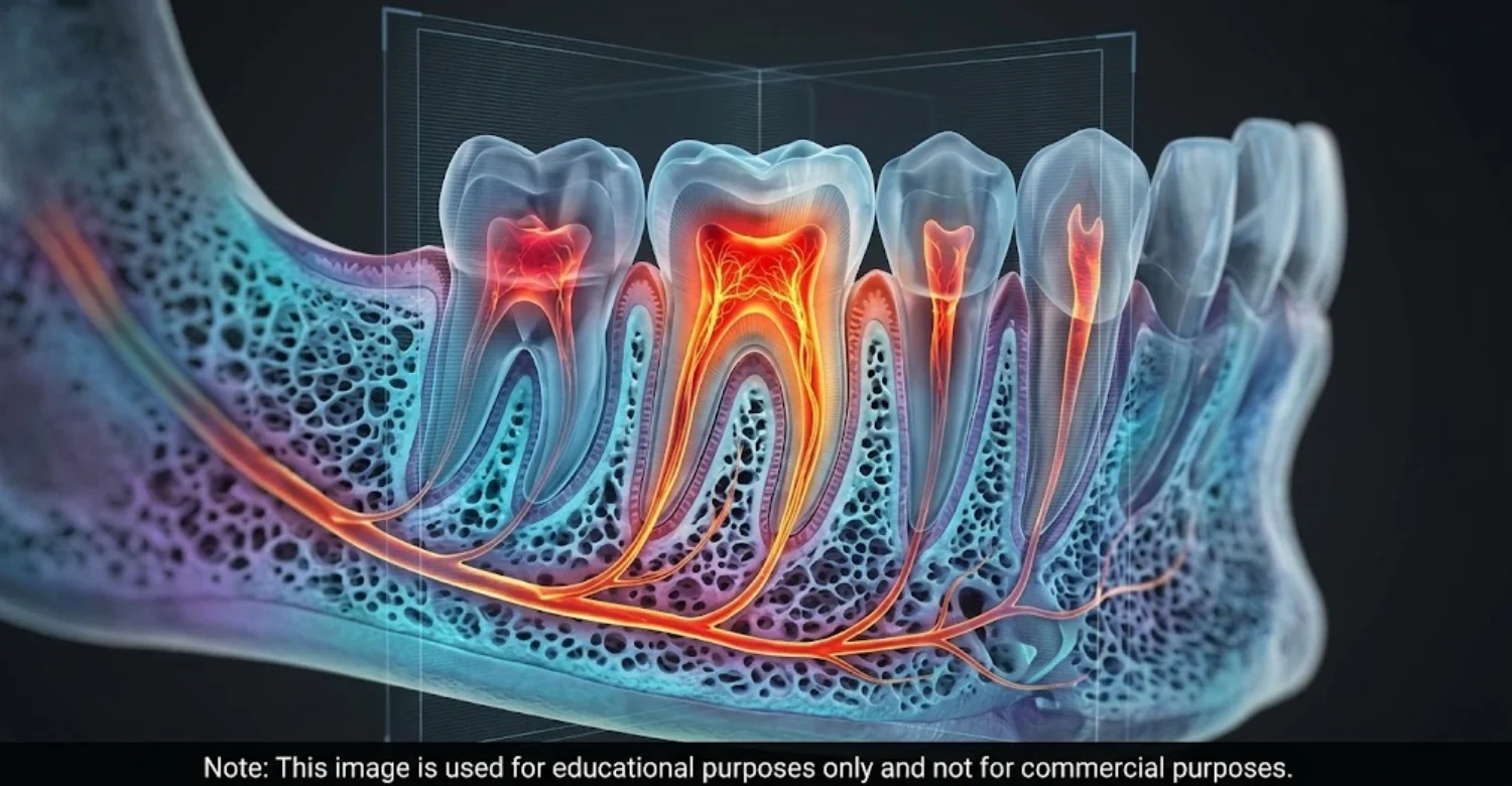
Soft tissue diagnosis is the main strength of MRI. On X-rays and CBCT, soft tissues look like similar shades of gray. MRI separates muscle, fat, fluid, and glands much more clearly.
Soft tissue inflammation and abnormal tissue changes
MRI shows fluid build-up and swelling within soft tissues. These signs often point to inflammation or infection. Fat-suppressed sequences make inflamed tissue appear brighter. This helps specialists judge how far inflammation has spread into nearby spaces.
Cysts, masses, and selected oral and maxillofacial lesions
MRI helps separate fluid-filled cysts from solid masses. Cysts usually look bright on T2 images. Solid masses show other patterns and may enhance with contrast. A biopsy is usually still needed for a final diagnosis.
Dental MRI for TMJ Disorders
Temporomandibular disorders are common. The US National Institute of Dental and Craniofacial Research estimates they affect about 5% to 12% of people. They occur more often in women. Some studies report a female-to-male ratio of about two to one in treatment-seeking patients. Most cases appear between the ages of 20 and 40.
Dental MRI for Oral and Maxillofacial Imaging is the preferred method for TMJ soft tissues. Studies report about 95% accuracy for disc position compared with surgical findings.
Displacement of the articular disc
The disc normally sits on top of the jaw condyle. In displacement, it moves forward or sideways. If it returns on opening, this is displacement with reduction. If it stays out of place, it is without reduction.
Joint effusion and inflammatory changes
Joint effusion means extra fluid inside the jaw joint. It appears bright on T2 images. Effusion is often linked to pain and active inflammation.
Degenerative changes and associated soft tissue abnormalities
Long-term TMJ problems may lead to degenerative joint disease. MRI may show disc deformity and condyle shape changes. CBCT shows bone erosion in more detail. Both scans may be combined in advanced or complex cases.
Dental MRI for Jaw Disorders
Jaw disorders involve the joint, muscles, and jawbone. MRI assesses their soft tissue parts. It also shows some changes inside the bone.
Conditions affecting the jaw joint and surrounding muscles
MRI can show inflamed or enlarged chewing muscles, such as the masseter. Muscle problems are a frequent cause of jaw pain. MRI can also rule out masses near these muscles.
Selected bone marrow abnormalities
Healthy jaw marrow contains fat and appears bright on T1 images. Infection or tumors can replace this fat with abnormal tissue. MRI detects these marrow changes early, which helps in suspected osteomyelitis.
MRI for Salivary Gland Disorders
Salivary gland disorders can cause swelling, pain, and dry mouth. MRI gives detailed images of gland tissue and nearby structures. It is often used after ultrasound when more detail is needed.
Salivary gland inflammation and structural abnormalities
MRI shows inflammation, enlargement, or scarring of the salivary glands. It may help assess conditions such as Sjögren's disease. Texture changes can suggest long-term inflammation. Sjögren's disease affects an estimated 0.1% to 0.6% of adults, mostly women.
Salivary gland masses and suspected duct-related conditions
Salivary gland tumors make up about 3% to 6% of head and neck tumors. Around 80% of parotid tumors are benign. MRI defines a mass's size, borders, and depth. MR sialography shows ducts without injected dye. This avoids the discomfort of traditional sialography, which requires cannulating the duct.
Magnetic Resonance Imaging for Oral Cancer
The American Cancer Society estimates nearly 60,000 new US oral cavity and pharynx cancers each year. Dental MRI for Oral and Maxillofacial Imaging helps show how far these tumors extend. A biopsy, not MRI, confirms cancer.
Assessing the extent of selected oral and maxillofacial tumors
MRI shows tumor size and depth of invasion. Depth of invasion is a key part of oral cancer staging. MRI measurements often match findings after surgery.
Evaluating the involvement of adjacent soft tissues
MRI shows spread into the tongue, muscles, or floor of the mouth. It can also show bone marrow involvement. Neck lymph nodes can be reviewed during the same scan.
Assessing possible nerve involvement when clinically indicated
Some oral tumors spread along nerves, called perineural spread. MRI is the best imaging method for detecting it. This finding can change the treatment plan significantly. It may widen the surgical field or the radiotherapy target.
Why MRI findings alone cannot confirm or exclude every oral cancer
Very small or surface lesions may not appear on MRI. Inflammation can also mimic tumor tissue. A persistent ulcer or lump needs examination, even after a normal scan.
Dental MRI for Trigeminal Nerve Disorders
The trigeminal nerve controls feeling in most of the face. Its disorders can cause sharp pain, burning, or numbness. MRI looks for structural causes along the nerve.
Investigating selected causes of facial pain and sensory changes
Trigeminal neuralgia affects about 4 to 13 people per 100,000 each year. It causes sudden, electric-shock-like pain. MRI looks for causes such as vessel contact, multiple sclerosis, or tumors.
Evaluating possible nerve compression or structural abnormalities
High-resolution sequences can show a blood vessel touching the nerve. This is called neurovascular compression. It may guide surgical decisions. Microvascular decompression surgery relieves pain in many carefully selected patients. Some studies report long-term pain relief in over 70% of cases.
What Are the Limitations of Dental MRI?
MRI does not show teeth and thin bones as well as X-rays or CBCT. It is also more expensive and less available.
Why MRI cannot replace every dental X-ray or CBCT scan
Enamel and dense bones contain little water, so they look dark on MRI. Cavities and small fractures are better seen on X-rays. Implant planning relies mainly on CBCT.
Why clinical examination and other diagnostic tests may still be necessary
Imaging shows structure, not the full clinical picture. Pain patterns and function must be assessed in person. Biopsies or blood tests may also be needed. Dental MRI for Oral and Maxillofacial Imaging works best as one part of a full assessment.
When Do Dentists Recommend an MRI Scan?
Dentists recommend MRI when the question involves soft tissues, discs, or nerves. It is rarely the first scan. Most patients first have an examination and, if needed, X-rays or CBCT.
Dental MRI for Oral and Maxillofacial Imaging is considered when symptoms persist or stay unexplained. It may also be needed before surgery on a lesion.
When Is an MRI Needed for Jaw Disorders?
MRI may be needed when jaw problems do not respond to first-line care. Research suggests up to 75% of TMD patients improve with simple measures. If symptoms continue, MRI can clarify the cause. It is also considered before joint injections or surgery. Surgeons need clear disc images before planning arthroscopy or open joint procedures.
When Is MRI Recommended for Persistent Jaw Pain?
Jaw pain lasting more than three months may need further investigation. MRI shows whether pain comes from the joint, disc, or muscles. It can also detect less common causes, such as masses or infection. Chronic TMD pain lasting over six months affects a smaller but significant group of patients. These patients often benefit most from detailed imaging.
When Is MRI Appropriate for Temporomandibular Joint Symptoms?
MRI is appropriate when symptoms suggest a disc problem. Signs include sudden locking, limited opening, or painful clicking. Mild painless clicking usually needs no imaging. About 30% of people without symptoms show disc displacement on MRI.
When Might MRI Be Recommended for Unexplained Facial Pain?
MRI may help when dental causes of facial pain have been ruled out. Pain may continue after a tooth is treated or removed. New numbness or tingling also suggests nerve involvement. In such cases, MRI is often preferred. Dental MRI for Oral and Maxillofacial Imaging can show nerve pathways that CBCT cannot display.
When Is MRI Used to Investigate Salivary Gland Abnormalities?
MRI is often used when ultrasound shows a salivary gland mass. It clarifies the mass's size, depth, and borders. This matters most for deep parotid masses. It may also assess chronic swelling and suspected autoimmune disease.
When Might MRI Be Part of an Oral Cancer Investigation?
MRI is often part of the workup after a biopsy confirms oral cancer. Dental MRI for Oral and Maxillofacial Imaging measures tumor depth and tissue involvement. It also shows possible nerve and lymph nodes spread. Neck lymph node status is one of the strongest predictors of oral cancer outcomes. Accurate staging helps teams choose surgery, radiotherapy, or combined treatment.
Can Dental MRI Help When Other Imaging Tests Are Inconclusive?
Yes, MRI can help when X-rays or CBCT do not explain symptoms. This is common when the problem lies in soft tissue. Dental MRI for Oral and Maxillofacial Imaging can act as a problem-solving tool. Scanning without a clear question may create confusing incidental findings.
Who Decides Whether a Dental MRI Is Necessary?
The treating dentist or specialist usually decides. Oral surgeons, oral medicine specialists, and ENT doctors may also request MRI. A radiologist may advise on the best protocol. Patients help by sharing symptoms, history, and safety concerns. Together, the team decides whether Dental MRI for Oral and Maxillofacial Imaging will add useful information.
What Symptoms May Lead to Oral and Maxillofacial MRI Imaging?
Certain symptoms may lead a dentist to consider MRI. They usually involve the jaw joint, facial soft tissues, or nerves. Most have common, non-serious causes. Persistent symptoms may lead to Dental MRI for Oral and Maxillofacial Imaging.
Jaw Pain, Stiffness, and Restricted Mouth Opening
Jaw pain and stiffness are among the most common TMD symptoms. Normal mouth opening is about 40 to 50 millimeters. Opening below 35 millimeters may suggest a joint or muscle problem. MRI can show a blocking disc or inflamed muscles.
Clicking, Locking, or Painful Movement of the Jaw
Jaw clicking is common and often harmless. Up to one-third of adults experience it at some point. Locking is different and may signal disc displacement. MRI shows disc position during these movements. A jaw that locks closed may need prompt assessment to restore normal opening.
Persistent Facial Pain, Tingling, or Numbness
Facial pain lasting weeks without a dental cause needs assessment. Tingling or numbness in the lip, chin, or cheek may point to nerve involvement. Chin numbness can rarely signal a serious condition. MRI often forms part of the investigation.
Swelling Near the Jaw or Salivary Glands
Swelling under the jaw or in front of the ear may involve the salivary glands. Swelling during meals may suggest a duct stone. A firm lump needs further assessment. Ultrasound comes first, and MRI may follow. Salivary stones cause many cases of recurrent gland swelling, especially in the submandibular gland.
Unexplained Soft Tissue Changes in the Mouth or Face
Lumps, thickened areas, or color changes in the mouth need examination. Any change lasting over two to three weeks should be checked. A biopsy often comes first. MRI then assesses the depth and extent of the lesion. Smokers and heavy drinkers face a higher risk and need closer monitoring. Tobacco and alcohol together raise oral cancer risk many times over.
Symptoms That Require Further Investigation Rather Than MRI Alone
Fever, weight loss, or general illness may need blood tests and medical review. A suspicious mouth lesion needs a biopsy. Nerve symptoms may need neurological examination. MRI is one piece of the diagnostic puzzle.
Ready to transform your smile?
Talk to our team and get a personal FREE treatment plan.
What Is the Difference Between Dental MRI, CBCT, and Dental X-Rays?
Each imaging method answers a different clinical question. X-rays and CBCT are best for teeth and bone. Dental MRI for Oral and Maxillofacial Imaging is best for soft tissues, discs, and nerves.
Feature | Dental X-Ray | CBCT | Dental MRI |
Radiation | Very low | Low to moderate | None (no ionizing radiation) |
Best for | Teeth, cavities, bone levels | 3D bone, implants, impacted teeth | TMJ disc, glands, nerves, soft tissue |
Scan time | Seconds | About 10 to 40 seconds | About 20 to 45 minutes |
Relative cost | Lowest | Moderate | Highest |
Dental MRI vs CBCT Scan: Which Is Better for Diagnosis?
Neither scan is better overall. CBCT gives detailed 3D images of bone and teeth. MRI gives detailed soft tissue images without radiation. For TMJ discs, MRI is clearly superior. For implants and impacted teeth, CBCT is preferred.
What Is the Difference Between Dental MRI and a Dental X-Ray?
A dental X-ray is quick, cheap, and available in most dental clinics. It shows cavities, bone loss, and root problems. MRI takes longer and is done at imaging centers. It shows soft tissues that X-rays cannot see.
Which Imaging Method Provides the Best Soft Tissue Detail?
Dental MRI for Oral and Maxillofacial Imaging provides the best soft tissue detail. It clearly separates muscle, fat, fluid, glands, and nerves. Ultrasound also shows salivary glands and neck nodes well. However, it cannot see the TMJ disc as clearly.
Which Imaging Method Is Better for Teeth and Jawbone Assessment?
Dental X-rays and CBCT are better for teeth and jawbone. X-rays are the first choice for cavities and gum disease. CBCT shows bone width, nerve canals, and impacted teeth in 3D.
When Is CBCT Preferred for Dental Implant Planning?
CBCT is the standard method for dental implant planning. It measures bone height, width, and nerve position in three dimensions. This helps reduce the risk of nerve injury. Nerve injury after lower jaw implant surgery is reported in roughly 1% to 13% of cases. MRI-based implant planning is still being researched.
When Is MRI Preferred for TMJ and Soft Tissue Assessment?
MRI is preferred when the disc, joint fluid, or muscles need assessment. It is also preferred for salivary gland masses and tumor staging. Nerve-related symptoms usually call for MRI as well.
Can a Patient Need Both MRI and CBCT?
Yes, some patients need both scans. In complex TMJ disorders, CBCT shows bone changes, and MRI shows the disc. For oral tumors, both may assess soft tissue and bone involvement together. Combining scans gives a more complete diagnostic picture.
Why the Best Imaging Method Depends on the Clinical Question
MRI will not find a hidden cavity. A dental X-ray will not show a displaced TMJ disc. Choosing the wrong scan can miss problems or add cost. Matching the scan to the question gives the most useful result.
How Is Dental MRI Used to Diagnose Oral and Maxillofacial Conditions?
Dental MRI for Oral and Maxillofacial Imaging follows a clear process before, during, and after the scan. Each step supports safety and accurate results. Knowing the process can reduce anxiety.
What Happens Before a Dental MRI Scan?
Preparation is essential for safety and accuracy. The team needs a clear picture of the patient's health and the clinical question.
Reviewing symptoms and medical history
The referring dentist explains the reason for the scan. The MRI team reviews allergies, kidney health, surgeries, and current medicines.
Examining previous dental images and clinical findings
Previous X-rays and CBCT scans help focus the examination. They show what is known and what needs answers. Patients should share these images when possible.
Checking implants, metal devices, and MRI safety considerations
Patients complete a safety questionnaire before the scan. It covers pacemakers, metal implants, and metal fragments. Dental implants and restorations are also recorded.
What Happens During an MRI Scan?
Patients lie on a table that slides into a tube-shaped scanner. A typical jaw or face MRI takes about 20 to 45 minutes.
Positioning the patient and obtaining diagnostic images
A coil is placed around the head or near the jaw. For TMJ scans, patients may open and close their mouth. A small bite block may hold the jaw steady.
Understanding the importance of remaining still
Movement blurs MRI images and reduces their diagnostic value. Each sequence may last a few minutes. Swallowing or jaw movement during scanning can affect results. If an image is blurred, the radiographer may repeat that sequence.
When contrast material may be considered
Contrast is a dye injected into a vein during some scans. It highlights inflammation, tumors, and blood vessels. Gadolinium agents are most common. Most TMJ disc scans need no contrast.
How Are Dental MRI Results Interpreted?
A radiologist reviews every sequence after the scan. They compare findings with the clinical question. The report includes an impression and may suggest further tests.
How Long Does It Take to Receive MRI Results?
In private settings, reports are often ready within 24 to 72 hours. Urgent cases may be reported the same day. Public systems may take longer. Patients should ask the imaging center when to expect their report.
What Happens After the MRI Report Is Available?
The dentist or specialist reviews the report and images. They compare findings with symptoms and examination results. Next steps may include treatment, further tests, or referral.
Why MRI Findings Must Be Interpreted Alongside Symptoms and Clinical Examination
MRI findings do not always match symptoms. About one-third of people without jaw symptoms show disc displacement on MRI. Treating images alone could lead to unnecessary treatment. Clinical correlation keeps findings relevant. The best results come when the dentist and radiologist share information.
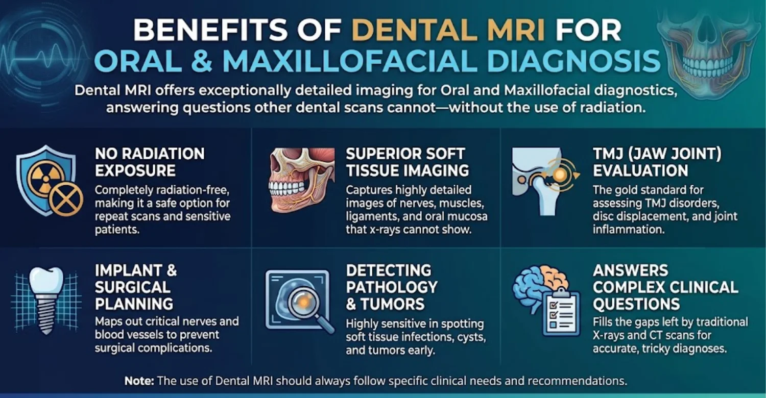
Detailed Imaging of Selected Soft Tissue Structures
MRI shows soft tissues with excellent contrast. It clearly separates muscles, fat, fluid, glands, and nerves. Surgeons can see a lesion's exact size and position before surgery.
Assessment of TMJ Disc Position and Joint Abnormalities
MRI is the only routine method that clearly shows the TMJ disc. It shows disc position in closed and open mouth positions. Studies report about 95% accuracy. This supports planning for splints, injections, or surgery.
Evaluation of Selected Salivary Gland and Facial Nerve-Related Conditions
MRI defines the depth of parotid tumors. This matters because the facial nerve runs through the parotid gland. MRI also shows nerve compression and tumor spread along nerves.
Radiation-Free Dental Imaging and Its Clinical Advantages
Dental MRI for Oral and Maxillofacial Imaging supports the ALARA principle. ALARA means keeping radiation "as low as reasonably achievable." This makes MRI suitable for repeated scans, children, and young adults.
When MRI May Provide Information That X-Rays Cannot
MRI shows disc displacement, joint effusion, and early marrow changes. These are invisible on X-rays. It can explain symptoms that stayed unclear after other imaging. This can lead to faster, more accurate diagnosis.
What Are the Limitations of MRI Compared With Other Imaging Methods?
MRI costs more, takes longer, and may involve waiting lists. Metal restorations can distort images near the teeth. It shows enamel and fine bone detail poorly. Some patients find the scanner noisy or confining.
What Are the Risks and Side Effects of Dental MRI?
MRI is a very safe imaging method for most patients. Serious complications are rare. Most risks relate to metal objects and contrast agents. Understanding these risks helps patients prepare and stay safe.
Is Dental MRI Safe for Most Patients?
Yes, Dental MRI for Oral and Maxillofacial Imaging is safe for most patients. More than 40 million MRI scans are performed in the US each year. Serious incidents are rare and usually linked to metal. Careful screening prevents most risks. Every patient is screened before entering the scanner room, even for short scans.
What Are the Common Discomforts During an MRI Scan?
Most discomforts during MRI are temporary. They relate mainly to noise, space, and staying still.
Noise, enclosed spaces, and anxiety
MRI scanners can exceed 100 decibels, so earplugs or headphones are provided. About 1% to 15% of patients experience claustrophobia during MRI. Open scanners or mild sedation may help. Research suggests about 2% of scans are stopped early because of anxiety.
Remaining still during image acquisition
Staying still for 20 to 45 minutes can be hard with jaw pain. Cushions improve comfort. Patients can speak with the team through an intercom.
What Are the Possible Side Effects of MRI Contrast Agents?
Contrast agents are generally safe. Most reactions are mild and short-lived.
Allergic-like reactions and other potential adverse effects
Mild effects include a cold feeling, headache, or nausea. Allergic-like reactions occur in roughly 0.1% to 0.2% of patients. Severe reactions occur in about 1 in 10,000 cases or fewer.
Kidney function considerations when gadolinium contrast is being considered
Severe kidney disease carries a small risk of nephrogenic systemic fibrosis. Modern agents have greatly reduced this risk. Kidney function may be checked before contrast.
Why contrast agents are not necessary for every examination
Many dental MRI scans need no contrast. TMJ disc assessment usually uses non-contrast images. Contrast is mainly used for tumors, infections, and some gland conditions.
Who Should Inform the MRI Team Before a Scan?
Some patients need extra safety checks. They should inform the team well before the appointment.
Patients with pacemakers or implanted electronic devices
Pacemakers and neurostimulators may be affected by the magnet. Many modern devices are "MR-conditional" and can be scanned under set conditions. The device must be checked first.
Patients with certain metallic implants or retained metal fragments
Some aneurysm clips and older implants may not be MRI safe. Metal fragments in the eye are a major concern. Metal workers may need an eye X-ray first.
Patients who are pregnant or may be pregnant
MRI without contrast is generally considered safe in pregnancy when needed. Non-urgent scans are often postponed. Gadolinium is usually avoided during pregnancy.
Patients with kidney disease or a history of contrast reactions
These patients should tell the team before any contrast scan. The team may change the protocol. Preventive medicine may also be considered.
Can Dental Implants Prevent a Patient From Having an MRI?
Dental implants usually do not prevent an MRI. Most are made of titanium or zirconia, which are not strongly magnetic. They may cause image artifacts nearby. Implants with magnetic denture attachments need special attention. Dental MRI for Oral and Maxillofacial Imaging can still be performed in most implant patients.
What Are the Safety Precautions for MRI With Metal Dental Restorations?
Fillings, crowns, and bridges are usually safe during MRI. Braces may distort images near the teeth. Removable dentures and retainers should be removed. Patients should report all dental work in advance.
What Can Patients Do for Relief While Waiting for a Dental MRI?
Simple self-care may ease mild discomfort while waiting. These steps support comfort but do not treat the cause.
How Can Patients Manage Mild Jaw Discomfort Safely?
Simple measures reduce strain on the jaw joint and muscles.
Choosing soft foods temporarily when chewing is uncomfortable
Soft foods reduce jaw workload. Good options include soups, yogurt, eggs, and mashed vegetables. Cutting food into small pieces also helps.
Avoiding excessive chewing, gum, and unnecessary jaw strain
Chewing gum places repeated stress on the joint. Nail or pen biting also strains the jaw. Avoid very wide mouth opening when possible.
Using a wrapped warm or cold compress when appropriate
A cold compress may ease swelling and acute pain. A warm compress may relax tight muscles. Apply either for 10 to 15 minutes, wrapped in cloth.
Which Medical Tips Can Help Reduce Jaw Strain?
Daily habits strongly affect jaw strain. Awareness of clenching is especially helpful.
Keeping the jaw relaxed and avoiding unnecessary clenching
At rest, teeth should be slightly apart. The tongue should rest gently on the palate. Many people clench during stress, so regular checks help.
Following professional advice about suitable pain relief
Over-the-counter pain relief may help some patients. Ask a dentist or pharmacist which option suits you. Health conditions and medicines affect the choice.
Avoiding unprescribed medication and unproven home remedies
Never take prescription medicines prescribed for someone else. Unproven remedies may delay proper care. Discuss new treatments with a professional first. Some herbal products can interact with prescribed medicines.
Can Jaw Exercises Help Before an MRI?
Gentle exercises may help muscle-related jaw pain. However, they are not suitable for everyone. Patients with a locked jaw should avoid them until assessed. A dentist or physiotherapist should guide any plan.
What Should Patients Avoid When the Cause of Jaw Pain Is Unknown?
Avoid forcing the jaw open when it feels locked. Avoid self-diagnosis from online sources. Irreversible treatments, such as major bite changes, should wait for a clear diagnosis.
Why Symptom Relief Does Not Replace a Diagnostic Assessment
Pain relief eases symptoms but does not treat the cause. Some serious conditions first feel like simple jaw pain. Patients should keep appointments even if symptoms improve. Symptoms that come and go can still have a significant underlying cause.
Ready to transform your smile?
Talk to our team and get a personal FREE treatment plan.
When Should Patients Seek Urgent Medical or Dental Care?
Some symptoms need urgent care rather than a routine scan. These red flags may signal serious infection or airway risk.
Which Jaw and Facial Symptoms Require Prompt Assessment?
The following warning signs need same-day or emergency assessment.
Rapidly increasing facial or jaw swelling
Swelling that grows over hours may signal spreading infection. Dental infections can reach deep spaces in the neck. They need urgent treatment. Untreated deep neck infections can become life-threatening within a short time.
Fever accompanied by facial swelling or worsening pain
Fever with facial swelling suggests significant infection. It may need antibiotics and drainage. Contact a dentist or doctor the same day.
Difficulty swallowing or breathing
This is a medical emergency. It may signal swelling that threatens the airway. Call emergency services immediately.
New or progressive facial numbness or weakness
Sudden facial weakness can signal a stroke. Seek emergency care immediately. Gradually worsening numbness also needs prompt assessment.
When Should a Persistent Oral Ulcer, Lump, or Tissue Change Be Examined?
Any ulcer, lump, or patch lasting over two to three weeks needs examination. Red or white patches are especially important. The five-year survival rate for localized oral cancer is about 85%. Early detection matters greatly. Survival drops to around 40% when cancer has spread to distant sites.
Why Patients Should Not Wait for an MRI When Red-Flag Symptoms Develop
An MRI appointment may be days or weeks away. Infections and airway problems can worsen within hours. Patients should seek urgent care even with a scan booked.
Which Symptoms May Require Emergency Care Rather Than a Routine Dental Appointment?
Breathing difficulty, rapid swelling, or sudden facial weakness need emergency care. Swelling around the eye with fever is also urgent. When in doubt, seek medical advice immediately.


.jpg)
What We Notice Clinically
Many patients arrive believing they need an MRI. In reality, most dental problems are diagnosed with examination and standard imaging. Careful assessment shows which test, if any, adds real value.
Why the Same Jaw Pain Symptoms Can Have Different Causes
Jaw pain can come from teeth, muscles, the joint, sinuses, or nerves. A cracked tooth can feel like TMJ pain. Sinus inflammation can mimic upper jaw pain. A structured examination comes first. Ear pain is another common source of confusion, since the ear sits close to the TMJ.
Common Misconceptions About Dental MRI
Several misconceptions about MRI are common among patients.
Why MRI is not automatically the best scan for every dental problem
The most advanced scan is not always the best one. MRI shows cavities and fine bone detail poorly. For many problems, X-rays or CBCT are more useful.
Why a normal MRI does not rule out every oral or dental condition
A normal MRI is reassuring but limited. Early cavities and some nerve conditions may not appear. Continued symptoms still need follow-up.
Why incidental imaging findings may require clinical interpretation
MRI often shows findings unrelated to the original question. Many are harmless, such as minor sinus thickening. A specialist decides whether action is needed.
Why Soft Tissue Assessment and Bone Assessment Require Different Imaging Approaches
X-rays and CBCT show dense structures like bone clearly. MRI shows water-rich tissues like muscles and glands. A TMJ problem may need MRI for the disc and CBCT for bone.
Why Imaging Results Must Be Matched to the Patient's Symptoms
A finding may matter greatly in one patient and not in another. Symptoms guide interpretation. This prevents over-treatment and missed diagnoses. It also helps patients understand what their results really mean.
Clinical Note: Dr. Rifat Alsaman's Perspective on Choosing the Right Dental Imaging Test
Choosing the right imaging test is a clinical decision. Each test should answer one clear question.
Dr. Rifat Alsaman, Head of the Medical Team at Vitrin Clinic and cosmetic dentist, on the importance of selecting imaging according to the clinical question
Dr. Rifat Alsaman stresses that diagnosis starts with listening to the patient. A detailed history and examination often point to the right test. Dental MRI for Oral and Maxillofacial Imaging is then chosen only when it fits.
Why an MRI referral should be based on clinical need rather than the assumption that advanced imaging is always better
An MRI without a clear question can add cost and confusion. A focused referral gives the radiologist clear goals. It also makes the results more useful for planning.
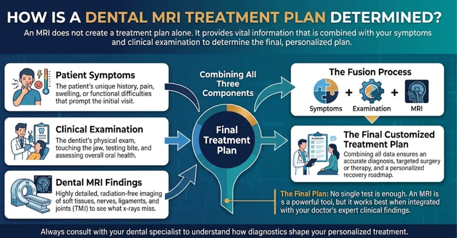
Does Every Patient With Jaw Pain Need an MRI?
No, most patients with jaw pain do not need an MRI. Many are diagnosed by examination alone. Dental MRI for Oral and Maxillofacial Imaging is reserved for persistent, severe, or unclear cases.
How Do Dentists and Specialists Decide Which Imaging Test to Request?
Dentists ask which tissue needs to be seen. Bone and teeth usually need X-rays or CBCT. Discs, glands, and nerves usually need MRI or ultrasound. Cost, safety, and availability also matter.
When Might a Dentist Refer a Patient to a Radiologist or Oral and Maxillofacial Specialist?
Referral is common for complex TMJ disorders, gland masses, or suspicious lesions. Persistent nerve symptoms may also need specialist input. Radiologists help choose and interpret advanced imaging.
How Do MRI Results Influence the Next Diagnostic or Treatment Step?
MRI results can confirm a diagnosis or reveal a new one. A disc displacement without reduction may change TMJ care. A deep parotid mass may need surgical planning.
What Happens if MRI Results Are Normal but Symptoms Continue?
A normal MRI does not mean symptoms are imagined. Many muscle and nerve pain conditions do not show on imaging. Referral to a pain specialist or neurologist may help. Dental MRI for Oral and Maxillofacial Imaging may also be repeated if symptoms change significantly.
Which Treatment Options May Be Considered After Diagnosis?
Treatment depends on the final diagnosis. It ranges from self-care to specialist surgery.
Conservative management for selected TMJ disorders
Conservative care is first-line for most TMJ disorders. It includes a soft diet, physiotherapy, splints, and stress management. Many patients improve within weeks to months. Surgery is needed in only a small minority of TMJ cases.
Condition-specific treatment for salivary gland disorders
Duct stones may be removed with minimally invasive procedures. Infections may need antibiotics and hydration. Tumors usually need surgical assessment.
Further investigation and specialist care for suspicious lesions
Suspicious lesions need a biopsy. If cancer is confirmed, a specialist team plans treatment. This may include surgery or radiotherapy.
Additional dental imaging or testing when clinically indicated
Some patients need CBCT after MRI to assess bone. Blood tests, nerve tests, or ultrasound may also be requested.
How Much Does Dental MRI Cost in the US, UK, and Turkey?
The cost of Dental MRI for Oral and Maxillofacial Imaging depends on the examination required, the facility, whether contrast is needed, and whether the radiologist's report is included. Prices below are based on published private-pay examples and are intended for budgeting, not as fixed quotes.
The term “dental MRI” does not describe one standardized scan. MRI is particularly useful for evaluating soft tissues around the temporomandibular joints (TMJs), while CBCT is commonly used to assess teeth and jawbone for specific dental indications. The appropriate examination depends on the clinical question.
What Is the Average Cost of a Dental MRI in the United States?
In the United States, published self-pay prices vary according to the examination and imaging provider. One imaging center lists a cash price of $523 for a TMJ MRI, while a New York-area pricing service lists $800 for a face, neck, or TMJ-related MRI without contrast and $1,100 with and without contrast.
TMJ MRI
$523
Published self-pay price
Cash price at MRI of Southfield
Includes the radiologist's fee
Face, orbit & neck MRI without contrast
$446–$800
Published self-pay price
Sample prices from published imaging providers
Face, orbit & neck MRI with and without contrast
$670–$1,100
Published self-pay price
Price varies by provider and protocol
These are examples from individual providers, not a nationwide average. Hospital charges may differ substantially from advertised self-pay prices. Request a written estimate that specifies the scan, contrast, and reporting fees.
What Is the Average Cost of a Dental MRI in the United Kingdom?
Private mouth and jaw MRI prices in the UK vary by provider, appointment type, and examination protocol. Published prices for dedicated TMJ imaging range from approximately $249 to $490.
TMJ MRI at other private providers
$250–$490
Published range
Other private providers
Some providers include the radiologist's report and access to digital images in the advertised price. Others may charge separately for consultations or additional services, so check the inclusions before booking.
NHS MRI scans are available when clinically indicated through the appropriate referral pathway and are generally free at the point of use. Waiting times depend on clinical priority, location, and service capacity.
What Is the Average Cost of a Dental MRI in Turkey?
In Turkey, private MRI prices depend on the facility, examination type, and whether contrast is required. Published price lists provide examples for TMJ-specific MRI, while medical travel platforms also report prices for general joint MRI examinations. These should not be treated as interchangeable tests.
General joint MRI in Istanbul
~$230
Bookimed-reported average
Average~$230
Reported range $150–$310
Not necessarily TMJ-specific
TMJ MRI at listed facilities
$330–$475
Published price
Published range $330–$475
Published price-list examples
The figures are published examples, not a confirmed national average for Dental MRI for Oral and Maxillofacial Imaging. Contact the imaging facility to confirm the exact scan, reporting arrangements, and total price.
Dental MRI Cost Comparison: US vs UK vs Turkey
The following table summarizes the published price benchmarks above. Currency conversions are not included because the underlying prices refer to different examinations and provider-specific packages.
United Kingdom
$360
Standard mouth and jaw scan
Private-pay benchmark Dedicated TMJ / mouth and jaw MRI $249–$490
Reference price $360
Check reporting and consultation inclusions
Turkey
$330
Lower end of listed TMJ range
Private-pay benchmark TMJ-specific price lists $330–$475
Reference price $330
Lower general joint MRI prices may not be the same exam
United States
$523
TMJ MRI
Private-pay benchmark TMJ and related face/neck MRI $523–$1,100
Reference price $523
Hospital charges and contrast protocols can increase costs
United States: estimated private-pay cost of a relevant MRI examination
Private-pay prices depend on the imaging provider and the examination ordered. Published examples range from $523 for a TMJ MRI to 800–1,100 for a related face, neck, or TMJ MRI in the New York area, depending on the contrast protocol.
Patients paying without insurance can ask the imaging center for a written self-pay estimate before booking. Confirm whether the quoted amount includes the radiologist's interpretation and any facility fees.
United Kingdom: estimated private-pay cost and potential NHS referral pathways
Dedicated private mouth and jaw MRI prices include published options from $249 to $380 at Vista Health, while Scan.com reports a range of $250–$490 for TMJ MRI.
An NHS scan may be available when clinically indicated through an appropriate referral. NHS England maintains a diagnostic waiting-time pledge, but actual waiting times vary and some patients wait longer than six weeks. Check current local information rather than assuming a particular appointment date.
Turkey: estimated private-pay cost at a private imaging facility
Published Turkish price lists include TMJ-specific MRI examples of 330–475. General joint MRI listings may be lower, but they do not establish the price of a comparable jaw examination.
Before booking, confirm whether the facility can perform the exact MRI protocol requested by your dentist or specialist, who will interpret the scan, and whether you will receive the report and digital images.
What Factors Affect the Cost of Oral and Maxillofacial MRI Imaging?
Understanding the main cost factors helps patients compare quotes fairly. The final fee may differ even when two facilities advertise similar starting prices.
Cost factor | How it may affect the price |
Anatomical area and scan protocol | A targeted TMJ examination may differ in price from a broader face or neck MRI |
Contrast material | Contrast may add medication, injection, and scanning costs when clinically needed |
Facility and scanner | Hospital and independent imaging-center fees can differ |
Radiologist's report | Interpretation may be included or charged separately |
Specialist consultation | A consultation to discuss the findings may add to the total |
Additional imaging | CBCT or another scan may be recommended if it answers a different clinical question |
The anatomical area and type of MRI examination
The anatomical area and imaging protocol influence the total cost. A dedicated TMJ MRI, for example, examines the jaw joints and may use sequences to assess the position and condition of the articular discs. A broader face or neck MRI examines a different area and may require a different protocol.
The scan should be selected according to the suspected condition, not simply by choosing the lowest-priced examination. MRI is useful for soft-tissue assessment, while CBCT is often more appropriate for evaluating dental and jawbone structures.
Whether contrast material is required
Some MRI examinations use contrast material to help assess particular conditions, such as suspected inflammation, infection, or a mass. Contrast is not routinely required for every TMJ MRI.
If contrast is recommended, ask whether the fee includes the contrast agent, its administration, any additional sequences, and relevant safety screening. The added cost varies by provider, so a universal percentage increase should not be assumed.
The facility, radiologist's expertise, and reporting arrangements
Hospitals and independent imaging centers may charge different fees for similar examinations. Costs can also vary depending on the scanner, availability of specialist radiology interpretation, and how quickly the report is required.
Before booking, confirm that the radiologist has appropriate experience with the requested head, neck, or maxillofacial examination. Check whether the written report, digital image access, and any expedited reporting fee are included.
Whether additional consultations or diagnostic tests are needed
An MRI fee may not cover the full diagnostic pathway. A dentist, oral and maxillofacial specialist, or other clinician may need to assess the results and recommend further care.
Additional imaging, such as CBCT, may sometimes be indicated because MRI and CBCT provide different information. These examinations should only be arranged when clinically justified. Request an itemized estimate for each test and consultation.
Does Insurance Cover Dental MRI in the United States?
Coverage depends on the reason for the MRI, the insurance policy, and whether the examination meets the insurer's medical-necessity criteria. A TMJ or facial MRI may be billed through medical insurance rather than a dental plan, but this is not universal.
Some insurers require prior authorization or documentation from a clinician. Deductibles, copayments, network restrictions, and exclusions can affect the amount you pay.
Before arranging a Dental MRI for Oral and Maxillofacial Imaging, ask your insurer whether the specific examination is covered, whether a referral or prior authorization is required, and what out-of-pocket costs apply.
Is an MRI Scan Available Through the NHS in the United Kingdom?
Yes. MRI is available through the NHS when a clinician considers it necessary and arranges the appropriate referral. Depending on the suspected condition, the referral may come through a GP, dentist, hospital specialist, or relevant clinical service.
Availability and waiting times vary by area and clinical priority. NHS England has a diagnostic waiting-time pledge, but actual waits can exceed six weeks. Urgent suspected-cancer pathways receive priority according to the applicable clinical pathway.
MRI is not necessary for every dental condition. Your clinician will decide whether MRI, dental X-rays, CBCT, or another examination is most suitable for the question being investigated.
What Should International Patients Check Before Arranging MRI in Turkey?
International patients should confirm the examination details and total fee before travelling or booking. Ask the facility to verify:
The exact anatomical area and MRI protocol requested by your clinician.
Whether contrast is required and included in the price.
Whether the radiologist's report and digital image copies are included.
Whether the report can be provided in English or another required language.
Whether the facility can share the images securely with your home dentist or specialist.
Whether a consultation, repeat scan, or additional test would incur a separate fee.
Also check the facility's credentials, the radiologist's relevant experience, and any appointment or cancellation conditions. Do not assume that a low advertised price includes every part of the diagnostic service.
Does the Cost of a Dental MRI Include the Consultation and Radiology Report?
Not always. Some imaging centers include the radiologist's report and digital image access in the scan price, while others may charge separately for consultations, additional imaging, contrast, or expedited reporting.
Service | What to confirm before booking |
MRI scan | Exact anatomical area and protocol |
Contrast, if required | Whether the agent and administration are included |
Radiologist's report | Whether interpretation and the written report are included |
Digital images | Whether you can download or share the images |
Specialist consultation | Whether discussing the results incurs an additional fee |
Additional tests | Whether another scan or examination may be charged separately |
Request a written, itemized quote before booking. Comparing the same examination and included services is the most reliable way to understand the real cost of Dental MRI for Oral and Maxillofacial Imaging.
Tips for Patients Preparing for a Dental MRI
Good preparation helps Dental MRI for Oral and Maxillofacial Imaging run safely and smoothly. It also improves image quality.
What Should Patients Tell the MRI Team Before Their Appointment?
Report all implants, devices, and metal in your body. Mention pregnancy, kidney problems, and allergies. Patients with claustrophobia should mention it early.
What Medical Records and Previous Dental Images Should Patients Bring?
Bring your referral letter and medicine list. Previous X-rays, CBCT scans, and reports are helpful. Bring device cards for pacemakers or implants.
Should Patients Remove Jewelry and Removable Dental Appliances?
Yes, remove jewelry, watches, hair clips, and hearing aids. Remove dentures, retainers, and removable orthodontic appliances. Mention any recent tattoos or permanent makeup.
Can Patients Eat and Drink Before an MRI?
Most patients can eat, drink, and take medicines normally. Sedation may require fasting for several hours. The imaging center will give specific instructions.
How Should Patients Prepare if They Have Claustrophobia?
Tell the team when booking. Some centers offer open or wide-bore scanners. Breathing exercises and music may help. Sedated patients need someone to drive them home. Closing your eyes before entering the scanner can also reduce anxiety.
What Questions Should Patients Ask About Contrast, Safety, and Results?
Ask whether contrast is needed and why. Ask about side effects and safety checks. Ask when results will be ready and who will explain them.
How Can Patients Make Sure They Understand Their MRI Report?
Ask your dentist or specialist to explain the findings. Ask what they mean for treatment. Request copies of the report and images.
At Vitrin Clinic: Choosing the Right Diagnostic Path for Your Oral Health
At Vitrin Clinic, diagnosis begins with understanding each patient's symptoms and goals. The right test matters more than the most advanced test. When MRI is needed, we help coordinate the referral.
Why the Initial Dental Assessment Matters Before Advanced Imaging
The initial assessment often reveals the cause of symptoms. It includes a detailed history and examination. It also decides whether Dental MRI for Oral and Maxillofacial Imaging is needed.
How Clinical Examination Helps Identify the Need for Additional Imaging
Examination checks jaw movement, joint sounds, muscle tenderness, and bite. It also looks for swelling and mouth changes. These findings guide the choice of imaging.
When Digital Dental Diagnostics May Be Appropriate
The right digital method depends on the clinical question.
Digital X-rays for selected tooth and dental structure assessments
Digital X-rays check cavities, roots, and bone levels. They use lower doses than film X-rays. Results appear within seconds.
CBCT for selected three-dimensional bone and implant-related assessments
CBCT provides 3D images of the jaws and teeth. It supports implant planning and impacted tooth assessment.
Referral for MRI when soft tissue or TMJ evaluation requires it
When soft tissue or TMJ assessment is needed, MRI may be recommended. It is usually performed at a specialized imaging facility.
How Personalized Treatment Planning Connects Diagnostic Findings With Clinical Needs
Each plan combines diagnostic findings with the patient's personal goals. Patients are involved in every decision. This ensures realistic and effective treatment. Patients understand the benefits and limits of each option before deciding.
Why MRI Referrals May Involve External Radiology or Specialist Services
MRI scanners are large, costly machines found in hospitals and imaging centers. Most dental clinics do not have one. External radiologists share their report with the treating dentist.
How Patients Can Discuss Their Diagnostic Options During a Consultation
Patients can ask why a test is recommended and what it will show. They can also ask about costs and alternatives. Our team explains each option clearly.






How Vitrin Clinic Supports Your Treatment Journey
Vitrin Clinic supports patients from the first consultation to follow-up. Each step keeps care clear and coordinated.
Step 1: Understanding the Patient's Symptoms and Treatment Goals
We listen to the patient's symptoms, their duration, and triggers. We also discuss treatment goals.
Step 2: Reviewing Existing Dental Records and Relevant Diagnostic Findings
We review existing X-rays, scans, and records. This avoids repeating tests unnecessarily.
Step 3: Identifying Whether Further Imaging or Specialist Assessment May Be Needed
We decide whether X-rays, CBCT, or Dental MRI for Oral and Maxillofacial Imaging is needed. If so, we explain why and help arrange it.
Step 4: Coordinating the Personalized Treatment Plan
Once the diagnosis is clear, we create a personalized plan. It considers the patient's health, goals, and timeline.
Step 5: Explaining Treatment Options, Expected Benefits, and Limitations
We explain each option in clear language. Patients learn the benefits, risks, and limitations.
Step 6: Organizing Relevant Follow-Up and Aftercare
We provide aftercare instructions and review appointments. International patients can receive remote follow-up support.
What International Patients Should Know About Diagnostic Coordination Before Traveling
Share existing dental images before traveling so our team can review them. If MRI is needed, ask where it will be done. Confirm report language and image copies in advance.
Ready to transform your smile?
Talk to our team and get a personal FREE treatment plan.
Need Expert Guidance on Dental MRI at Vitrin Clinic?
Do you have jaw pain, facial swelling, or unexplained oral symptoms? Our team starts with a careful clinical assessment. We then recommend from our treatments the most suitable diagnostic path, including Dental MRI for Oral and Maxillofacial Imaging when needed. Contact Vitrin Clinic today to book your FREE treatment.
Reference
FAQs
Dr. Rifat Alsaman has more than 5 years of clinical experience in dentistry and currently serves as the Head of the Medical Team at Vitrin Clinic. He is dedicated to providing exceptional patient care, overseeing treatment planning, and ensuring the highest clinical standards across the team. His expertise, attention to detail, and commitment to continuous professional development have helped countless patients achieve healthier, more confident smiles.


