General Dentistry
September 2, 2026
Gum Anatomy: What Are the Different Parts of the Gums?
 Dr. Rifat Alsaman
The gums are not one uniform tissue. They are several distinct zones working together: the free gingiva that forms a loose collar around each tooth, the attached gingiva bonded firmly to the underlying bone, the junctional epithelium that fuses directly to the tooth surface, and the interdental gingiva filling the space between teeth. Understanding gum anatomy helps explain why healthy gums are essential for supporting teeth, protecting tooth roots, and maintaining long-term oral health.
What Is Gum Anatomy?
Gum anatomy refers to the structured layers and zones of tissue that surround and support each tooth. It includes the free gingiva, attached gingiva, junctional epithelium, and interdental gingiva. Understanding what makes up gum anatomy starts with recognizing these distinct, purpose-built regions rather than treating the gums as a single uniform tissue.
Understanding the Basic Anatomy of Gums
The basic anatomy of gums includes a movable outer collar around the tooth, a firm inner cuff bonded to bone, and a molecular attachment point that fuses gum tissue to the tooth itself. Each zone has a distinct texture, width, and function. Together they form the protective barrier surrounding every tooth. This layered design is central to how the different parts of the gums work as a whole.
Why Gum Tissue Is Important for Oral Health
Gum tissue seals around each tooth. This prevents bacteria from reaching the underlying bone and root surface. Healthy gum tissue also anchors the tooth's supporting structures in place. It acts as a frontline defense against infection, inflammation, and eventual tooth loss.
What Are the Main Parts of the Gums?
The main parts of the gums include the free gingiva, attached gingiva, interdental gingiva, and the gingival sulcus. Each plays a distinct role in protecting and supporting the teeth. Together, these parts of gum anatomy form a continuous, functional barrier around every tooth in the mouth.
Free Gingiva
The free gingiva is the movable, unattached portion of gum tissue. It forms the visible collar around the neck of each tooth, typically measuring 1 to 2 millimeters wide. Free gingiva is continuous with the attached gingiva but separated from it by a shallow groove.
Where Is the Free Gingiva Located?
Free gingiva sits directly around the visible base of each tooth. It forms the gum margin you see when smiling. It sits just above the gingival sulcus and remains unattached to the tooth surface. This allows slight movement without disrupting the tighter attachment found further down.
Attached Gingiva
Attached gingiva is the firm, dense tissue bound tightly to the underlying alveolar bone. It shows a stippled, orange-peel texture when healthy. Its width varies significantly between people, ranging from about 1 millimeter to over 10 millimeters. It provides resistance against the mechanical forces of chewing.
How Does Attached Gingiva Support the Teeth?
Attached gingiva anchors firmly to the underlying bone. This creates a stable cuff that resists the friction and pressure generated during chewing and brushing. This tight bond helps prevent the gum margin from being pushed away or damaged during normal daily function.
Interdental Gingiva
Interdental gingiva fills the triangular space between adjacent teeth. It typically forms a pyramid shape in a healthy mouth. Between the buccal and lingual peaks of that pyramid sits a shallow depression called the gingival col. This area is often the first site to show signs of inflammation.
What Is the Role of Gum Tissue Between the Teeth?
Gum tissue between the teeth is known as the interdental papilla. It fills the space beneath the contact point of neighboring teeth, preventing food from packing into the gap. This tissue also supports the appearance of a healthy smile by filling what would otherwise be visible black triangles.
Gingival Sulcus
The gingival sulcus is the shallow groove formed where the free gingiva meets the tooth surface. It typically measures 1 to 3 millimeters deep in a healthy mouth. This space shouldn't bleed when gently probed. It is lined by a distinct, non-keratinized layer of sulcular epithelium.
Why Is the Gingival Sulcus Important?
The gingival sulcus is one of the first things dentists check during a periodontal exam. A sulcus deeper than the typical healthy range often signals early gum disease. This small groove is a key diagnostic marker of overall gum health.
What Is the Difference Between Free Gingiva and Attached Gingiva?
The difference between free gingiva and attached gingiva lies mainly in mobility and attachment. Free gingiva is unattached and moves slightly. Attached gingiva is firmly bonded to the bone beneath it. This distinction is central to understanding gum anatomy: what are the different parts of the gums overall.
Free Gingiva vs Attached Gingiva
Free gingiva sits above the attachment line and moves independently. Attached gingiva below it stays firmly fixed to the underlying bone. The two are separated by a shallow groove rather than a visible boundary.
How Can You Identify the Different Gum Areas?
You can identify the different gum areas by looking for the mucogingival junction. This scalloped line separates the firm, stippled attached gingiva from the smoother, looser alveolar mucosa beyond it. The free gingiva sits closest to the tooth, just above this attachment boundary.
How Does Gum Anatomy Connect With Tooth Anatomy?
Gum anatomy connects with tooth anatomy through the junctional epithelium. This is the only part of the gum structurally fused directly to the tooth surface. Combined with the surrounding periodontal ligament and alveolar bone, it forms the complete support system that keeps every tooth stable and protected.
Understanding the Anatomy of Teeth and Gums
The anatomy of teeth and gums works together as one integrated system. Gum tissue seals around the tooth, while underlying bone and ligament provide structural support. Gum and tooth anatomy cannot really be separated, since each tissue layer depends on the others for overall function.
How Do Gums Protect the Roots of Teeth?
Gums protect the roots of teeth by covering and sealing the area where the root meets the crown. This prevents bacteria and food debris from reaching sensitive root surfaces. Healthy attached gingiva shields the root from friction and pressure during normal chewing.
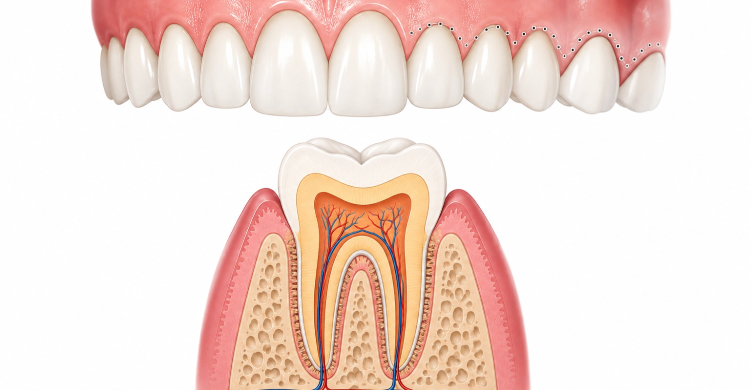
What Is the Anatomy of the Mouth and Gums?
The anatomy of the mouth and gums includes not just the gum tissue itself, but its relationship to the alveolar mucosa, palate, and surrounding oral structures. Gum anatomy varies slightly by location. The palate, for example, shows continuously keratinized tissue rather than a distinct mucogingival boundary.
Understanding Gum Anatomy in the Mouth
Gum anatomy changes subtly from one region to another. The palate lacks the usual mucogingival junction found elsewhere, because its tissue is uniformly keratinized. Understanding these regional differences helps explain why gum appearance and texture can vary across different areas of the mouth.
How Do the Gums Connect to Other Oral Structures?
The gums connect to other oral structures through the alveolar mucosa, periosteum, and underlying bone. Together they form a continuous soft-tissue lining throughout the mouth. This connectivity allows changes in one area, such as inflammation, to sometimes influence the health and appearance of neighboring tissue as well.
What Does Gum Tissue Anatomy Look Like?
In a healthy mouth, gum tissue typically appears pink, firm, and slightly stippled over the attached gingiva. The free gingival margin has a smoother, more mobile texture. This visual and structural pattern helps dentists quickly assess whether gum tissue appears healthy or inflamed.
Layers and Structure of Gum Tissue
Gum tissue has an outer epithelial layer and an underlying connective tissue layer that anchors to bone. It also includes the junctional epithelium at the base of the sulcus. This layer physically attaches the gum to the tooth surface itself.
What Is Healthy Gum Tissue Made Of?
Healthy gum tissue is made of keratinized epithelium over the attached gingiva, non-keratinized epithelium lining the sulcus, and dense underlying connective tissue rich in collagen fibers. This combination gives it a firm texture, while still allowing flexibility around the more mobile free gingiva.
Clinical Note
Understanding gum tissue anatomy helps patients recognize subtle changes in texture, color, or firmness that may signal early gum disease. Because gum anatomy involves several distinct zones, changes often begin in just one area before becoming more widespread.
What Does a Gum Anatomy Diagram Show?
A gum anatomy diagram typically shows the free gingiva, attached gingiva, gingival sulcus, interdental papilla, and mucogingival junction, all in relation to the tooth and underlying bone. Reviewing a gum anatomy diagram makes it easier to visualize how these separate zones work together as one system.
Understanding a Gum Anatomy Diagram
Understanding a gum anatomy diagram means identifying where each labeled zone sits relative to the tooth. This runs from the visible gum margin down to the base of the sulcus and the crest of bone beneath it. This visual reference makes the abstract anatomy far easier to follow.
Which Gum Structures Should You Identify?
On a diagram, you should be able to identify the free gingival margin, gingival sulcus, junctional epithelium, attached gingiva, mucogingival junction, and interdental papilla. Recognizing each of these labeled areas builds a clearer picture of how healthy gum anatomy is organized around every tooth.
How Do Healthy Gums Support Your Teeth?
Healthy gums support your teeth by sealing tightly around each tooth, anchoring firmly to underlying bone, and resisting the mechanical stress of daily chewing. This combined support protects both the visible crown and the hidden root structure from bacteria, trauma, and ongoing wear.
The Role of Gums in Tooth Stability
The role of gums in tooth stability centers on the attached gingiva's firm bond to bone. This bond helps resist shifting and movement during chewing and speech. Alongside the periodontal ligament, healthy gum tissue keeps each tooth securely positioned within its socket over the long term.
How Do Gums Protect the Tooth Roots?
Gums cover the area where enamel ends and root surface begins. This area is far more vulnerable to decay and sensitivity than the enamel-covered crown. When gum tissue recedes, exposed roots become significantly more susceptible to both decay and temperature sensitivity.
Tips for Patients
Brush gently along the gumline twice daily and floss once a day to keep gum tissue healthy. Schedule regular dental checkups so subtle changes can be caught early. Avoid tobacco products, which are strongly linked to gum recession and progressive periodontal disease.
What Happens When Gum Anatomy Is Affected by Gum Disease?
When gum disease affects gum anatomy, the normally tight seal around each tooth begins to break down. This allows bacteria to reach deeper tissue and bone. Left untreated, this progression can permanently alter the structure of the gums in ways that are hard to reverse.
How Gum Disease Changes Gum Tissue
Gum disease triggers inflammation that causes swelling, bleeding, and eventual loss of the stippled texture typical of healthy attached gingiva. As inflammation progresses, the sulcus often deepens into a periodontal pocket. This creates more space for bacteria to accumulate and cause further damage.
Can Gum Disease Affect Tooth Support?
Gum disease can significantly affect tooth support by breaking down the attachment between gum tissue and the tooth, along with the underlying bone that anchors the root. As this support weakens, teeth may become loose, shift position, or eventually require extraction if the disease isn't treated.
Clinical Note
Early changes in gum tissue, such as slight bleeding during brushing, often signal the start of gum disease before more serious symptoms appear. Recognizing these early signs and seeking prompt evaluation gives patients the best chance of protecting their gum anatomy and supporting bone.
What Causes Changes in Gum Anatomy?
Changes in gum anatomy are most often caused by gum disease, hormonal shifts, certain medications, or mechanical trauma from aggressive brushing. Gum tissue is relatively thin and responsive. It tends to show visible changes earlier than many other structures in the mouth when something goes wrong.
Gum Recession
Gum recession occurs when the gum margin pulls away from the tooth, exposing more of the root surface than is typical in a healthy mouth. Global data suggests gingival recession of at least 1 millimeter affects roughly 81% of people. This makes it an extremely common finding overall.
Why Do Gums Pull Away From Teeth?
Gums pull away from teeth due to a combination of factors. These include periodontal disease, overly aggressive brushing, teeth grinding, and naturally thin gum tissue in some individuals. Genetics also plays a role, since some people are simply predisposed to thinner, more recession-prone gum tissue.
Gum Enlargement
Gum enlargement refers to gum tissue that becomes swollen or overgrown, sometimes covering more of the tooth surface than usual. This can result from chronic inflammation, certain medications, hormonal changes during pregnancy, or, less commonly, underlying systemic conditions that affect tissue growth throughout the body.
What Can Cause Swollen or Enlarged Gums?
Swollen or enlarged gums can be caused by plaque-related inflammation, certain blood pressure or seizure medications, hormonal fluctuations, or poor oral hygiene that lets bacteria accumulate along the gumline. Identifying the underlying cause helps determine whether treatment should focus on hygiene, medication review, or both.
Periodontal Disease
Periodontal disease is a progressive infection that damages both gum tissue and the underlying bone supporting the teeth. It often begins as mild gingivitis. Left untreated, this disease can destroy significant portions of the attachment apparatus, fundamentally altering the healthy structure of the gums.
How Can Periodontal Disease Affect Gum Structure?
Periodontal disease destroys the connective tissue and bone that normally anchor teeth in place. It deepens the sulcus into pockets that harbor more bacteria. Over time, this structural breakdown leads to recession, tooth mobility, and, in advanced cases, eventual tooth loss.
How Can You Keep Your Gum Tissue Healthy?
You can keep your gum tissue healthy through consistent daily hygiene, regular professional cleanings, and prompt attention to any changes in color, texture, or bleeding. These combined habits protect the delicate balance of gum anatomy and help prevent the progression of gum disease over time.
Daily Oral Hygiene for Healthy Gums
Daily oral hygiene for healthy gums includes gentle brushing along the gumline, flossing between every tooth, and using an antimicrobial rinse if recommended by your dentist. Consistency matters more than intensity, since overly aggressive brushing can actually contribute to gum recession over time.
Which Habits Help Protect Gum Anatomy?
Habits that help protect gum anatomy include using a soft-bristled toothbrush, flossing daily, avoiding tobacco products, and attending regular dental checkups for professional cleaning. Managing conditions like diabetes and reducing stress-related teeth grinding also support healthier gum tissue over the long term.
Tips for Patients
Use gentle, circular brushing motions rather than aggressive back-and-forth scrubbing to protect delicate gum tissue. Clean between teeth daily with floss or an interdental brush. Avoid tobacco entirely, and keep up with regular dental checkups so any early changes can be caught quickly.
Can Cosmetic Dentistry Affect Gum Appearance?
Cosmetic dentistry can significantly affect gum appearance, since gum contour and symmetry play a major role in how balanced and attractive a smile looks overall. Procedures that reshape or adjust gum tissue are often planned alongside restorative or cosmetic dental work for a more harmonious result.
The Importance of Gum Contours in Smile Aesthetics
Gum contours matter for smile aesthetics because uneven or excessive gum tissue can make teeth appear shorter, smaller, or asymmetrical, even when the teeth themselves are perfectly healthy. Adjusting gum shape can dramatically improve how balanced and proportional a smile appears, without changing the teeth at all.
Why Is Gum Symmetry Important for a Balanced Smile?
Uneven gum lines draw attention away from the teeth and can make an otherwise healthy smile look irregular. Achieving symmetrical gum contours helps create the visual harmony most people associate with an attractive, natural-looking smile.
When Should You See a Dentist About Changes in Your Gums?
You should see a dentist about changes in your gums whenever you notice bleeding, persistent swelling, recession, color changes, ongoing bad breath, loose teeth, or shifts in the gumline. These signs often indicate early gum disease that responds far better to prompt rather than delayed treatment.
Warning Signs You Should Not Ignore
Warning signs you should not ignore include gums that bleed during brushing or flossing, persistent swelling, visible recession, unusual color changes, and loose or shifting teeth. Any combination of these symptoms warrants a dental evaluation rather than waiting to see if the issue resolves on its own.
When Do Bleeding or Receding Gums Need Professional Evaluation?
Bleeding or receding gums need professional evaluation as soon as they appear consistently rather than occasionally. Persistent symptoms usually indicate an active underlying problem. Watch especially for persistent swelling, changes in gum color, persistent bad breath, loose teeth, or noticeable shifts in the gumline.
Gum Care and Cosmetic Dentistry at Vitrin Clinic
Vitrin Clinic provides gum evaluation and treatment planning for patients considering cosmetic dental work, since gum health and shape directly influence overall smile results. The clinical team assesses gum anatomy carefully before recommending any restorative or cosmetic procedure, to ensure a stable, healthy foundation.
Understanding Gum Health Before Cosmetic Dental Treatment
Understanding gum health before cosmetic dental treatment allows Vitrin Clinic to identify any underlying issues, such as recession or inflammation, that could affect the outcome of restorative work. Addressing these concerns first helps ensure cosmetic results remain stable and attractive over the long term.
Why Is Gum Evaluation Important Before Improving a Smile?
Uneven or unhealthy gum tissue can undermine even the most carefully planned cosmetic work. Assessing gum anatomy first allows the treatment plan to address both function and appearance together, rather than focusing on teeth alone.
How Vitrin Clinic Considers Gum Anatomy During Smile Planning
Vitrin Clinic considers gum anatomy during smile planning by evaluating gum symmetry, contour, and overall tissue health alongside tooth shape and alignment. This comprehensive approach reflects why gum anatomy matters just as much as tooth appearance in cosmetic planning.
How Can Healthy Gum Contours Improve Smile Aesthetics?
Healthy gum contours create a balanced, symmetrical frame around the teeth that enhances their natural shape and proportion. Even minor adjustments to gum tissue can noticeably improve how harmonious and polished a smile appears overall.
What We Notice Clinically
At Vitrin Clinic, the clinical team notes that gum anatomy is an essential consideration when planning cosmetic and restorative dental treatments. Healthy, well-proportioned gum tissue supports not only the function of the teeth, but also the overall symmetry and appearance of the finished smile.
Clinical Note
At Vitrin Clinic, evaluating gum shape, symmetry, and the condition of the gumline helps create a more accurate and personalized treatment plan. This attention to gum anatomy supports both healthier gums and more predictable cosmetic outcomes.
Tips for Patients
Maintain consistent oral hygiene, avoid tobacco, and attend regular dental checkups to protect your gum tissue over time. If you're considering cosmetic dental treatment, ask your dentist to evaluate your gum anatomy first, since healthy gum contours are an essential part of a balanced, lasting smile.
Resource:
Understanding gum anatomy and the different parts of the gums is essential for maintaining healthy teeth and supporting tissues. Healthy gums play an important role in protecting the oral environment and preventing inflammation and periodontal problems. As highlighted in a study published on PMC, understanding periodontal tissues and their functions can help support better oral health and improve awareness of gum-related conditions.
https://pmc.ncbi.nlm.nih.gov/articles/PMC8370901/
https://pubmed.ncbi.nlm.nih.gov/28551728/
https://www.sciencedirect.com/topics/immunology-and-microbiology/gingiva
FAQs
Dr. Rifat Alsaman has more than 5 years of clinical experience in dentistry and currently serves as the Head of the Medical Team at Vitrin Clinic. He is dedicated to providing exceptional patient care, overseeing treatment planning, and ensuring the highest clinical standards across the team. His expertise, attention to detail, and commitment to continuous professional development have helped countless patients achieve healthier, more confident smiles.

.webp&w=3840&q=75)


