Dental Crowns
September 23, 2026
What Is Microdontia? Causes, Types, Treatment, and Cost
 Dr. Rifat Alsaman
Microdontia is a developmental dental anomaly involving teeth that are smaller than expected. It can affect one tooth, several teeth, or an entire dentition. The condition may influence tooth proportions, spacing, appearance, and sometimes oral function. Maxillary lateral incisors are commonly associated with localized cases. Peg-shaped lateral incisors are particularly recognized in orthodontic and cosmetic dentistry. A meta-analysis estimated their pooled prevalence at 1.8%. Diagnosis requires clinical examination and appropriate imaging. Treatment depends on tooth size, shape, position, bite, and remaining tooth structure. Options can include composite bonding, veneers, crowns, orthodontics, or combined treatment. Vitrin Clinic uses digital assessment and personalized planning to evaluate suitable restorative and orthodontic approaches.
What Is Microdontia?
Microdontia describes teeth that are smaller than normal for the patient's dentition. The condition can involve tooth width, length, or overall proportions. Some cases affect one tooth, while others involve multiple teeth. The clinical significance varies considerably between patients. Some people experience mainly cosmetic concerns. Others may develop spacing or alignment problems. Diagnosis should distinguish true small teeth from teeth that only appear small. Jaw dimensions and neighboring tooth proportions can influence appearance. A dentist may use measurements, photographs, and radiographs during assessment. Treatment is not automatically necessary. Conservative options can often improve tooth proportions when treatment is desired.
Microdontia Definition and Meaning
The term describes an abnormal reduction in tooth size. Dental professionals assess tooth dimensions against expected anatomical proportions. The condition can involve crowns, roots, or overall tooth development. Localized cases commonly involve specific permanent teeth. Generalized cases can affect multiple teeth across both arches. The clinical diagnosis should consider age and dentition stage. Small-looking teeth are not always developmentally undersized. Relative proportions between teeth and jaws can create a similar appearance. Accurate diagnosis therefore requires more than visual inspection. Treatment decisions depend on the cause and clinical presentation.
What Do Microdontia Teeth Look Like?
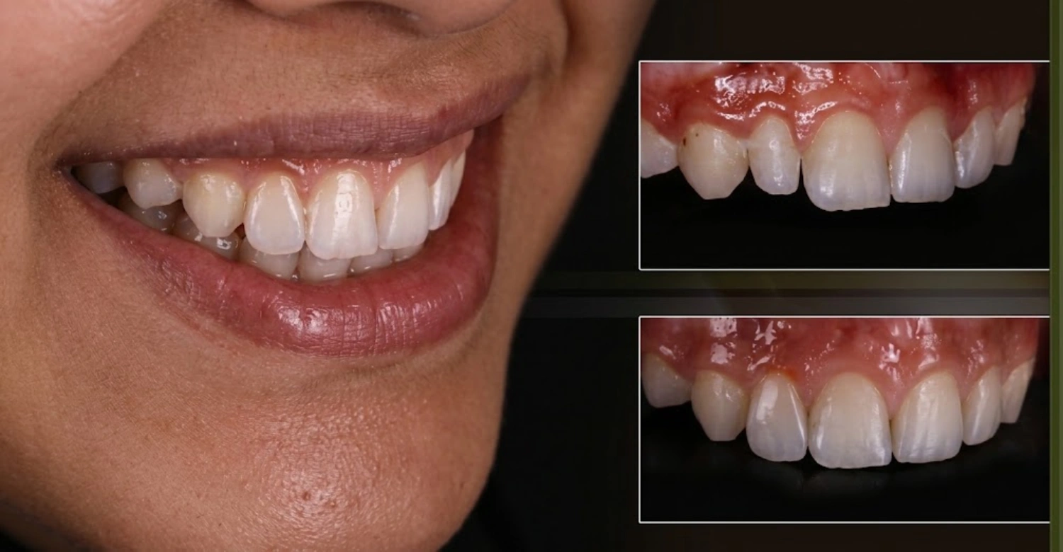
Affected teeth may appear noticeably shorter or narrower than neighboring teeth. Their shape can also look tapered, conical, or peg-like. Maxillary lateral incisors may show a particularly recognizable peg-shaped appearance. Small crowns can create visible spaces between adjacent teeth. The effect becomes more noticeable when neighboring teeth have typical dimensions. Tooth color and enamel quality may remain normal. However, some developmental dental anomalies can occur alongside altered tooth size. Photographs can help document appearance before treatment. A dentist should confirm the diagnosis before cosmetic reshaping begins.
Microdontia Clinical Features
Clinical features vary according to the affected tooth and severity. Common findings include reduced crown dimensions and altered tooth proportions. Some affected teeth have unusual shapes or tapered contours. Spacing can develop when tooth width is insufficient for the dental arch. Occlusal relationships may also change when multiple teeth are involved. Localized cases may cause few functional problems. Generalized cases can produce more noticeable changes. Clinical examination should assess tooth size, position, contacts, and occlusion. Radiographs can provide additional information about roots and surrounding structures.
Microdontia Images and Common Tooth Shapes
Clinical images can help patients understand how developmental tooth-size differences appear. Common appearances include tapered lateral incisors and unusually narrow crowns. Peg-shaped lateral incisors are one recognizable presentation. Images should not replace professional diagnosis. Tooth shape can vary substantially between individuals. Dentists compare affected teeth with neighboring teeth and expected anatomical proportions. Before-and-after photographs can also demonstrate possible restorative changes. At Vitrin Clinic, digital photographs can support treatment planning and smile analysis. The goal is to assess proportion rather than simply enlarge a tooth.
Explore angular stomatitis and cheilosis
What Causes Microdontia?
Causes can involve genetic, developmental, and environmental influences. In many localized cases, a single definitive cause cannot be identified. Tooth development involves complex interactions between genes and developmental signaling. Variations affecting these processes may influence tooth size and shape. Developmental conditions can also involve multiple dental anomalies. Environmental influences may affect tooth formation during development. However, individual causation should not be assumed without appropriate assessment. Family history can provide useful context. A dentist may recommend additional evaluation when multiple abnormalities occur. Treatment focuses on the clinical presentation rather than simply identifying a suspected cause.
Microdontia Causes and Etiology
Etiology can be multifactorial rather than linked to one factor. Genetic influences are important in tooth development and size. Developmental disturbances can alter crown morphology and dimensions. Some syndromic conditions include smaller teeth among their dental findings. Localized anomalies may occur without an identifiable systemic condition. Family patterns can sometimes suggest inherited susceptibility. However, having an affected relative does not prove a specific genetic cause. Dental history and clinical findings should guide evaluation. When several dental abnormalities occur together, broader assessment may be appropriate.
Is Microdontia Genetic or Hereditary?
Genetic factors can contribute to differences in tooth size. Dental development involves numerous genes and signaling pathways. Family patterns have been reported for several dental developmental anomalies. However, inheritance is not always straightforward. A patient can have isolated small teeth without a known family history. Conversely, similar dental findings can occur among relatives. Genetic syndromes may also include altered tooth size as one feature. A dentist should therefore consider the complete clinical picture. Genetic evaluation is not routinely required for every isolated case.
What Causes Small Teeth?
Small teeth can result from developmental variation or localized dental anomalies. Genetic influences may affect tooth formation and dimensions. Some systemic or developmental conditions can also alter dental morphology. Another possibility is relative smallness caused by larger jaw dimensions. A tooth may therefore look small without being truly undersized. Tooth position can further change its apparent proportions. Accurate diagnosis should distinguish anatomical size from visual perception. Dental measurements and radiographic assessment can help clarify the situation. Treatment should address the underlying dental relationship rather than appearance alone.
Developmental Factors That Affect Tooth Size
Tooth size develops through coordinated biological processes during dental formation. Disturbances during development can influence crown dimensions and morphology. Genetic signaling contributes to the number, shape, and size of teeth. Environmental factors may also influence developing dental tissues. The timing and severity of developmental disruption can affect the final appearance. Different teeth may respond differently to developmental influences. This explains why localized abnormalities can occur. A complete dental examination helps determine whether the finding is isolated. It also helps identify associated abnormalities requiring further assessment.
Find out more about dental trauma and possible complications
What Are the Types of Microdontia?
Dental literature commonly describes localized and generalized patterns. Localized cases affect one tooth or a limited group of teeth. Generalized cases involve multiple teeth across the dentition. Generalized presentations can be classified further as true or relative. True generalized cases involve genuinely small teeth. Relative generalized cases involve normal-sized teeth appearing small compared with large jaws. Some descriptions also use terms such as partial or isolated cases. Classification helps dentists understand the distribution and likely treatment needs. The treatment approach depends more on individual anatomy than terminology alone.
Localized Microdontia
Localized disease affects one tooth or a small number of teeth. Maxillary lateral incisors are a common clinical example. Affected teeth may appear tapered or peg-shaped. The remaining teeth can have normal dimensions. Localized cases are often mainly aesthetic concerns. However, spacing and alignment can also develop. Orthodontic assessment may be useful when significant gaps are present. Restorative treatment can increase the visible tooth dimensions. Conservative approaches are generally considered when adequate enamel and tooth structure remain.
Generalized Microdontia
Generalized cases involve multiple teeth across the dentition. They can occur with developmental or genetic conditions. The appearance may involve reduced tooth dimensions throughout both arches. Diagnosis should distinguish true generalized findings from relative proportions. A comprehensive examination is important when multiple teeth appear unusually small. Radiographs can help assess tooth development and associated abnormalities. Treatment often requires coordinated restorative and orthodontic planning. The number of affected teeth can significantly influence treatment complexity and cost.
True Generalized Microdontia
True generalized disease means multiple teeth are genuinely smaller than expected. This pattern can occur with certain developmental conditions. The entire dentition may show reduced dimensions. Diagnosis requires comparison with appropriate dental norms. A dentist should also consider the patient's age and dentition stage. Additional clinical findings can help identify associated conditions. Treatment may require several restorative procedures. Orthodontic planning can sometimes improve spacing and tooth relationships. Management should remain individualized rather than based only on the classification.
Relative Generalized Microdontia
Relative generalized disease describes teeth that appear small because the jaws are comparatively large. The teeth themselves may have dimensions within normal ranges. The apparent discrepancy results from tooth-to-jaw proportions. This distinction is important before restorative treatment begins. Enlarging every tooth may not always be appropriate. Orthodontic assessment can help evaluate spacing and occlusion. Digital smile analysis can demonstrate proportion changes before treatment. Conservative treatment should address the actual anatomical relationship.
Partial and Isolated Microdontia
Partial or isolated presentations involve limited tooth groups. The terms may be used differently across dental literature. A single peg-shaped lateral incisor is a typical localized presentation. Such cases often have a straightforward aesthetic component. Nevertheless, associated missing teeth or developmental anomalies should be considered. Radiographs can help identify unerupted or absent teeth. Treatment may involve bonding, veneers, orthodontics, or a combination. Conservative planning can preserve healthy dental tissue.
Compare dental implants vs. bridges
Which Teeth Are Commonly Affected by Microdontia?
The condition can affect different teeth, but some teeth are recognized more frequently. Maxillary lateral incisors are especially important in localized cases. Third molars can also demonstrate reduced dimensions. Other teeth may occasionally be involved. The distribution differs between individuals and populations. Tooth size should always be considered alongside shape and position. A small tooth may also coexist with missing neighboring teeth. This combination can significantly affect orthodontic planning. Current systematic-review evidence supports individualized management when a small lateral incisor accompanies missing tooth development.
Microdontia of the Lateral Incisor
Small maxillary lateral incisors are among the most recognizable presentations. They may have a narrow crown and tapered shape. Peg-shaped lateral incisors are particularly common examples. The altered width can create visible spaces beside the tooth. Treatment may involve direct composite, ceramic veneers, or orthodontics. Space management is important before restorative enlargement. The appropriate option depends on enamel, bite, tooth position, and aesthetic goals. A 2013 meta-analysis found a pooled prevalence of 1.8% for peg-shaped maxillary permanent lateral incisors.
Microdontia of the Molar
Molar involvement can occur but is less visually obvious. Small molars may affect chewing relationships when substantially reduced. Their shape and occlusal anatomy should be examined carefully. A dentist should also evaluate caries risk and existing restorations. Treatment depends on tooth strength and functional requirements. Crowns may be considered when significant structural restoration is necessary. Conservative restorations may be suitable for minor size differences. Radiographs can help evaluate roots and surrounding bone.
Microdontia of the Third Molar
Third molars can show considerable variation in size and shape. Smaller third molars may not create an obvious cosmetic concern. Their clinical significance depends on eruption, position, and associated pathology. A dentist may evaluate them during routine radiographic examination. Removal is not automatically required because of reduced size. Treatment decisions depend on symptoms, pathology, and functional considerations. Radiographic findings can guide management when third molars are impacted.
Microdontia in Baby Teeth
Developmental tooth-size differences can occur in primary dentition. Small primary teeth may affect spacing and tooth proportions. Their significance depends on the number of affected teeth. Dentists should consider normal developmental variation in young children. A child's dentition changes substantially during growth. Treatment is therefore carefully timed. Preventive care remains important regardless of tooth size. Monitoring can help determine whether intervention is necessary.
Microdontia in Adult Teeth
Adult teeth with reduced dimensions can create persistent aesthetic concerns. Gaps may remain after the eruption is complete. Small lateral incisors are particularly noticeable within the smile. Treatment can often be planned once growth and tooth development are appropriate. Orthodontics may precede restorative enlargement. Veneers or composite can modify visible proportions. Crowns are generally considered when greater structural coverage is clinically justified. Treatment should preserve healthy tooth structure whenever possible.
Discover the relationship between oral health and overall well-being
What Conditions Are Associated With Microdontia?
Small teeth can occur as isolated findings or alongside other dental abnormalities. Some developmental and genetic conditions include altered tooth size. Missing teeth can also occur alongside small lateral incisors. This relationship is particularly important during orthodontic planning. A comprehensive examination helps identify whether the finding is isolated. The presence of one dental anomaly does not automatically indicate a systemic disorder. Diagnosis should rely on documented clinical findings. Broader evaluation may be considered when multiple abnormalities appear.
Microdontia and Hypodontia
Small teeth and missing teeth can occur together. This association is particularly relevant with maxillary lateral incisors. A patient may have one missing lateral incisor and a small opposite lateral incisor. Such cases can require multidisciplinary orthodontic and restorative planning. A recent systematic review evaluated 35 studies involving this clinical situation. It found that both orthodontic space opening and closure can be viable approaches. The best approach depends on occlusion, aesthetics, periodontal factors, and individual anatomy.
Microdontia and Down Syndrome
Altered tooth size and shape can occur among the dental findings associated with Down syndrome. Patients may also experience delayed eruption and other dental differences. Dental care should therefore consider the broader clinical context. Treatment planning should be individualized and coordinated appropriately. A diagnosis of small teeth alone does not indicate Down syndrome. Additional clinical features would be necessary for any broader diagnosis. Dental professionals should avoid drawing systemic conclusions from tooth size alone.
Microdontia and Cleidocranial Dysplasia
Cleidocranial dysplasia can involve several dental developmental abnormalities. Delayed eruption and numerous unerupted teeth are characteristic dental concerns. Tooth-size changes may occur alongside these findings. Management can require prolonged multidisciplinary dental care. Radiographic evaluation is particularly important. Treatment planning may involve oral surgery and orthodontics. A small tooth by itself does not establish this diagnosis.
Microdontia and Other Developmental Conditions
Several developmental conditions can involve altered tooth size. Dental anomalies may include changes in number, shape, eruption, and morphology. The combination of findings is often more informative than one isolated feature. A dentist may recommend additional assessment when several anomalies occur. Radiographs can help identify unerupted or missing teeth. Treatment should address functional and aesthetic needs separately. A coordinated plan can reduce unnecessary procedures.
Is Microdontia Associated With Autism?
There is no basis for diagnosing autism from tooth size alone. Dental findings can vary among individuals with autism for many reasons. Oral hygiene, sensory preferences, diet, and access to dental care may influence oral health. A small tooth is not a diagnostic marker for autism. Dental professionals should therefore avoid linking isolated tooth-size findings with autism. Treatment should focus on the patient's actual dental needs.
Learn more about food allergies and oral health
What Is False or Relative Microdontia?
False or relative smallness occurs when teeth appear smaller than expected. The teeth may actually have normal dimensions. Large jaw dimensions can create increased spaces around normal-sized teeth. Neighboring tooth proportions can also alter visual perception. Dentists can distinguish these situations through measurements and clinical examination. Treatment should not begin with cosmetic enlargement before confirming the diagnosis. Digital planning can help demonstrate the effect of different tooth proportions.
Relative Microdontia vs True Microdontia
True small teeth have reduced anatomical dimensions. Relative smallness results from surrounding proportions. The distinction matters because treatment can differ substantially. True small teeth may benefit from restorative enlargement. Relative cases may require orthodontic or comprehensive aesthetic planning. Tooth measurements can help establish the difference. A dentist also evaluates jaw size and neighboring teeth. The diagnosis should reflect the entire dental relationship.
How Jaw Size Can Make Teeth Appear Smaller
Large jaws can create greater spacing between normally sized teeth. This increased space can make individual teeth look disproportionately small. The issue is therefore one of proportion rather than tooth development. Orthodontic examination can identify this relationship. Digital smile analysis can further demonstrate tooth-to-arch proportions. Restorative enlargement may not be necessary for every relative case. Treatment should be based on objective findings.
How Dentists Identify False Microdontia
Dentists compare tooth dimensions with expected anatomical proportions. They also assess neighboring teeth and arch dimensions. Clinical photographs can provide useful visual documentation. Radiographs can help evaluate tooth development and morphology. Digital scanning can provide detailed three-dimensional measurements. The findings are interpreted alongside age and dentition stage. This comprehensive approach reduces the risk of unnecessary cosmetic treatment.
Microdontia vs Macrodontia: What Is the Difference?
Microdontia and macrodontia describe opposite tooth-size abnormalities. One involves unusually small teeth. The other involves unusually large teeth. Both can affect tooth proportions and dental alignment. Diagnosis requires assessment against expected tooth dimensions. Localized and generalized patterns can occur with either condition. Treatment depends on the functional and aesthetic effects. Conservative planning is important for both conditions. A dentist should identify the actual anatomical difference before treatment.
Microdontia and Macrodontia
Macrodontia involves teeth that are larger than expected. The condition can affect individual teeth or several teeth. Small teeth represent the opposite size variation. Both conditions can influence spacing and occlusion. Their appearance may also affect smile proportions. Treatment approaches differ because the biological problems are opposite. Restorative enlargement is relevant to small teeth. Reduction or reshaping may sometimes be considered for large teeth.
Differences in Tooth Size and Appearance
Small teeth often create spaces between neighboring teeth. Large teeth can reduce available space or cause crowding. Shape can also differ from normal anatomy. The visual effect depends on tooth position and neighboring proportions. Photographs can help identify obvious differences. Dental measurements provide more reliable diagnostic information. Treatment should address both size and function.
Microdontia and Macrodontia Drawing
A simple dental diagram can illustrate the difference between small and large teeth. A small lateral incisor can be compared with a normal lateral incisor. A large tooth can then demonstrate the opposite relationship. Such diagrams are educational rather than diagnostic. Clinical photographs and radiographs provide more useful patient-specific information. Vitrin Clinic can use digital smile planning to visualize proposed restorative changes.
Explore the connection between thyroid eye disease and oral health
Is Microdontia Bad for Your Teeth?
Small teeth are not automatically unhealthy or dangerous. Many patients have no pain or functional symptoms. The main concern may be appearance or spacing. However, abnormal proportions can sometimes contribute to alignment problems. Associated developmental anomalies can also require assessment. Treatment is therefore based on clinical need rather than the diagnosis alone. Regular dental care remains important. A dentist can monitor changes and determine whether intervention is appropriate.
Does Microdontia Affect Oral Function?
Function is usually determined by the overall dental relationship. One small tooth may have little effect on chewing. Multiple affected teeth can influence occlusal contacts. Spacing may also affect how neighboring teeth contact each other. Severe developmental differences can require orthodontic assessment. The presence of symptoms should guide further evaluation. Treatment aims to improve function when function is actually affected.
Can Microdontia Cause Gaps Between Teeth?
Reduced tooth width can create visible spaces. This is especially noticeable with small lateral incisors. Gaps can become more apparent when neighboring teeth have normal dimensions. Orthodontic treatment can reposition teeth and redistribute available space. Restorative treatment can increase the visible width of selected teeth. Combined treatment may sometimes provide better proportions. The appropriate sequence depends on individual anatomy.
Does Microdontia Cause Loose Teeth?
Small tooth size alone does not usually cause tooth mobility. Loose teeth can result from periodontal disease, trauma, infection, or other conditions. Mobility should therefore be assessed separately. A dentist can examine periodontal tissues and supporting bones. Radiographs may be needed when mobility has an unclear cause. Patients should not assume mobility results from tooth size.
When Microdontia Is Mainly a Cosmetic Concern
A localized small tooth may cause primarily aesthetic concerns. This is particularly common with visible front teeth. Patients may notice uneven proportions or spaces while smiling. Composite bonding can sometimes provide a conservative cosmetic change. Veneers may be considered when greater shape modification is appropriate. Orthodontics can address associated spacing. Treatment should remain proportional to the actual concern.
Discover more about polyarteritis nodosa and oral health
How Is Microdontia Diagnosed?
Diagnosis combines visual assessment, tooth measurements, and appropriate imaging. Dentists evaluate crown dimensions, tooth shape, position, and surrounding structures. They also assess occlusion and spacing. Radiographs can identify developmental abnormalities that are not visible clinically. Digital scanning can provide detailed measurements. Diagnosis should also consider whether the finding is localized or generalized. Children require age-appropriate interpretation because their dentition is developing. Adults can generally receive more definitive restorative planning.
Dental Examination and Tooth Measurements
A dental examination begins with visual inspection and clinical measurements. The dentist compares affected teeth with neighboring teeth. Tooth width and length can be evaluated. Proportions are considered rather than size alone. Photographs can document the initial appearance. Occlusal relationships should also be assessed. This information helps determine whether treatment is necessary. Measurements become especially useful before cosmetic restoration.
Microdontia Radiograph and Dental X-Rays
Radiographs can reveal root development and associated dental abnormalities. They may identify missing, unerupted, or malformed teeth. Imaging can be especially important when multiple anomalies are suspected. However, radiographs should be selected according to clinical indication. Routine imaging should not be performed without justification. The dentist determines which imaging method provides useful diagnostic information. Findings are interpreted alongside the clinical examination.
Digital Dental Imaging
Digital imaging can improve visualization during treatment planning. Intraoral scanning can capture detailed dental surfaces. Digital photographs can support smile analysis. Digital X-rays can provide information about roots and surrounding structures. These technologies do not replace clinical diagnosis. They complement examination and patient history. At Vitrin Clinic, digital assessment can support personalized restorative and orthodontic planning.
Diagnosing Microdontia in Children and Adults
Diagnosis differs according to developmental stage. Children have teeth that are still erupting and changing. Dentists must distinguish normal variation from developmental abnormalities. Adults generally have a more stable dentition. Treatment can therefore be planned around established tooth dimensions. Orthodontic and restorative needs can still vary significantly. Age should always be considered alongside tooth development.
What Is the Treatment for Microdontia?
Treatment depends on severity, location, tooth structure, spacing, and occlusion. Not every small tooth requires intervention. Composite bonding can add material conservatively. Veneers can alter visible tooth shape when appropriate. Crowns provide more extensive coverage but require greater tooth preparation. Orthodontics can manage spacing and tooth position. Some patients benefit from combined orthodontic and restorative treatment. Recent evidence supports both direct composite and indirect ceramic approaches for reshaping small or peg-shaped lateral incisors.
Does Microdontia Always Need Treatment?
No. Treatment is not automatically required. Some small teeth function normally and create minimal aesthetic concern. Monitoring may be appropriate when there are no functional problems. Treatment becomes more relevant when spacing, appearance, or occlusion causes concern. A dentist should discuss benefits and limitations. Conservative options should generally be considered first when suitable. The final decision depends on the individual clinical presentation.
Composite Bonding for Microdontia
Composite bonding adds tooth-colored resin to increase visible tooth dimensions. It can reshape small or tapered crowns. The technique is often conservative because material can be added directly. It may require less tooth preparation than veneers. Results depend on bonding quality, occlusion, hygiene, and maintenance. Composite can also be repaired or modified more easily. It may be suitable for younger patients in selected cases.
Veneers for Microdontia
Veneers can change the visible width, length, and shape of selected teeth. They are commonly considered for aesthetic front-tooth concerns. Ceramic veneers provide a laboratory-made restoration with precise morphology. Tooth preparation varies according to the material and clinical situation. Healthy enamel should be preserved whenever clinically possible. The final design should consider facial proportions and neighboring teeth. A dentist should assess bite forces before treatment.
Crowns for Microdontia
Crowns cover more of the tooth than veneers. They may be considered when substantial structural restoration is needed. A healthy small tooth may not require full coverage. Crown preparation removes more tooth structure than additive bonding. The decision should therefore be based on clinical necessity. Material selection also affects appearance, strength, and cost. At Vitrin Clinic, restorative planning considers tooth structure and the intended treatment outcome.
Orthodontic Treatment for Microdontia
Orthodontics can manage spacing and tooth position. It may be used before restorative enlargement. Space can be redistributed to create appropriate proportions. Orthodontics alone does not increase the anatomical size of a tooth. Restorative treatment may therefore follow orthodontic alignment. The sequence depends on the patient's bite and dental anatomy. A coordinated plan can reduce unnecessary restorative preparation.
Can Braces Fix Microdontia?
Braces cannot physically enlarge a small tooth. They can improve alignment and manage surrounding spaces. This may create a better foundation for bonding or veneers. In some cases, orthodontics alone may provide an acceptable result. The decision depends on tooth size and spacing. Clear aligners can also be considered for selected cases. Treatment planning should focus on both position and proportion.
How to Fix Microdontia in Adults
Adult treatment can combine orthodontic and restorative approaches. Composite bonding is one conservative option. Veneers can provide more durable ceramic reshaping in suitable cases. Crowns may be appropriate when structural damage requires full coverage. Orthodontics can correct spacing before restoration. Digital planning can help visualize tooth proportions before treatment. Vitrin Clinic can assess the available options through a personalized consultation.
Explore the science behind ferroptosis in periodontitis
How Is Lateral Incisor Microdontia Treated?
Small lateral incisors often require careful aesthetic planning. Their position strongly affects smile symmetry. The dentist evaluates space, tooth shape, gum levels, and neighboring teeth. Orthodontics may be used to create or redistribute space. Composite or ceramic restoration can then modify tooth proportions. The treatment sequence should protect healthy tooth structure. Recent systematic-review evidence supports individualized treatment rather than one universal approach.
Treatment for Peg-Shaped Lateral Incisors
Peg-shaped lateral incisors can often be reshaped with restorative treatment. Composite resin may provide a conservative approach. Ceramic veneers can provide an alternative for selected adults. Orthodontics may be needed when spacing is significant. The dentist should evaluate the bite before increasing tooth width. Restorative contours should match adjacent teeth naturally. The final design should consider smile symmetry and facial proportions.
Creating Space Before Restorative Treatment
Space management can be important before restoring a small lateral incisor. Insufficient space may produce an overly wide restoration. Orthodontics can reposition neighboring teeth when necessary. The amount of required space depends on the planned restoration. Digital planning can help estimate the intended tooth dimensions. This approach can improve predictability. The final plan should preserve periodontal health and stable contacts.
Composite vs Veneers for Small Lateral Incisors
Composite is generally more conservative and easily repairable. Veneers can offer laboratory-controlled shape and ceramic aesthetics. Composite may require less preparation. Ceramic restorations may involve greater preparation depending on the case. Recent evidence found comparable effectiveness between direct composite and indirect ceramic reshaping in relevant lateral-incisor cases. Material choice should therefore consider tooth structure, bite, aesthetics, maintenance, and patient preference.
Dr. Rifat Alsaman's Clinical Perspective on Microdontia Treatment
Dr. Rifat Alsaman, Head of the Medical Team at Vitrin Clinic and cosmetic dentist, emphasizes individualized planning. Tooth size should be evaluated alongside spacing, occlusion, and surrounding structures. Enlarging a tooth without considering these factors can create unnatural proportions. Conservative treatment should be considered when adequate tooth structure remains. Orthodontic preparation may improve the restorative result in selected cases. Digital planning can help patients understand proposed changes before treatment.
Microdontia Treatment in Children and Adults
Age strongly influences treatment timing. Children may still have developing teeth and changing occlusion. Permanent restorative treatment may therefore be delayed in selected cases. Monitoring can allow the dentist to evaluate eruption and growth. Adolescents may require orthodontic space management. Adults generally have stable tooth positions. Restorative options can therefore be planned more definitively. Treatment should always consider the patient's stage of dental development.
Microdontia Treatment for Children
Children may require monitoring rather than immediate cosmetic treatment. Dentists consider tooth development, eruption, and future orthodontic needs. Preventive care remains important for all affected teeth. Composite restoration may be considered in selected cases. Extensive permanent restorations may be postponed when appropriate. Parents should receive clear information about expected development. Treatment should avoid unnecessary removal of healthy dental tissue.
Microdontia Treatment During Mixed Dentition
Mixed dentition includes both primary and permanent teeth. Tooth positions change considerably during this period. Orthodontic space analysis can therefore be particularly important. Small permanent teeth may become more noticeable after an eruption. Treatment timing should account for remaining growth and eruption. Temporary restorative measures may sometimes be appropriate. A dentist can monitor the developing dentition before definitive treatment.
Microdontia Treatment in Adults
Adults can consider restorative and orthodontic treatment according to clinical needs. Composite bonding may be suitable for minor size differences. Veneers can reshape visible front teeth. Crowns are generally reserved for teeth requiring broader coverage. Orthodontics can manage spacing and alignment. Combined treatment can address both position and tooth proportions. Digital smile planning can help compare possible approaches.
Why Age and Tooth Development Matter
Developing teeth require different treatment considerations from fully mature teeth. Tooth eruption and jaw growth can change the final result. Permanent restorations may therefore require careful timing. Orthodontic treatment can also change available space. A dentist should consider growth, root development, and occlusion. Age alone does not determine treatment eligibility.
Microdontia Before and After
Before-and-after results depend on the treatment selected. Restorations can change visible tooth width, length, and shape. Orthodontics can improve spacing and alignment. Combined treatment can produce more comprehensive changes. Results depend on existing tooth position and facial proportions. Photographs should be taken under consistent conditions. Digital simulations can demonstrate potential changes before treatment. Actual outcomes may differ from simulations.
What Can Microdontia Treatment Change?
Treatment can modify tooth shape and visible dimensions. Composite and veneers can increase apparent width or length. Orthodontics can change spacing and alignment. Crowns can provide broader structural coverage when indicated. Treatment cannot change the underlying biological development of the original tooth. The objective is to create appropriate function and proportions. Patients should understand what each treatment can realistically achieve.
Improving Tooth Size and Proportion
Increasing tooth size is not always the only objective. Proportion between teeth can be more important than absolute dimensions. The dentist considers neighboring teeth and smile symmetry. Tooth width-to-length relationships also influence appearance. Digital planning can help visualize these relationships. The final restoration should blend with adjacent teeth naturally.
Why Results Differ Between Patients
Results depend on tooth structure, spacing, bite, and treatment selection. Different materials also produce different aesthetic characteristics. Oral hygiene and maintenance affect restoration longevity. Orthodontic movement can change treatment timing. Patients may also have different expectations. Personalized planning helps account for these variables. No single before-and-after result represents every case.
Note : All images used are for editorial and illustrative purposes only and may not originate from the original news provider or associated company.
What Are the Possible Complications of Microdontia?
The condition itself does not necessarily create complications. Problems may develop when tooth proportions affect spacing or occlusion. Associated developmental anomalies can also complicate treatment. Restorative procedures introduce their own considerations. Orthodontic treatment may require additional planning. Diagnosis should identify existing risks before treatment begins. Regular monitoring can help detect changes early.
Tooth Gaps and Alignment Problems
Reduced tooth width can create visible spaces. Neighboring teeth may move into available space over time. Alignment can therefore change gradually. Orthodontic assessment can determine whether movement is needed. Restorative enlargement can close selected spaces. Treatment sequencing is important when both approaches are used. Stable contacts should be established before final restoration.
Bite and Occlusion Considerations
Tooth enlargement can change occlusal contacts. The dentist should evaluate the bite before adding restorative material. Excessive restoration width can interfere with normal contacts. Orthodontics may be required when tooth position contributes to the problem. Occlusion should be checked after restoration. Proper adjustment can reduce unnecessary functional stress.
Possible Functional and Cosmetic Concerns
Cosmetic concerns commonly involve visible spacing or unusual tooth proportions. Functional concerns are more dependent on the number and location of affected teeth. Multiple small teeth may influence chewing contacts. Individual front teeth often have primarily aesthetic implications. Treatment should address the patient's actual concern. Aesthetic treatment should not compromise function.
Microdontia and Other Dental Anomalies
Small teeth can occur alongside missing or malformed teeth. This combination can influence orthodontic treatment planning. Radiographs may reveal abnormalities that are not visible clinically. A complete dental assessment can identify associated findings. The presence of multiple anomalies may justify broader evaluation. Treatment should be coordinated when several dental problems coexist.
Are There Side Effects or Risks From Microdontia Treatment?
Every dental treatment has potential limitations. Conservative procedures generally preserve more natural tooth structure. Restorations can require maintenance or replacement over time. Veneers may chip or debond. Crowns require greater tooth preparation. Orthodontic treatment can cause temporary discomfort and requires good hygiene. The dentist should explain risks before treatment. Long-term maintenance should be included in treatment planning.
Risks of Composite Bonding
Composites can chip or wear under heavy biting forces. Staining may occur depending on material and dietary habits. Repairs are often possible when minor damage occurs. Proper bonding technique influences durability. Bruxism can increase mechanical stress. Regular dental reviews help monitor the restoration. Patients should follow recommended maintenance instructions.
Risks and Limitations of Veneers
Veneers can chip, fracture, or debond. Tooth preparation can permanently alter enamel when preparation is required. Existing bite problems can increase mechanical stress. Good oral hygiene remains important around veneer margins. Veneers also require maintenance and eventual replacement. The dentist should determine whether veneers are appropriate before preparation. Conservative alternatives should be considered when suitable.
Risks of Dental Crowns
Crowns require greater tooth reduction than additive bonding. This makes case selection particularly important. A healthy tooth may not need full coverage. Crown margins also require careful hygiene. Chipping or loosening can occur over time. The underlying tooth remains susceptible to decay if oral hygiene is poor. Regular dental care supports long-term restoration health.
Orthodontic Treatment Considerations
Orthodontic treatment can improve spacing and alignment. It requires patient cooperation and regular appointments. Poor hygiene can increase the risk of enamel demineralization. Retention is also necessary after active treatment. Treatment duration varies according to the complexity of tooth movement. The orthodontic plan should coordinate with any planned restoration.
Clinical Note: Preserving Healthy Tooth Structure
Healthy enamel should be preserved whenever clinically possible. Adding composite may require less preparation than full-coverage restorations. Veneers may also provide a conservative option in suitable cases. Crowns should generally have a clear structural indication. The treatment choice should reflect biological and functional considerations. Aesthetic goals should not override long-term dental health.
What We Notice Clinically
Patients often focus on tooth size without considering surrounding proportions. A small lateral incisor can look more noticeable when neighboring teeth are broad. Spacing can also make the tooth appear smaller. Some patients assume orthodontics alone can increase tooth size. Others assume veneers are always necessary. Careful diagnosis often reveals several possible treatment pathways. Digital planning can help compare these options. Vitrin Clinic evaluates tooth proportions before recommending restorative treatment.
Common Patient Patterns With Microdontia
Localized small lateral incisors are a common clinical presentation. Patients may primarily notice gaps or uneven smile proportions. Some patients have no functional symptoms. Others require orthodontic space management. Associated missing teeth can complicate treatment. A full examination helps identify these differences. Treatment is then planned according to the complete dental relationship.
Common Misconceptions About Small Teeth
Small teeth are not necessarily unhealthy. They also do not automatically require crowns or veneers. Braces can move teeth but cannot enlarge them. Cosmetic treatment should not begin before diagnosis. A small tooth can sometimes be managed conservatively. The appropriate treatment depends on anatomy and patient goals. Professional assessment prevents unnecessary intervention.
Why Tooth Proportions Matter Before Treatment
Tooth size should be considered relative to neighboring teeth. Enlarging one tooth excessively can create an unnatural appearance. Smile width, tooth length, and gingival levels also matter. Occlusion must be considered before restorative enlargement. Digital planning can help assess these relationships. Proportion-based planning supports more natural results.
Clinical Note: Dr. Rifat Alsaman, Head of the Medical Team at Vitrin Clinic and Cosmetic Dentist
Dr. Rifat Alsaman emphasizes that treatment should begin with diagnosis rather than restoration selection. Small teeth require evaluation of space, bite, enamel, and surrounding structures. A conservative option may be appropriate when a healthy structure is available. Orthodontic preparation can improve restorative proportions in selected cases. Digital assessment also helps visualize potential changes before treatment. The treatment plan should balance aesthetics, function, and long-term maintenance.
How Much Does Microdontia Treatment Cost?
Treatment cost depends on the number of teeth and selected procedure. Composite bonding generally costs less than ceramic veneers or crowns. Orthodontic treatment has separate costs because treatment duration varies. Countries and clinics also influence pricing. Published 2026 market guides show substantial international variation. For example, one current Turkey guide lists composite bonding around $100–$250 per tooth. The same source lists porcelain or E-max veneers around $200–$400 per tooth. These are market indications rather than Vitrin Clinic quotations. Patients should request an individualized treatment plan before comparing prices.
What Determines Microdontia Treatment Cost?
Several factors influence the final price. The number of affected teeth is one major factor. Material selection also affects the total. Composite bonding usually requires less laboratory work. Ceramic veneers involve laboratory fabrication and additional clinical steps. Crowns require more extensive preparation and restoration. Orthodontic costs depend on treatment complexity and duration. Imaging and diagnostic appointments may also affect the final fee.
Average Microdontia Treatment Cost in the United States
Published 2026 cosmetic dentistry guides show wide U.S. price ranges. Composite bonding can range around $100–$500 per tooth. Porcelain or E-max veneers can range around $900–$2,500 per tooth. Orthodontic treatment varies substantially according to case complexity. These figures should be considered indicative private-market ranges. Insurance may not cover treatment performed primarily for cosmetic reasons. Patients should request a complete written estimate.
Average Microdontia Treatment Cost in the United Kingdom
UK private costs vary between clinics and treatment types. Current published market guidance lists composite bonding around $200–$500 per tooth. Porcelain or E-max veneers can range around $900–$1,500 per tooth. Orthodontic treatment costs vary according to appliance and complexity. Diagnostic fees may also be separate. Patients should confirm exactly what each quotation includes.
Average Microdontia Treatment Cost in Turkey
Current 2026 market guides show lower published ranges in Turkey. Composite bonding is commonly listed around $100–$250 per tooth. Porcelain or E-max veneers are listed around $200–$400 per tooth. A current Vitrin Clinic pricing page lists composite bonding at approximately $80–$120 per tooth. It lists E-max veneers around $180–$280 per tooth. Actual treatment costs depend on individual assessment and treatment design.
Microdontia Cost by Treatment Type
Treatment | United States | United Kingdom | Turkey |
Composite bonding | $100–$500/tooth | $200–$500/tooth | $100–$250/tooth |
Porcelain/E-max veneer | $900–$2,500/tooth | $900–$1,500/tooth | $200–$400/tooth |
Crown | $1,000–$3,500/tooth* | $550–$1,500/tooth* | $200–$500/tooth* |
Orthodontic treatment | $3,500–$7,500+ | $3,000–$5,500+ | $1,800–$4,000+* |
*Indicative ranges vary by material, provider, complexity, and treatment plan. Current market sources show substantial differences between clinics.
Composite Bonding Cost
Composite bonding generally has the lowest per-tooth cost among these restorative options. Current Turkey market guidance lists approximately $100–$250 per tooth. The final amount depends on the number of teeth and complexity. Minor reshaping may require less material and chair time. More extensive additions can increase the fee. Patients should confirm whether polishing, adjustments, and follow-up are included.
Veneer Cost
Ceramic veneer prices vary significantly between countries and materials. Current published guides list approximately $200–$400 per tooth in Turkey. UK figures can reach $900–$1,500 per tooth. U.S. figures can reach $900–$2,500 per tooth. Material selection and laboratory quality affect the final price. Patients should compare the complete treatment rather than the veneer alone.
Crown Cost
Crown prices vary according to material and clinical complexity. Zirconia and E-max crowns can have different laboratory costs. Current Turkish market guides show ceramic crown prices from approximately $200 upward. Crowns may cost more when additional treatment is required. A crown should be recommended because of clinical need, not simply tooth size.
Orthodontic Treatment Cost
Orthodontic treatment costs depend heavily on complexity and treatment duration. Clear aligners and fixed braces can have different pricing structures. Current market guidance places clear aligner treatment around $1,800–$4,000 in Turkey. UK examples can range around $3,000–$5,500. U.S. prices can reach approximately $3,500–$7,500 or more. These figures are broad market estimates.
At Vitrin Clinic
Vitrin Clinic approaches Dental Crowns through digital assessment and individualized treatment planning. The focus is on evaluating tooth size, shape, proportions, and surrounding structures before crown treatment. Digital diagnostics can support accurate assessment and help guide the design of the final restoration. Dental crowns may be considered for teeth affected by microdontia when additional coverage is clinically appropriate. The treatment plan is tailored to the patient's dental condition, aesthetic goals, and overall smile proportions. International patients can receive coordinated consultations and personalized treatment planning at Vitrin Clinic. A personalized quotation is provided after clinical assessment rather than relying on generic online prices.
Digital Assessment of Tooth Size and Proportions
Digital assessment can help evaluate tooth width, length, and symmetry. Intraoral scanning captures detailed dental surfaces. Digital photographs can support smile analysis. These records help compare affected teeth with neighboring structures. The information can support restorative design and orthodontic planning. Digital planning does not replace clinical examination. It provides additional information for treatment decisions.
Digital X-Rays and Clinical Diagnostics
Digital X-rays can provide information about roots and surrounding structures. Imaging may help identify associated dental abnormalities. The dentist selects imaging according to clinical indications. Additional diagnostic records may be required for complex cases. Combining clinical and digital findings improves treatment planning. Vitrin Clinic uses digital diagnostics as part of personalized dental assessment.
Personalized Microdontia Treatment Planning
Treatment planning should consider tooth size, space, bite, enamel, and aesthetics. One patient may benefit from composite bonding. Another may require orthodontics before ceramic restoration. Some patients may need no treatment at all. Digital planning can help compare possible approaches. The final recommendation should follow clinical assessment. Patient preferences should also be discussed.
Choosing the Appropriate Restorative or Orthodontic Approach
Treatment selection depends on the underlying dental relationship. Bonding can be appropriate for conservative reshaping. Veneers may suit selected adult aesthetic cases. Crowns can address teeth requiring broader structural coverage. Orthodontics can manage spacing and alignment. Combined approaches can address both tooth position and size. The safest option depends on individual anatomy.
How Vitrin Clinic Supports Your Treatment Journey
Vitrin Clinic supports international patients through coordinated dental planning. The process can begin with digital records and an initial consultation. The dental team evaluates the patient's needs before recommending treatment. Digital planning can help organize restorative and orthodontic procedures. Treatment coordination helps patients understand the expected sequence. Follow-up and aftercare remain part of the treatment journey. International patients can also receive support with appointment coordination and logistics.
Initial Consultation and Assessment
The consultation establishes the patient's dental concerns and treatment objectives. Clinical examination evaluates tooth structure and surrounding tissues. Photographs and digital records may support assessment. Additional imaging can be requested when clinically indicated. The findings guide the treatment discussion. A personalized plan can then be developed.
Digital Treatment Planning
Digital planning can help visualize proposed restorative changes. Scans and photographs can support tooth-shape analysis. The plan can also coordinate orthodontic and restorative stages. Digital workflows can improve communication between clinical and laboratory teams. Treatment remains dependent on clinical findings. Digital visualization should therefore be considered a planning tool.
Coordinating Orthodontic and Restorative Treatment
Some patients need orthodontics before restorative enlargement. Space may need to be created or redistributed first. Restorative treatment can then establish final tooth proportions. Coordination can prevent unnecessary restoration width. It can also improve alignment and symmetry. The sequence should be determined by the clinical team.
Treatment Follow-Up and Aftercare
Follow-up allows the dental team to assess treatment stability. Restorations can be checked for margins, contacts, and occlusion. Orthodontic patients require retention and monitoring. Good oral hygiene supports long-term dental health. Patients should follow individualized maintenance instructions. International patients can receive coordinated aftercare support.
Tips for Patients
Patients should understand the diagnosis before selecting cosmetic treatment. Different causes of small-looking teeth can require different approaches. Conservative options should be discussed first. Orthodontic needs should also be evaluated. Good oral hygiene remains important before and after restoration. Regular dental checkups help monitor both natural teeth and restorations. A second opinion can be useful for complex treatment decisions.
Ask What Type of Microdontia You Have
Ask whether the tooth is genuinely small or relatively small. Ask which teeth are affected. Also ask whether missing or abnormal teeth are present. Understanding the diagnosis helps explain treatment recommendations. Radiographs may be useful in complex cases. Clear diagnosis should precede irreversible treatment.
Discuss Conservative Treatment Options
Ask whether composite bonding could achieve the desired change. Discuss enamel preservation before choosing veneers or crowns. Ask how much tooth preparation is required. Understand the expected maintenance requirements. Conservative treatment may be suitable for selected cases. The final choice should follow clinical assessment.
Ask Whether Orthodontics Is Needed
Spacing may need correction before restorative treatment. Orthodontics can redistribute space and improve alignment. Ask whether braces or aligners are necessary. Also ask how orthodontic treatment affects the final restoration. Proper sequencing can improve the final proportions. Treatment duration should also be discussed.
Maintain Good Oral Hygiene
Brush regularly with appropriate fluoride toothpaste. Clean carefully around restorations and orthodontic appliances. Floss or use suitable interdental cleaning methods. Limit frequent exposure to sugary foods and drinks. Attend recommended dental reviews. Good hygiene supports both natural teeth and restorations.
Keep Regular Dental Checkups
Regular examinations help monitor tooth health and restorations. Dentists can identify changes before they become significant problems. Children may need closer monitoring during dental development. Adults should maintain routine preventive care. Follow-up intervals depend on individual risk factors. Your dentist should provide personalized recommendations.
Medical Tips for Patients
Dental treatment should be based on professional diagnosis. Patients should provide accurate medical and dental histories. Existing decay or gum disease should be addressed before cosmetic treatment. Restorations also require long-term maintenance. Patients should understand potential repairs and replacement needs. Conservative treatment can help preserve healthy dental structure.
Do Not Reshape Small Teeth at Home
Home filing can permanently damage enamel. It can also alter tooth sensitivity and shape. Uneven filing may create further aesthetic problems. Professional assessment should precede any reshaping. Dentists have controlled instruments and diagnostic information. Do not attempt irreversible dental modification yourself.
Treat Existing Dental Problems Before Cosmetic Treatment
Decay and gum disease should be managed first. Active dental problems can compromise restorative outcomes. Infection may also require separate treatment. A healthy foundation supports better long-term results. Your dentist can establish the appropriate treatment sequence. Cosmetic procedures should not replace necessary dental care.
Discuss Relevant Medical and Family History
Family history can provide useful information about developmental dental patterns. Tell your dentist about previous dental diagnoses. Mention relevant medications and allergies. Multiple dental abnormalities may require additional assessment. Accurate history helps support appropriate treatment planning. Not every isolated small tooth has a hereditary cause.
Ask About Long-Term Maintenance of Restorations
Ask how often the restoration may need professional review. Discuss possible repair or replacement requirements. Ask whether your bite creates additional mechanical stress. Understand recommended hygiene practices. Maintenance requirements differ between composite, ceramic, and crowns. Knowing these factors helps support informed treatment decisions.
When Should You See a Dentist About Microdontia?
A dental consultation is appropriate when a tooth looks significantly different. New spacing or bite changes also warrant assessment. Pain or damage should not be attributed to tooth size automatically. Multiple dental abnormalities may require comprehensive evaluation. Early assessment can clarify whether treatment is necessary. It can also prevent inappropriate cosmetic intervention.
When a Tooth Looks Significantly Smaller Than Neighboring Teeth
A noticeable difference may represent a developmental tooth-size variation. It may also reflect surrounding tooth proportions. A dentist can determine the actual cause. Photographs and measurements can support the assessment. Treatment may not be necessary. If treatment is desired, several options may be available.
When New Gaps or Bite Changes Appear
New spacing can indicate changes in tooth position. Bite changes may also affect function. Orthodontic assessment can identify the cause. Restorative treatment may be considered after alignment is addressed. Do not assume the gap requires immediate veneers. Diagnosis should come first.
When a Small Tooth Is Painful or Damaged
Pain is not a normal consequence of tooth size alone. It may indicate decay, trauma, fracture, or another condition. A dental examination should identify the cause. Radiographs may be required. Treatment should address the underlying problem before cosmetic reshaping. Prompt assessment is particularly important when symptoms worsen.
When Multiple Dental Abnormalities Are Present
Multiple abnormalities may indicate broader developmental variation. Missing teeth, unusual shapes, and eruption problems can occur together. A comprehensive dental examination can identify the pattern. Radiographs may provide additional information. Orthodontic planning may become more complex. Coordinated care can help address several problems efficiently.
Conclusion About Microdontia
Small teeth can occur as isolated or generalized developmental findings. Their significance depends on tooth location, size, shape, spacing, and occlusion. Some patients need no treatment. Others may benefit from bonding, veneers, crowns, orthodontics, or combined care. Diagnosis should precede irreversible cosmetic treatment. Digital assessment can support personalized planning. Treatment costs vary according to country, material, number of teeth, and complexity. Vitrin Clinic can assess individual dental proportions and develop a treatment plan based on clinical findings.
Microdontia Is a Tooth-Size Developmental Anomaly
The condition involves teeth that are smaller than expected. It can affect individual teeth or multiple teeth. Localized lateral-incisor cases are especially recognizable. The appearance can range from subtle to obvious. Diagnosis requires consideration of tooth proportions and dental development.
Localized and Generalized Microdontia Require Different Considerations
Localized cases may require limited restorative treatment. Generalized cases can require broader multidisciplinary planning. Associated dental abnormalities may also influence treatment. Classification helps describe distribution. Clinical findings ultimately determine management.
Treatment Depends on Tooth Size, Position, Bite, and Oral Health
Treatment should consider the complete dental relationship. Tooth enlargement without space assessment can create poor proportions. Orthodontics may be needed before restoration. Existing decay or gum disease should be addressed first. Conservative options should be considered when appropriate.
Professional Assessment Helps Determine the Appropriate Treatment
A dentist can distinguish true tooth-size abnormalities from relative appearance. Clinical measurements and imaging can support diagnosis. Digital planning can demonstrate potential restorative changes. The treatment plan should reflect individual anatomy. No single treatment is appropriate for every patient.
Treatment Costs Vary by Procedure, Materials, and Number of Teeth
Composite bonding generally has lower per-tooth costs. Ceramic veneers and crowns involve additional laboratory and clinical processes. Orthodontic treatment depends on case complexity. Country and clinic prices also affect pricing. Patients should compare complete treatment plans rather than procedure-only prices.
Reference
FAQs
Dr. Rifat Alsaman has more than 5 years of clinical experience in dentistry and currently serves as the Head of the Medical Team at Vitrin Clinic. He is dedicated to providing exceptional patient care, overseeing treatment planning, and ensuring the highest clinical standards across the team. His expertise, attention to detail, and commitment to continuous professional development have helped countless patients achieve healthier, more confident smiles.


