General Dentistry
September 1, 2026
Neck of tooth: What Is the Tooth Neck and Why Is It Important?
 Dr. Rifat Alsaman
The neck of the tooth is the cervical area connecting the crown with the root. It sits close to the gumline and includes the important transition between enamel and root tissues. Understanding this region helps explain sensitivity, gum recession, cervical decay, and tooth wear. The cervical area also contains the cementoenamel junction, commonly called the CEJ. NCBI describes the tooth cervix as the constricted portion at the junction of the crown and root. At Vitrin Clinic, dentists assess this region when evaluating gum health, sensitivity, recession, and cervical tooth damage. The following guide explains its anatomy, function, common problems, diagnosis, prevention, and treatment. It also explains why protecting this region can support long-term oral health.
What Is the Neck of the Tooth?
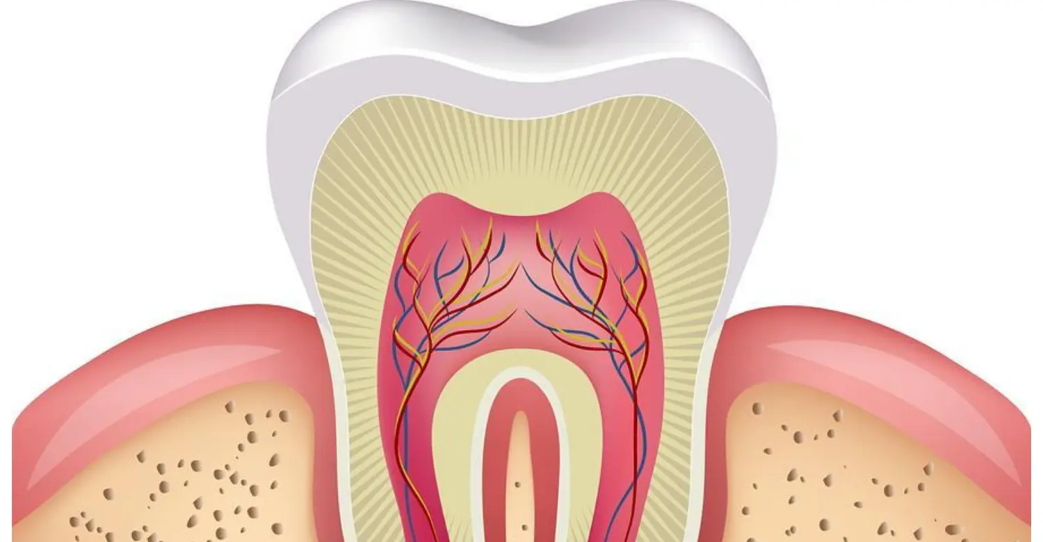
The neck of tooth is the anatomical transition between the crown and root. It is located near the gingival margin in a healthy mouth. This region involves the relationship between enamel, dentin, cementum, and surrounding gum tissue. The cementoenamel junction, or CEJ, provides an important landmark between crown and root surfaces. Healthy gingiva normally protects much of this area from direct exposure. When gum recession occurs, previously covered surfaces can become visible. This may contribute to sensitivity, cervical wear, or root-surface decay. The anatomy also varies among incisors, canines, premolars, and molars. Dentists therefore examine the cervical area together with surrounding periodontal tissues. Understanding this anatomy helps patients recognize early changes and understand why gum health and tooth structure are closely connected.
Neck of Tooth Definition
The neck of tooth refers to the constricted cervical area where the crown transitions toward the root. It is closely associated with the cementoenamel junction, where enamel and cementum meet. The term cervical region can describe a broader area surrounding this anatomical transition. The CEJ itself represents a specific boundary rather than an entire region. This distinction becomes useful when dentists describe recession, abrasion, erosion, or cervical cavities. For example, a cervical lesion may affect the area near the CEJ without directly damaging the junction itself. Understanding these anatomical terms can make dental explanations easier to follow. It also helps patients distinguish between normal tooth anatomy and changes caused by disease, gum recession, or mechanical wear.
Where Is the Neck of the Tooth?
The neck of tooth is located between the crown and root, close to the gumline. In healthy conditions, the gingival margin generally covers or closely surrounds this transition. Its exact position can vary according to tooth anatomy and periodontal conditions. The CEJ serves as an important reference point when dentists evaluate gum position and attachment. When gum recession occurs, more of the cervical root surface may become visible. This can make a tooth appear longer than neighboring teeth. Exposed surfaces may also look darker because dentin and cementum have different colors from enamel. Patients may mistake this appearance for sudden tooth damage. However, the underlying change may result from recession, cervical wear, erosion, or another dental condition.
Neck of a Tooth vs Crown vs Root
The neck of tooth represents the transition between two major anatomical portions of the tooth. The crown is the portion generally visible above the gumline. It is primarily covered by enamel and is designed to withstand chewing forces. The root is positioned within the jaw and provides support through the periodontal ligament and surrounding bone. Cementum covers much of the root surface. Dentin forms the internal hard tissue beneath enamel and cementum. The neck is therefore not a separate fourth tooth tissue. Instead, it describes an anatomical region where different structures transition. Understanding this distinction helps patients interpret dental diagrams and clinical explanations. It also explains why cervical problems can involve enamel, dentin, cementum, and nearby gum tissue simultaneously. Read more about Is A Subperiosteal Implant Painful
What Is the Neck of a Tooth Made Of?
The neck of the tooth involves relationships among several mineralized dental tissues. These include enamel, dentin, and cementum. Their arrangement around the cementoenamel junction can vary between teeth. Research has identified different patterns involving cementum overlapping enamel, direct tissue contact, or small areas where dentin may remain exposed. This anatomical variation can influence sensitivity when protective tissues are lost. Cementum is thinner and less mineralized than enamel, making exposed root surfaces more vulnerable to mechanical and chemical challenges. Dentin can also become sensitive when its protective covering is removed. Dentists consider these tissue differences when evaluating cervical lesions. Treatment therefore depends on whether the problem involves sensitivity, recession, decay, erosion, abrasion, or another condition.
Enamel at the Cervical Region
Enamel extends from the crown toward the cervical margin, although its thickness varies across the tooth. The neck of tooth may therefore contain a thinner enamel region compared with areas farther toward the crown. Enamel is highly mineralized and provides strong protection against everyday mechanical and chemical challenges. However, repeated acid exposure can cause mineral loss over time. Excessive brushing may also contribute to cervical wear when combined with other risk factors. Patients should therefore clean the gumline thoroughly without applying unnecessary pressure. A soft-bristled toothbrush and appropriate brushing technique can help reduce mechanical stress. Maintaining fluoride exposure can also support enamel mineralization. Regular dental examinations can identify early cervical changes before substantial structural damage develops.
Dentin Around the Neck of the Tooth
The neck of tooth contains dentin beneath the outer protective tissues. Dentin forms much of the internal tooth structure and surrounds the dental pulp. It contains microscopic dentinal tubules that extend toward the pulp. When cervical dentin becomes exposed, external stimuli can produce sensitivity. Cold drinks, air, touch, and certain foods may trigger short, sharp discomfort. This response can occur even when no obvious cavity exists. The movement of fluid within dentinal tubules is associated with the mechanism of dentin hypersensitivity. Therefore, exposed cervical dentin can become clinically significant. Dentists evaluate the cause of exposure before recommending treatment. Management may involve fluoride, desensitizing products, behavioral changes, or restorative treatment when structural loss is present.
Cementum and the Tooth Neck
The neck of tooth transitions toward the root, where cementum covers the external root surface. Cementum provides an attachment surface for periodontal ligament fibers. These fibers help connect the tooth to the surrounding alveolar bone. Compared with enamel, cementum is thinner and less resistant to mechanical and chemical challenges. When gum recession exposes the root, cementum can become directly exposed to brushing, acids, plaque, and other environmental factors. This can increase the risk of cervical wear or root-surface decay. However, exposed cementum does not always indicate active disease. Dentists assess its condition alongside gum health and tooth structure. Professional monitoring can help determine whether the exposed surface requires preventive care, desensitization, restoration, or periodontal treatment.
Cementoenamel Junction (CEJ)
The neck of tooth is closely associated with the cementoenamel junction, commonly called the CEJ. The CEJ represents the anatomical area where enamel and cementum meet. It provides an important landmark for understanding the transition between crown and root tissues. The junction is not necessarily a perfectly uniform line around every tooth. Research has identified several patterns involving cementum overlapping enamel, meeting enamel directly, or leaving small areas of exposed dentin. One frequently cited study reported approximately 60% overlap, 30% edge-to-edge contact, and 10% gaps exposing dentin. These findings demonstrate that cervical anatomy varies naturally. The CEJ is also clinically valuable because dentists can use it as a reference when evaluating periodontal attachment and gum recession. Learn more about When To Stop Drinking Alcohol Before Wisdom Teeth Surgery
What Is the Function of a Tooth Neck?
The neck of tooth provides an important anatomical transition between the crown and root. Although it is not an independent functional organ, its position contributes to the continuity of tooth structure. The cervical region also lies close to the gingival margin and periodontal attachment. Healthy surrounding tissues help protect this area from bacterial accumulation and mechanical irritation. The CEJ provides an anatomical reference that dentists can use during periodontal evaluation. When cervical surfaces become exposed, dentin or cementum may face greater exposure to acids, plaque, and mechanical forces. Protecting this area therefore contributes to overall dental health. Good oral hygiene, gentle brushing, appropriate dietary habits, and regular dental examinations can help maintain cervical tooth integrity.
Function of the Neck of a Tooth
The neck of tooth primarily provides structural continuity between the crown and root. It represents the region where different mineralized tissues transition into one another. The cervical area also interacts closely with the gingival margin and surrounding periodontal tissues. Unlike a tooth cusp, it does not directly perform a specific chewing function. However, its structural integrity contributes to the overall strength and shape of the tooth. Damage in this area can expose dentin or create defects that collect plaque. Cervical changes can also contribute to sensitivity when protective tissues are lost. Dentists therefore consider the cervical region during routine examinations. Identifying changes early can help preserve healthy tooth structure and prevent avoidable progression.
How the Neck Supports Tooth Stability
The neck of tooth is connected anatomically to structures that contribute to overall tooth support. However, tooth stability primarily depends on the periodontal ligament, cementum, alveolar bone, and surrounding periodontal tissues. Cementum provides the surface where periodontal ligament fibers attach to the tooth root. These fibers connect the tooth with supporting bone and help distribute functional forces. When periodontal disease causes attachment and bone loss, more root structure can become exposed. This may alter the visible appearance around the gumline. CDC identifies periodontitis as a disease involving inflammation and damage to the tissues supporting teeth. Therefore, changes around the cervical area can sometimes indicate broader periodontal problems requiring professional assessment.
Why the Neck of the Tooth Matters for Oral Health
The neck of tooth matters because several dental and periodontal problems can develop near this transition. Common concerns include sensitivity, gum recession, cervical decay, erosion, abrasion, and non-carious cervical lesions. The cervical region can also become more vulnerable when protective tissues are lost. Plaque accumulation around the gumline may further increase the risk of inflammation and decay. Early detection is valuable because smaller cervical problems can often be managed more conservatively. Patients should report persistent sensitivity, visible recession, bleeding, swelling, discoloration, or notches near the gumline. At Vitrin Clinic, dentists can evaluate both tooth structure and surrounding periodontal tissues. This combined assessment helps determine whether the problem originates primarily from the tooth, gums, or both.
Clinical Note
The cervical region deserves attention during routine dental examinations. Its appearance can reflect both tooth structure and periodontal conditions. A dentist should distinguish recession from enamel wear, erosion, abrasion, and cervical caries. These conditions can look similar but require different management. Research confirms considerable anatomical variation at the CEJ. This means one patient's cervical anatomy may look different from another's without representing disease. Clinical diagnosis should therefore rely on examination rather than photographs alone. At Vitrin Clinic, a comprehensive assessment can combine periodontal evaluation, tooth inspection, and appropriate imaging. Discover details of Cholesterin High Cholesterol And Oral Health
Neck of Tooth Anatomy in Different Types of Teeth
Cervical anatomy varies between tooth groups because each tooth has a different crown and root shape. Incisors generally have relatively narrow cervical contours. Canines often have strong root anatomy and prominent cervical transitions. Premolars have more complex crown forms and variable root structures. Molars have broader cervical regions and multiple roots. The CEJ itself can also vary around individual teeth. Research on permanent teeth demonstrates substantial morphological diversity in cervical tissue relationships. These differences influence how dentists examine recession, cervical wear, and root exposure.
Neck of Incisors
Incisors have relatively narrow crowns designed primarily for cutting. Their cervical contours are usually easier to visualize from the front. Recession around incisors can therefore become noticeable quickly. Exposed cervical surfaces may appear darker than the surrounding enamel. Sensitivity can also develop when dentin becomes exposed. Careful brushing is particularly important because excessive horizontal brushing can concentrate force near the gumline. Dentists assess cervical changes alongside gum position and tooth wear. Cosmetic concerns may also arise when recession changes the apparent length of front teeth.
Neck of Canines
Canines have prominent crowns and generally strong roots. Their cervical areas transition from a prominent crown toward a substantial root structure. Because canines often guide certain jaw movements, their surrounding tissues experience functional forces. Recession may make the cervical area visibly prominent. Cervical wear can also occur when brushing forces or acidic exposure are excessive. Dentists evaluate canine recession by measuring the gum margin relative to anatomical landmarks. Treatment depends on whether the primary concern involves sensitivity, tissue loss, esthetics, or periodontal disease.
Neck of Premolars
Premolars have diverse crown and root configurations. Their cervical region can show considerable variation in enamel and cementum relationships. One study of 67 premolars found edge-to-edge enamel-cementum contact in 55.1% of examined specimens. Approximately one-third showed gaps associated with exposed dentin. This demonstrates that cervical anatomy can differ even within one tooth group. Premolars may therefore develop sensitivity or cervical lesions for multiple reasons. Professional examination helps determine whether symptoms involve recession, wear, erosion, or decay.
Neck of Molars
Molars have broad crowns and commonly possess multiple roots. Their cervical region surrounds the transition toward these roots. Because molars have more complex anatomy, cervical areas may be harder to inspect directly. Plaque accumulation can also occur around posterior gumlines when cleaning is incomplete. Exposed root surfaces may become susceptible to decay and sensitivity. Dentists may use mirrors, periodontal probes, and radiographs to assess posterior cervical conditions. Maintaining interdental cleaning is especially important around molars because toothbrush bristles cannot clean every surface effectively. Your complete guide to Silver Diamine Fluoride
What Is an Exposed Neck of Tooth?
An exposed neck of tooth occurs when previously protected cervical surfaces become visible. Gum recession is one of the most common reasons for this exposure. Cervical wear can also remove enamel, cementum, or dentin. Exposed surfaces may appear yellowish, beige, or darker than surrounding enamel. Some patients experience sensitivity when drinking cold beverages or brushing. Exposure does not automatically indicate serious dental disease. However, professional evaluation can identify the underlying cause. Possible causes include gum recession, mechanical abrasion, acid erosion, cervical decay, or tooth-position factors. Early assessment can help prevent further structural damage. Treatment depends on whether the problem affects tooth tissues, periodontal tissues, or both. Vitrin Clinic can evaluate exposed cervical surfaces and recommend treatment based on the patient's clinical findings.
What Causes an Exposed Neck of Tooth?
An exposed neck of tooth can develop from several interacting factors. Gum recession may uncover previously protected root surfaces. Aggressive brushing can contribute to gingival trauma and cervical abrasion. Periodontal inflammation can cause attachment loss and progressive recession. Acidic foods and beverages may contribute to cervical erosion. Tooth position can also influence how much protective gum tissue surrounds the area. Grinding and excessive functional forces may contribute to some cervical defects. The cause is often multifactorial rather than a single habit. CDC identifies poor oral hygiene and smoking as important risk factors for gum disease. Periodontal disease can also damage tissues supporting the teeth. A dental examination can identify the dominant contributing factors and guide appropriate prevention or treatment.
Symptoms of an Exposed Tooth Neck
An exposed neck of tooth may produce several noticeable changes. Common symptoms include sensitivity, visible root exposure, and an apparently longer tooth. Cold water may cause a short, sharp sensation. Brushing may also trigger localized discomfort. The exposed surface can appear yellow, beige, or darker than enamel. Some patients develop a notch or indentation near the gumline. Symptoms depend on the amount and type of tissue exposed. Dentin hypersensitivity is relatively common but prevalence estimates vary considerably between populations. One systematic review reported a best prevalence estimate of 11.5%. The average prevalence across included studies was approximately 33.5%. These differences reflect variations in populations and diagnostic methods. Persistent sensitivity should therefore receive professional evaluation rather than self-diagnosis.
Is an Exposed Tooth Neck a Serious Problem?
An exposed neck of tooth is not always an emergency. However, it should not automatically be considered harmless. The underlying cause determines its potential significance. Stable gum recession may require monitoring and preventive care. Progressive recession can increase root exposure and sensitivity. Cervical decay can gradually remove tooth structure. Exposed dentin can also cause persistent discomfort. Early evaluation helps distinguish stable exposure from active disease. A dentist can assess gum position, tooth structure, plaque accumulation, and periodontal health. Prompt examination is particularly important when exposure occurs with bleeding, swelling, worsening sensitivity, or visible structural loss. Treatment can then focus on the underlying cause rather than simply covering the exposed area.
Clinical Note
An exposed neck of tooth should be diagnosed according to its cause rather than appearance alone. Two teeth can show similar cervical exposure but require different treatment. One may have a stable recession without active disease. Another may have cervical decay or progressive mechanical wear. Periodontal probing can assess gum attachment and surrounding tissue health. Clinical inspection can identify surface defects, discoloration, or cavitation. Radiographs may reveal deeper decay or supporting bone changes when indicated. Sensitivity testing can provide additional information when pain is present. This comprehensive approach helps avoid unnecessary treatment. It also allows dentists to identify conditions that require early intervention. Vitrin Clinic can assess both the tooth and surrounding periodontal tissues during evaluation. Important tips for Alcohol And Oral Cancer Risk
Neck of Tooth and Tooth Sensitivity
Sensitivity around the neck of tooth can occur when protective tissues are lost. Enamel and cementum normally provide protection over underlying dentin. When dentin becomes exposed, external stimuli can affect dentinal tubules. Cold drinks, air, touch, and certain foods may produce short, sharp discomfort. Sensitivity does not automatically mean that a cavity exists. Gum recession, erosion, abrasion, and cervical wear can all expose dentin. Dentin hypersensitivity research shows considerable variation between populations. Diagnosis is therefore important before treatment begins. Dentists evaluate the tooth surface and surrounding gums to identify the cause. Treatment may include fluoride, desensitizing products, behavior changes, periodontal care, or restorative procedures. Managing the cause can provide more sustainable relief.
Why Can an Exposed Tooth Neck Become Sensitive?
An exposed neck of the tooth can become sensitive when dentin loses its protective covering. Dentin contains microscopic tubules extending toward the dental pulp. External stimuli can cause fluid movement within these tubules. This movement can stimulate nerve responses and produce short, sharp pain. Cold temperatures are common triggers. Touch and airflow can also cause discomfort. Cervical sensitivity may result from gum recession, erosion, abrasion, or periodontal treatment. Not every exposed surface becomes sensitive. Individual anatomy and the degree of tubule exposure influence symptoms. Dentists should identify the cause before recommending treatment. Desensitizing products may help suitable patients. Fluoride treatments can also support vulnerable surfaces. Persistent or unusual pain requires further diagnostic evaluation.
Neck of Tooth Sensitivity vs Enamel Sensitivity
Sensitivity involving the neck of the tooth usually relates to exposed dentin rather than healthy enamel itself. Enamel is highly mineralized and does not contain the same sensory structures found in dentin. When enamel remains intact, external stimuli generally do not directly produce dentin hypersensitivity. Cervical sensitivity can develop when recession or wear exposes underlying dentin. Enamel demineralization may reduce protection and contribute indirectly. However, sensitivity can also result from decay, cracks, or pulpal inflammation. Pain duration and triggers can provide useful diagnostic information. Brief pain after cold stimulation may suggest hypersensitivity. Lingering or spontaneous pain requires closer evaluation. A dentist can determine whether the symptoms originate from exposed dentin or another dental condition.
When Should Tooth Sensitivity Be Checked by a Dentist?
Sensitivity involving the neck of tooth should be evaluated when symptoms persist or worsen. Occasional brief sensitivity may have a relatively simple cause. Repeated discomfort can indicate exposed dentin, recession, erosion, abrasion, or decay. Pain that occurs without an obvious trigger deserves professional attention. Lingering pain after hot or cold stimulation may indicate pulpal involvement. Sensitivity accompanied by swelling, spontaneous pain, or biting discomfort also requires examination. A dentist can perform clinical inspection and appropriate sensitivity testing. Radiographs may be recommended when deeper pathology is suspected. Early diagnosis can help prevent unnecessary progression. Patients should avoid assuming that every sensitive tooth has the same cause. Revealing facts about How Eating Disorders Damage Teeth
Neck of Tooth and Gum Recession
The neck of the tooth can become visible when the gingival margin moves toward the root. This condition is commonly called gum recession. Recession exposes portions of the root that were previously covered by gingival tissue. The exposed surface may appear darker or more yellow than enamel. Some patients develop sensitivity because underlying dentin becomes exposed. Recession can result from several factors, including periodontal inflammation, traumatic brushing, tooth position, and anatomical characteristics. It does not automatically mean that active periodontal disease is present. Professional examination can determine whether recession is stable or progressing. Dentists also evaluate periodontal attachment and supporting bone. Early assessment can help identify appropriate preventive or periodontal treatment.
How Gum Recession Exposes the Tooth Neck
The gum normally surrounds and protects the cervical transition between crown and root. When the gingival margin moves apically, more of the neck of the tooth becomes visible. This process can expose cementum and underlying dentin. The tooth may consequently appear longer than neighboring teeth. Recession can occur with or without active periodontal disease. Periodontitis can cause attachment and supporting bone loss, which may increase root exposure. CDC describes periodontitis as an inflammatory disease affecting tissues supporting the teeth. Accurate diagnosis therefore requires more than visual inspection. Dentists may measure recession and periodontal pockets during examination. They can also evaluate plaque, inflammation, tooth position, and supporting bone. These findings help determine the most appropriate treatment approach.
Common Causes of Gum Recession Around the Tooth Neck
Several factors can contribute to recession around the neck of tooth. Traumatic brushing can place excessive mechanical pressure on the gingival margin. Periodontal inflammation can damage supporting tissues and contribute to attachment loss. Thin gingival tissue may increase susceptibility in certain areas. Tooth position can also influence the amount of tissue covering the root. Inadequate plaque control can increase gingival inflammation and periodontal risk. Smoking is another recognized risk factor for periodontal disease. Orthodontic tooth movement may influence soft-tissue conditions in susceptible patients. Recession can therefore result from multiple interacting factors. Dentists should identify the primary contributors before recommending treatment. Controlling inflammation and modifying harmful habits can help reduce future progression.
Can an Exposed Tooth Neck Grow Back?
An exposed neck of tooth does not normally become naturally covered again after permanent gum recession. Lost gingival tissue generally does not regenerate spontaneously to its previous position. However, selected recession cases can be treated using periodontal soft-tissue procedures. Root coverage techniques may improve tissue protection and appearance. Treatment suitability depends on recession depth, tissue thickness, root anatomy, periodontal health, and patient expectations. Some mild cases may require only monitoring and preventive care. Others may benefit from surgical treatment when sensitivity or cosmetic concerns are significant. The objective is not always complete root coverage. A dentist first determines whether treatment is necessary and what outcome is realistic. Long-term maintenance remains important after treatment. Everything you need to know about 3D Printed Vs Milled Titanium Implant
Neck of Tooth and Cervical Tooth Decay
Cervical decay develops near the gumline. It can affect enamel, exposed root surfaces, or both. Root surfaces become particularly vulnerable when recession exposes cementum and dentin. These tissues are less mineralized and more susceptible to acid-mediated damage than enamel. Plaque accumulation around the gumline can increase caries risk. Patients with dry mouth may also face increased risk because saliva normally helps protect tooth surfaces. Early cervical lesions may be managed conservatively. More advanced lesions may require restorative treatment.
What Is Cervical Tooth Decay?
Cervical tooth decay is caries occurring near the cervical portion of a tooth. It can appear as a white, brown, yellow, or darkened area. Root-surface caries may progress differently from enamel caries. Lesions can be difficult to clean when they occur below or near the gumline. Early detection improves the opportunity for minimally invasive management. Dentists evaluate lesion activity, depth, location, and accessibility. Treatment may involve fluoride, preventive changes, or restoration.
Why Is the Tooth Neck Vulnerable to Decay?
Cervical surfaces can become vulnerable when enamel protection is reduced. Exposed root surfaces are especially susceptible because cementum and dentin have lower mineral density than enamel. Plaque can accumulate around irregular recession margins. Reduced saliva can further impair natural remineralization. Dietary acids and frequent sugar exposure can increase the challenge. The risk is therefore influenced by several factors. Good plaque control and regular dental examinations help identify early changes.
How Dentists Treat Cervical Cavities
Treatment depends on lesion depth and activity. Early non-cavitated lesions may benefit from fluoride and improved plaque control. Cavitated lesions may require tooth-colored restorative materials. The dentist first removes diseased tissue when necessary. The restoration is then shaped to restore function and facilitate cleaning. If gum recession contributed to the problem, periodontal management may also be needed. Treating the cavity without addressing plaque retention can increase recurrence risk.
Neck of Tooth Problems: Signs You Should Not Ignore
Cervical changes can be subtle during their early stages. Patients should pay attention to changes around the gumline. Visible recession, persistent sensitivity, bleeding, swelling, notches, and discoloration deserve evaluation. These signs do not automatically indicate severe disease. However, they can signal conditions requiring preventive or restorative care. Early assessment is especially useful when symptoms are localized to one tooth. Vitrin Clinic can evaluate the cervical area and surrounding tissues as part of a comprehensive examination.
Visible Recession Around the Tooth Neck
Visible recession can make a tooth appear longer. The exposed root may look darker than the surrounding enamel. Some patients also notice a triangular space between neighboring teeth. Recession can be stable or progressive. The dentist can measure its extent and assess periodontal attachment. Treatment depends on symptoms, progression, tissue anatomy, and cosmetic concerns. Patients should avoid aggressive brushing while awaiting professional assessment.
Persistent Cervical Sensitivity
Sensitivity that repeatedly occurs around the gumline should be evaluated. It may result from exposed dentin, recession, erosion, abrasion, or decay. The pain pattern can help distinguish hypersensitivity from pulpal disease. Short pain after cold stimulation is more typical of dentin hypersensitivity. Pain that lingers may require deeper evaluation. A dentist can perform appropriate tests before recommending treatment.
Bleeding or Swollen Gums
Bleeding gums are commonly associated with gingival inflammation. Swelling can also indicate plaque accumulation or periodontal disease. CDC identifies red, swollen, or easily bleeding gums as common signs of gingivitis. Persistent bleeding should not be considered normal. Professional cleaning and improved home care may resolve gingivitis. Advanced periodontal disease requires more comprehensive management.
Notches or Wear Near the Gumline
A notch near the cervical area may indicate non-carious tooth wear. Possible contributors include abrasion, erosion, stress concentration, or combinations of factors. The appearance alone cannot identify the cause. Dentists evaluate brushing habits, dietary acids, occlusion, and tooth anatomy. Treatment may involve behavior modification or restorative repair. Unnecessary drilling should be avoided when active disease is absent.
Discoloration Around the Tooth Neck
Discoloration near the gumline can have multiple causes. It may represent exposed root structure, plaque, calculus, staining, or dental caries. Yellowish coloration is common when dentin or cementum becomes visible. Darkening can occur with caries or staining. A dentist should inspect the surface and determine whether it is hard, soft, smooth, or cavitated. Visual color alone cannot establish a diagnosis.
Clinical Note
Cervical findings should always be interpreted within the patient's overall oral condition. A visible root may be stable recession rather than active disease. Conversely, a small dark area can represent developing caries. Professional examination allows dentists to separate these possibilities. Periodontal measurements and radiographs can provide additional information. This approach reduces unnecessary treatment while identifying problems early. Key details on Subperiosteal Vs Endosteal Implants
How Dentists Examine the Neck of a Tooth
A cervical assessment typically begins with visual inspection. The dentist then evaluates gum position, plaque, calculus, tooth structure, and symptoms. Periodontal probing helps measure pocket depth and attachment relationships. Sensitivity testing can determine whether exposed dentin responds normally. Radiographs may be used when deeper structures require assessment. The examination is tailored to the patient's symptoms and risk factors. Not every cervical complaint requires imaging.
Visual Examination
The dentist inspects the tooth under appropriate lighting and magnification. They look for recession, discoloration, wear, cracks, plaque, calculus, and cavitation. The cervical contour is compared with neighboring teeth. The dentist also evaluates whether the exposed surface is smooth or irregular. Photographs may document changes over time. Visual examination provides the first step toward identifying the underlying problem.
Periodontal Examination
Periodontal examination evaluates the gingiva and supporting tissues around the tooth. Probing measurements help identify pocket depth and attachment changes. The CEJ provides an important anatomical reference during periodontal assessment. Bleeding during probing can indicate inflammation. Mobility and gum recession may also be assessed. These findings help determine whether cervical exposure is associated with periodontal disease.
Dental X-Rays
Radiographs can reveal structures that are not visible during clinical inspection. They may help identify interproximal decay, bone loss, or deeper structural changes. Imaging is selected according to the clinical question. A small superficial cervical lesion may not require extensive imaging. Deeper or uncertain lesions may justify radiographic evaluation. The dentist balances diagnostic value with radiation exposure.
Sensitivity and Pulp Testing
Sensitivity testing helps determine how the tooth responds to stimuli. Cold testing is commonly used when pulpal status requires evaluation. Percussion and bite testing may also help identify other sources of pain. Results are interpreted alongside symptoms and clinical findings. A sensitive cervical surface does not automatically mean the pulp is diseased. Persistent or spontaneous pain requires broader diagnostic assessment. Medical advice regarding Subperiosteal Implant Care
How to Protect the Neck of Your Teeth
Protecting the cervical area requires both tooth care and gum care. Gentle brushing reduces unnecessary mechanical stress. Cleaning the gumline helps control plaque accumulation. Limiting frequent acidic exposures reduces chemical challenges. Fluoride supports enamel and root-surface protection. Patients with sensitivity should seek assessment rather than repeatedly masking symptoms. Prevention is particularly important after gum recession exposes root surfaces.
Use a Gentle Brushing Technique
A soft-bristled toothbrush can help reduce mechanical trauma. Brushing should use controlled movements rather than aggressive horizontal scrubbing. Excessive pressure can contribute to cervical wear and gingival irritation. The toothbrush should reach the gumline without forcing bristles into the tissue. Electric toothbrush pressure sensors may help some patients control force. Dentists can demonstrate an appropriate technique during routine visits.
Maintain the Gumline Clean
Plaque control around the gumline helps reduce gingival inflammation. Patients should brush twice daily using fluoride toothpaste. Interdental cleaning removes plaque from spaces that toothbrush bristles cannot reach. Regular professional cleanings may also help manage calculus. CDC emphasizes that good oral hygiene and professional care are important for preventing and managing gum disease. Consistency matters more than aggressive cleaning.
Reduce Acid Exposure
Frequent acidic drinks and foods can contribute to enamel and cervical erosion. Common sources include soft drinks, energy drinks, fruit juices, and acidic snacks. Reducing frequency can decrease repeated acid attacks. Drinking water after acidic exposure can help clear the mouth. Patients should avoid immediately brushing after strong acid exposure. Waiting allows saliva to help restore surface conditions.
Treat Tooth Sensitivity Early
Sensitivity should not simply be ignored. Early evaluation can identify whether the cause involves recession, wear, erosion, or decay. Desensitizing toothpaste may help suitable patients. Fluoride treatments can also strengthen vulnerable surfaces. However, persistent symptoms require diagnosis. Treatment should address the underlying cause whenever possible.
Tips for Patients
Use a soft toothbrush and gentle pressure every day. Clean carefully along the gumline without aggressive scrubbing. Use fluoride toothpaste unless your dentist recommends another product. Limit frequent acidic drinks and rinse with water afterward. Do not ignore persistent sensitivity or bleeding. Schedule professional examinations when recession or cervical changes appear. Keep track of changes in appearance or symptoms. At Vitrin Clinic, patients can receive personalized recommendations based on their tooth and gum findings.
How Is an Exposed Neck of Tooth Treated?
Treatment depends on the reason for exposure. Mild sensitivity may respond to fluoride or desensitizing products. Structural defects may require tooth-colored restorations. Gum recession may require periodontal treatment or soft-tissue procedures. Cervical wear requires removal of contributing factors before restoration is considered. Active decay requires caries management and possibly restoration. A successful treatment plan should therefore identify the cause first.
Fluoride and Desensitizing Treatments
Fluoride can help strengthen vulnerable mineralized surfaces. Desensitizing products may reduce the response of exposed dentin. Some formulations work by reducing dentinal tubule fluid movement. Others create protective deposits over sensitive areas. Treatment effectiveness varies between patients. Dentists may recommend professional fluoride applications when home products are insufficient. Persistent symptoms still require diagnostic evaluation.
Tooth-Colored Cervical Restorations
Composite resin can restore localized cervical defects. It may improve appearance and protect exposed dentin. Restorative treatment is particularly useful when a lesion is cavitated or structurally significant. The dentist prepares the area and bonds the material to the tooth. Proper isolation and occlusal evaluation can improve longevity. Restorations should not replace preventive treatment when the underlying cause remains active.
Treatment of Gum Recession
Management depends on recession severity and periodontal conditions. Stable, asymptomatic recession may simply require monitoring and preventive care. Sensitive areas may benefit from desensitization and improved brushing technique. Selected cases may be treated with periodontal plastic surgery. Root coverage procedures can improve tissue protection and esthetics. Patient-specific anatomy strongly influences the expected result.
Treatment for Cervical Tooth Wear
Cervical wear treatment begins with identifying the contributing mechanism. The dentist may evaluate brushing, dietary acids, grinding, and bite forces. Behavior modification can prevent further progression. Restorative materials may be considered when defects are deep or symptomatic. Treatment should preserve as much healthy tooth structure as possible. Monitoring can be appropriate for stable, shallow lesions.
Why Treating the Cause Matters
Treating only the visible defect may not prevent recurrence. A restoration can fail if aggressive brushing continues. Sensitivity may persist if gum recession remains active. Cervical caries may recur when plaque control is inadequate. Acid erosion can continue despite restoration if dietary exposure remains high. Identifying causative factors therefore forms an essential part of treatment planning.
What We Notice Clinically
The cervical region often provides useful information about both tooth and periodontal health. Dentists look for patterns rather than isolated findings. Recession, sensitivity, wear, plaque, and cervical defects may occur together. The relationship between these findings helps identify the dominant cause. Research describes the cervical region as susceptible to several physical and chemical challenges. This makes preventive examination particularly valuable. Vitrin Clinic focuses on evaluating the entire oral environment before recommending treatment.
Why the Cervical Area Deserves Special Attention
The cervical area contains several different tissues close to the gumline. It can therefore be affected by both periodontal and dental problems. The CEJ also has naturally variable morphology. Studies demonstrate that enamel-cementum relationships can differ around individual teeth. Exposed dentin may become sensitive when protective tissue is lost. Cervical decay can also progress if plaque remains undisturbed. Early professional assessment helps preserve healthy tissue.
Clinical Note
A cervical lesion should not automatically be restored. Dentists first determine whether it is active, progressing, symptomatic, or stable. Preventive management may be more appropriate for some lesions. Restoration is useful when structural protection or symptom control is necessary. Periodontal treatment may be essential when recession or inflammation is involved. This cause-based approach supports conservative dental care.
Neck of Tooth vs Cervical Region: Are They the Same?
The terms are related but can have slightly different meanings. The tooth cervix refers specifically to the constricted transition between crown and root. The cervical region can describe the broader area surrounding this transition. NCBI associates tooth cervix with the crown-root junction and CEJ. Dental professionals may use these terms differently depending on context. Understanding the terminology helps patients interpret dental explanations more accurately.
Neck of Tooth
The tooth neck describes the anatomical transition between crown and root. It is commonly associated with the cervical portion of the tooth. The term is useful when explaining basic tooth anatomy. It does not refer to a separate tissue layer. Instead, it identifies a location where different tissues meet. Clinical problems occurring there can involve enamel, dentin, cementum, or surrounding gingiva.
Cervical Region of the Tooth
The cervical region is a broader descriptive term. It generally refers to the area around the crown-root transition. It can include cervical enamel, CEJ, cementum, dentin, and adjacent periodontal tissues. Dental conditions such as cervical caries and cervical lesions use this terminology. The region is clinically important because several different tissues are exposed to environmental challenges.
Cervix of Tooth
Cervix of tooth is an anatomical term used for the neck region. NCBI's MeSH terminology includes both “Tooth Cervix” and “Cervix, Tooth.” It describes the constricted part connecting the crown and root. The term is particularly useful in anatomical and academic literature. Patients may encounter it when reading dental anatomy resources.
Cementoenamel Junction vs Tooth Neck
The CEJ is a specific anatomical boundary. The tooth neck is a broader anatomical concept surrounding the crown-root transition. They are closely related but should not always be treated as identical terms. The CEJ marks where enamel and cementum meet. The cervical region extends around this landmark. This distinction becomes important during periodontal measurements and cervical pathology assessment.
When Should You See a Dentist About Your Tooth Neck?
A dental visit is recommended when cervical changes are new, worsening, or symptomatic. Persistent sensitivity deserves assessment. Visible recession should also be evaluated, especially if it progresses. Bleeding or swollen gums may indicate gingival inflammation. Notches, cavities, or discoloration can indicate structural damage. Early diagnosis often allows more conservative treatment. Waiting until pain becomes severe can allow underlying disease to progress.
Signs That Require a Dental Examination
Seek professional assessment for persistent sensitivity or spontaneous pain. Report bleeding or swelling around the gumline. Visible root exposure should be evaluated when it appears suddenly. Dark, soft, or enlarging cervical areas require examination. Notches that continue to deepen also deserve attention. A dentist can determine whether the problem involves tooth structure, periodontal tissue, or both.
Clinical Note
Cervical symptoms should be interpreted within the patient's complete dental history. Previous periodontal treatment can influence gum position. Orthodontic history may affect tooth position and tissue thickness. Brushing habits can contribute to cervical wear. Dietary patterns can influence erosion and caries risk. A detailed history therefore improves diagnostic accuracy.
Why Choose Vitrin Clinic for Tooth and Gum Evaluation?
Vitrin Clinic provides a comprehensive approach to concerns involving the neck of the tooth. A proper evaluation considers teeth, gums, supporting tissues, symptoms, and contributing habits. Cervical changes can have several possible causes, including recession, wear, erosion, and decay. Professional diagnosis helps identify the actual source before treatment begins. Dentists can assess whether the condition is stable or progressing. They can also recommend preventive measures based on individual risk factors. Protecting healthy tooth structure remains an important treatment goal. Patients can receive guidance designed around their specific oral health needs. A detailed assessment can also help identify problems before they require more extensive treatment. Vitrin Clinic focuses on combining diagnosis, prevention, and personalized dental care.
Comprehensive Dental Assessment
A comprehensive assessment of the neck of tooth can include clinical examination, periodontal evaluation, sensitivity testing, and imaging when necessary. Dentists inspect the cervical surface for recession, discoloration, wear, plaque accumulation, and decay. Periodontal measurements can help determine gum health and attachment levels. Sensitivity testing may identify exposed dentin or other sources of discomfort. Dental X-rays can provide additional information when deeper structures require evaluation. Examining teeth and gums together helps distinguish periodontal exposure from tooth-surface loss. This approach can support more accurate diagnosis and treatment planning. Early detection may also reduce the need for extensive restorative procedures. Vitrin Clinic can tailor the assessment according to the patient's symptoms and clinical findings.
Personalized Treatment Planning
Treatment planning for the neck of tooth should address the patient's specific condition and contributing factors. Some patients may need preventive care rather than restorative treatment. Others may require periodontal management, desensitizing treatment, or cervical restorations. Dentists consider symptoms, recession, structural changes, anatomy, esthetic concerns, and long-term maintenance. This approach helps avoid unnecessary procedures while addressing active problems. Treatment may also involve modifying brushing techniques or dietary habits. Each recommendation should reflect the patient's individual dental findings. Vitrin Clinic can develop a personalized plan focused on protecting healthy tissues and controlling the underlying cause. Regular follow-up can help monitor treatment results and identify any future changes.
Why Choose Vitrin Clinic for Exposed Tooth Neck Treatment?
Treatment for an exposed neck of tooth should focus on more than improving appearance. The underlying cause needs to be identified before selecting an appropriate procedure. Gum recession, erosion, abrasion, cervical decay, and periodontal disease can require different treatments. Some patients may benefit from preventive care or desensitizing products. Others may require tooth-colored restorations or periodontal procedures. Vitrin Clinic evaluates the surrounding gums and tooth structure before recommending treatment. This helps create a plan based on the patient's specific clinical condition. The goal is to protect exposed surfaces, manage symptoms, and reduce the risk of further damage. Treatment decisions can also consider functional and esthetic concerns.
Treating the Cause, Not Just the Symptom
Treating the neck of tooth effectively requires identifying why the problem developed. Sensitivity relief alone may not prevent further structural changes. Continued aggressive brushing can contribute to cervical wear. Uncontrolled periodontal inflammation may contribute to progressive recession. Frequent acidic exposure can continue damaging vulnerable surfaces. Active decay may also return if underlying risk factors remain unmanaged. Cause-based treatment addresses these contributing factors alongside the visible problem. Dentists may recommend brushing modifications, fluoride, dietary changes, periodontal treatment, or restorative procedures. This comprehensive approach can provide better long-term protection. Vitrin Clinic focuses on identifying contributing factors before selecting treatment whenever clinically appropriate.
Patient-Focused Dental Care
Patient-focused management of the neck of tooth combines accurate diagnosis with clear communication. Patients should understand what caused their cervical problem and how to protect the affected area. Dentists can explain available treatment options and their expected benefits. They can also discuss limitations and possible maintenance requirements. Preventive recommendations may include gentler brushing, improved gumline cleaning, fluoride use, and dietary adjustments. Follow-up appointments can monitor recession, sensitivity, or cervical wear when necessary. Vitrin Clinic can provide individualized guidance based on clinical findings and patient concerns. The objective is to create practical care that supports long-term tooth and gum health.
Reference
FAQs
Dr. Rifat Alsaman has more than 5 years of clinical experience in dentistry and currently serves as the Head of the Medical Team at Vitrin Clinic. He is dedicated to providing exceptional patient care, overseeing treatment planning, and ensuring the highest clinical standards across the team. His expertise, attention to detail, and commitment to continuous professional development have helped countless patients achieve healthier, more confident smiles.

.webp&w=3840&q=75)


