General Dentistry
October 5, 2026
What Is Oral Cancer Screening and How Does It Detect Mouth Cancer?
 Dr. Rifat Alsaman
An oral examination can help identify suspicious changes before a confirmed cancer diagnosis. Dentists inspect oral tissues and surrounding areas for unusual findings. These findings can include ulcers, patches, lumps, thickened tissue, bleeding, or altered sensation. A clinical examination does not prove that cancer exists. Vitrin Clinic emphasizes individualized dental assessment when patients report concerning oral changes.
What Is Oral Cancer Screening?
An oral cancer screening is a clinical examination for suspicious changes in oral tissues. A dentist typically evaluates visible and palpable tissues inside and around the mouth. The examination may occur during a routine dental appointment. The goal is to identify abnormalities that deserve closer assessment.
What does an oral cancer screening check for?
The examination checks for visible or palpable changes that appear unusual. Dentists may look for sores, patches, swelling, masses, bleeding, or thickened tissue. They also consider color, surface texture, symmetry, and tissue mobility. Some findings may appear red, white, mixed, ulcerated, or irregular.
What is included in an oral cancer exam?
A comprehensive examination usually includes both visual inspection and tactile assessment. The clinician examines oral surfaces systematically rather than checking only the area causing symptoms. Lips, cheeks, gums, tongue, palate, and floor of the mouth all receive attention. The examination itself usually needs no special preparation.
Examination of the lips, cheeks, gums, tongue, palate, and floor of the mouth
These tissues are examined for changes in color, texture, thickness, ulceration, or shape. The inner cheeks are often inspected after gently retracting the soft tissues. The tongue is examined on several surfaces because different areas can show different findings.
Examination of the back of the mouth, throat, jaw, face, and neck
The examination can extend beyond the front of the mouth. Dentists may inspect accessible areas near the throat and back of the oral cavity. They can also examine facial symmetry and jaw movement. Gentle palpation of the neck can help identify enlarged or unusual lymph nodes.
Is an oral cancer screening the same as a dental screening?
The terms are related, but they describe different scopes of evaluation. A routine dental examination assesses teeth, gums, bite, restorations, and periodontal health. It also includes a general look at oral tissues. A cancer-focused examination specifically considers suspicious mucosal changes and related structures.
Why is oral cancer screening important for early detection?
Early evaluation can identify suspicious abnormalities before a patient understands their significance. Some concerning lesions may produce little or no pain. NIDCR lists persistent sores, patches, numbness, bleeding, swallowing problems, and neck lumps as warning symptoms. Statistics and insight: NIDCR reports that oral cancer incidence rises with age, particularly after age 50.
How long does an oral cancer examination take?
A basic clinical examination can be completed relatively quickly. NIDCR describes the examination as taking only a few minutes. ADA materials reference an examination process of approximately five minutes. The exact appointment length varies according to the patient's symptoms and dental history. At Vitrin Clinic, the clinical approach remains individualized when symptoms require additional assessment.
Can a Dentist Detect Oral Cancer?
Dentists can identify abnormalities that may warrant further investigation. They cannot reliably confirm malignancy through visual inspection alone. A dentist can recognize suspicious sores, patches, masses, thickened tissue, bleeding, and unusual changes. They may also detect altered sensation or movement.
How dentists look for early abnormalities during a dental visit
Dentists generally combine visual inspection with gentle palpation. They compare tissue appearance across different areas of the mouth. They assess color, texture, symmetry, thickness, and surface changes. The patient's history helps interpret whether an abnormality appears temporary or persistent.
Can a dentist detect cheek cancer, tongue cancer, or cancer on the floor of the mouth?
A dentist can inspect these areas during a comprehensive examination. The inner cheeks, tongue, and floor of the mouth are accessible to clinical inspection. Dentists can identify suspicious changes requiring additional investigation. They may also palpate tissues for firmness, thickness, or masses.
Can a dentist detect throat or oropharyngeal cancer?
Dentists can examine accessible portions of the mouth and throat. They may identify signs that warrant further head and neck evaluation. However, some areas of the oropharynx are difficult to inspect fully in a dental setting. HPV has a particularly important relationship with some oropharyngeal cancers.
What can a dentist identify before a cancer diagnosis is confirmed?
Dentists can document clinical abnormalities that require closer evaluation. They may identify potentially malignant disorders or lesions with uncertain behavior. They can also recognize non-cancerous conditions that resemble lesions. Documentation can include location, size, color, surface, symptoms, and duration.
Suspicious sores, lumps, thickened tissue, and unexplained bleeding
A persistent sore deserves attention when it lacks an obvious explanation. A lump can also require evaluation when it remains present or changes. Thickened tissue can feel different from surrounding mucosa. Unexplained bleeding may become more concerning when it occurs repeatedly.
Persistent red or white patches
Red and white patches can develop for many reasons. Some represent irritation, infection, inflammation, or other benign processes. Others can represent potentially malignant mucosal changes. Their appearance alone cannot establish the diagnosis. Persistence is an important clinical consideration.
Changes in movement, sensation, swallowing, chewing, or speech
Functional changes can provide important clinical information. Difficulty moving the tongue or jaw can indicate deeper tissue involvement or another oral condition. NIDCR lists difficulty chewing, swallowing, speaking, or moving the jaw or tongue as symptoms requiring evaluation.
Why a visual examination cannot confirm cancer by itself
A visual examination identifies abnormalities but does not determine their cellular behavior. Cancer diagnosis requires examination of tissue or cells using appropriate diagnostic methods. A biopsy allows a pathologist to assess sampled tissue microscopically. NCI and NHS sources identify biopsy as a key diagnostic test.
What Are the Early Signs of Mouth Cancer?
Early oral cancer can produce visible changes or subtle functional symptoms. Common warning signs include persistent ulcers, red or white patches, lumps, thickened areas, and bleeding. Pain and numbness can also occur. Difficulty chewing, speaking, or swallowing may develop as well.
What are the first signs of cancer in the mouth?
A persistent mouth sore may be an early warning sign. Red or white tissue changes can also deserve examination. A lump or thickened area can be another concerning finding. Some patients experience unexplained bleeding or numbness. Functional changes may appear as disease progresses. Statistics and insight: these symptoms overlap with many common dental conditions.
What are the most common early signs of mouth cancer?
Common warning signs include ulcers, persistent patches, lumps, unexplained bleeding, and unusual tissue thickening. Pain can occur, but some abnormalities never hurt. Numbness can sometimes occur with oral malignancy. Changes in chewing, swallowing, speaking, or jaw movement may also matter. Persistent lesions still require professional evaluation.
A mouth ulcer or sore that does not heal
Many mouth ulcers heal without treatment. A sore that continues beyond the expected healing period deserves clinical assessment. NHS guidance recommends seeing a healthcare professional for an ulcer lasting more than three weeks. NIDCR similarly advises evaluation when symptoms persist beyond two weeks.
A red or white patch
Red or white patches can have several possible causes. Some relate to irritation or inflammation. Some may represent changes requiring biopsy. The appearance, location, duration, and surface texture all matter. A patient should not attempt to classify the patch using internet photographs.
A lump, thickened area, or persistent irritation
A new lump can arise from trauma, infection, cysts, glandular changes, or benign growths. Persistent thickening may also have multiple causes. Clinical evaluation helps determine whether the area has an obvious explanation. The dentist may assess consistency, mobility, tenderness, and boundaries.
Unexplained pain, bleeding, or numbness
Pain has many possible dental and oral causes. Persistent unexplained pain should not be automatically attributed to cancer. Bleeding can result from gum disease, trauma, infection, or other conditions. Numbness deserves special attention when it lacks an obvious local explanation.
Difficulty chewing, swallowing, speaking, or moving the jaw or tongue
Functional changes can affect daily activities and may have several causes. Dental infection can make chewing painful. Temporomandibular disorders can affect jaw movement. Neurological or muscular problems can also alter function. Persistent or unexplained changes deserve clinical assessment.
What are the signs of mouth cancer in women?
The major warning signs are generally similar across sexes. Persistent ulcers, unusual patches, lumps, bleeding, pain, numbness, and functional changes remain important. NHS and NIDCR guidance does not define a completely separate symptom pattern for women. HPV-related cancers can occur independently of traditional tobacco-related risk patterns.
How do you know if you have mouth cancer?
You cannot reliably confirm mouth cancer at home. Symptoms can raise concern, but they cannot establish a diagnosis. A dental or medical examination is necessary for clinical assessment. A suspicious lesion may require biopsy for definitive diagnosis. Self-checking can help patients notice changes earlier.
How do you know if you get cancer in the mouth or throat?
Mouth and throat cancers can present differently. Oral cavity symptoms may involve visible sores, patches, lumps, or persistent irritation. Oropharyngeal symptoms can include sore throat, swallowing pain, hoarseness, ear pain, or enlarged neck nodes. Some patients have minimal visible symptoms.
Are oral cancer symptoms always painful?
No. Some suspicious oral lesions can be completely painless. A painless patch or lump may still require evaluation. NHS guidance lists painless tissue changes among warning symptoms that deserve attention. Pain can appear in some patients, especially when tissues become irritated or disease progresses.
Do these symptoms always mean cancer?
No. Many symptoms have benign explanations. Mouth ulcers can result from trauma, stress, infection, or inflammatory conditions. Red or white patches can arise from several oral disorders. Lumps can also be cysts, infections, or benign growths. The correct response is professional evaluation, not panic.
What Is the Difference?
Oral Cancer
Oropharyngeal Cancer
The terms describe different anatomical regions. Oral cavity cancer develops in structures of the mouth. Oropharyngeal cancer develops farther back, in the oropharynx. CDC defines oral cavity structures separately from the oropharynx. The distinction matters because risk factors and treatment pathways can differ.
What is oral cavity cancer?
Oral cavity cancer refers to malignancy arising within mouth structures. These include the lips, inner cheeks, gums, front tongue, floor of the mouth, and hard palate. Most oral cavity cancers are squamous cell carcinomas arising from the lining of oral tissues.
What is oropharyngeal cancer?
Oropharyngeal cancer affects the back of the throat. Structures include the tonsils, base of the tongue, soft palate, and posterior pharyngeal wall. HPV plays an important role in many of these cancers. CDC estimates that HPV causes about 60% to 70% of U.S. oropharyngeal cancers.
Where can oral cancer develop?
Cancer can develop in several oral structures. These include the tongue, gums, cheeks, lips, palate, and floor of the mouth. Each site can present differently. The oral cavity includes the front two-thirds of the tongue, gingiva, hard palate, floor, inner cheeks, and lips.
Tongue
The tongue requires examination of several surfaces. The sides and underside deserve careful attention. Lesions there can be hard for patients to see without good lighting and positioning. A dentist may ask the patient to extend the tongue.
Gums
The gums can develop sores, patches, swelling, or localized thickening. These changes are sometimes mistaken for ordinary gum disease. Periodontal disease also causes redness and bleeding, so clinical context is essential. The clinician evaluates both the gum tissue and adjacent teeth.
Inner cheeks and buccal mucosa
The inner cheek commonly experiences trauma from biting, teeth, fillings, or dentures. Repeated irritation can produce persistent changes, so clinical history is especially important. The dentist inspects the surface and looks for a mechanical source. Removing that source may allow healing.
Lips
Lip cancer can be associated with chronic ultraviolet exposure. NIDCR lists sun exposure as a risk factor for lip cancer. A persistent lip sore, thickened area, crusted lesion, or changing patch deserves assessment. Outdoor workers should pay particular attention to these changes.
Roof of the mouth
The roof of the mouth includes the hard palate and adjacent tissues. Persistent swelling or unusual patches may arise from several causes. Dental appliances, trauma, infections, and benign growths can all alter the palate. A clinician can compare the area with surrounding tissue.
Floor of the mouth
The floor of the mouth is difficult to inspect at home. The tissue is mobile and lies beneath the tongue. A dentist examines it with the tongue raised and assesses both appearance and firmness. Persistent ulcers, patches, swelling, or numbness here deserve professional evaluation.
Which symptoms can suggest oropharyngeal cancer?
Persistent throat symptoms can signal the need for medical assessment. Warning signs include prolonged sore throat, painful or difficult swallowing, hoarseness, ear pain, neck swelling, and weight loss. None of these is specific to cancer. Reflux, infection, allergies, and muscular problems can produce similar complaints.
Why HPV matters more strongly for some oropharyngeal cancers
HPV has a strong relationship with cancers arising in the oropharynx. HPV-positive disease can occur in people without any smoking history. Absence of tobacco use therefore does not remove the need for evaluation. Statistics and insight: CDC reports that HPV is thought to cause about 70% of the U.S.
Explore the benefits and fit of the Maryland bridges
Who Is at Higher Risk of Oral Cancer?
Risk is shaped by several factors rather than one exposure. Tobacco and alcohol are major established risks. HPV contributes strongly to many oropharyngeal cancers. Ultraviolet exposure affects lip cancer risk. Age, genetics, and immune factors can also play a role.
Does smoking increase the risk of oral cancer?
Yes. Tobacco exposure is a major risk factor for oral cancer. NIDCR states that tobacco in its different forms increases risk. This includes both smoked and smokeless products. Stopping tobacco use reduces future exposure, although earlier exposure may still influence risk.
How does alcohol affect oral cancer risk?
Heavy alcohol consumption increases oral cancer risk. The NHS lists high alcohol intake among important risk factors. Alcohol may contribute to tissue damage and interact with other carcinogenic exposures. Patients should discuss their drinking honestly during clinical history.
Why combined tobacco and alcohol exposure matters
Tobacco and alcohol together present greater concern than either exposure alone. NIDCR explicitly states that combined use increases risk further. Patients often underestimate this cumulative effect. Statistics and insight: USPSTF's evidence review identified tobacco and alcohol as major oral cancer risk factors.
Is HPV linked to oral or oropharyngeal cancer?
Yes, especially oropharyngeal cancer. CDC identifies HPV as a cause of cancers involving the tonsils and base of the tongue. These cancers can occur without any tobacco exposure. HPV vaccination prevents new infections but does not treat existing infection or established disease.
Can sun exposure increase lip cancer risk?
Yes. Long-term ultraviolet exposure can increase the risk of lip cancer. NIDCR specifically identifies sun exposure as a lip cancer risk factor. Outdoor workers may accumulate substantial exposure over many years. Regular lip sun protection reduces this exposure.
Does age increase oral cancer risk?
Risk generally increases with age. NIDCR reports that oral cancers occur most often in people over 40. Its incidence data show a stronger rise after age 50. Rates are highest among adults aged 65 and older.
Can people who do not smoke still develop oral cancer?
Yes. Tobacco is an important risk factor, but cancer can develop without it. HPV contributes to many oropharyngeal cancers. Age, genetics, immune status, alcohol, and ultraviolet exposure may also matter. Nonsmokers should never dismiss persistent symptoms. Risk history supplements the examination rather than replacing it.
Do betel quid, areca nut, or smokeless tobacco increase risk?
Yes. These exposures have recognized associations with oral cancer. The NHS lists tobacco, betel nut, and paan among important risk factors. Smokeless tobacco exposes oral tissues directly to carcinogens. Areca nut matters even in preparations without tobacco.
Can family history or genetics affect oral cancer risk?
Genetic factors can influence susceptibility to some cancers. NIDCR notes that inherited gene defects can increase risk of mouth and middle-throat cancers. Family history does not make cancer inevitable. It simply adds context for clinicians. Persistent symptoms still need examination even without a known genetic condition.
How Is Oral Cancer Screening Performed?
A clinical examination generally begins with history and a discussion of symptoms. The dentist then visually inspects oral tissues. Gentle palpation follows when appropriate. The clinician may also assess the face, jaw, and neck. NIDCR describes this as a systematic examination of extraoral and intraoral tissues.
What happens during an oral cancer screening at the dentist?
The dentist typically asks about symptoms and relevant history first. The mouth is then inspected under good lighting. Lips, cheeks, gums, tongue, palate, and floor are evaluated in turn. Selected tissues may be felt for unusual firmness. Neck and jaw examination may follow.
What does the dentist look for during the examination?
Dentists look for changes in appearance and texture. These include ulcers, patches, swelling, thickening, lumps, bleeding, and unusual surfaces. Asymmetry and functional changes are also noted. Duration helps separate brief irritation from persistent lesions. Statistics and insight: NIDCR advises professional evaluation when symptoms persist beyond two weeks.
How does palpation help identify abnormal tissue?
Palpation lets the clinician feel tissues that may look normal. An area can be unusually firm, thickened, fixed, tender, or enlarged. Lymph nodes can also be assessed. ADA guidance emphasizes conventional visual and tactile assessment. Palpation cannot diagnose cancer alone.
Why are the neck and jaw also examined?
The neck contains lymph nodes that can enlarge for many reasons. Infection is common, but persistent or unexplained enlargement deserves assessment. Jaw examination can reveal swelling, tenderness, or movement changes. NIDCR includes the face and neck in its examination approach.
Is an oral cancer screening painful?
The routine clinical examination is generally painless. NIDCR describes it as painless. Some patients feel mild discomfort when tissues are retracted or pressed. A sensitive ulcer may feel tender during inspection. No incision occurs during routine examination. Patients should mention sensitive areas beforehand.
Do you need X-rays for every oral cancer screening?
No. X-rays are not automatically required. The initial evaluation relies mainly on visual and tactile examination. Imaging may help when symptoms suggest bone or deeper involvement. Dental X-rays can also reveal common causes of pain or swelling.
When might additional imaging be considered?
Imaging may help when examination cannot fully assess a suspected area. It can also evaluate bone changes, deeper extension, or alternative diagnoses. NHS lists X-ray, ultrasound, CT, and MRI among possible tests after referral. The modality depends on the anatomical question.
When a lesion extends beyond what can be assessed clinically
Some abnormalities seem to extend into deeper tissues. A surface examination cannot define deep boundaries. Imaging can provide extra anatomical information in these cases. The clinician may refer the patient for CT or MRI. The choice depends on the suspected location and diagnostic question.
When bone involvement or another dental condition needs evaluation
Dental X-rays assess teeth and supporting bone. They can reveal infections, periodontal changes, or impacted teeth that explain symptoms. If a lesion seems linked to bone, broader imaging may be appropriate. The aim is accurate diagnosis without unnecessary tests. Vitrin Clinic uses imaging only when clinically appropriate.
Learn about the procedure and recovery for a gingivectomy
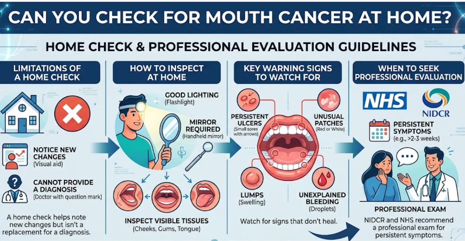
How to check your mouth for unusual changes at home
Choose a well-lit area and use a clean mirror. Look at the lips and inner cheeks first. Examine the gums and visible palate gently. Stick out the tongue and inspect the top and sides. Raise it to view the floor of the mouth.
Can you tell if you have mouth cancer without seeing a dentist?
No. You can recognize warning signs, but you cannot confirm their cause. Several common conditions closely resemble suspicious lesions. A dentist or doctor can assess the tissue directly. Biopsy provides tissue for pathological examination when cancer is suspected.
How reliable is a home mouth cancer check?
A home check helps detect visible changes, but its diagnostic reliability is limited. Some areas are hard to see, and normal anatomy varies. Small lesions can hide behind teeth or the tongue. Mayo Clinic notes that even professional examinations cannot detect every cancer.
Can an oral cancer test kit diagnose cancer?
Home test kits cannot independently diagnose cancer. Some products collect cells or flag possible abnormalities. Diagnostic interpretation still requires proper laboratory methods and clinical context. NCI describes cytology and brush methods as ways to collect cells for microscopic review. A worrying result always needs professional evaluation.
Can a brush test for oral cancer replace a biopsy?
No. Brush sampling collects cells from an oral lesion. It may add useful information in selected situations. NCI describes brush biopsy as collecting cells from multiple lesion layers. The ADA's 2026 guideline treats cytology as an adjunct. Tissue biopsy remains the diagnostic standard when indicated.
Can a light-based oral cancer screening device replace a clinical examination?
No. Light-based systems highlight tissue features that may be less obvious under normal light. They are adjunctive tools rather than definitive cancer tests. The ADA's 2026 living guideline evaluates them alongside other adjuncts. Conventional visual and tactile examination remains central.
VELscope and other light-based technologies
VELscope and similar systems use specialized illumination to show tissue patterns. Their purpose is to supplement clinical inspection. They may make certain changes easier to see. They cannot establish malignancy on their own. Patients can reasonably ask whether a device result would change their management.
Why adjunctive devices should not be presented as definitive cancer tests
No adjunctive device should be marketed as a guaranteed cancer detector. Suspicious tissue may need biopsy whatever the device shows. Mayo Clinic notes that additional tests may not add clear benefit. The ADA's 2026 guidance keeps these tools within an evidence-based framework.
What should you do if you notice an abnormal area at home?
Book a dental or medical appointment. Describe when the change started and whether it has grown, bled, or become painful. Mention any swallowing, speech, jaw, or neck symptoms. Avoid irritating the area or applying unproven remedies. Continue gentle oral hygiene unless advised otherwise.
What Happens If an Oral Cancer Screening Finds Something Abnormal?
An abnormal finding does not automatically mean cancer. Dentists regularly see benign conditions that resemble serious disease. The next step depends on appearance, duration, location, symptoms, and history. Some lesions are observed after an irritant is removed. Others need prompt referral or biopsy.
Does an abnormal oral exam mean you have cancer?
No. Clinical abnormalities have many possible causes. Trauma, infection, inflammation, medication effects, and benign growths can all look suspicious. NHS notes that common mouth cancer symptoms often have other causes. A responsible clinician neither dismisses a persistent lesion nor labels it cancer without evidence.
What conditions can resemble oral cancer?
Several common disorders can resemble malignant lesions. Traumatic ulcers, fungal infections, inflammatory diseases, and benign growths can all alter oral tissues. A sharp tooth may explain repeated cheek ulcers. Denture friction can cause chronic irritation. Persistent unexplained lesions may still need biopsy.
Traumatic ulcers and irritation
Mechanical trauma can cause ulcers and thickened tissue. Common sources include sharp teeth, broken fillings, dentures, and cheek biting. The dentist can correct the irritant and monitor healing. Persistence after removing the cause changes the clinical picture.
Infections and inflammatory conditions
Infections can cause redness, ulceration, swelling, and pain. Inflammatory conditions can produce persistent mucosal changes. History and examination help narrow the possibilities. Treatment may be appropriate when the cause is clear. A lesion that fails to improve should be reassessed rather than repeatedly treated.
Benign growths and other non-cancerous lesions
Benign growths can appear as lumps, nodules, or thickening. Some look concerned despite being harmless. Clinical assessment helps decide whether observation is reasonable. Some lesions are removed for diagnosis or comfort. A benign label should come from examination and, when necessary, pathology.
When is follow-up observation appropriate?
Observation may suit lesions with a clear benign cause. A traumatic ulcer, for example, may heal once the irritant is removed. Follow-up should be scheduled, not open-ended. A lesion that persists, enlarges, or changes needs reconsideration. Mild symptoms are not a reason to keep waiting.
When should a dentist refer a patient to an oral medicine or head and neck specialist?
Referral suits lesions that remain suspicious, unexplained, or hard to characterize. Deep, extensive, recurrent, or function-affecting abnormalities may also need specialist input. Persistent neck masses or throat symptoms may require ENT evaluation. Referral does not mean cancer is confirmed. It brings extra expertise to the diagnosis.
When is a biopsy recommended?
Biopsy may be recommended when a lesion appears clinically suspicious. Its goal is obtaining tissue for pathological assessment. NCI and NHS both describe biopsy as a central diagnostic step. Some lesions are managed first through observation or removal of an obvious cause.
Discover how to identify and treat salivary gland stones
How Is Oral Cancer Diagnosed?
Diagnosis involves more than looking at the mouth. Clinicians combine symptoms, history, physical findings, and appropriate tests. Tissue sampling is often necessary. NCI identifies biopsy and pathology as central elements. Imaging helps evaluate the extent of disease.
What is the definitive test for oral cancer?
A tissue diagnosis is generally required to confirm malignancy. A biopsy removes a sample of the suspicious lesion. A pathologist then examines it under a microscope. Mayo Clinic states that biopsy is how oral cancer is confirmed. Examination and imaging cannot substitute for pathological confirmation.
How does a biopsy confirm oral cancer?
A biopsy provides tissue containing cells from the suspicious area. Pathologists study its microscopic structure. They identify abnormal cellular features and decide whether malignancy is present. The report may describe tumor type and other characteristics. Screening finds abnormalities, while pathology establishes their nature.
What happens during a biopsy of a suspicious oral lesion?
The procedure depends on lesion size, location, and clinical circumstances. Local anesthetic usually numbs the area. The clinician removes part or all of the suspicious tissue. The sample goes to a pathology laboratory. Patients should follow aftercare instructions and report unexpected bleeding, pain, or swelling.
What other tests may be used after a diagnosis?
Additional tests depend on the confirmed diagnosis and stage. Imaging may assess local extension or distant spread. Lymph node assessment helps determine spread. NHS lists X-ray, ultrasound, CT, and MRI among possible investigations. NCI explains that staging considers tumor size, nodes, and spread.
Histopathology
Histopathology examines tissue under a microscope. It confirms cancer and identifies the tumor's cellular characteristics. Reports may describe tumor type, differentiation, and margins. This step connects a clinical finding to a formal diagnosis.
Imaging such as CT, MRI, or other clinically indicated scans
Imaging characterizes anatomy and disease extent. CT gives useful structural detail. MRI offers detailed soft-tissue evaluation. Other scans answer specific clinical questions. Not every patient needs every imaging method.
Assessment of lymph nodes and possible spread
Lymph nodes can contain cancer cells that have spread. Their involvement influences staging and treatment planning. Clinicians assess the neck by examination, imaging, or specialist procedures. NCI staging resources emphasize tumor extent and node involvement.
How is oral cancer staged after diagnosis?
Staging describes how extensive the cancer is. NCI explains that lip and oral cavity cancers commonly use the TNM system. It considers the primary tumor, lymph nodes, and distant spread. Stage guides treatment choices and informs prognosis. The oncology team should explain each individual report.
Why diagnosis and screening are not the same process
Screening looks for abnormalities before any confirmed diagnosis. Diagnosis identifies what an abnormal finding actually represents. A visual examination may detect a suspicious lesion. A biopsy shows whether cancer cells are present. A normal clinical exam does not guarantee that cancer will never develop.
When Should You See a Dentist or Doctor About Mouth Symptoms?
Persistent oral changes should not be ignored. NHS recommends assessment for ulcers lasting more than three weeks. NIDCR advises seeing a dentist or doctor for several symptoms lasting over two weeks. Lumps, patches, bleeding, numbness, and swallowing difficulty also deserve prompt evaluation.
Which mouth symptoms should not be ignored?
Persistent sores and unexplained red or white patches deserve evaluation. Lumps, thickened tissue, bleeding, numbness, and ongoing pain also matter. Difficulty swallowing, speaking, or moving the tongue is important too. The goal is identifying unexplained abnormalities early, not assuming cancer.
When does a persistent ulcer need professional evaluation?
An ulcer that outlasts normal healing needs assessment. The NHS uses three weeks as a practical threshold. NIDCR uses two weeks for several persistent symptoms. Large, recurrent, or unexplained ulcers may need earlier review.
When should a lump, red patch, white patch, or bleeding area be checked?
These findings need evaluation when they persist, recur, enlarge, or lack an obvious explanation. NHS specifically advises assessment for lumps and red or white patches. Bleeding without injury also deserves attention.
When do swallowing, speaking, jaw movement, or persistent throat symptoms require evaluation?
Persistent functional changes deserve professional assessment. NIDCR lists chewing, swallowing, speaking, and jaw or tongue movement problems as warning symptoms. Ongoing sore throat, hoarseness, or neck lumps may need ENT evaluation.
When should you seek urgent medical attention?
Seek urgent care for symptoms affecting breathing or swallowing. Rapidly increasing swelling, severe bleeding, or a fast-growing neck mass also need prompt attention. Emergency symptoms should go through local emergency services.
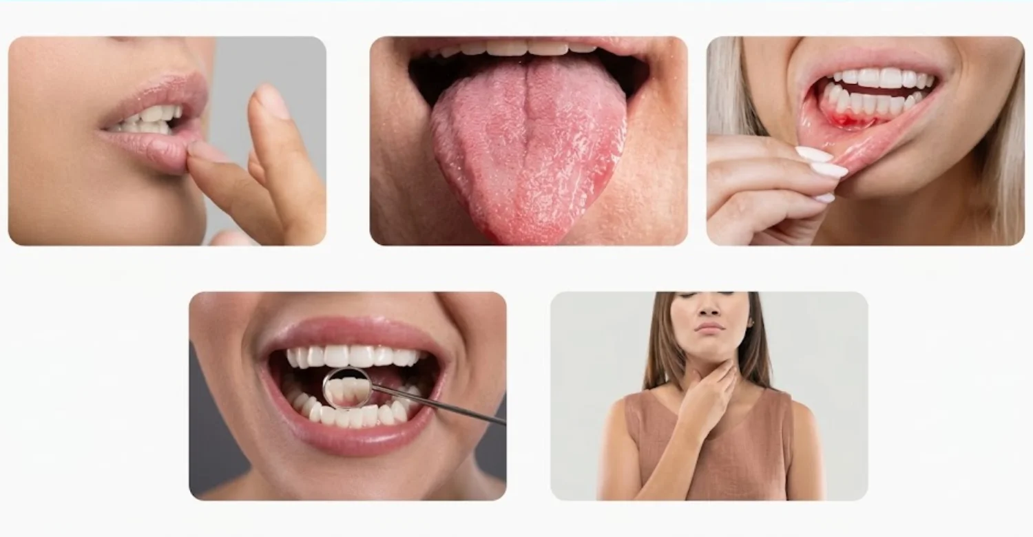
Note : All images used are for editorial and illustrative purposes only and may not originate from the original news provider or associated company.
What We Notice Clinically
Patient behavior strongly influences how quickly oral abnormalities reach a professional. People often wait because symptoms are painless. Others assume a lesion comes from a minor injury. Symptoms that improve and then return can also delay care. At Vitrin Clinic, individualized assessment helps separate routine dental concerns from findings needing further evaluation.
Common patient patterns that can delay oral cancer evaluation
A common delay involves waiting for pain that may never come. Another involves repeated home remedies without assessment. Some patients feel low-risk because they do not smoke. Others permanently blame dentures or appliances for persistent irritation. Each pattern can postpone a useful examination.
The difference between a common irritation and a persistent unexplained lesion
A common irritation usually has a credible local cause. Examples include cheek biting, sharp teeth, or loose appliances. Healing after removing the cause supports a benign explanation. Persistence despite removal changes the clinical picture.
Why symptom duration and change over time matter clinically
Time provides useful clinical information. A short-lived lesion behaves differently from one lasting weeks. Enlargement or surface change can raise concern. Patients should note when a lesion started and whether it recurs.
Why absence of smoking does not completely rule out oral cancer
Smoking is a major risk factor, but cancer can occur without it. HPV drives many oropharyngeal cancers. Alcohol, sunlight, age, and genetics also contribute. A nonsmoker with a persistent abnormality still needs evaluation.
Why a suspicious lesion needs assessment rather than self-diagnosis
Online images cannot reproduce clinical lighting, palpation, or patient history. Similar-looking lesions can have very different causes. A clinician weighs trauma, infection, inflammation, and neoplastic disease. Biopsy provides certainty when needed.
Clinical Note
Persistent oral abnormalities deserve structured clinical assessment. A qualified dental professional can identify concerning findings and decide whether referral is required. Findings should be interpreted alongside duration, location, examination, and risk history.
Dr. Rifat Alsaman’s Clinical Perspective on Oral Cancer Screening
Dr. Rifat Alsaman, Head of the Medical Team at Vitrin Clinic and Cosmetic Dentist, explains the approach. Clinical assessment should begin with careful examination rather than assumptions. Persistent oral changes should be documented and evaluated by location and behavior. A painless lesion can still deserve examination.
Learn how to detect a tooth infection on x-ray
How Often Should You Get an Oral Cancer Screening?
No single interval suits every patient. Risk factors, age, symptoms, previous lesions, and findings all shape the approach. Many dental practices include mucosal evaluation in routine visits. Patients with symptoms should never wait for a routine appointment.
Is there a universal oral cancer screening schedule?
No universal schedule fits everyone. Evidence on organized screening of asymptomatic adults remains limited. Dentists still examine oral tissues during routine care. Higher-risk patients may receive closer attention.
Should oral cancer screening be part of routine dental visits?
A comprehensive dental examination can include mucosal evaluation. ADA includes oral cancer evaluation within routine examination codes. Routine visits are therefore practical opportunities to find abnormalities. Symptoms between visits should still be reported promptly.
How risk factors can influence the clinical approach
Tobacco, alcohol, HPV, age, sunlight, previous cancer, and immune status can shape clinical attention. These factors do not establish cancer. They help clinicians decide how closely to reassess findings.
Why screening frequency should be individualized
People differ in risk and clinical history. Someone with previous suspicious lesions may need closer follow-up. New symptoms can also change timing. Individual scheduling makes more sense than one rigid interval.
What to do if you have symptoms between routine dental appointments
Book an appointment promptly. Explain the symptom and how long it has lasted. Mention swelling, bleeding, numbness, swallowing trouble, or neck changes. Do not wait for your next routine cleaning.
Oral Cancer Screening Benefits and Limitations
Clinical examination offers a simple way to find suspicious oral changes. It can also prompt referral and diagnostic testing. However, no examination detects every lesion. Mayo Clinic notes that some cancers can be missed because certain areas are hard to see.
What are the potential benefits of early detection?
Earlier recognition shortens the delay before diagnostic evaluation. Treatment planning can then start sooner when cancer is confirmed. CDC notes that cancers found earlier generally have better survival. Still, no examination can promise a better outcome for everyone.
What can a clinical oral cancer screening detect?
It can identify visible or palpable abnormalities. These include ulcers, patches, lumps, thickened tissue, and swelling. Functional changes may also prompt referral. It works best when systematic and combined with patient history.
What can an oral cancer screening not tell you?
It cannot reliably determine whether a lesion is benign or malignant. It cannot rule out microscopic disease. A normal result eases immediate concern but cannot remove future risk. Suspicious lesions may still need biopsy or imaging.
Why screening does not guarantee that cancer is absent
Some areas are difficult to see, and small lesions can be subtle. Some cancers cause few obvious symptoms. Mayo Clinic states that not all mouth cancers are detectable by examination. Persistent symptoms deserve reassessment even after a normal result.
What the current evidence says about screening asymptomatic adults
USPSTF finds insufficient evidence for screening asymptomatic adults through primary care. The recommendation specifically excludes dentists and otolaryngologists. Routine dental examinations and population screening policies are therefore different concepts.
.jpg)
.jpg)
Oral Cancer Screening Devices: What Do They Actually Do?
Adjunctive technologies aim to add information during lesion assessment. They include light-based systems, cytology tests, and salivary methods. The ADA's 2026 living guideline evaluates several of these approaches. None should replace conventional examination or indicated biopsy.
What are oral cancer screening devices?
These devices and tests supplement clinical examination. They may highlight tissue characteristics or collect cell samples. Examples include light-based systems and brush cytology. Their value depends on clinical context and expert interpretation.
What are light-based screening technologies?
These tools use special illumination to reveal optical differences in tissue. Unusual optical patterns are not the same as cancer cells. Clinical correlation remains necessary. The ADA's 2026 guideline specifically reviews these adjuncts.
What are cytology-based or brush-based tests?
Brush tests collect cells from an oral lesion for microscopic review. NCI describes exfoliative cytology and brush biopsy as cell collection methods. Cytology may help in selected cases. It does not replace biopsy when tissue diagnosis is needed.
What are salivary tests?
Salivary tests are being studied as possible adjuncts. The ADA's 2026 guideline includes them in its evidence review. Research is still defining their role. Patients should ask what a result can and cannot establish.
Can screening devices diagnose oral cancer?
Adjunctive devices generally cannot replace pathological diagnosis. A concerning lesion may still need biopsy. The ADA identifies biopsy as the diagnostic standard. Device results alone should not guide major decisions.
When should an adjunctive technology be considered?
Use should rest on clinical judgment and evidence. The device should answer a specific clinical question. It should never delay an indicated biopsy. It should never create false reassurance after a suspicious examination.
Find out why no exercise after dental implant
Oral Cancer Treatment After a Confirmed Diagnosis
Treatment depends on location, stage, pathology, overall health, and goals. NCI recommends planning with a team experienced in head and neck cancers. Oral cavity and oropharyngeal cancers may need different strategies.
How is treatment selected according to cancer type and stage?
Stage describes disease extent, including tumor size, nodes, and spread. Planning also considers tumor biology, general health, and patient preferences. Multidisciplinary teams may combine surgery, radiation, and systemic therapy.
What treatments may be used for oral cavity cancer?
Options include surgery and radiation therapy. Some patients also receive systemic therapy. NCI lists immunotherapy among options for selected cases. The combination depends on the individual diagnosis.
Surgery
Surgery may remove the primary tumor and, when needed, nearby lymph nodes. Reconstruction may follow. Rehabilitation can support speech, swallowing, appearance, and function.
Radiation therapy
Radiation uses high-energy beams to damage cancer cells. It may be used alone or after surgery. Planning depends on location, stage, and earlier treatment.
Systemic therapies when clinically appropriate
Systemic treatment can include immunotherapy and other drug therapies. NCI lists immunotherapy for selected recurrent or metastatic disease. Oncology teams decide when these are appropriate.
How is oropharyngeal cancer treated?
Options include surgery, radiation, chemotherapy, targeted therapy, and immunotherapy. HPV status can influence how the disease is characterized and managed. A head and neck team individualizes treatment.
Surgery
Selected patients may undergo tumor removal. Transoral procedures may suit appropriate cases. The approach depends on location, stage, and anatomy.
Radiation therapy
Radiation can be used alone or with chemotherapy. NCI describes several radiation-based approaches. Schedules depend on disease characteristics.
Chemotherapy, targeted therapy, or immunotherapy when indicated
These therapies depend on stage, recurrence, biomarkers, and treatment goals. NCI lists all three among options. Each requires oncology supervision.
Why multidisciplinary head and neck cancer care matters
Treatment can affect chewing, speech, swallowing, breathing, and oral health. Teams may include surgeons, oncologists, dentists, speech therapists, and nutrition specialists. NCI recommends planning through experienced head and neck teams.
What dental care may be needed before cancer treatment?
Pre-treatment dental assessment finds problems that could complicate therapy. NIDCR recommends this evaluation when possible. Infections, gum disease, or problem teeth may need treatment first. Timing should be coordinated with the oncology team.
What follow-up care is needed after treatment?
Follow-up monitors recurrence, treatment effects, swallowing, nutrition, and oral health. NCI notes that ongoing follow-up may be needed. Regular dental review helps manage long-term oral complications.
Oral Cancer Treatment Side Effects and Oral Health
Cancer treatment can affect the mouth in several ways. Chemotherapy may cause mouth sores and taste changes. Radiation can cause dry mouth and raise cavity risk. Dental care should be coordinated with oncology care.
What oral side effects can occur during treatment?
Patients may develop sores, dryness, taste changes, sensitivity, infections, or eating difficulty. Effects vary by therapy and treatment site. Reporting new symptoms early improves comfort.
How can radiation or cancer therapy affect the mouth?
Radiation can damage tissues within the treatment field. Reduced saliva increases discomfort and decay risk. Long-term effects depend on dose, location, and individual susceptibility.
What dental complications may require preventive management?
Possible complications include decay, gum problems, dry mouth, sensitivity, and infection. Preventive assessment before therapy helps catch these early. Patients should receive individualized preventive advice.
How can dentists support oral health before, during, and after cancer treatment?
Dentists treat oral disease before therapy and coordinate with oncologists. They help manage pain, infection, dryness, and prevention. Afterward, they support long-term hygiene and function.
When should patients contact their oncology or dental team about new symptoms?
Report severe pain, persistent ulcers, swelling, bleeding, or infection signs promptly. New swallowing problems also need quick communication. Timing depends on the therapy and symptoms.
Discover all about endosteal implants
Relief for Mouth Symptoms While Waiting for Evaluation
Temporary relief can ease discomfort before an appointment. It should never replace diagnostic evaluation. Reducing irritation and keeping gentle hygiene usually help.
What can help relieve minor mouth irritation safely?
Gentle brushing limits plaque buildup. Soft foods reduce trauma to tender areas. Avoiding very hot, spicy, or rough foods may help. Patients in cancer treatment should follow their team's advice.
How can patients avoid further trauma to a sore or irritated area?
Avoid biting or touching the lesion. Check whether a sharp tooth or denture edge rubs the area. A dentist can smooth or adjust the source.
Why symptom relief should not replace professional evaluation
Pain relief does not explain why a lesion exists. A suspicious sore may improve briefly while the cause remains. Persistent symptoms still require assessment.
What should you avoid applying to a suspicious oral lesion?
Avoid caustic substances, strong chemicals, and unproven concentrated remedies. Never burn, cut, or scrape the lesion. These actions worsen injury and complicate assessment.
Medical Tips for Reducing Oral Cancer Risk
Risk reduction targets modifiable exposures. Avoiding tobacco is especially important. Limiting alcohol, protecting the lips, and considering HPV vaccination also help.
Stop smoking and avoid smokeless tobacco
Tobacco is a major preventable risk factor. NIDCR notes that all tobacco forms raise risk. Patients needing cessation support should ask a healthcare professional.
Reduce or avoid excessive alcohol consumption
Heavy drinking increases risk. NHS advises reducing alcohol and avoiding excess. Combined tobacco and alcohol use deserves particular attention.
Discuss HPV vaccination with an appropriate healthcare professional
HPV vaccination prevents new infections from cancer-linked types. CDC recommends it routinely during adolescence. Adults should discuss their circumstances with a clinician. The vaccine does not treat existing infections.
Protect the lips from prolonged ultraviolet exposure
Ultraviolet exposure can increase lip cancer risk. Use suitable lip sun protection outdoors. Persistent lip changes still need examination.
Maintain regular dental care and oral hygiene
Regular dental visits create chances for examination and prevention. Good hygiene supports gum and tooth health. CDC recommends regular dental and medical visits.
Report persistent mouth or throat changes promptly
Do not wait for severe pain. Persistent symptoms deserve professional review. NHS and NIDCR give practical duration thresholds.
Prevention and Early Detection
Prevention and detection work together. Risk reduction addresses exposures, while examinations address suspicious changes. No strategy removes all risk.
Can oral cancer be prevented?
Not every case is preventable. However, several risk factors can be modified. Tobacco avoidance, less alcohol, lip protection, and HPV vaccination can lower certain risks.
How tobacco and alcohol prevention can lower risk
Avoiding tobacco and limiting alcohol reduces exposure to recognized carcinogens. Combined use deserves special attention because the risks interact.
Why regular dental examinations remain important
Dental visits allow inspection of oral tissues. They also reveal dental sources of irritation. Routine care should never replace symptom-driven appointments.
Why early detection should not depend on symptoms alone
Some lesions cause little discomfort. Patients may notice only a subtle visual change. Dental examinations can find abnormalities patients overlook.
The role of population-level oral cancer screening programs
Population programs require evidence that benefits outweigh harms. USPSTF currently finds insufficient evidence for asymptomatic adults in primary care. This differs from opportunistic dental examination.
How oral cancer screening programs differ by country
Healthcare systems use different detection approaches. Some rely on routine dental care. Others depend on specialist referral pathways. Policy reflects evidence, resources, and prevalence.
What combined cancer screening programs can and cannot tell patients
Programs target specific diseases and risk groups. A normal result does not exclude every cancer. Patients should know exactly what a program covers.
See the risks and timeline of smoking after tooth extraction
What Does Oral Cancer Screening Cost?
Prices vary widely by country and clinic. Some practices include the check in routine examination fees. Others charge separately for an oral cancer screening. Biopsy, pathology, imaging, or specialist review add further costs.
Country | Average cost | What it usually covers | Notes |
United States | About 100–150 | Oral exam, often bundled with a comprehensive dental exam | Based on advertised clinic examples, not a national average |
United Kingdom (NHS England) | $27.90 (Band 1) | Examination, diagnosis, preventive advice, and needed X-rays | Private practice fees are usually higher |
Turkey | From about $ 2,666 | Private-clinic examination, depending on the package | Marketplace listing; prices vary by clinic |
Average Oral Cancer Screening Cost in the United States
There is no single national private fee. Advertised examples vary considerably. Some clinics bundle oral evaluation with comprehensive exams and X-rays. One practice lists a $125 examination that includes cancer assessment.
Standalone screening fees versus screening included in a dental examination
Many dentists include the evaluation within periodic or comprehensive examinations. ADA coding supports this approach. Standalone services may be priced differently. Ask exactly what the fee covers.
How insurance, new-patient exams, X-rays, and adjunctive technologies can affect the price
New-patient packages may combine several services. X-rays or special devices can add cost. Insurance coverage varies by plan. Confirm what the quote includes.
Planning reference: approximately 100–150 for advertised U.S. examination/screening examples, not a national average
Treat this 100–150 range as a planning reference only. It reflects advertised examples, not a national tariff. Actual costs vary by location, insurance, and bundled services.
Average Oral Cancer Screening Cost in the UK
NHS dental pricing differs from private pricing. In England, NHS charges follow treatment bands. Private fees are set by each practice.
NHS England dental examination and diagnosis costs
NHS Band 1 covers examination, diagnosis, and preventive advice. Clinically needed X-rays are included. It is not a private standalone cancer examination.
Current 2026 NHS England Band 1 charge: $27.90
From 1 April 2026, Band 1 costs $27.90 in England. Band 2 costs $76.60, and Band 3 costs $332.10.
Why private dental fees can differ from NHS charges
Private practices set their own prices. Fees reflect clinician time, location, diagnostics, expertise, and technology. Compare service scope, not just numbers.
Average Oral Cancer Screening Cost in Turkey
Private prices vary by clinic and package. A current marketplace listing shows services from about $ 2,666. This is a planning reference, not a national average.
Private-clinic screening prices and why they vary
Prices depend on whether the visit is standalone. Specialist input, imaging, or extra tests also matter. Request a written breakdown of what is included.
Planning reference: listings from approximately $ 2,666, with clinic-specific pricing varying by examination and additional tests
Listings start at about $ 2,666 across multiple clinics. This is not a standardized rate. Vitrin Clinic pricing should be confirmed according to clinical needs.
What Can Increase the Cost Beyond a Basic Screening?
The examination itself is usually simple. Additional diagnostic services can change the total significantly.
Specialist consultations
Oral medicine, oral surgery, or ENT consultations add separate fees.
Biopsy and pathology
Biopsy involves procedure costs and laboratory pathology fees. These are separate from a basic examination.
Imaging when clinically indicated
CT, MRI, ultrasound, or dental imaging can add expense. NHS lists several of these modalities.
Follow-up examinations
Persistent lesions may need repeat assessments. Extra visits raise the overall cost.
Why Oral Cancer Screening Prices Are Not Directly Comparable Between Countries
Healthcare financing differs dramatically between countries. Headline prices rarely describe identical services.
Examination-only versus bundled dental checkups
One clinic may charge for the examination alone. Another may bundle X-rays, cleaning, or consultation.
Public versus private healthcare pricing
NHS England uses standardized bands. Private clinics set their own fee structures.
Insurance, VAT, laboratory, pathology, and specialist fees
Insurance, taxes, and laboratory charges differ by country. Compare total clinical scope rather than headline prices.
How to Find an Oral Cancer Screening Dentist Near You
Choose a dentist based on clinical suitability first. Location matters, but diagnostic competence matters more. Ask whether an oral cancer screening is part of every routine examination. Also ask how referrals are coordinated.
What to look for in an oral cancer screening dentist
Look for experience in oral mucosal examination. Ask how suspicious findings are documented and managed. The clinic should explain when referral or biopsy is appropriate.
Questions to ask before booking an oral cancer examination
Ask whether oral and extraoral tissues are both examined. Ask what happens if something abnormal appears. Confirm whether the fee covers additional tests.
When to seek a dentist versus an oral medicine, oral and maxillofacial, or ENT specialist
Dentists suit most initial oral findings. Oral medicine and oral surgery specialists manage complex mucosal lesions. ENT specialists handle persistent throat or deeper oropharyngeal concerns.
How local searches such as “oral cancer screening near me” should guide appointment decisions
Local search helps identify accessible clinics. It should not decide clinical suitability alone. Review qualifications, services, referral pathways, and communication.
Why city-based search terms should not replace clinical suitability
A nearby clinic helps only if it provides appropriate evaluation. A further specialist may suit a complex lesion better.
Oral Health Assessment and Personalized Care at Vitrin Clinic
Vitrin Clinic keeps every dental assessment individualized. Concerning findings receive clinical evaluation before any treatment is recommended.
Individualized oral assessment for patients with concerning oral findings
Symptoms, dental history, tissues, and findings are assessed together. The aim is understanding the likely cause before planning treatment.
Clinical examination of teeth, gums, oral tissues, tongue, and surrounding structures
A comprehensive assessment reviews teeth, gums, mucosa, tongue, jaw, and nearby structures. This identifies dental causes and findings needing referral.
Digital X-rays, CBCT, or 3D scanning only when clinically appropriate
Advanced imaging is chosen only when the clinical question justifies it. CBCT is not used automatically for every symptom.
Identifying the likely dental or oral cause before recommending treatment
Pain or irritation may come from infection, trauma, or gum disease. Diagnosis always comes before treatment planning.
Referral or specialist coordination when a finding requires evaluation beyond routine dentistry
Some findings need oral medicine, oral surgery, pathology, or ENT input. Appropriate referral supports safe diagnosis.
Internal link opportunity: Vitrin Clinic Treatments page
A natural link can guide relevant patients to the Vitrin Clinic treatment directory. Anchor text should describe the treatment without implying cancer care.
How Vitrin Clinic Supports Your Treatment Journey
Vitrin Clinic offers a structured dental journey for international patients. Clinical suitability always remains the first priority.
Initial consultation and review of symptoms and dental history
The first consultation documents symptoms, timing, previous treatment, and relevant history. Clear records guide the next step.
Personalized assessment and treatment planning
Planning follows diagnosis. Cosmetic or restorative work never comes before unresolved pathology is addressed.
Coordinating appropriate diagnostic appointments for international patients
International patients may need coordinated dental and specialist visits. Vitrin Clinic helps schedule them along the clinical pathway.
Digital records and clinically indicated imaging to support treatment planning
Digital records organize diagnostic information clearly. Imaging is used only when clinically indicated.
Treatment coordination, follow-up, and aftercare support
Follow-up confirms healing and addresses remaining dental needs. International patients receive coordinated aftercare communication.
Ready to transform your smile?
Talk to our team and get a personal FREE treatment plan.
Key Takeaways
A careful clinical examination is an important chance to detect suspicious oral changes. On its own, it does not confirm cancer.
Oral cancer screening begins with a careful clinical examination
Visual and tactile examination remains central. ADA supports conventional examination as the foundation of any oral cancer screening.
Persistent sores, lumps, red or white patches, bleeding, numbness, and functional changes need professional assessment
NIDCR and NHS list these findings as symptoms requiring evaluation.
A screening examination cannot confirm cancer; suspicious findings may require biopsy and specialist evaluation
Biopsy provides tissue for pathological diagnosis.
Tobacco, alcohol, HPV, ultraviolet exposure, and other risk factors should be considered when assessing risk
Risk depends on cancer site and personal history. HPV matters especially for oropharyngeal cancers.
Home checks can help you notice changes but cannot replace professional oral cancer screening
Self-checks build awareness. They cannot diagnose malignancy.
Screening costs vary substantially between the US, UK, and Turkey depending on the examination, healthcare system, and additional testing
U.S. private examples vary widely. NHS England uses fixed bands. Turkish listings differ by clinic.
Evidence and source opportunities for the article
The strongest sources are authoritative clinical and government organizations.
American Dental Association 2026 oral cancer living guideline and clinical guidance
The guideline evaluates cytology, light-based, salivary, and other adjuncts.
National Institute of Dental and Craniofacial Research oral cancer examination guidance
NIDCR covers examination, symptoms, risk, and diagnosis for patients.
CDC oral cavity, pharyngeal, and HPV-related cancer resources
CDC provides risk, symptom, HPV, prevention, and U.S. statistics information.
National Cancer Institute diagnosis, staging, treatment, and follow-up resources
NCI explains diagnosis, staging, surgery, radiation, systemic therapy, and follow-up.
NHS guidance on mouth cancer symptoms, referral, and dental care
NHS gives patient-focused symptom thresholds and diagnostic pathways.
USPSTF evidence regarding screening asymptomatic adults and the distinction between primary-care screening and dental examinations
USPSTF finds insufficient evidence for primary-care screening of asymptomatic adults. Its recommendation excludes dentists and otolaryngologists.
Recommended internal-link architecture for Vitrin Clinic
Internal links should support user intent without promoting unrelated treatments.
Vitrin Clinic Treatments page for relevant dental assessment and treatment pathways
Link only where a treatment is clinically relevant to the discussion.
Related Vitrin educational articles only where the existing article directly addresses a symptom, differential diagnosis, or oral-health concern
Use contextual anchors that accurately describe the linked topic.
Reference
FAQs
Dr. Rifat Alsaman has more than 5 years of clinical experience in dentistry and currently serves as the Head of the Medical Team at Vitrin Clinic. He is dedicated to providing exceptional patient care, overseeing treatment planning, and ensuring the highest clinical standards across the team. His expertise, attention to detail, and commitment to continuous professional development have helped countless patients achieve healthier, more confident smiles.


