
The question “Are teeth part of the integumentary system?” has a surprisingly nuanced answer. Standard anatomical classification places teeth within the oral and stomatognathic structures. The integumentary system includes skin and its appendages, such as hair, nails, and glands. Teeth share embryological similarities with several ectoderm-derived structures. However, embryological origin does not determine adult anatomical system classification. Teeth also differ chemically and structurally from skin, hair, and nails. Enamel is highly mineralized rather than keratinized. Therefore, teeth are best discussed as specialized oral structures rather than integumentary appendages.
Are Teeth Part of the Integumentary System?
The standard answer is generally no. The integumentary system is defined by the skin and its appendages. Teeth are not listed among those conventional structures. Instead, teeth function as specialized structures of the stomatognathic system. MeSH places tooth components under the dentition and mouth. Some textbooks mention teeth alongside ectodermal derivatives, which refers to embryology. So when students ask, “Are teeth part of the integumentary system?” the textbook answer is no. Teeth are also not bones, despite containing mineralized tissues. For patients, classification provides understanding but does not determine treatment needs.
The Direct Answer: Are Teeth Part of the Integumentary System?
The direct answer is no under standard adult anatomical classification. The integumentary system includes skin, hair, nails, sebaceous glands, and sweat glands. Teeth are specialized mineralized structures within the oral cavity. They work with the jaws, periodontal tissues, and muscles during chewing and speech. The confusion comes mainly from embryology. Teeth develop from oral epithelium interacting with neural crest-derived mesenchymes. Anyone asking “Are teeth part of the integumentary system?” should separate origin from adult function. Tooth development is a classic organogenesis model in developmental biology.
Why Anatomy Textbooks Usually Separate Teeth From the Integumentary System
Anatomy textbooks organize body systems according to adult structure and function. The integumentary system concerns the body's external covering and its accessory structures. Skin provides a barrier, while glands support thermoregulation and surface maintenance. Teeth have a different position and purpose. They sit within the oral cavity and interact closely with the jawbone. Their functions include biting, grinding, and supporting speech. For that reason, textbooks rarely answer “Are teeth part of the integumentary system?” with yes. Standard teaching keeps teeth in a separate oral category.
Why Some Sources Discuss Teeth as Ectodermal or Integumentary Derivatives
Some sources emphasize developmental relationships rather than adult system classification. Tooth development begins from a localized thickening of the oral ectoderm. This forms the dental lamina and later the tooth buds. Dental placodes share features with hair follicles and certain glands. Simplified sources may therefore group teeth with other ectodermal derivatives. This is why “Are teeth part of the integumentary system?” sometimes receives mixed answers online. Developmental literature describes odontogenesis as coordinated epithelial and mesenchymal organogenesis.
Standard Anatomical Classification vs. Embryological Classification
Standard anatomical classification focuses on location and function. Embryological classification focuses on developmental origin and cellular interactions. Under standard anatomy, teeth are oral structures of the stomatognathic system. Embryology presents a more complicated picture. The enamel-forming component comes from ectodermal epithelium. Other dental tissues develop through ectomesenchymal contributions. Both descriptions can be accurate within their intended context. The question “Are teeth part of the integumentary system?” is really an anatomy question, not an embryology one.
Why Definitions Can Differ Between Medical and Dental Sources
Medical and dental references sometimes use different organizational frameworks. General anatomy resources organize structures by major body systems. Dental sources focus on the oral cavity, dentition, and periodontium. This creates different emphasis without creating a scientific contradiction. MeSH lists skin and appendages under the integumentary system. It lists tooth components separately within the stomatognathic system. A reader searching “Are teeth part of the integumentary system?” may meet both framings. Definitions should always be interpreted according to the source's purpose.
What Body System Do Teeth Belong To?
Teeth are best discussed as specialized structures of the oral cavity and stomatognathic system. This system includes the mouth, jaws, teeth, chewing muscles, and temporomandibular structures. Teeth also interact with the digestive system by cutting and grinding food. That functional link does not make them digestive organs. Teeth connect with alveolar bone, yet they are not bones. That is the clearest reply to “Are teeth part of the integumentary system?” and related questions. Teeth support mastication, speech, and overall oral function.
Teeth as Specialized Structures of the Oral Cavity
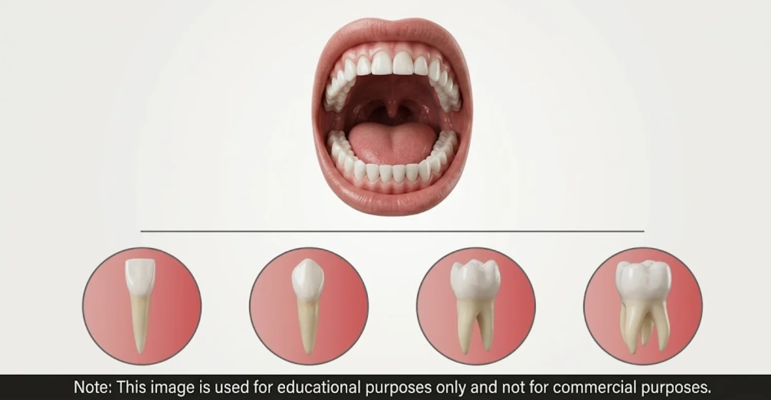
A tooth contains several tissues arranged into one functional structure. Enamel covers the crown, and cementum covers the root. Dentin forms most of the internal hard structure. Pulp occupies the central chamber and contains nerves and blood vessels. Periodontal tissues connect the root to surrounding alveolar bone. Teeth are embedded in jawbone sockets rather than attached to skin. Location alone helps answer “Are teeth part of the integumentary system?” for many readers. Human teeth contain multiple tissues with different developmental origins. Each tooth type, from incisor to molar, has a shape suited to its job.
How Teeth Relate to the Skeletal and Digestive Systems Without Being Bones or Skin
Teeth relate closely to both skeletal and digestive functions. Their roots sit inside alveolar bone, held by periodontal ligaments. However, teeth are not classified as bones. Their role in digestion is mechanical. Teeth reduce food size and prepare it for swallowing. Saliva begins chemical digestion while teeth perform mechanical breakdown. Asking “Are teeth part of the integumentary system?” is similar to asking whether they are bones. In both cases, the accurate answer points to specialized oral anatomy.
Understand the causes and treatment for trench mouth
What Is the Integumentary System?
The integumentary system is the body's external covering and its associated structures. NCBI's MeSH definition identifies skin and skin appendages as its main components. These appendages include hair, nails, sebaceous glands, sweat glands, and their ducts. The system limits water loss and protects deeper tissues. It also supports sensation, temperature regulation, and vitamin D synthesis. Understanding these components makes the tooth classification question easier. Before answering “Are teeth part of the integumentary system?” it helps to know what the system contains.
What Structures Make Up the Integumentary System?
The integumentary system contains skin plus several accessory structures. The skin includes the epidermis and dermis. The epidermis forms the outer nonvascular layer. The dermis provides deeper connective tissue support. Hair develops from follicles, and sebaceous glands often open into them. Sweat glands occupy the dermis and discharge through ducts. Nails are keratinized structures over the fingers and toes. Teeth simply do not appear on this list. That absence settles much of the “Are teeth part of the integumentary system?” discussion.
Skin
Skin is the primary component of the integumentary system. NCBI defines skin as the body's outer covering that protects against the environment. It consists of epidermis and dermis. The epidermis is a stratified epithelial layer. The dermis contains connective tissue, hair follicles, and glands. Skin provides protection and sensory input. It also supports thermoregulation and vitamin D synthesis. Unlike teeth, skin is primarily a surface barrier. Its thickness varies widely, from thin eyelid skin to thick skin on the soles.
Hair and Hair Follicles
Hair and hair follicles are classic integumentary structures. Hair is a filament extending from a root within a follicle. Follicles are epithelial invaginations with epidermal root sheaths. Hair shafts consist mostly of keratinized cells. Hair grows in cycles and can regenerate throughout life. Teeth differ considerably from hair. Their hard tissues are mineralized rather than keratinized. Adult teeth also do not regrow after loss. Comparing hair with teeth is a useful way to approach “Are teeth part of the integumentary system?” in class. Hair is shed and replaced constantly. A tooth, once fully formed, stays in place for decades.
Nails
Nails are specialized integumentary structures formed from keratinized cells. Their primary role is protecting the fingertips and toes. They also help with gripping small objects. Teeth may seem similar because both are hard. However, hardness alone does not determine biological classification. Mature enamel contains approximately 95% mineral by weight in published research. Nails, in contrast, are keratinized epithelial structures. Tissue composition provides a stronger basis for classification. Damaged nails grow out within months. Damaged enamel, in contrast, needs professional repair.
Sweat Glands and Sebaceous Glands
Sweat and sebaceous glands are classic appendages of the integumentary system. Sweat glands lie within the dermis and produce sweat. Eccrine glands discharge sweat directly onto the skin surface. Sebaceous glands secrete sebum, usually into hair follicles. Sweat helps cooling, while sebum maintains the skin surface. No equivalent glandular system exists within teeth. The dental pulp contains vessels and nerves, not secretory glands. This further supports separate classification.
What Does the Integumentary System Do?
The integumentary system performs essential protective and regulatory functions. It protects deeper tissues and reduces water loss. It supports touch, pressure, and temperature perception. Sweat and skin blood flow help regulate body heat. Skin also contributes to vitamin D synthesis. Teeth have different primary responsibilities. They process food mechanically and support speech. Function offers another lens on “Are teeth part of the integumentary system?” and again points to no.
Protection
Protection is one of the most important integumentary functions. Skin forms a physical barrier between internal tissues and the environment. The epidermis contributes strongly to this barrier. Hair and nails add specialized mechanical protection. Teeth also protect, but differently. Enamel shields deeper tooth tissues from mechanical and chemical challenges. It works within the mouth rather than as a body-wide barrier. Similar protective roles do not establish identical classification.
Temperature Regulation
Temperature regulation is strongly linked to the integumentary system. Sweat cools the body as it evaporates from the skin. Skin blood vessels also widen or narrow to manage heat. Teeth have no role in whole-body temperature control. They may sense temperature, but they do not regulate it. This difference is useful when comparing teeth with skin appendages. Sweat glands are distributed widely across almost the entire skin surface. Very cold air, however, can trigger sharp tooth sensitivity.
Sensory Function
The integumentary system plays a major sensory role. Skin receptors detect touch, pressure, vibration, temperature, and pain. Teeth also contain sensory structures. The pulp holds nerves, and dentin tubules transmit stimuli toward it. The periodontal ligament senses pressure during biting. Shared sensory function does not make two structures part of one system. Tooth sensitivity reflects dental tissue physiology, not skin appendage behavior.
Prevention of Water Loss and Other Barrier Functions
Skin is critical for limiting water loss from the body. Its layers separate internal fluids from the external environment. Skin also interacts constantly with microorganisms and outside substances. Teeth have a barrier function at a much smaller scale. Enamel shields deeper dental structures from the oral environment. However, enamel lacks the layered cellular architecture of epidermis. Similar protective outcomes do not establish the same system classification.
Vitamin D Production
The skin contributes to vitamin D synthesis after ultraviolet B exposure. Ultraviolet radiation converts a precursor molecule in the skin. The liver and kidneys then complete activation. Teeth do not perform this function. Dental tissues have very different biological roles. Teeth meet food, drinks, saliva, and oral microorganisms instead of sunlight. Shared environmental exposure does not imply shared system membership. Vitamin D still matters for teeth, because it supports calcium absorption.
Which Structures Are Not Normally Included in the Integumentary System?
Many structures are excluded from standard integumentary classification. Teeth are one example and belong with oral structures. Bones belong to the skeletal system. Muscles, nerves, blood vessels, and internal organs are separate categories too. The lens of the eye and parts of the nervous system also arise from ectoderm. Yet nobody classifies them as skin appendages. An anatomical system is not simply a list sharing one developmental feature. Classification depends on organization, location, function, and accepted convention. Teeth fit the same pattern.
How Are Teeth Different From Skin and Its Appendages?
Teeth differ from skin in composition, organization, location, and function. Skin consists of epidermal and dermal tissues. Hair and nails are keratinized derivatives. Teeth contain enamel, dentin, pulp, and cementum. Periodontal tissues connect them to alveolar bone. Skin renews itself constantly, while enamel cannot regenerate. A side-by-side comparison settles “Are teeth part of the integumentary system?” quite clearly. These differences support separate adult anatomical classification.
What Are Teeth Made Of?
A tooth is a complex structure containing several tissues. Enamel covers the crown. Dentin lies beneath enamel and forms most of the tooth. The central pulp contains connective tissue, blood vessels, and nerves. Cementum covers the root and anchors the periodontal ligament. The ligament attaches the tooth to the alveolar bone. Each tissue differs in composition and biological activity. Together, they form an organ adapted to chewing and oral sensation.
Enamel
Enamel forms the outer covering of the crown. It is the hardest tissue in the human body. Hydroxyapatite crystals give it exceptional hardness. Research estimates mature enamel at roughly 95% mineral by weight. Ameloblast cells produce enamel before eruption and are then lost. Therefore, mature enamel has very limited repair capacity. Enamel is not keratin. Its high mineral content explains both its hardness and its limited self-repair. Enamel also has no blood supply and no nerves. That is why early enamel wear often causes no pain at all. Regular checks help detect thinning before dentin becomes exposed.
Dentin
Dentin forms most of the tooth's internal hard structure. It lies beneath enamel in the crown and cementum in the root. Dentin is less mineralized than enamel. It contains microscopic tubules that contribute to sensitivity. Odontoblasts at the pulp edge produce dentin. Unlike enamel, dentin stays connected to living cells. This allows limited reparative responses over time. Dentin shows that teeth are complex mineralized organs, not hardened skin. Exposed dentin is a common reason for sensitivity to cold drinks. The tubules allow fluid movement that nerves inside the pulp can detect.
Dental Pulp
The dental pulp is the living central tissue of the tooth. It fills the pulp chamber and root canals. It contains connective tissue, blood vessels, and nerves. The pulp nourishes dentin and supports defense and repair. Pulp inflammation can cause significant sensitivity or pain. Root canal treatment targets diseased or irreversibly inflamed pulp. Hair and nails have no comparable vascularized central chamber. Because pulp sits inside rigid walls, swelling there has little room to expand. This explains why pulp inflammation can feel so intense.
Cementum and Periodontal Tissues
Cementum covers the external surface of the tooth root. Periodontal ligament fibers attach to it and to alveolar bone. The ligament provides support and sensory feedback. Gingiva and alveolar bone complete the supporting structures. Together, these form the periodontium. Periodontal disease can weaken support around teeth. This architecture makes a tooth a coordinated biological unit. The ligament also acts like a shock absorber during heavy chewing.
Why Are Teeth Hard but Not Bones?
Teeth and bones are both mineralized, but they remain distinct. Bone contains a collagen-rich matrix and constantly remodels itself. Mature enamel cannot remodel after an eruption. Enamel forms from epithelial ameloblasts, while dentin forms from odontoblasts. Bone can heal a fracture, but a cracked tooth cannot. Alveolar bone belongs to the skeleton, yet the tooth inside it does not. People asking “Are teeth part of the integumentary system?” often raise this question next. “Hard tissue” and “bone” are not interchangeable terms.
How Do Teeth Develop Compared With Skin, Hair, and Nails?
Teeth and integumentary structures share some developmental principles. Tooth development starts with oral ectoderm and neural crest-derived mesenchyme. It passes through bud, cap, bell, apposition, and root stages. Skin and hair follicles also form through epithelial-mesenchymal interactions. Similar signaling pathways guide placodes in teeth, hair, and some glands. However, hair becomes keratinized while enamel becomes mineralized. This is the heart of the “Are teeth part of the integumentary system?” debate.
The Role of Oral Epithelium and Dental Tissue Development
Oral epithelium plays a central role in starting tooth formation. A localized thickening forms the dental lamina. Signals then pass between epithelium and the underlying mesenchyme. These signals control tooth shape, position, and tissue differentiation. The enamel organ forms enamel. The dental papilla forms dentin and pulp. The dental follicle forms cementum, ligament, and supporting bone. The resulting tooth becomes specialized for chewing and oral sensation.
Ectodermal Contributions to Tooth Development
Ectoderm makes an important contribution to tooth development. Enamel-forming epithelium originates from oral ectoderm. Neural crest-derived mesenchyme forms dentin, pulp, cementum, and periodontal tissues. Ectodermal dysplasias can affect hair, nails, sweat glands, and teeth together. That shared vulnerability shows a real developmental link. However, ectodermal contribution is not adult system membership. Embryology and anatomy answer different questions.
Are Teeth Considered Organs?
Teeth can be described as organs because they contain coordinated tissues. Each includes enamel, dentin, pulp, cementum, and supporting structures. These tissues have different properties and origins. Tooth development is a widely studied model of organogenesis. Calling a tooth an organ does not place it in the integumentary system. Dentists evaluate each tooth as a complete unit. Problems may involve any of its tissues. Humans develop two sets of these organs, primary teeth and permanent teeth.
Learn about causes and symptoms of oral stomatitis
Why Do People Think Teeth Are Part of the Integumentary System?
The confusion usually comes from developmental biology. Teeth arise from oral epithelium through ectodermal signaling. Hair follicles and glands form through similar interactions. Teeth and nails also look alike because both are hard. That similarity misleads when tissue composition is ignored. Quiz questions such as “Are teeth part of the integumentary system?” add to the confusion. Short answer keys rarely explain the embryology behind them. The phrase “ectodermal derivative” is another frequent source of mixed messages.
Similarities Between Teeth, Nails, Hair, and Other Ectoderm-Derived Structures
Teeth, hair, and nails share developmental similarities involving epithelial tissues. All three begin with epithelial thickening and mesenchymal signaling. All three also become mechanically durable. Yet durability comes from different materials. Hair and nails rely on keratin. Teeth rely on mineralized enamel, dentin, and cementum. A broken nail grows back, while a chipped tooth needs dental repair. The similarities explain confusion without proving identical classification.
How Keratin Can Cause Confusion About Tooth Classification
Keratin is a structural protein found in hair and nails. Because teeth are hard and visible, some people assume enamel is keratin. That assumption is incorrect. Mature enamel is dominated by hydroxyapatite mineral crystals. Its organic component is very small. Hair and nails get their hardness from organized keratin. A simple rule helps students remember. Keratinization does not define enamel, but mineralization does. That rule quickly resolves “Are teeth part of the integumentary system?” for many learners.
Why Enamel Should Not Be Confused With Keratinized Skin or Nails
Enamel and keratinized tissues have different materials and properties. Enamel is about 95% inorganic mineral by weight. Keratinized structures contain large amounts of keratin proteins. Ameloblasts form enamel before eruption and then disappear. This limits enamel's natural repair ability. Nails and hair keep growing from living epithelial regions. Lost enamel must be protected or restored by a dentist. Similar hardness does not mean similar tissue origin.
What We Notice Clinically
Patients rarely ask about anatomical classification without a reason. Some learned about ectodermal tissues in school. Others compare tooth hardness with nails or bones. Dental symptoms can also trigger these questions. When a patient asks “Are teeth part of the integumentary system?” we also ask about symptoms. At Vitrin Clinic, discussions focus first on symptoms and oral findings. Classification stays educational rather than diagnostic. Tooth pain can involve decay, cracks, pulp inflammation, gum disease, grinding, or sensitivity.
Common Patient Misconceptions About Teeth and Body Systems
Several misconceptions appear repeatedly. One says teeth are bones because both contain calcium and phosphate. Another says enamel is keratin because nails and enamel are hard. A third says ectodermal origin makes teeth skin appendages. Patients may also believe tooth pain signals a systemic disease. Most tooth pain has a local dental explanation. NHS guidance lists decay, abscesses, cracks, damaged fillings, gum disease, grinding, and sensitivity. Clearing up these myths makes dental information easier to understand.
Why Tooth Pain or Sensitivity Is a Dental Issue Rather Than a Sign That Teeth Belong to the Integumentary System
Tooth pain usually reflects problems in dental or nearby oral tissues. Decay can irritate deeper tooth structures. Cracks can expose sensitive dentin. Pulp inflammation can cause significant pain. Gum disease can cause pain and swelling around teeth. Having nerves does not make a tooth a skin structure. Pain is evaluated by finding its source, not by assigning a system label. A dentist examines the tooth, gums, bite, and nearby tissues.
When a Question About Tooth Classification May Actually Reflect an Oral Health Concern
An anatomy question may sometimes hide a practical symptom. A person asking “Are teeth part of the integumentary system?” may actually notice a change. That change might be sensitivity, discoloration, cracking, gum bleeding, or pain. Persistent toothache, swelling, fever, or painful biting needs dental evaluation. NHS guidance recommends urgent assessment for persistent mouth lumps, patches, or ulcers. At Vitrin Clinic, clinicians assess teeth, gums, oral tissues, muscles, and jaw function.
Tooth Pain
Tooth pain can arise from several dental conditions. Causes include decay, abscesses, cracks, broken fillings, gum disease, grinding, and sensitivity. Some people notice brief twinges, while others feel prolonged throbbing. Pain on biting may suggest damage to tooth structure or supporting tissues. Swelling can indicate infection. NHS guidance recommends dental assessment when pain lasts longer than two days. The classification question does not change this need. Keeping a short pain diary can make your appointment more productive.
Tooth Sensitivity
Tooth sensitivity often produces short, sharp discomfort. Triggers include cold drinks, hot foods, sweets, air, or brushing. Exposed dentin tubules make these stimuli more noticeable. Sensitivity can also follow some dental procedures. A dentist may check enamel wear, gum recession, cracks, or decay. Tell your dentist whether pain is brief or lingering. Persistent or worsening sensitivity warrants professional evaluation. Students revising “Are teeth part of the integumentary system?” sometimes link sensitivity to skin nerves. In reality, dental sensitivity follows its own dentin and pulp pathways.
Broken, Cracked, or Worn Teeth
Teeth can crack, chip, or wear through several mechanisms. Trauma, grinding, and acid exposure are common causes. A cracked tooth may hurt on biting or with temperature changes. The visible size of a defect may not show its depth. A small chip may need only a simple restoration. A deeper crack may affect the pulp or supporting tissues. Examination and radiographs help determine the extent of damage.
Gum Swelling or Bleeding
Swollen or bleeding gums can indicate gingivitis or periodontal disease. Plaque buildup commonly causes gum inflammation. Advanced disease can affect deeper supporting tissues. Gums are oral soft tissues, not evidence of integumentary membership. Persistent swelling or spontaneous bleeding should not be ignored. A dentist may check plaque, pocket depths, tooth mobility, and bone support. Early evaluation helps treat problems before they become complex.
Clinical Note
“From a clinical perspective, teeth should be understood as specialized oral structures. Their developmental relationship with ectoderm does not make them conventional skin appendages.” The key distinction is between developmental origin and adult classification. A tooth contains enamel, dentin, pulp, and cementum. Periodontal tissues connect it with the jaw. Symptoms should therefore be evaluated through dental anatomy. Pain, sensitivity, discoloration, cracks, and gum changes may reflect different conditions. At Vitrin Clinic, individualized evaluation connects symptoms with appropriate dental findings.
Find out how to quit smoking with nicotine gum
What Symptoms Are Related to Actual Dental Problems?
Dental problems often present through changes patients can recognize. Pain, sensitivity, swelling, bleeding, discoloration, cracks, and chewing discomfort all matter. One symptom may have several possible causes. Some conditions cause few symptoms at first. Regular examinations can detect issues before discomfort appears. Facial swelling, fever, and breathing or swallowing difficulty need urgent care. Symptoms should be judged through clinical findings, not through “Are teeth part of the integumentary system?”
Can Tooth Problems Cause Pain or Sensitivity?
Yes, several dental conditions can cause pain or sensitivity. Decay can reach deeper tooth tissues. Cracks can expose dentin or irritate the pulp. Gum disease can cause discomfort around the tooth. Inflammation inside the confined pulp space can be painful. The pattern matters for diagnosis. Brief pain after cold differs from lingering spontaneous pain. A dental examination can distinguish among these possibilities.
What Can Cause Tooth Discoloration, Cracks, or Enamel Damage?
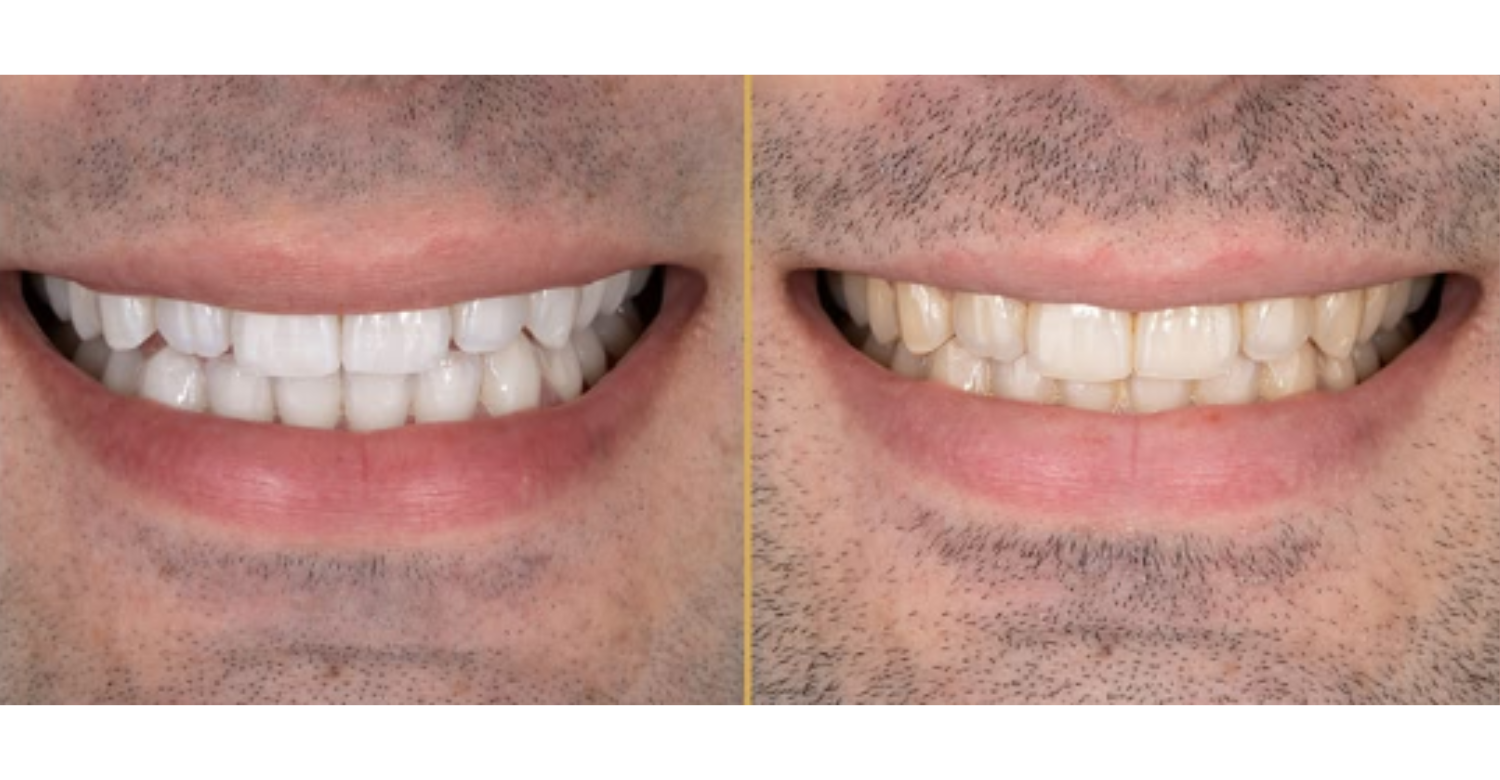
Tooth discoloration has several possible causes. Extrinsic stains come from food pigments, tobacco, or surface deposits. Intrinsic changes may involve development, trauma, medications, or internal changes. Cracks can follow trauma, heavy biting, large fillings, or grinding. Enamel damage can involve decay, erosion, abrasion, or developmental defects. Some stains respond to cleaning or whitening. A single dark tooth after trauma may need pulp assessment. Treatment must always be diagnosis-driven. Acidic drinks, frequent snacking, and reflux can all contribute to enamel erosion.
What Oral Symptoms Should Not Be Ignored?
Persistent oral symptoms deserve appropriate assessment. A toothache lasting more than two days should be checked by a dentist. Pain that continues despite painkillers also needs attention. Growing lumps and red or white patches require assessment. An ulcer lasting two weeks or longer warrants evaluation. Swelling affecting breathing or swallowing requires emergency care. These signs mean assessment should not be delayed.
Persistent or Severe Toothache
Persistent toothache may indicate an unresolved dental problem. Causes include decay, pulp inflammation, infection, cracks, gum disease, or trauma. Pain that remains despite painkillers needs professional attention. Severe pain affecting sleep may require urgent dental care. Pain relief reduces symptoms but does not remove the cause. Describe timing, triggers, location, and severity to your dentist. Pain that wakes you at night is a common sign of pulp involvement.
Facial or Jaw Swelling
Facial or jaw swelling can accompany dental infection or inflammation. A swollen cheek with tooth pain warrants dental assessment. Swelling near the eye or neck is more concerning. Large swelling can affect nearby structures. The cause cannot be confirmed from appearance alone. Mention how quickly swelling developed and any fever. Rapidly worsening swelling requires urgent evaluation. Cold compresses on the outside of the face may ease discomfort temporarily. They do not treat an underlying infection.
Fever With Dental Symptoms
Fever can occur with dental infection or other inflammatory conditions. Fever with tooth pain, swelling, bad taste, or painful biting needs assessment. It may mean the problem extends beyond a local surface issue. Do not delay care just because pain temporarily improves. A dentist can assess the tooth and surrounding tissues. Imaging may help when deeper infection is suspected.
Difficulty Swallowing or Breathing
Difficulty breathing or swallowing with oral swelling is an emergency warning sign. NHS guidance advises immediate emergency care in this situation. Severe swelling near the throat can become dangerous. Home remedies are not appropriate here. Contact emergency services according to your location. The priority is protecting the airway. Do not wait for a routine dental appointment.
Unexplained Oral Bleeding or a Persistent Oral Lesion
Unexplained oral bleeding can have dental or soft-tissue causes. Gum inflammation and trauma commonly cause local bleeding. Persistent lesions need a different level of attention. NHS guidance recommends urgent assessment for growing or persistent lumps and patches. An ulcer lasting two weeks or longer should also be checked. These signs do not automatically mean serious disease. A clinician can decide on monitoring, treatment, or referral.
Understand the function and treatments for a frenum in the mouth
Note : All images used are for editorial and illustrative purposes only and may not originate from the original news provider or associated company.
How Are Dental Problems Diagnosed?
Dental diagnosis usually begins with a clinical examination. The dentist checks teeth, gums, bite, jaw function, and soft tissues. History matters equally, including pain timing, triggers, trauma, and medications. Imaging is added when examination alone is not enough. Diagnosis never depends on answering “Are teeth part of the integumentary system?” At Vitrin Clinic, diagnostic steps follow individual findings rather than routine over-testing. The aim is finding the true source of symptoms.
What Does a Dentist Check During an Oral Examination?
A dental examination covers more than visible tooth surfaces. The dentist checks enamel, restorations, cracks, decay, and tooth mobility. Gums and surrounding tissues are also assessed. The bite is evaluated for abnormal contacts. Jaw movement and muscles may be checked when symptoms suggest them. Oral soft tissues are inspected too. Combining history with findings separates tooth problems from gum, muscle, or joint problems. The visit is also a good moment for anatomy questions. A dentist can explain “Are teeth part of the integumentary system?” using your own mouth as an example.
Teeth and Enamel
The dentist examines teeth for decay, fractures, wear, discoloration, and defects. Enamel loss can expose dentin and increase sensitivity. Margins around fillings and crowns are also inspected. Visual examination finds obvious defects. Deeper problems may require radiographs. Wear patterns can suggest grinding, erosion, or abrasion. These findings guide diagnosis and planning.
Gums and Periodontal Tissues
The examination includes gingiva and periodontal tissues. The dentist looks for redness, swelling, bleeding, recession, and mobility. A small probe measures pocket depth around each tooth. Periodontal disease can gradually weaken tooth support. Bleeding is often the first sign patients notice. Routine periodontal checks therefore offer valuable preventive information. Healthy pockets are usually shallow, while deeper pockets may indicate disease.
Bite and Jaw Function
The bite affects how teeth meet during chewing. Abnormal contacts can cause wear, fractures, or muscle discomfort. The dentist observes jaw opening, closing, and chewing patterns. Muscle tenderness can separate muscular pain from tooth pain. This matters when symptoms occur without obvious decay. The plan can then address the actual source. Night grinding often shows as flattened tooth edges and morning jaw stiffness.
Oral Soft Tissues
Oral examinations also include soft tissues. Dentists inspect the cheeks, lips, tongue, palate, and floor of mouth. Persistent lesions, swelling, ulcers, or color changes may need further assessment. The duration of a lesion matters. Many early changes are painless. This makes routine checks valuable even without symptoms. Your dentist may also feel the neck and jaw for swollen lymph nodes.
When Are Dental X-Rays Used?
Dental X-rays are used when internal information is needed. They reveal decay between teeth, roots, bone levels, and infections. NHS England's Band 1 can include clinically needed X-rays. A periapical image focuses on one tooth and its root. Bitewings show crowns and nearby bone levels. Imaging should always be clinically justified. Not every visit requires new X-rays. Students who wonder “Are teeth part of the integumentary system?” often find radiographs helpful. X-rays clearly show enamel, dentin, pulp, and surrounding bone as separate layers.
When Might Panoramic Imaging or CBCT Be Considered?
Panoramic imaging shows both jaws in one broad view. It suits selected orthodontic, surgical, eruption, or implant assessments. CBCT provides three-dimensional information. It is considered when standard images cannot answer a key question. The 2026 Turkish Dental Association tariff lists panoramic imaging and tomography separately. CBCT should not be performed simply because it is available. At the Vitrin Clinic, imaging choices are individualized.
Can a Dentist Determine Whether a Symptom Comes From a Tooth or Surrounding Tissue?
Often, a structured examination can narrow the source. Tools include history, percussion, bite testing, vitality testing, periodontal checks, and imaging. Tooth pain can overlap with gum, muscle, or jaw joint pain. Pain can also feel far from its true source. Upper tooth pain may even feel like sinus pressure. The final assessment combines all findings together.
Read about the oral symptoms of leprosy and oral health
What Treatment Is Needed for a Tooth Problem?
Treatment depends on the underlying dental diagnosis. Healthy teeth may need only preventive care. Decay may need restoration, and pulp disease may need root canal treatment. Gum disease requires periodontal management. Non-restorable teeth may need extraction and replacement. The answer to “Are teeth part of the integumentary system?” never decides a treatment plan. At Vitrin Clinic, planning is based on individual examination findings.
Treatment Depends on the Underlying Dental Diagnosis
Dental treatment should address the actual cause. A cavity needs decay control and restoration. Pulp inflammation may need endodontic treatment. Cracks require evaluation of depth, location, and restorability. Replacement is considered only when a tooth is missing or must be removed. Two people with tooth pain may need different treatments. Accurate diagnosis avoids unnecessary procedures and preserves natural tooth structure.
Preventive Dental Care
Preventive care helps maintain healthy teeth and gums. Routine examinations catch early changes. NHS guidance recommends brushing twice daily for about two minutes with fluoride toothpaste. Cleaning between teeth removes plaque and debris. Reducing frequent sugary snacks and drinks supports oral health. Some patients need closer monitoring because of higher risk. Prevention depends on habits, not on body-system labels. Professional cleaning removes hardened deposits that brushing cannot reach. Fluoride varnish may also benefit patients with higher decay risk.
Restorative Dentistry
Restorative dentistry repairs or replaces damaged tooth structure. Options include fillings, inlays, onlays, and crowns. The choice depends on remaining tooth structure and function. Small cavities may need conservative restorations. Heavily damaged teeth may need more protection. Restorations should preserve healthy tissue whenever possible. At Vitrin Clinic, restorative planning can use digital examination records. Material choice can include composite, ceramic, or other options. Each option balances strength, appearance, and the amount of tooth preparation required.
Root Canal Treatment
Root canal treatment addresses diseased or irreversibly inflamed pulp. The procedure removes affected pulp tissue and cleans the canals. The canals are then shaped, disinfected, and sealed. The goal is preserving a restorable natural tooth. Symptoms can include lingering sensitivity, spontaneous pain, or pain on biting. A crown often protects the treated tooth afterward. Not every painful tooth needs a root canal. Modern anesthesia makes the procedure comparable to having a filling for most patients.
Periodontal Treatment
Periodontal treatment addresses disease affecting gums and supporting structures. Early gingivitis may improve with cleaning and better plaque control. Periodontitis can involve deeper pockets and bone loss. Treatment depends on disease severity. A dentist or periodontist may recommend non-surgical or surgical care. Regular maintenance keeps results stable. Healthy teeth depend on healthy supporting tissues. Smoking and poorly controlled diabetes can make gum disease harder to manage. Addressing these factors often improves periodontal treatment outcomes.
Tooth Extraction When Clinically Necessary
Extraction may be considered when a tooth cannot be predictably restored. Severe fracture, extensive decay, or advanced bone loss can cause this. Extraction should not be the automatic first option. The dentist should consider whether the tooth can be saved. Replacement options can be discussed when extraction is needed. Clear aftercare instructions support healing.
Dental Implants or Other Tooth-Replacement Options
Missing teeth can be replaced with implants, bridges, or dentures. Implants use a fixture placed within the jawbone. A crown then replaces the visible tooth. Suitability depends on bone, gum health, and medical factors. Digital imaging helps evaluate bone before implant treatment. At Vitrin Clinic, individualized assessment supports planning for international patients. Bridges rely on neighboring teeth, while dentures are removable and often more affordable.
Is There Any Treatment Needed Because Teeth Are or Are Not Part of the Integumentary System?
No treatment is required simply because of anatomical classification. Treatment becomes appropriate only when a dental condition exists. Examples include decay, pulp disease, gum disease, fractures, wear, or tooth loss. Classification is mainly educational. It does not create a disease state. So “Are teeth part of the integumentary system?” is a learning question, not a treatment question.
.jpg)
Relief, Medical Tips, and Side Effects
Temporary relief can help while arranging dental care. However, relief does not replace diagnosis. NHS guidance suggests ibuprofen or paracetamol when appropriate. A pharmacist can advise on suitability. Soft foods and avoiding temperature extremes may reduce discomfort. Persistent pain still requires professional evaluation. Whatever the answer to “Are teeth part of the integumentary system?” real pain needs real care.
What Can Help Relieve Mild Tooth-Related Discomfort Before a Dental Appointment?
Mild discomfort can sometimes be managed temporarily. Ibuprofen or paracetamol may help when suitable. Soft foods reduce pressure on a sore tooth. Avoid very hot, cold, or sugary foods. Keep brushing gently. NHS guidance describes a rinse of half a teaspoon of salt in warm water. Children should not use salt-water rinses because they may swallow them. These measures provide temporary relief only.
What Should Patients Avoid When Experiencing Tooth Pain or Sensitivity?
Avoid actions that worsen irritation. Very hot, cold, or sugary foods may trigger pain. Avoid chewing heavily on the painful tooth. NHS guidance recommends avoiding smoking during toothache. Never place aspirin directly on the gum, as it can burn tissue. Avoid unverified home treatments that may damage enamel. Delayed treatment can make a tooth harder to restore.
When Should Pain Relief Not Replace a Dental Evaluation?
Pain relief should never be the only strategy for persistent symptoms. See a dentist when a toothache lasts more than two days. Fever, swelling, painful biting, or bad taste strengthen that need. Emergency care is needed when swelling affects breathing, swallowing, or speaking. Medication can hide infection, decay, or cracks without treating them. Sometimes pain fades because the pulp has died while infection continues.
What Side Effects Can Occur With Common Dental Treatments?
Dental procedures can produce temporary effects. Sensitivity may follow restorative, whitening, or periodontal procedures. Mild tenderness can follow injections or extractions. Root canal treatment can leave short-term discomfort. Medication side effects depend on the drug, dose, and patient. Dentists should explain expected effects beforehand. Persistent or severe symptoms should always be reported.
Temporary Sensitivity After Certain Dental Procedures
Temporary sensitivity can follow some dental treatments. Tooth preparation, whitening, and periodontal care are common triggers. Symptoms often improve as tissues recover. Duration varies between people and procedures. Persistent sensitivity should be checked rather than assumed normal. Describing the trigger and duration helps your dentist.
Post-Treatment Discomfort
Some discomfort after dental treatment is expected. Soreness may follow extraction, periodontal care, or injections. Severity should generally match the procedure. Increasing pain, swelling, or fever may signal a complication. Patients should know when to contact the dental team. At Vitrin Clinic, international patients can receive planned follow-up and aftercare support. Most expected soreness eases gradually over a few days.
Possible Medication Side Effects and Why Professional Guidance Matters
Dental care may involve pain medicines, antibiotics, or anesthetics. Every medicine has potential side effects. Risk depends on health, allergies, interactions, dose, and duration. Antibiotics should not be taken just because a tooth hurts. Unnecessary use contributes to antibiotic resistance. A dentist or pharmacist can guide appropriate use. Always disclose medications and allergies before treatment.
Discover the impact of the oral virome in health and disease
How to Maintain Healthy Teeth and Gums
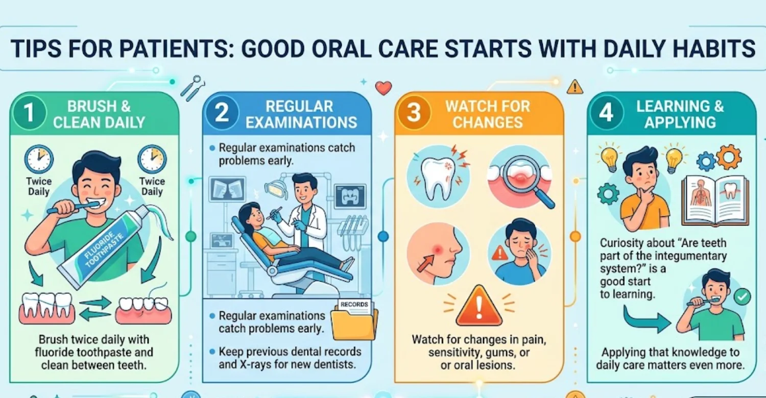
Brush twice daily with fluoride toothpaste. Clean between teeth daily with floss or interdental brushes. Limit frequent sugary foods and drinks. Attend regular dental examinations based on your risk. Avoid using teeth to open packages. Wear a mouthguard during contact sports. Rinse with water after acidic drinks. Consistent habits make prevention most effective. Replace your toothbrush every three months or sooner if bristles fray.
What to Tell Your Dentist About New or Persistent Oral Symptoms
Give your dentist a clear symptom history. Explain when the problem started. Describe whether pain is sharp, dull, throbbing, or temperature-related. Mention pain on biting, swelling, bleeding, fever, or bad taste. Report previous treatment, trauma, grinding, and current medications. Seemingly minor details can matter. A complete history speeds accurate diagnosis.
When to Ask for Dental Imaging
Ask about imaging when findings suggest hidden problems. Imaging can assess roots, bone, developing teeth, and decay between teeth. The best image depends on the clinical question. Not every visit needs new X-rays. Ask what each image is meant to show. Ask whether previous images can be used. CBCT should be chosen only when three-dimensional detail is needed.
Questions to Ask During a Dental Consultation
Direct questions improve understanding. Ask what the suspected diagnosis is and which findings support it. Ask whether imaging is necessary. Ask what treatment options exist, with their benefits and limits. Ask about recovery, side effects, and complications. Ask whether the tooth can be saved. Ask what happens if treatment is delayed. International patients should also ask what aftercare is included. You may also ask anatomy questions, including “Are teeth part of the integumentary system?” if you are curious.
Average Cost of a Dental Examination in
US
UK
Turkey
Dental examination prices vary considerably by country and provider. These figures are planning references, not fixed quotes. A 2026 US pricing guide places examination-only costs around $65 to $165. In England, NHS Band 1 costs $27.90 from April 1, 2026. The Turkish Dental Association lists a dentist examination at $ 1,650 including VAT. These figures describe different, non-equivalent services.
What Does a Dental Examination Cost in the United States?
A 2026 US pricing reference places examination-only costs around $65 to $165. The midpoint is about $115. Prices vary by city, provider, insurance, and visit type. New-patient examinations may cost more than recall visits. X-rays and cleaning may be billed separately. Ask for the full visit cost and check insurance coverage. These figures are not guaranteed.
What Does a Dental Check-Up Cost in the United Kingdom?
UK pricing depends on whether care is private or NHS. In England, the NHS Band 1 charge is $27.90 from April 1, 2026. Band 1 covers examination, advice, and clinically needed X-rays. Private check-ups vary by provider. One 2026 comparison places a private UK check-up around $65. Ask whether imaging or hygiene treatment is included.
What Does a Dental Examination Cost in Turkey?
The Turkish Dental Association's 2026 tariff lists a dentist examination at $ 1,650 including VAT. The pre-VAT amount is $ 1,500. Specialist examination is $ 2,035 including VAT. Periapical and bitewing radiographs are $ 830 each. Panoramic imaging is $ 2,040. These are reference figures, not guaranteed private-clinic prices. Exchange rates also affect how these amounts compare for visiting patients.
Average Dental Examination Cost Comparison
Country | 2026 planning reference | What it represents |
US | 65–165; midpoint about $115 | Dental exam without insurance; X-rays and cleaning may be additional. |
UK | $65 typical private check-up | Private dental check-up; X-rays and hygiene treatment may be separate. NHS England Band 1 is $27.90 from April 1, 2026. It can include examination, clinically needed X-rays, advice, and scale and polish when needed. |
Turkey | $ 1,650 incl. VAT | 2026 dentist examination reference. Specialist examination is listed at $ 2,035 incl. VAT in the cited Istanbul tariff reference. |
US pricing comes from a published 2026 guide. England's NHS charges are officially listed for 2026/27. Turkey's figure comes from the Turkish Dental Association's 2026 tariff. These values reflect different pricing systems, so direct comparison can mislead.
Important Cost Disclaimer: Published Planning Ranges Are Not Fixed Treatment Quotes
Published costs are for planning only. Fees vary by location, provider, scope, materials, and diagnostic needs. A routine examination differs from a specialist consultation. Private clinics may package services differently. Request an itemized written quotation. Confirm whether X-rays, CBCT, cleaning, planning, and follow-up are included. The Turkish tariff is a guide, not a universal price.
Factors That Can Increase the Cost: X-Rays, CBCT, Specialist Consultation, and Treatment Complexity
Additional diagnostic services can raise the total cost. Some patients need no imaging at all. Others need periapical, panoramic, or CBCT imaging. Specialist consultations may carry separate fees. Complex conditions may need multiple appointments. Treatment planning can be priced separately in some systems. Compare complete treatment plans rather than one headline price.
Personalized Dental Evaluation at Vitrin Clinic
A personalized evaluation connects symptoms with specific dental findings. It considers teeth, gums, soft tissues, chewing muscles, jaw function, and history. Digital X-rays support diagnosis when indicated. CBCT adds three-dimensional detail for selected complex cases. 3D scanning supports restorative and orthodontic workflows. The goal is the most useful diagnostic pathway, not every test. Vitrin Clinic can coordinate this process for international patients.
How a Dental Evaluation Can Clarify Tooth Anatomy and Oral Health Concerns
A dental evaluation begins with individualized assessment. The dentist reviews symptoms and examines teeth and supporting tissues. It can show whether symptoms start within a tooth or around it. Patients curious about “Are teeth part of the integumentary system?” can ask their dentist directly. Seeing your own X-rays makes enamel, dentin, and pulp easier to understand. At Vitrin Clinic, the aim is a justified plan, not a generic package.
When Digital X-Rays, CBCT, or 3D Scanning May Be Clinically Appropriate
Digital X-rays help when internal tooth or bone information is needed. CBCT may suit implant, surgical, orthodontic, or complex diagnostic cases. 3D scanning captures oral surfaces digitally. The best technology depends on the clinical question. Technology supports diagnosis but does not replace examination. Ask what information each scan should provide.
Personalized Treatment Planning When a Dental Problem Is Identified
A personalized plan links diagnosis with realistic options. The dentist should explain the current condition clearly. Plans may include prevention, restoration, root canal treatment, gum care, extraction, or replacement. Sequence matters as well. Disease control often comes before cosmetic work. Periodontal stability may be needed before implants. Vitrin Clinic organizes treatment steps around clinical requirements.
Internal-Link Opportunity: Vitrin Clinic Treatments Page
This section suits an internal link to the clinic's treatment catalogue. Readers may continue into restorative, implant, orthodontic, periodontal, or cosmetic information. A suitable anchor phrase is “Vitrin Clinic Treatments.” The destination should not imply that every patient needs treatment. Internal links help search engines connect educational and treatment pages. This creates a logical path from education to care.
How Vitrin Clinic Supports Your Treatment Journey
Dental treatment involves more than procedures. Patients may need diagnostic coordination, scheduling, transport, accommodation, and follow-up. This matters especially for international patients. The clinical component always remains the priority. Vitrin Clinic coordinates examinations and planning around individual findings. Digital workflows support communication between clinical and laboratory teams. The journey should stay transparent from assessment through aftercare.
Individualized Clinical Assessment Before Treatment
An individualized assessment sets the clinical starting point. It considers teeth, gums, soft tissues, muscles, and jaw function. Digital X-rays, CBCT, or scanning may be added when appropriate. A cosmetic concern may first reveal periodontal or restorative needs. A pain complaint may need endodontic assessment. Treatment begins after diagnosis and planning are complete.
Coordinating Appropriate Diagnostic Steps for International Patients
International patients often need efficient scheduling. Previous records and imaging can be shared before travel. This may reduce unnecessary duplication. Extra imaging is requested only when clinically justified. Vitrin Clinic's coordination team helps organize appointments and logistics. Clinical decisions remain with qualified dental professionals. Travel plans should never override necessary diagnostic steps. Sending recent X-rays in advance can also shorten the first appointment.
Translating Examination Findings Into a Personalized Treatment Plan
Findings should become clear, understandable treatment options. Patients need to know what was found and why treatment is advised. The plan should separate necessary care from optional cosmetic work. It should also show the order of procedures. Digital planning supports visualization and laboratory communication. A clear plan supports more informed decisions.
Follow-Up and Aftercare Following Dental Treatment
Aftercare is part of responsible dental treatment. Patients should receive hygiene, diet, medication, and healing instructions. Follow-up visits check healing and outcomes. International patients may need support after returning home. Remote communication can answer questions between visits. Significant symptoms may still require local evaluation. Vitrin Clinic coordinates follow-up around the treatment performed. Written aftercare instructions are especially helpful after long-distance travel.
See the risks of ignoring tooth sensitivity and progressive tooth wear
Evidence-Based Sources and EEAT Opportunities
This topic benefits from authoritative anatomy and dental sources. Strong evidence combines anatomical databases, textbooks, clinical guidance, and peer-reviewed research. Classification claims can rely on MeSH and anatomy references. Developmental claims should use embryology research. Warning signs should follow recognized health-system guidance. Cost claims need current, clearly identified references. Good sourcing turns “Are teeth part of the integumentary system?” into a verifiable answer.
National Library of Medicine and MeSH Definitions of the Integumentary System
NCBI MeSH provides a standardized integumentary system definition. It describes the body's outer covering of skin and skin appendages. Listed appendages include hair, nails, sebaceous glands, sweat glands, and ducts. MeSH places tooth components separately under the stomatognathic system. This offers consistent terminology rather than informal internet descriptions. Researchers worldwide use MeSH to index medical literature.
NIDCR Resources for Tooth Anatomy and Oral Health
NIDCR is a strong institutional source for oral health information. Its resources explain dental anatomy, diseases, prevention, and treatment in plain language. NCBI Bookshelf adds detailed structural information. StatPearls describes enamel, dentin, pulp, cementum, and periodontal relationships. Together, these offer accessible and technical views of the same structures.
OpenStax Anatomy and Physiology for Standard Integumentary System Classification
OpenStax gives a clear educational description of integumentary anatomy. It lists skin, hair, nails, sweat glands, and sebaceous glands as components. It also explains protection, sensation, temperature regulation, and vitamin D synthesis. The textbook is free, peer-reviewed, and widely used. Pairing it with MeSH strengthens classification coverage.
PubMed and Peer-Reviewed Literature for Tooth Development and Ectodermal Origins
Peer-reviewed literature is essential for developmental claims. Research describes tooth formation through oral epithelium and neural crest-derived mesenchyme. Recent reviews describe it as coordinated organogenesis. These sources explain the ectodermal link without calling teeth skin appendages. Ectodermal dysplasia studies add clinical evidence for shared pathways.
ADA, AAE, and Other Professional Dental Sources for Clinical Information
Professional dental organizations support the clinical sections. The ADA provides dental education and professional guidance. The AAE covers pulp disease and root canal care. Warning signs should be cross-checked with national health services. NHS guidance covers persistent toothache and swelling emergencies. Multiple sources avoid relying on one organization for every claim.
Ready to transform your smile?
Talk to our team and get a personal FREE treatment plan.
Key Takeaways
The anatomy question becomes easier once classification and development are separated. Standard references define the integumentary system as skin and its appendages. Teeth are not listed among these components. They are specialized oral structures within the stomatognathic system. Their ectodermal development explains why some sources link them with skin derivatives. In short, “Are teeth part of the integumentary system?” has a clear anatomical answer: no.
The Main Answer About Teeth and the Integumentary System
The conventional anatomical answer is no. Teeth are not classified as part of the integumentary system. That system includes skin, hair, nails, sweat glands, and sebaceous glands. Teeth belong within oral and stomatognathic anatomy. Their developmental origin explains most of the confusion. An ectodermal start does not make a tooth a skin appendage.
Why Tooth Classification Can Vary by Anatomical Context
Classification depends on the question being asked. Anatomy texts classify teeth by adult location and function. Embryology texts emphasize ectodermal origin and signaling. Dental pathology texts focus on tooth tissues and the periodontium. These descriptions can coexist without contradiction. The key is separating adult anatomy from developmental biology. Reading the source's purpose usually explains its chosen label.
What Patients Should Know About Tooth and Oral Health Symptoms
Patients should focus on symptoms and examination findings. Pain, sensitivity, cracks, discoloration, bleeding, and swelling have different causes. A dentist can assess the tooth and nearby tissues. Imaging helps when more information is needed. NHS guidance recommends assessment for toothache lasting more than two days. Early care often means simpler and less costly treatment.
Relief and Medical Tips When a Dental Symptom Is Present
Temporary relief can include suitable pain medication, soft foods, and avoiding temperature triggers. Warm salt-water rinses may help appropriate adults. These measures do not treat the cause. Arrange a dental evaluation when symptoms persist. Follow professional guidance and label instructions for medicines.
Warning Signs That Need Prompt Professional Dental or Medical Evaluation
Warning signs include severe persistent pain, fever, facial swelling, and worsening symptoms. Swelling that affects breathing or swallowing needs emergency care. Persistent ulcers, lumps, and abnormal patches also need evaluation. These signs do not establish a diagnosis. They show that delay may be unsafe.
When to See a Dentist Even if the Question Started as an Anatomy Question
An anatomy question can reveal an unresolved dental concern. See a dentist when pain persists or sensitivity worsens. Repeated gum bleeding or a changing tooth also needs review. Seek urgent care when swelling or fever appears. The anatomical label never changes the clinical need. Early evaluation usually offers clearer treatment choices.
Reference
FAQs

Dr. Rifat Alsaman has more than 5 years of clinical experience in dentistry and currently serves as the Head of the Medical Team at Vitrin Clinic. He is dedicated to providing exceptional patient care, overseeing treatment planning, and ensuring the highest clinical standards across the team. His expertise, attention to detail, and commitment to continuous professional development have helped countless patients achieve healthier, more confident smiles.


