General Dentistry
October 5, 2026
CT Scan for Salivary Gland: What Can It Detect and When Is It Needed?
 Dr. Rifat Alsaman
A CT scan for salivary gland problems is often requested when swelling, pain, or a lump needs a clearer explanation. A dentist, oral and maxillofacial specialist, or physician may recommend it for a suspected stone, infection, or mass. CT produces detailed cross-sectional images of the glands and nearby tissues. Still, the right test depends on the suspected condition. Findings are read alongside symptoms, examination, medical history, and sometimes ultrasound, MRI, or biopsy. Patients usually ask three things. Can CT detect a stone? Can it show a tumor? Does it diagnose cancer?
CT Scan for Salivary Gland: What Is It and Why Do Dentists Request It?
A CT-scan for salivary gland assessment uses X-rays and computer processing to build detailed, slice-by-slice images. It covers the three major pairs: the parotid, submandibular, and sublingual glands. Dentists and physicians may request it for persistent swelling, suspected stones, infection or abscess, a suspicious mass, trauma, or deep glandular disease. A routine dental X-ray is flat and focuses on teeth and bone. A salivary gland CT scan shows soft tissues and deep spaces in three dimensions.
What Does a Salivary Gland CT Scan Show?
A salivary gland CT scan shows the size and shape of each gland. It reveals calcifications and stones clearly. It also shows inflammation and changes in the surrounding fat and muscle. Fluid collections and abscesses are usually visible, especially with contrast. When a mass is present, the scan maps its location and borders. It can show whether changes extend into nearby structures. Enlarged lymph nodes and other incidental findings may also appear. Some of these need further assessment.
When Might Contrast Dye Be Used?
Contrast dye is an iodine-based liquid given through a small vein in the arm. It makes blood vessels, inflamed tissue, and certain masses stand out more clearly. Contrast is helpful when an abscess or tumor is suspected. It is not required for every CT scan for salivary gland concerns. A suspected stone, for example, is often seen well without it. The clinician considers the diagnostic question first. The radiology team then selects the protocol that answers it with the least exposure.
Learn about different types of partial dentures
What Can a CT Scan for Salivary Gland Detect?
A CT scan for salivary gland disease can detect several conditions in one examination. These include calcified stones, gland swelling, infection, abscess formation, and masses. It also shows how far a problem extends into the neck or jaw. That makes it valuable when symptoms are severe or the cause is unclear. However, detection is not the same as diagnosis. The scan describes what is present and where. Your clinician then decides what it means for you. The sections below explain each finding.
CT-Scan for Salivary Gland Stone
Sialolithiasis means stones forming inside a gland or its duct. They can block saliva flow, most often in the submandibular gland. Common symptoms include swelling around meals, pain or pressure, recurrent gland swelling, and dry mouth. A CT scan for salivary gland stone detection is particularly useful because calcified stones appear bright and distinct. The scan pinpoints the stone's position, size, and number. It also shows inflammation around the gland. This information helps the specialist plan removal or conservative care.
CT Scan for Swollen Salivary Gland
Salivary gland swelling has many possible causes. Viral or bacterial infection, a blocked duct, autoimmune inflammation, cysts, and tumors can all enlarge a gland. A CT scan for swollen salivary gland assessment helps distinguish between these. It can separate infection from obstruction, inflammatory change, a cystic lesion, or a solid mass. The pattern and location guide the next step. Importantly, swelling does not automatically indicate cancer. Most swollen glands turn out to be inflammatory or obstructive problems that respond to treatment.
CT Scan Salivary Gland Infection
Bacterial infection, called sialadenitis, usually affects the parotid or submandibular gland. The gland becomes painful, warm, and swollen. On a CT scan salivary gland infection typically appears as an enlarged gland with inflamed surrounding tissue. Contrast can reveal an abscess, which may need drainage. Severe infection can spread into the deep neck spaces, so prompt medical assessment matters. Never ignore fever, rapidly increasing swelling, difficulty swallowing, or difficulty breathing. These symptoms need urgent attention rather than a routine appointment.
CT-Scan for Salivary Gland Tumor
A persistent lump in or near a gland deserves imaging. A CT scan for salivary gland tumor evaluation shows where the mass sits and how large it is. It also shows its relationship to the jawbone, vessels, and deep neck spaces. This helps surgeons plan treatment and decide whether further tests are needed. Certain features, such as irregular borders, can raise suspicion. Yet imaging characteristics alone do not establish whether a tumor is benign or malignant. A tissue sample provides that answer.
Tips for Patients
Before Your Scan
Tell the imaging team about relevant medical conditions.
Mention previous reactions to contrast agents.
Follow preparation instructions if contrast or fasting is required.
Bring previous imaging reports when available.
Ask what type of CT is being performed and why.
After Your Scan
Follow the instructions provided by the imaging team.
Ask when and how the results will be reviewed.
Do not interpret isolated phrases from the radiology report as a diagnosis.
Explore options and fit for a bottom dentureSeek medical attention promptly if swelling worsens or severe symptoms develop.
Does a CT Scan Show Salivary Gland Cancer?
Does a CT scan show salivary gland cancer? It can reveal findings that raise suspicion, but CT alone generally cannot confirm cancer. Concerning features include irregular margins, invasion of nearby tissue, bone erosion, and abnormal lymph nodes. None of these is definitive. Further evaluation usually involves clinical examination, ultrasound, MRI in selected cases, and fine-needle aspiration or another biopsy. Pathology gives the final answer. A suspicious result on a CT-scan for salivary gland disease does not mean you definitely have cancer.
Can CT Tell Whether a Salivary Gland Tumor Is Benign or Malignant?
Not reliably. Benign and malignant tumors can look similar on imaging. Some cancers have smooth, well-defined borders. Some benign tumors look irregular. CT therefore establishes the next diagnostic step rather than the final diagnosis. It shows size, depth, and spread, which guides biopsy and surgical planning. When malignancy is suspected, tissue examination is required. A pathologist studies the cells under a microscope. Only then can the tumor type be named with confidence. Imaging and pathology work together, not in competition.
Will a Blocked Salivary Gland Show on CT Scan?
Many patients ask, "will blocked salivary gland show on CT scan?" Often, yes. Obstruction is usually caused by a stone, and calcified stones are clearly visible. A CT scan for salivary gland blockage may also show secondary signs. These include an enlarged gland, a widened duct, or surrounding inflammation. Visibility depends on the obstruction's location, size, composition, and the technique used. Narrowed ducts, mucus plugs, and poorly calcified stones can be missed. A normal CT does not eliminate every duct disorder.
Understand the causes and treatment for gum pockets treatment
Salivary Gland Ultrasound Scan vs CT Scan: Which Is Better?
Neither test is better in every situation. A salivary gland ultrasound scan is quick, widely available, and free of radiation. It is often the first test for a lump or swelling near the surface. A CT scan for salivary gland problems offers a wider view of deep tissues and bone. It is often preferred for deep infection or complex disease. Many patients have both tests at different stages. The choice depends on your symptoms, examination findings, and the question being asked.
What Can Salivary Gland Ultrasound Detect?
Ultrasound uses sound waves rather than X-rays, so it involves no radiation. It works well for superficial gland abnormalities. It can detect certain stones, cysts, and solid masses. It also shows blood flow and gland texture in real time. Ultrasound is commonly used to guide needle procedures, including fine-needle aspiration. That makes it especially useful for palpable or superficial lesions. Its main limits are deep structures and areas hidden behind bone. Results also depend on the operator's experience.
CT Scan vs Ultrasound for Salivary Gland Problems
Feature | CT Scan | Ultrasound |
Radiation | Yes | No |
Calcified stones | Very useful | Can detect some stones |
Superficial lesions | Useful | Often very useful |
Deep structures | Strong advantage | More limited |
Abscess/infection assessment | Useful | Useful in selected cases |
Needle guidance | Not usually the primary role | Commonly useful |
Detailed anatomical relationships | Strong | More limited |
The table summarizes the main differences. There is no single "best" scan for every salivary gland problem. Ultrasound suits superficial lumps and needle guidance. A CT scan for salivary gland disease suits deep spaces, calcified stones, and detailed anatomy. The appropriate test depends on the clinical question. Sometimes one test is enough. Sometimes the first result leads to a second, complementary test. Your clinician weighs radiation, availability, and the information needed before deciding.
.webp)
Salivary Gland Scan for Sjogren's: Is CT the Right Test?
Sjögren's disease is an autoimmune condition that commonly reduces saliva and tear production. Patients with persistent dry mouth may therefore undergo salivary gland evaluation. CT is not automatically the first or only salivary gland scan for Sjogren's. Diagnosis usually relies on clinical assessment, blood tests, and often a salivary gland ultrasound scan. Other imaging is added when clinically indicated. A minor salivary gland biopsy helps in selected cases. Any imaging finding must be interpreted within the wider diagnostic assessment.
Read about how to relieve throbbing tooth pain
What Happens During a CT Scan of the Salivary Glands?
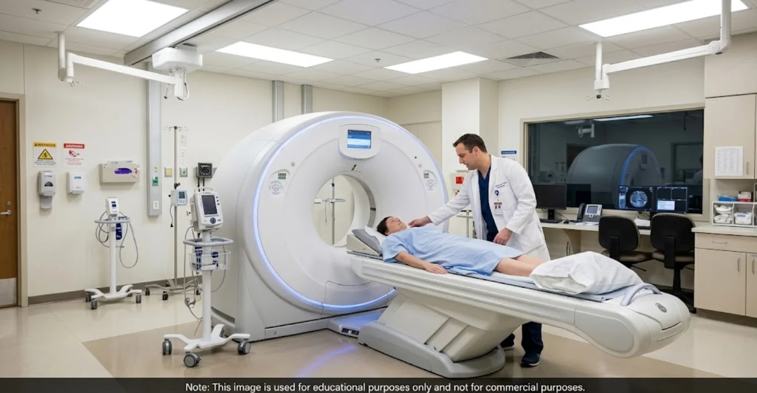
A CT scan for salivary gland evaluation is a quick, painless outpatient test. You lie on a table that slides through a ring-shaped scanner. The machine is open at both ends, so most people tolerate it well. A radiographer operates the scanner from the next room and can speak to you throughout. Some patients receive contrast through a vein. The whole process follows three simple stages. Each is explained below, so you know what to expect.
Before the Scan
The imaging team will ask about your symptoms and medical history. Expect questions about allergies, kidney problems, pregnancy, and current medications. These matter most when contrast is planned. Some centers request a recent kidney blood test first. You may be asked to avoid food for a few hours. Tell the team about previous contrast reactions, even mild ones. You will also remove metal items near the head and neck. A small cannula may be placed in your arm.
During the Scan
You lie on your back with your head resting in a shaped support. The table moves slowly through the scanner. You must stay still, and you may be asked not to swallow briefly. Scanning itself takes only a short time. If contrast is given, it is injected through the cannula. Many people feel a warm flush or a metallic taste. Some feel a brief sensation of needing to urinate. These feelings pass within a minute or two.
After the Scan
Most patients go home and resume normal activities immediately. If contrast was used, the team may observe you briefly and suggest extra fluids. A radiologist then studies the images and writes a report. The report goes to the clinician who requested the scan. Results are usually reviewed at a follow-up appointment. The radiologist's report is always interpreted together with your clinical findings. The images alone never tell the whole story. Ask who will contact you with results.
What Happens After an Abnormal Salivary Gland CT Scan?
The next step depends on what the scan shows. Options include clinical monitoring, ultrasound, or MRI for more detail. Antibiotics may be prescribed when infection is diagnosed clinically. A salivary stone may need removal or conservative treatment. An abscess may need needle aspiration. A mass may need fine-needle aspiration or biopsy. Referral to an appropriate specialist is common. An abnormal CT scan for salivary gland symptoms does not automatically mean invasive treatment. Many findings are managed simply.
Find out the answer to is root canal painless?
What We Notice Clinically
Dr. Rifat Alsaman, Head of the Medical Team at Vitrin Clinic and a cosmetic dentist, emphasizes diagnosis before treatment. In his view, imaging should answer a specific diagnostic question. It should not be requested simply because a scan is available. When a patient presents with unexplained swelling, pain, or a suspected glandular abnormality, history and examination come first. They determine whether a CT-scan for salivary gland assessment, ultrasound, or another test is appropriate. CT findings should never be interpreted in isolation.
When Dr. Rifat Alsaman Would Encourage Further Investigation
Dr. Rifat Alsaman, Head of the Medical Team at Vitrin Clinic and a cosmetic dentist, would encourage further investigation in these situations:
Persistent swelling
A firm or unexplained mass
Recurrent swelling associated with meals
Signs of infection
Symptoms that do not resolve as expected
Imaging findings requiring clarification
In these cases, a CT-scan for salivary gland evaluation or another test may be arranged through the appropriate specialist. Early referral usually makes treatment simpler.
Clinical Note
Clinical note: A CT scan is an important diagnostic tool, but it is not a substitute for a clinical examination or pathological assessment when cancer or another significant salivary gland disorder is suspected. The same symptom can have several possible causes. Treatment should target the confirmed or most likely underlying condition. Patients should not self-diagnose from an imaging report. Always review the radiology report with your treating clinician. They can explain what each finding means for you.
When Should You Seek Urgent Assessment for Salivary Gland Symptoms?
Some symptoms should not wait for a routine CT scan for salivary gland appointment. Seek urgent assessment if you notice:
Rapidly increasing facial or neck swelling
Difficulty breathing
Difficulty swallowing
High fever with significant swelling
Severe or rapidly worsening pain
Signs of a spreading infection, such as redness moving down the neck
A persistent or enlarging mass
New neurological symptoms, such as facial weakness, with a facial or glandular mass
Breathing or swallowing difficulty is an emergency. Go to the nearest emergency department immediately.
Read about the procedure for intraoral periapical first
Can Salivary Gland Problems Be Prevented?
Not every problem can be prevented, but good habits lower the risk. Maintain good oral hygiene and attend routine dental examinations. Stay adequately hydrated, since thick saliva encourages stones and infection. Address recurrent oral infections promptly. Discuss recurrent meal-related gland swelling with a dentist or physician. Avoid assuming that recurrent swelling is simply dehydration. Manage underlying conditions, such as diabetes or autoimmune disease, according to medical advice. These steps also support early detection when something does change.
How Much Does a Salivary Gland CT Scan Cost?
There is no universal price for a salivary gland CT scan. Costs vary by country and location, imaging center, CT protocol, use of contrast, and specialist interpretation. The imaging fee is also separate from any follow-up investigations or treatment. Before booking, ask whether the quoted price includes the radiologist's report. Ask about contrast charges when applicable. Comparing quotes for a CT scan for salivary gland assessment is only meaningful when the same items are included. Request a written breakdown.
How Long Does a Salivary Gland CT Scan Take?
The imaging portion is usually brief, often just a few minutes on the table. The total visit takes longer. Allow time for registration and preparation, contrast administration if required, the actual scanning, and waiting for the report. With contrast, a short observation period may be added. A CT scan for salivary gland assessment therefore often fits within an hour at the center. Reporting time varies between imaging centers. Some report the same day, while others take several days.
Vitrin Clinic: A Patient-Centered Approach to Dental and Oral Health
Vitrin Clinic in Istanbul, Turkey, builds every plan around individualized assessment. Dr. Rifat Alsaman, Head of the Medical Team at Vitrin Clinic and a cosmetic dentist, leads this approach. Oral health is assessed before cosmetic or restorative treatment when clinically indicated. Diagnostic technology, such as CBCT/3D imaging and intraoral scanning, is selected according to each patient's needs. When findings fall outside routine cosmetic dentistry, the team coordinates with the relevant specialties. The focus is education, realistic expectations, and individualized care, never a promised diagnosis or outcome.
Book a Consultation for an Individualized Assessment
Persistent salivary gland symptoms or concerning oral findings deserve professional assessment. Do not wait for them to settle on their own. The appropriate diagnostic pathway depends on your symptoms and clinical findings. You may need a dental examination, a CT scan for salivary gland evaluation, or a specialist referral. The first step is a conversation.
Ready to transform your smile?
Talk to our team and get a FREE treatment plan.
References:
FAQs
Dr. Rifat Alsaman has more than 5 years of clinical experience in dentistry and currently serves as the Head of the Medical Team at Vitrin Clinic. He is dedicated to providing exceptional patient care, overseeing treatment planning, and ensuring the highest clinical standards across the team. His expertise, attention to detail, and commitment to continuous professional development have helped countless patients achieve healthier, more confident smiles.


