General Dentistry
October 5, 2026
What Is Oral Lichen Planus? Symptoms, Causes, Diagnosis, and Treatment
 Dr. Rifat Alsaman
.webp&w=3840&q=75)
Oral Lichen Planus of the mouth is a chronic inflammatory disorder affecting the mucous membranes inside the mouth. It can appear as white lines, red areas, erosions, ulcers, or mixed patterns. Some people notice nothing, while others feel burning, soreness, or difficulty eating. The condition may occur alone or alongside skin or other mucosal involvement. Accurate diagnosis matters because several oral disorders look similar. A biopsy may confirm the diagnosis or exclude dysplasia, infection, or malignancy. Treatment usually focuses on symptom control and reducing inflammation, and long-term monitoring remains important. This guide covers symptoms, causes, diagnosis, treatment, home care, monitoring, and expected costs. It also separates established evidence from areas where research remains uncertain.
What Is Oral Lichen Planus?
This section explains what the condition is and how it behaves inside the mouth. It is a chronic, immune-mediated inflammatory disorder of the oral lining. It is not contagious and is not cancer. Some people never notice it, while others live with burning and painful erosions. Understanding its nature helps patients set realistic expectations about control, monitoring, and long-term care.
What is oral lichen planus disease?
It is a long-term inflammatory condition of the oral mucosa. It reflects an abnormal immune response against the cells lining the mouth. The exact trigger often remains unknown. White lace-like lines are common, while other forms cause redness, erosions, or ulcers. Symptoms range from none at all to persistent burning. The condition is not an infection, and treatment aims at control rather than guaranteed cure.
How does lichen planus affect the oral cavity?
The immune response injures the surface cells lining the mouth. This can create white striations, redness, thinning, and superficial breakdown. Reticular lesions look like fine lace, while erosive areas lose part of the protective surface. Exposed tissue may burn, especially during meals or brushing. Several patterns can appear together and fluctuate over months or years. Local trauma and irritating foods may worsen existing inflammation.
Is it an autoimmune or inflammatory condition?
It is best described as a chronic immune-mediated inflammatory condition. Cytotoxic CD8-positive T cells and inflammatory cytokines drive injury to epithelial cells. However, the precise antigen behind this response is still unknown. Lichenoid reactions differ because a trigger, such as a medicine or dental material, may be identified. Removing that trigger can improve a reaction. Idiopathic disease usually needs symptom control and monitoring instead.
Is oral lichen planus contagious?
No, this condition is not contagious. It does not spread through kissing, shared utensils, or household contact. The problem comes from the body's own immune process rather than a transmissible germ. However, erosions can resemble infections such as candidiasis or viral ulcers. That similarity is a good reason to seek examination rather than guess. Persistent or recurring lesions deserve a professional look.
Is it the same as lichen planus on the skin?
Both belong to the same broader disease spectrum. However, the mouth can be the only affected site. DermNet notes that oral involvement occurs in about half of patients with skin disease. Oral lesions usually look like white lace-like lines, while skin lesions form scaly papules. Your clinician may therefore ask about past skin, nail, or genital changes. Those details can support the overall diagnosis.
Find out why no exercise after dental implant
What Does Oral Lichen Planus Look Like?
The condition can look very different from one person to another. Fine white lace-like lines are the classic sign, usually on both inner cheeks. Other forms appear as red, thin, eroded, or blistered areas. Lesions may affect the tongue, gums, lips, and, less often, the palate. Knowing these patterns helps patients describe changes accurately, although appearance alone never confirms the diagnosis.
What are the most common symptoms of this condition?
Symptoms range from complete silence to significant daily discomfort. Burning is common when tissue becomes thin or eroded. Pain may increase with eating, brushing, or speaking, especially after spicy, acidic, or rough foods. Gum involvement can bleed lightly after brushing. Some people notice taste changes when larger areas are inflamed. Severity does not always match how extensive the lesions look.
What does reticular oral lichen planus look like?
The reticular form appears as fine white lines forming a lace-like network called Wickham striae. It usually affects both inner cheeks, though the tongue and gums can show similar markings. Surrounding tissue often looks fairly normal, and symptoms may be absent. A bilateral, symmetrical pattern can support a clinical diagnosis. Still, other disorders produce white streaks, so history and examination matter.
What is erosive oral lichen planus?
The erosive form involves areas where the surface lining has broken down. These areas look red, raw, or shallowly ulcerated, often with thin white borders. Pain is usually greater than with the reticular form. Gums, tongue, and inner cheeks are common sites. Blistering diseases and cancer can look similar. Persistent erosions therefore deserve careful evaluation and sometimes a biopsy.
What does the atrophic form look like?
Atrophic lesions appear as smooth, thin, bright red areas. White peripheral lines or erosions may sit alongside them. On the gums, this can create a striking red appearance. Patients often describe burning, especially with food or brushing. Atrophic changes overlap with other immune disorders, so appearance alone may not settle the diagnosis. A review of medicines and dental materials also helps.
What are the bullous and annular forms?
The bullous form produces small fluid-filled blisters that rupture quickly and leave painful erosions. It is less common than the reticular form. It can closely resemble mucous membrane pemphigoid or pemphigus. Specialized testing such as direct immunofluorescence may therefore be needed. Annular lesions form ring-shaped patterns. Mixed patterns often appear in the same mouth, and atypical blistering warrants specialist assessment.
Where can lesions appear in the mouth?
The inner cheeks are the most typical location, followed by the tongue and gums. Lips may also be involved, while the palate is uncommon. Symmetry and distribution give useful diagnostic clues. Different sites can show different patterns at the same time. A full examination should therefore check every surface, not only the painful area.
Lichen planus on the tongue
Tongue lesions may appear as white lines, plaques, redness, or erosions. Rough or spicy foods can make them burn. Plaque-like or one-sided lesions deserve extra caution because other keratotic conditions look similar. Some studies link tongue involvement with higher transformation risk. That finding supports careful surveillance, not a prediction of cancer. Persistent ulcers on the tongue borders should always be examined.
Lichen planus on the gums
Gum involvement causes redness, peeling, and soreness, a pattern called desquamative gingivitis. The tissue may bleed during brushing. Pain can discourage cleaning, allowing plaque to build and inflammation to worsen. Gentle professional cleaning can help break that cycle. However, desquamative gingivitis is a sign rather than a diagnosis. Pemphigoid and pemphigus can look identical.
Lichen planus on the lips
Lip involvement may appear as white lines, redness, scaling, or crusted erosions. Cold weather and irritating foods can worsen symptoms. Similar changes may come from cosmetic products or toothpaste flavorings. A medication review is also useful when lesions persist. Avoid peeling the skin, and use a bland lip balm to reduce friction. Persistent lip ulcers need professional review.
Lichen planus on the roof of the mouth
The hard palate is an uncommon site for this condition. A palatal lesion therefore raises other possibilities, including trauma, burns, candidiasis, denture irritation, or lupus. The lesion's history and duration matter greatly. Unexplained palatal changes may justify specialist referral and biopsy. Meanwhile, avoid repeated irritation from hot foods or poorly fitting appliances.
Oral mucosal lichen planus on the inner cheeks
The inner cheeks are the classic site for reticular disease. White lines usually appear on both sides in a symmetrical, lace-like network. Some patients feel nothing, while others develop painful erosions between the lines. Cheek biting and nearby fillings can also change the surface. One-sided plaques therefore need broader consideration before a diagnosis is accepted.
Pictures and photos: what can images show?
Clinical pictures can help patients recognize white striations, red erosions, or peeling gums. Clinicians also use photos to compare changes over time. However, lighting and camera angle alter color and hide surface detail. Images cannot show firmness, history, or microscopic changes. Online examples also tend to show severe cases. Use pictures for education, never for self-diagnosis.
Images of reticular lesions
Reticular images usually show thin, intersecting white lines resembling lace or branches. Cheek photos often show the same pattern on both sides. This explains why symmetry is clinically useful. Yet similar white patterns can come from friction, medicines, or dental materials. An image cannot reveal medication history or hidden dysplasia. It supports recognition rather than confirmation.
Oral erosive lichen planus photos
Erosive photos usually show irregular red or ulcerated surfaces bordered by faint white lines. The tissue may look glossy and thin. Photos often understate the pain a small erosion can cause. They also cannot show how long a lesion has lasted. Serial images taken in similar lighting are more useful than one snapshot. Changing lesions still need direct examination.
Photos of the tongue and gums
Tongue photos may show lines, plaques, redness, or erosions, while gum photos often show red, peeling tissue. These images help document distribution for later comparison. They cannot detect firmness, infection, or dysplasia. Good lighting and gentle lip retraction improve quality. Clinicians may also photograph healthy areas to create a clearer baseline for monitoring.
Why pictures cannot replace an examination
A photograph shows only the surface. An examination adds texture, tenderness, symmetry, and the feel of surrounding tissue. Gentle palpation can reveal firmness invisible on camera. The clinician also reviews medicines, fillings, and symptom timing. When needed, a biopsy or immunofluorescence test adds microscopic evidence. This is why persistent ulcers or changing lesions must be examined in person.
Discover all about endosteal implants
What Causes Oral Lichen Planus?
The exact cause remains unknown in most patients. An abnormal immune response, led by T cells, damages the cells lining the mouth. Certain medicines and dental materials can trigger lookalike lichenoid reactions. Stress, trauma, and irritating foods may worsen flares without causing the disease. Separating true triggers from coincidence helps guide treatment and avoids unnecessary changes.
What causes oral lichen planus?
The exact cause of this condition is not fully understood. Most cases are considered idiopathic, meaning no single trigger is found. CD8-positive T cells and inflammatory mediators appear to target the mouth's lining cells. Some lookalike lesions come from medicines, dental materials, or contact allergens. Hepatitis C is associated in some regions, though the link varies. Identifying a true trigger shapes treatment.
What triggers flare-ups?
Triggers differ between individuals. Stress, mechanical trauma, and sharp teeth are commonly reported. Spicy or acidic foods can intensify pain without causing the disease itself. Strongly flavored toothpaste and poorly fitting appliances may also irritate tissue. Tobacco and alcohol add irritation and raise cancer risk. A simple symptom diary can help separate real triggers from coincidence.
Can stress make the condition worse?
Many patients report flare-ups during stressful periods. However, stress is not proven to cause the condition by itself. Stress can disturb sleep, eating, and oral hygiene, which may indirectly worsen symptoms. Breathing exercises, regular sleep, and physical activity may support overall wellbeing. These measures complement treatment rather than replace it. Persistent lesions still need professional review.
Can medications cause lichen planus-like oral lesions?
Yes, some medicines cause oral lichenoid reactions that closely resemble the idiopathic condition. Reported groups include certain blood pressure medicines, anti-inflammatories, antimalarials, and diabetes drugs. The timing between starting a medicine and the lesion appearing is a useful clue. Never stop taking medicine on your own. Your prescriber can consider alternatives, and improvement may take weeks or months.
Can dental materials or restorations trigger lichenoid reactions?
Yes, localized reactions may develop beside certain fillings, especially amalgam. Composite, acrylic, and resin materials have also been reported. A key clue is a lesion directly touching the restoration. Replacing the material may help, though improvement can take months. Patch testing has limits. Replacement should therefore follow careful clinical reasoning rather than assumption.
Can acid reflux cause it?
Acid reflux is not an established cause of this condition. However, stomach acid can irritate already sensitive tissue and increase burning. Treating reflux may improve comfort for affected patients. It does not replace proper diagnosis of a persistent white or red lesion. Avoid strong home alkalizing products. See a doctor when reflux is frequent or severe.
Is it caused by poor oral hygiene?
No, poor hygiene does not cause the underlying immune disorder. However, plaque can aggravate inflamed gums. Sore gums may discourage brushing, which allows more plaque and more irritation. A soft toothbrush, gentle technique, and careful interdental cleaning help break this cycle. Professional cleaning supports gum health. Good hygiene eases symptoms but does not cure the condition.
See the risks and timeline of smoking after tooth extraction
Who Is More Likely to Develop Oral Lichen Planus?
The condition most often affects adults over 45, and women slightly more than men. However, it can appear at almost any age, including in younger adults. Some medicines, dental materials, and hepatitis C have been associated with it. Many patients have no identifiable risk factor at all. Risk factors offer context, while diagnosis still depends on examination.
What are the known risk factors for oral lichen planus?
Many patients have no clear risk factor. The condition most often affects middle-aged and older adults, and women slightly more than men. Some medicines and dental materials cause similar lichenoid reactions. Hepatitis C associations vary by population. Stress and trauma may aggravate symptoms, while tobacco and alcohol raise oral cancer risk. These are associations, not guaranteed causes.
Does it affect women and men differently?
Women are affected somewhat more often, with DermNet citing a ratio of roughly 1.4 to 1. Men can still develop every clinical pattern. Current evidence does not link sex alone to cancer risk. Smoking, alcohol, lesion type, and site appear more relevant. Monitoring should therefore follow clinical findings rather than demographic assumptions.
Can it develop at any age?
Yes, although it is most common after age 45. DermNet reports an average diagnosis age around 50 to 60 years. Younger adults and, less often, children can also be affected. Age alone cannot confirm or exclude the diagnosis. Persistent lesions in a younger patient still deserve careful assessment and, when atypical, possible biopsy.
Can it occur in the mouth without skin lichen planus?
Yes, it often occurs without any skin involvement. In some people, the mouth is the only affected site. A dentist may therefore be the first to notice it. Clinicians may still ask about past skin or genital symptoms. The absence of skin disease does not make oral lesions harmless. It also does not remove the need for monitoring.
What We Notice Clinically
In daily practice, the classic bilateral white pattern is the most familiar presentation. Erosive, red, and gum lesions tend to cause more symptoms and deserve closer attention. Many other disorders can mimic these appearances. These include drug reactions, contact reactions, candidiasis, pemphigoid, and leukoplakia. Careful history, examination, and selective biopsy help separate them safely.
Which patterns are most often seen clinically?
The reticular pattern on both inner cheeks is the most familiar presentation. Erosive and atrophic patterns are also common and usually more painful. Gum involvement often appears as desquamative gingivitis. Mixed patterns are frequent, and one patient may have painless and painful areas together. Recent reviews list the cheeks, tongue, and gums as the most common sites.
Why white lacy patterns should not automatically be assumed to be lichen planus
Several conditions produce white lacy or streaky changes. These include frictional keratosis, contact reactions, medication reactions, leukoplakia, and lupus. Symmetry, location, and history all help separate them. One-sided or plaque-like lesions deserve particular caution. When findings are atypical or persistent, a biopsy is often the safest way forward.
Why erosive and red lesions deserve closer evaluation
Red and erosive lesions reflect greater damage to the protective lining. They cause more pain and can affect nutrition. They also overlap with potentially serious disorders. Several analyses link red lesions with higher transformation risk, though most remain benign. Enlarging, firm, or bleeding lesions need prompt review. Smoking and alcohol history should always be considered.
Oral lichen planus vs leukoplakia: what is the difference?
Lichen planus in the mouth typically shows symmetrical white lines on both sides. Leukoplakia is a white patch diagnosed only after other causes are excluded. It may be uniform or irregular and can contain dysplasia. Lichenoid features sometimes complicate the microscopic picture. A biopsy is often needed for persistent plaques, especially in smokers. Photos alone cannot reliably separate the two.
What other conditions can look like oral lichen planus?
Many conditions mimic its white, red, or eroded patterns. Examples include lichenoid reactions, lupus, graft-versus-host disease, pemphigoid, and pemphigus. Candidiasis, leukoplakia, and frictional keratosis also belong on the list. Proliferative verrucous leukoplakia and early cancer require particular caution. Accurate classification needs more than one visual feature. Atypical cases therefore benefit from specialist input.
Oral lichenoid drug reactions
Drug-related lesions can look almost identical to the idiopathic condition. They may appear after starting a medicine or changing its dose. Some reactions affect both the mouth and skin. Improvement after switching medicines may take weeks or months. Always change medicines only with your prescriber. Bring a full list, including over-the-counter products and recent changes.
Contact reactions and dental-material-related lesions
A contact reaction typically sits directly against a filling or crown. Amalgam is the most frequently reported material, though composites and acrylics can also be involved. Replacing a suspected material may help the lesion settle. Patch tests do not perfectly predict mouth reactions. Lesion location and timing should guide decisions, avoiding unnecessary replacement costs.
Candidiasis and other inflammatory conditions
Candidiasis can cause burning and white patches that sometimes wipe away to reveal red tissue. It needs antifungal treatment rather than anti-inflammatory therapy. Steroids can actually encourage fungal overgrowth, which makes correct diagnosis essential. Nutritional deficiencies and contact reactions can also cause soreness. Repeated trial treatments without reassessment should be avoided.
Mucous membrane pemphigoid and pemphigus
Both conditions cause painful oral erosions. Pemphigoid often affects the gums, while pemphigus can produce widespread fragile blisters. Blisters often burst before an appointment, so the result resembles erosive inflammation. Biopsy with direct immunofluorescence is usually key. Treatment differs, and some cases need systemic therapy. Widespread erosion deserves prompt specialist evaluation.
Leukoplakia and other potentially malignant oral lesions
Leukoplakia is diagnosed only after other causes are excluded. Persistent white lesions require consideration of dysplasia and cancer. Proliferative verrucous leukoplakia carries notable malignant potential. Firmness, rapid growth, or mixed red and white areas raise concern. A biopsy determines whether dysplasia or invasion exists. Tobacco and alcohol history should always be reviewed.
Clinical Note: Dr. Rifat Alsaman on why appearance alone is not enough
A sound diagnosis combines appearance with history and hands-on examination. A typical pattern supports a working diagnosis, while atypical features call for wider thinking. Persistent ulcers should never be explained away without reassessment. Lesion texture, medication timing, and nearby fillings all add information. A practical principle is to diagnose before escalating treatment. Vitrin Clinic applies this individualized approach when reviewing oral mucosal changes.
Learn about the anatomy and health of the dental pulp
What Are the Symptoms of Oral Lichen Planus?
Symptoms range from none at all to persistent daily pain. Burning and soreness are common with erosive or atrophic forms. Spicy, acidic, or rough foods often intensify discomfort. Gum involvement can make brushing painful and cause light bleeding. Severe flares may affect eating, drinking, and speaking. Symptom severity does not always match how extensive the lesions look.
Can it cause burning or soreness?
Yes, burning and soreness are common, especially with erosive or atrophic forms. Reticular lesions may cause little or no discomfort. Symptoms often flare with food contact, brushing, stress, or dry mouth. Severe pain does not automatically indicate cancer. Persistent or worsening symptoms still deserve reassessment. Treatment usually targets inflammation and comfort.
Can it cause painful ulcers or erosions?
Yes, the erosive form can produce shallow, painful ulcers. These may sting with eating, brushing, or acidic foods, and they can recur. However, trauma, infection, immune disorders, and cancer also cause ulcers. A non-healing ulcer should never be self-treated indefinitely. Report any change in size, surface, or bleeding. A biopsy may follow if uncertainty remains.
Can it make the gums sore or peel?
Yes, gum involvement can cause marked soreness, redness, and peeling. The gums may look shiny and bleed with brushing. Discomfort can lead patients to brush less, allowing plaque to worsen inflammation. Gentle hygiene therefore remains important. Other immune disorders cause similar peeling. Blistering or widespread changes therefore warrant specialist assessment.
Can it affect eating, drinking, or speaking?
Severe disease can affect all three. Chewing may hurt, and acidic or spicy foods can trigger sharp burning. Extensive erosions may make swallowing uncomfortable, and tongue lesions can affect speech. Some patients unintentionally eat less. Soft foods and good hydration help during flares. Persistent swallowing difficulty needs medical evaluation, and treatment should restore normal function.
Can it cause sensitivity to spicy or acidic foods?
Yes, sensitive lesions often react to chili, citrus, vinegar, salt, and acidic drinks. These reactions reflect exposed tissue, not food causing the disease. Symptoms usually ease once the irritant is removed. Test tolerance by avoiding one food at a time. A symptom diary helps, while overly restrictive diets are rarely necessary.
Can it be mild with no symptoms?
Yes, many people have mild diseases without any symptoms. Painless reticular lines often need no medication at all. Monitoring remains useful because appearance can change over time. New ulcers or changes in a stable lesion should prompt review. Annual checks are common for mild disease, with closer review for higher-risk patterns.
Explore types and benefits of dental prosthesis
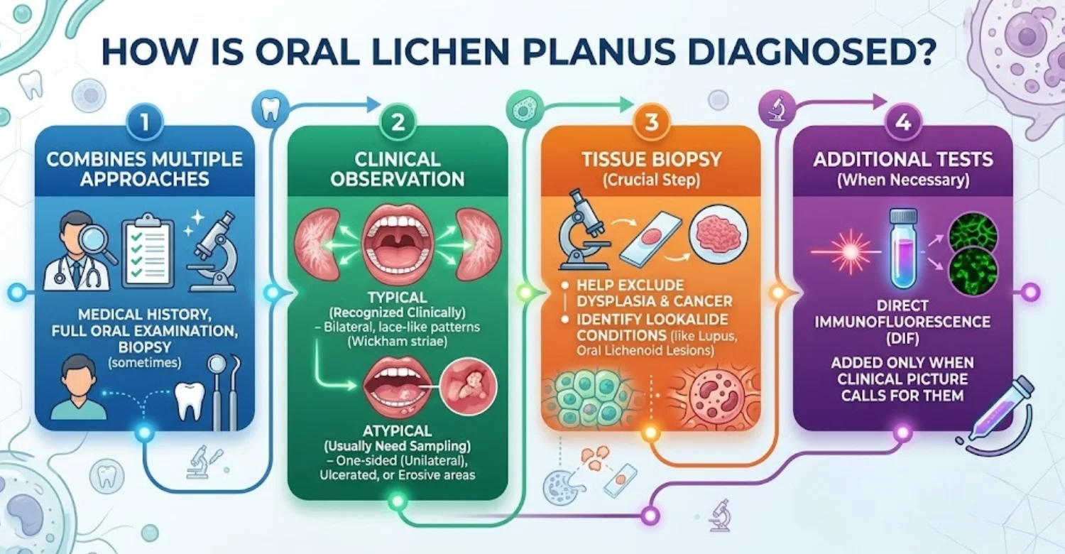
How does a dentist or oral medicine specialist diagnose oral lichen planus?
Diagnosis starts with a detailed history and oral examination. The clinician assesses color, texture, symmetry, and distribution. Medicines, fillings, and skin findings are also reviewed. Typical bilateral reticular lesions can sometimes be diagnosed clinically. Atypical lesions usually need a biopsy to confirm the pattern and exclude mimics. No single test is perfect, so evidence is combined.
What happens during an oral examination?
The clinician first asks about symptoms, duration, medicines, tobacco, and alcohol. The mouth is then inspected under good lighting. This includes the cheeks, tongue, gums, palate, lips, and floor of the mouth. Suspicious areas may be gently felt for firmness. Photographs may be taken for comparison. Further tests are chosen only if the findings justify them.
When is an oral biopsy needed?
A biopsy is not needed for every classic reticular lesion. It becomes valuable for atypical, one-sided, or persistent ulcerated lesions. Firmness, rapid growth, or a new lump also justify sampling. A biopsy helps exclude dysplasia, cancer, and lookalike conditions. Immunofluorescence may be added when blistering disease is suspected.
What does an oral lichen planus biopsy show?
A biopsy often shows a band of lymphocytes beneath the surface lining. Damaged basal cells and scattered dying keratinocytes may also appear. These findings must be interpreted alongside clinical details because lichenoid reactions look similar. Extra stains may check for candidiasis. The report should also comment on any dysplasia.
Can a biopsy distinguish lichen planus from leukoplakia or cancer?
A biopsy provides critical information but still needs context. It can identify dysplasia, invasive cancer, or a lichenoid pattern. Some conditions overlap microscopically, and sampling location matters. One biopsy may miss part of a varied lesion. When clinical concern persists, repeat sampling may be advised. Pathology reduces uncertainty but does not replace clinical judgment.
Are blood tests needed for diagnosis?
Routine blood tests are not needed for every patient with this condition. Hepatitis C testing may be considered in selected populations. Tests for anemia or nutritional deficiency may help when symptoms suggest them. Blood work is also used before some systemic medicines. Testing should answer a clear clinical question rather than add confusion.
When might additional testing be considered?
Additional tests suit atypical or uncertain presentations. Direct immunofluorescence helps assess blistering disorders, and patch testing may examine suspected contact reactions. Swabs can check for infection. Repeat biopsy may be considered when clinical and pathology findings disagree. Imaging is rarely needed for surface lesions. Each test should have a specific purpose.
Who treats oral lichen planus?
Several professionals may help manage the condition, depending on complexity. General dentists often spot lesions first. Oral medicine specialists manage chronic mucosal disorders, while oral surgeons perform biopsies. Dermatologists help when skin is involved, and physicians may adjust medicines. Do not delay care while searching for the perfect specialist. The first qualified clinician can begin assessment and refer onward.
Dentist or general dental practitioner
General dentists often notice mucosal changes during routine visits. They document location, appearance, symptoms, and possible triggers. They can smooth sharp teeth, adjust appliances, and support plaque control. Stable lesions can be monitored with photographs. Suspicious or atypical findings should be referred to with a clear note covering history and medicines.
Oral medicine specialist
Oral medicine specialists focus on diseases of the mouth's soft tissues. They evaluate chronic white, red, ulcerated, and blistering lesions. They also plan biopsies, coordinate immunofluorescence, and adjust treatment. Referral is especially useful when lesions are atypical or treatment fails. This specialty is often the most directly relevant for persistent mucosal disease.
Oral and maxillofacial specialist
Oral and maxillofacial specialists perform incisional and excisional biopsies. They assess lesions with uncertain depth, firmness, or bone involvement. Most inflammatory cases do not need surgery. However, these specialists become important when a lesion looks suspicious. They can also coordinate with pathology and oncology teams when required.
Dermatologist or other medical specialist when appropriate
Dermatologists help when skin lichen planus is also present. Prescribing physicians may review medicines suspected of causing lichenoid reactions. Hepatology input becomes relevant if hepatitis C is confirmed. Systemic treatments may need laboratory monitoring. Clear communication across the care team improves safety. The mouth still needs direct dental assessment.
Is Oral Lichen Planus Dangerous?
For most patients, the condition remains a manageable inflammatory disorder. It is not cancer, but research shows a small long-term transformation risk in some cases. Red or erosive lesions, tongue involvement, tobacco, and alcohol may raise that risk. Regular monitoring helps detect warning changes early. Persistent ulcers, new lumps, bleeding, or numbness always need prompt assessment.
Is oral lichen planus cancer?
No, this condition is not cancer. It is an inflammatory disorder of the mouth's lining. However, it is classified among oral potentially malignant disorders because of a small long-term transformation risk. Most patients never develop cancer. Attention should focus on persistent or changing lesions. Regular monitoring and avoiding tobacco and alcohol remain sensible steps.
Can oral lichen planus increase oral cancer risk?
Evidence suggests a small increased risk in some patients. A 2024 meta-analysis reported a pooled transformation rate of 1.43%. Stricter analyses reported lower figures, reflecting different diagnostic criteria. Risk appears higher with red or erosive lesions, tongue involvement, tobacco, alcohol, and hepatitis C. Dysplasia is particularly important. These figures guide surveillance rather than predict individual outcomes.
What cancer-related symptoms need evaluation?
Warning signs include a persistent ulcer, a new lump, or a thickened area. Unexplained bleeding, steadily worsening pain, or numbness also need attention. Rapid growth or a change in color or texture is concerning. Difficulty swallowing and neck swelling require prompt review. These signs do not prove cancer, but they do justify assessment.
Which lesion changes are concerning?
Concern rises when a previously stable lesion changes. Firmness, persistent ulceration, rapid growth, and unexplained bleeding are key examples. New mixed red and white areas or numbness also matter. Tongue lesions, tobacco, and alcohol increase concern further. A changing lesion should be reassessed, not simply treated again.
A persistent ulcer or erosion
Most ordinary mouth ulcers heal within days. An ulcer lasting about three weeks deserves examination. The clinician assesses size, edges, surface, and surrounding tissue. Obvious trauma should be removed first, but persistent lesions may still need biopsy. Pain does not reliably separate benign from serious causes. Tongue and floor-of-mouth ulcers need particular care.
A new lump or thickened area
A new lump should be examined promptly. Firmness, called induration, cannot be judged from photographs. The clinician will feel the area and surrounding tissue. Scar tissue, benign growths, and cancer can all cause lumps. Rapid enlargement is especially concerning. A previous inflammatory diagnosis never explains a new finding automatically.
Unexplained bleeding
Inflamed tissue may bleed after brushing or eating, which is often harmless. Spontaneous or unexplained bleeding deserves closer attention. The clinician checks for ulcers, fragile tissue, and gum disease. Persistent bleeding from one focal area may need biopsy. Always disclose blood thinners, and never stop them without medical advice.
Increasing pain or persistent soreness
Flares can temporarily increase pain, but steady progression needs reassessment. A new ulcer, secondary infection, or dental trauma may explain the change. Sometimes a biopsy becomes necessary. Pain relief can continue during investigation. Report any new difficulty eating or swallowing. Symptoms affecting nutrition deserve prompt support.
Numbness or altered sensation
Numbness is not typical of simple surface inflammation. The new altered sensation therefore deserves assessment, even without pain. Dental nerve irritation or previous surgery can explain it. However, numbness near a mucosal lesion is more concerning. The clinician may examine nearby lymph nodes and consider imaging or biopsy.
A lesion that changes in color, size, or surface
Any changing lesion deserves reassessment, regardless of its earlier diagnosis. Increasing redness, mixed red and white areas, roughness, thickening, or ulceration all matter. Rapid change is particularly important. Record when you first noticed it, and bring photographs. Direct examination remains essential, and biopsy may follow if suspicion increases.
Cancer pictures: why internet images can be misleading
Online images usually show advanced or dramatic cases. Early cancers can look subtle, while benign inflammation can look severe. Screens also distort color, and images cannot capture firmness or history. Never delay care because your lesion looks different online. Persistent ulcers need examination regardless of photographic comparisons.
Why regular oral examinations matter even when symptoms improve
Symptoms can settle while visible lesions remain. The condition often fluctuates rather than disappearing. Regular examinations create a stable baseline and help detect new changes. They matter most for erosive or atrophic forms. Many sources suggest at least yearly review for mild disease. Return sooner whenever you notice a change.
Check safety tips on using peroxide to help with tooth pain
Can Oral Lichen Planus Go Away?
Some mild cases improve or fade over time, but permanent disappearance is unpredictable. The condition often follows a relapsing course, with quiet periods and flares. Pain may resolve while white lines remain visible. There is no guaranteed cure for idiopathic disease. The realistic goal is long-term control with comfortable function and safe follow-up.
Does oral lichen planus go away on its own?
Mild disease sometimes improves or becomes less noticeable. However, complete spontaneous resolution cannot be predicted. The condition usually waxes and wanes over time. Pain may vanish while white lines remain. Asymptomatic disease often needs monitoring rather than medication. Treatment aims at control, and follow-up confirms that the pattern stays stable.
Will it go away permanently?
Permanent resolution cannot be guaranteed. Many patients experience remission followed by recurrence. Some keep mild, painless lines for years, while others have repeated painful erosions. Treatment often improves symptoms substantially without removing the underlying tendency. Avoiding irritants and maintaining good hygiene help. The realistic goal is stable disease with minimal symptoms.
Can it go into remission and return later?
Yes, symptoms and visible lesions may fade for months before returning. Stress, trauma, or new medicines sometimes coincide with recurrence. A recurrence should resemble the previously documented pattern. A different appearance deserves fresh assessment. Photographs help compare episodes, but significant changes still need clinical review.
Is there a permanent oral lichen planus cure?
There is currently no reliable permanent cure for idiopathic disease. Treatment suppresses inflammation and controls symptoms, mostly with topical corticosteroids. Other immunomodulators help when response is inadequate. Removing a true trigger can resolve lichenoid reactions, which differ from idiopathic disease. Treatment can usually be reduced after control is achieved.
What does successful long-term control look like?
Success means fewer symptoms and stable clinical findings. Eating and brushing become comfortable, and erosions heal or shrink. Triggers become easier to recognize and avoid. Medicine use stays proportionate to severity. Regular reviews document a stable appearance. Stopping tobacco and limiting alcohol further reduce avoidable risk.
How Is Oral Lichen Planus Treated?
Treatment focuses on calming inflammation, healing erosions, and relieving pain. Painless lesions may need only monitoring. Topical corticosteroids are the main first-line option for symptomatic disease. Tacrolimus or systemic medicines are considered for resistant or severe cases. Removing triggers and local irritants supports every plan. Treatment intensity should always match symptoms and clinical findings.
What is the main goal of treatment?
The main goal of treatment is symptom control and tissue healing. It also aims to reduce inflammatory activity and maintain comfortable function. Treatment should match severity, so painless lesions may need no medicine. Aggravating factors and secondary infections should be addressed. Suspicious changes still require biopsy when indicated.
When does mild disease require treatment?
Painless mild disease often needs observation only. Treatment becomes relevant when symptoms affect eating, brushing, or daily comfort. Lesions turning erosive may change the plan. Extensive painless disease may need less treatment than small painful erosions. Removing trauma and controlling plaque help either way, and monitoring continues regardless.
What are the first-line treatments for symptomatic oral lichen planus?
Topical corticosteroids remain the main first-line treatment for symptomatic disease. Options include triamcinolone, fluocinolone, fluocinonide, and clobetasol. Adhesive pastes suit localized areas, while rinses help widespread lesions. Clinicians monitor for candidiasis during use. Good hygiene and trigger avoidance support treatment. Systemic therapy is reserved for extensive or resistant disease.
How are topical corticosteroids used for this condition?
They are applied directly to affected areas, concentrating the effect while limiting whole-body exposure. Applying after cleaning the mouth improves contact. Avoid eating or drinking briefly afterward. Your clinician may taper the dose after improvement. Do not use skin creams inside the mouth without advice. Response is judged clinically, not by pain alone.
Topical corticosteroid gels, ointments, and pastes
Adhesive pastes stay on the tissue longer and suit localized painful lesions. Gels allow broader or spot application. Gently drying the area first helps the product stick. Using more than prescribed does not speed healing and raises side-effect risk. Reported increased burning. Ask about alternatives if taste or texture affects adherence.
Topical steroid mouth rinses
Rinses spread medicine across several areas and suit widespread lesions. Patients usually swish the solution and spit it out without swallowing. Food and drink may be avoided briefly afterward. Follow your prescription rather than online dosing advice. Repeated use can raise candidiasis risk, so regular review allows safe adjustment.
Topical steroid sprays and other delivery methods
Sprays can reach sites where pastes are hard to apply. Different devices deposit medicine differently, so follow the prescribed technique exactly. Some nasal steroid sprays are used off-label in the mouth under professional guidance. Never substitute products independently. The aim is effective local treatment with the lowest necessary exposure.
What is clobetasol propionate used for?
Clobetasol is a potent topical corticosteroid used for painful or erosive lesions. Systematic reviews support its ability to reduce symptoms and visible disease. Concentration and duration must be set by a clinician. Long-term use raises the risk of oral thrush. Report new white removable patches promptly. A diagnosis should come before prolonged potent steroid use.
When might tacrolimus or another steroid-sparing treatment be considered?
Tacrolimus may help when topical steroids fail or are unsuitable. It is a calcineurin inhibitor with local immune-modulating effects. Evidence supports benefit, but it has not consistently outperformed steroids. Temporary burning is common at first. Pimecrolimus is another option. Specialist supervision is preferable for repeated or prolonged use.
When are systemic medicines considered?
Systemic medicines are reserved for severe, widespread, or treatment-resistant disease. Short courses of oral steroids may provide rapid control. Other immunomodulators may be used under specialist care. These carry greater risks, so blood tests and interaction checks may be required. The diagnosis should be secure before escalation. Mild painless disease does not need this level of treatment.
What are treatment options for erosive oral lichen planus?
Treatment for the erosive form usually starts with potent topical steroids. Topical anesthetics before meals can ease pain. Unexpected worsening may signal candidiasis. Reducing trauma and avoiding trigger foods support healing. Resistant cases may need tacrolimus or, rarely, systemic therapy. Lesions that change or fail to respond may need biopsy.
How is treatment adjusted when the condition does not respond?
First, the diagnosis should be reconsidered, because nonresponse can reveal a different condition. Clinicians also check for medication triggers, candidiasis, and ongoing trauma. The formulation or strength may change. Biopsy or specialist referral may follow. Tacrolimus or systemic therapy suits selected cases. Repeatedly switching medicines without review is not ideal.
What does an individualized treatment plan include?
A personalized plan begins with a confirmed or likely diagnosis. It considers lesion type, site, extent, and symptoms. Medicines, fillings, tobacco, alcohol, and hygiene are reviewed. Treatment is chosen to match distribution, with pain relief added when needed. Follow-up reflects risk and response. Vitrin Clinic emphasizes this individualized approach rather than one fixed regimen.
Clinical Note: Dr. Rifat Alsaman on matching treatment intensity to symptoms and clinical findings
Treatment intensity should reflect both symptoms and objective findings. A painless white pattern may need observation, while painful erosions usually need active therapy. Atypical lesions may need biopsy before escalation. Correcting trauma and reviewing medicines come before adding more immunosuppression. Medicine should be reduced after improvement, and recurrence planned for. Vitrin Clinic uses structured assessment to support these decisions.
Learn how to care for swollen gum from wisdom tooth
What Relief Can Help With Oral Lichen Planus Symptoms?
Several simple measures can ease burning between treatments. Soft foods, cool drinks, and good hydration often help. Anesthetic or analgesic rinses can reduce pain before meals. Bland, alcohol-free mouthwashes usually feel more comfortable. Gentle oral hygiene limits extra irritation from plaque. These measures support prescribed treatment rather than replace it.
What can help relieve burning and mouth soreness?
Avoiding known irritants often brings quick relief. Soft foods, cool drinks, and good hydration can soothe sensitive tissue. Topical anesthetics may help before meals. Brush gently and never scrub painful areas. Some people find alcohol-free, bland rinses more comfortable. Persistent pain should prompt a treatment review.
Can an analgesic or anesthetic mouthwash help before meals?
Yes, topical anesthetic or analgesic rinses can reduce pain temporarily. Benzydamine and lidocaine preparations are common options. Use them exactly as advised and avoid swallowing unless instructed. Numbness raises the risk of biting your cheek or burning yourself on hot food. They ease symptoms but do not replace anti-inflammatory treatment.
What type of mouthwash may be more comfortable for sensitive oral tissues?
Bland, alcohol-free rinses usually feel most comfortable. Mild saline or diluted bicarbonate rinses suit many people. Strongly flavored or alcohol-based products may sting erosions. Chlorhexidine can be useful for limited periods but may stain teeth and alter taste. No rinse replaces brushing. Stop any product that clearly increases irritation.
Can oral rinses help when the gums are affected?
Rinses provide supportive comfort when gums are painful. However, they complement rather than replace gentle brushing. Alcohol-free formulas tend to sting less. Chlorhexidine may be recommended for short-term plaque control. Persistent gum peeling still needs diagnosis. Blistering should prompt specialist referral rather than more rinsing.
How can good oral hygiene reduce additional irritation?
Gentle brushing removes plaque without traumatizing tissue. Use a soft toothbrush, light pressure, and careful interdental cleaning. Professional hygiene visits help when home cleaning becomes difficult. Sharp fillings and pressure points from appliances should be corrected. Switch toothpaste if one causes burning. Good hygiene supports treatment, though it cannot remove the immune cause.
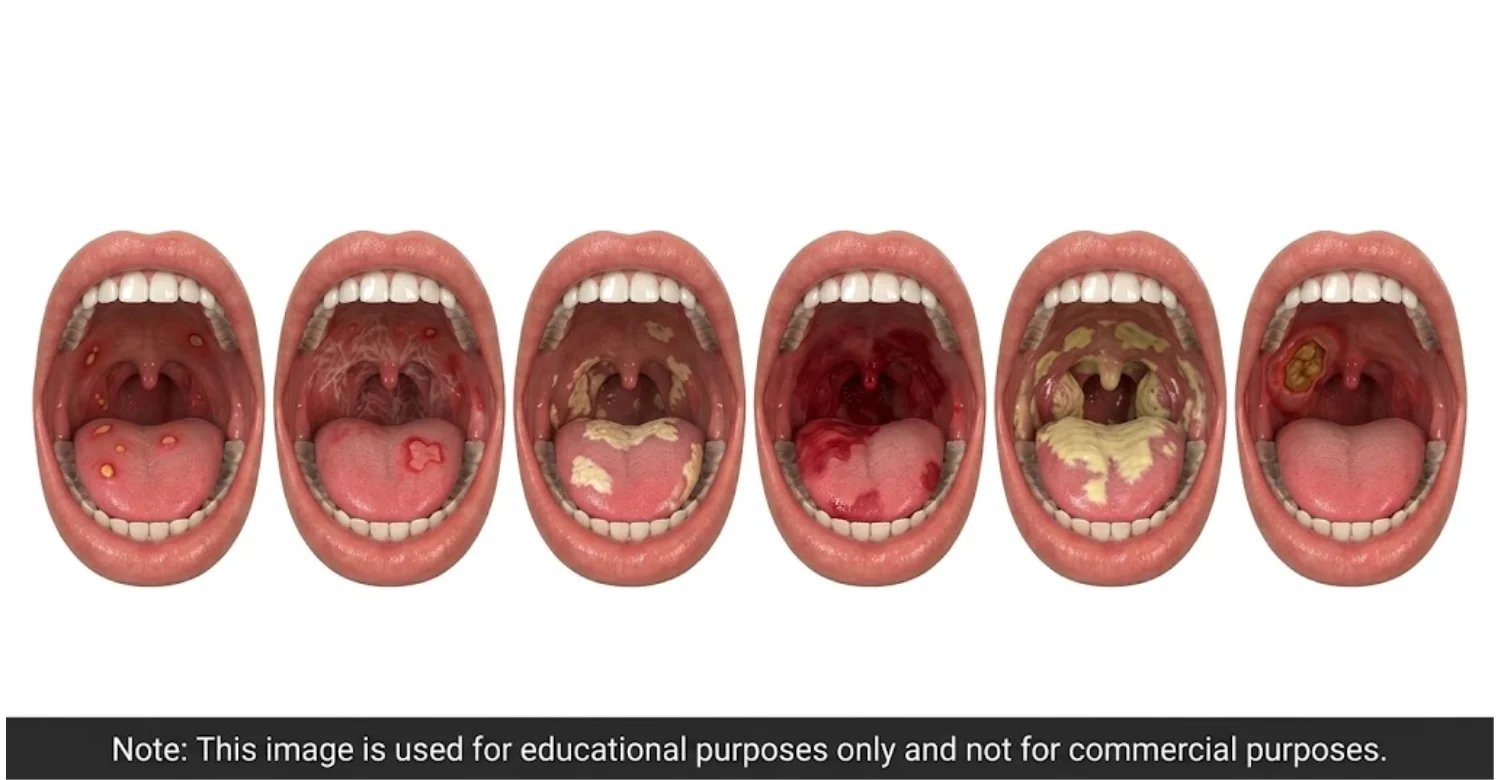
Note : All images used are for editorial and illustrative purposes only and may not originate from the original news provider or associated company.
What Are the Side Effects of Oral Lichen Planus Treatments?
Most topical treatments are well tolerated when used as prescribed. Oral thrush is the most important side effect of corticosteroids. Tacrolimus commonly causes temporary burning and sometimes taste changes. Systemic medicines carry broader risks and often need blood tests. Reporting unexpected effects early allows clinicians to adjust treatment safely and effectively.
What side effects can occur with topical corticosteroids?
Topical steroids are generally well tolerated when prescribed correctly. Oral thrush is the most important side effect. Local irritation and taste changes can occur. Prolonged use of strong preparations may rarely thin the tissue. Systemic absorption is usually low. Follow the prescribed frequency, and report unexpected worsening rather than assuming another flare.
Can topical steroids cause oral thrush?
Yes, steroids can encourage candida overgrowth. Thrush may appear as removable white patches or new diffuse soreness. Not every new white area is thrush, so examination helps. Antifungal treatment is used when infection is confirmed. Your clinician may adjust the steroid rather than stop it abruptly. Persistent symptoms after antifungal treatment need reassessment.
Can oral treatments cause burning, dryness, or taste changes?
Yes, some products cause temporary irritation. Tacrolimus often burns initially, and certain rinses alter taste. Using several products frequently may worsen dryness. A medicine reaction can mimic a flare, so timing offers clues. Keep a simple diary of applications and symptoms. Persistent effects deserve review and possible formulation changes.
What side effects can occur with tacrolimus or other second-line treatments?
Topical tacrolimus commonly causes temporary burning and sometimes taste changes. These effects are usually manageable. Long-term use should have a clear purpose, since it has not consistently outperformed steroids. Systemic immunomodulators carry broader risks, including infection. Regular follow-up balances benefits against harms, ideally under specialist supervision.
What should patients discuss before using systemic medicines?
Share all current medicines, past infections, and chronic illnesses. Discuss pregnancy plans and vaccination status, since both may affect choices. Blood tests may be needed before and during treatment. Ask about expected duration, warning symptoms, and drug interactions. Never take leftover immunosuppressive medicines on your own initiative.
When should a treatment side effect be reported to a clinician?
Report any persistent or unexpected side effects. Facial swelling or breathing difficulty needs urgent medical care. Worsening pain, new white plaques, persistent taste disturbance, or severe dryness should also be reported. Systemic symptoms after systemic treatment need prompt attention. Note when symptoms began relative to doses. This helps your clinician adjust treatment safely.
Understand potential reasons if you have a tooth hurts no cavity
What Medical Tips Can Make Oral Lichen Planus Easier to Manage?
Small daily habits can make a meaningful difference. Choose soft, mild foods during flares and avoid personal triggers. Use a soft toothbrush and a mild, flavor-light fluoride toothpaste. Alcohol-free rinses usually sting less. Keep a balanced diet rather than following restrictive plans. A symptom diary helps identify patterns worth discussing with your clinician.
How should patients protect irritated oral tissues?
Choose soft foods during flares and avoid sharp, crunchy textures. Use a soft toothbrush with gentle pressure. Drink water regularly and protect your lips from dryness. Never pick or peel lesions. Ask your dentist to smooth sharp tooth edges. Follow prescriptions exactly, record changes, and seek care when a stable lesion changes.
What foods should be avoided during a flare?
During a flare, reduce foods that clearly cause burning. Common culprits include chili, citrus, vinegar, salty snacks, crusty bread, and very hot dishes. Alcohol and acidic drinks may also sting. Tolerance varies, so a diary helps identify personal triggers. Soft, nutrient-rich foods maintain nutrition while tissues settle.
What is the best diet for oral lichen planus?
No single diet is proven to cure this condition. A balanced, symptom-aware diet works best. Include adequate protein, fruits, and vegetables that you tolerate. Soft textures help during flares, and hydration matters. Very restrictive diets risk deficiencies, and supplements are not cures. A dietitian can help if eating becomes significantly limited.
Should patients avoid spicy, acidic, salty, or hard foods?
These foods often worsen symptoms during active inflammation. Spice and acid can sting exposed tissue, and hard textures cause friction. Avoidance should follow personal tolerance rather than strict rules. Temporary restriction during flares is often enough. Foods can usually return gradually after healing. Keep nutritional balance as the priority.
What toothpaste is best for oral lichen planus?
A mild fluoride toothpaste is usually most comfortable for sensitive mucosa. Some people react to strong cinnamon or mint flavors. Sodium lauryl sulfate irritates certain individuals, though avoidance is not essential for everyone. Whitening toothpastes may increase irritation during flares. Avoid products marketed as cures, and pair your choice with a soft toothbrush.
Can toothpaste for sensitive mouths reduce irritation?
It may help some people, depending on ingredients and personal tolerance. Flavoring agents often matter more than the label. Try a new toothpaste during a calm period and stop if burning starts. Keep fluoride protection for your teeth. Toothpaste cannot treat the underlying inflammation, but a comfortable product makes consistent brushing easier.
What should patients look for in an oral rinse for lichen planus?
Choose a rinse that does not sting. Alcohol-free and mildly flavored products are often best during flares. Saltwater or gentle bicarbonate rinses soothe some people. Use prescription rinses exactly as directed, and limit chlorhexidine to recommended periods. Avoid concentrated homemade mixtures. Stop any product that worsens symptoms.
Can Home Remedies Help Oral Lichen Planus?
Some home measures may soothe symptoms, but none cures the condition. Mild saltwater or bicarbonate rinses can feel comfortable. Aloe vera has limited and inconsistent supporting evidence. Many herbal or homeopathic products lack reliable research. Caustic substances, such as lemon juice or undiluted oils, can damage tissue. Home remedies should never delay diagnosis or prescribed care.
What home remedies may provide symptom relief?
Simple home care can reduce irritation. Soft foods, cool water, and mild saltwater or bicarbonate rinses may soothe burning. These steps do not treat the underlying inflammation. Natural products can still cause allergic or irritant reactions, so avoid strong essential oils. Home care should support, not replace, diagnosis and professional review.
Can baking soda help?
A mild baking soda rinse may soothe symptoms temporarily. There is limited evidence that it treats the disease itself. Always dilute it properly, and never apply dry powder to erosions. Stop if burning increases. Baking soda does not replace prescribed steroids or the need to assess persistent ulcers.
Can aloe vera help oral lichen planus?
Aloe vera has been studied as a supportive treatment for this condition. One randomized trial showed improvement compared with placebo. However, later reviews rated the overall evidence as weak and inconsistent. Product quality varies widely, and cosmetic gels may contain irritants. Aloe should be considered an add-on rather than a proven alternative to steroids.
Is aloe vera gel or aloe vera mouthwash safe to use?
Short-term studies suggest reasonable tolerability. However, formulations differ greatly. Cosmetic gels may contain alcohol, fragrance, or preservatives that irritate damaged tissue. Choose oral-use products when advised. Stop if swelling, rash, or worsening burning appears. Long-term safety is less clear, and aloe should never delay prescribed treatment.
What is known about aloe vera juice for this condition?
Evidence for drinking aloe vera juice is weak, since most research uses topical products. Ingested aloe can upset digestion, and some products have laxative effects. Interactions with prescription medicines are possible. Avoid unregulated concentrates. Topical findings should not be assumed to apply to juice. Discuss any supplement with your clinician first.
Do herbal, Ayurvedic, homeopathic, or natural treatments work?
Evidence varies greatly. Some topical plant extracts have small studies, while many products lack controlled evidence. Homeopathy has not shown reliable benefit for this condition. Ayurvedic mixtures may cause irritation or interactions. Natural remedies can also delay biopsy. Bring product details to appointments, and keep evidence-based therapy as the foundation.
What alternative treatments should be discussed with a clinician first?
Discuss anything affecting the immune system before starting it. This includes herbal supplements, laser treatments, and photodynamic therapy. Avoid strong topical acids or essential oils without advice. Removing dental fillings should follow proper assessment. Supplements can interact with prescribed medicines. A clinician can help separate genuine evidence from marketing claims.
Which home remedies should be avoided because they may irritate oral tissue?
Avoid applying lemon juice, vinegar, raw garlic, or undiluted essential oils to lesions. Repeated hydrogen peroxide rinses and strong salt solutions can also damage tissue. Never pour alcohol onto sores or use abrasive powders. Pain from a remedy does not mean it is working. Persistent lesions need diagnosis, not repeated chemical exposure.
.jpg)
.jpg)
.jpg)
.jpg)
What Should Patients Avoid During an Oral Lichen Planus Flare?
During flares, reducing irritation helps tissues settle. Avoid tobacco and limit alcohol, since both irritate tissue and raise cancer risk. Spicy, acidic, salty, and hard foods often worsen burning. Alcohol-based mouthwashes may sting erosions. Sharp teeth and ill-fitting appliances should be corrected. Never stop prescribed medicines just because symptoms improve.
Should patients avoid tobacco and alcohol?
Yes, avoiding tobacco is strongly advised, and limiting alcohol is sensible. Both independently raise oral cancer risk, and together the risk climbs further. Each can also irritate inflamed tissue. Quitting tobacco improves general dental health. Seek cessation support if needed. Keep these changes beyond flare periods.
Can spicy and acidic foods worsen symptoms?
Yes, they often intensify burning during active disease. Chili stimulates pain receptors, while acid stings exposed tissue. Very hot foods can add further irritation. Responses vary, so avoidance can be temporary and symptom-based. These foods worsen symptoms but do not cause the disorder. A diary helps clarify your personal triggers.
Should alcohol-containing mouthwash be avoided when it causes irritation?
Yes, if it stings, switch to an alcohol-free product. Many people with erosions find alcohol-based rinses uncomfortable, though reactions vary. Chlorhexidine may suit specific clinical needs for limited periods. Avoid concentrated homemade substitutes. Choose rinses with a clear purpose, prioritizing comfort and plaque control.
Can rough foods, sharp teeth, or dental appliances aggravate lesions?
Yes, mechanical trauma often worsens symptoms. Sharp tooth edges, broken fillings, dentures, and orthodontic appliances can rub inflamed tissue. Hard foods may scrape erosions. A dentist can smooth edges and adjust appliances safely. Do not remove appliances without advice. Monitoring should continue even after irritants are corrected.
Should patients stop prescribed medication because symptoms improve?
No, do not stop prescribed medicine on your own. Some treatments need gradual tapering, and abrupt stopping may cause rebound flares. Improvement is a reason to review treatment with your clinician. They can decide whether reduction is safe. Never replace a prescription with a supplement without discussion.
Discover the signs to help answer should I get a root canal?
What Happens When Oral Lichen Planus Affects the Gums?
Gum involvement often appears as red, fragile, peeling tissue called desquamative gingivitis. Brushing may become painful and cause bleeding. Patients then tend to clean less, allowing plaque to worsen inflammation. Gentle plaque control and professional cleaning help break this cycle. Because other immune disorders look similar, persistent gum peeling deserves proper diagnosis.
What is desquamative gingivitis?
Desquamative gingivitis describes red, fragile, peeling gums. It is a clinical sign, not a single disease. Lichen planus is one possible cause. Mucous membrane pemphigoid and pemphigus can look identical. Brushing may hurt and cause bleeding. Blistering suggests other diagnoses, so biopsy and immunofluorescence are sometimes needed.
How can it affect gum health?
When the condition involves the gums, they become painful and fragile. This often reduces brushing quality. Plaque then accumulates, adding secondary inflammation and bleeding. Over time, periodontal maintenance becomes harder. Gentle professional cleaning helps, even during flares. Gum disease should be assessed separately, because plaque treatment alone cannot control the mucosal disorder.
Why is plaque control important when the gums are inflamed?
Plaque bacteria provoke further gum inflammation. Already inflamed gums respond more strongly to bacterial build-up. Effective plaque control removes this extra irritant. Soft brushes and gentle interdental cleaning make it easier. Do not stop cleaning because of bleeding. Good plaque control protects periodontal health while other treatments control immune activity.
How are painful gingival lesions managed?
Management depends on the confirmed cause. Topical steroids are common for inflammatory disease, sometimes applied with custom trays. Pain relief before brushing can help. Gentle plaque control remains essential, and sharp fillings should be corrected. Worsening after steroids may indicate thrush. Blistering or persistent erosions often need specialist referral.
How Can Oral Lichen Planus Be Monitored Long Term?
Long-term monitoring helps confirm stability and catch changes early. Review intervals depend on lesion type, symptoms, and risk factors. Mild stable disease is often checked yearly, while higher-risk cases need closer review. Consistent photographs support comparison over time. Patients should report new ulcers, lumps, bleeding, or numbness without waiting for routine visits.
How often should oral lichen planus be reviewed?
There is no single universal review interval for this condition. Mild stable disease is often reviewed at least yearly. Active disease may need visits every three to six months. Recent guidance suggests three to four months for high-risk findings. Dysplasia may shorten intervals further. Return earlier whenever something changes.
What should patients monitor between appointments?
Watch for changes in color, size, or surface. Report new ulcers, lumps, firm areas, unexplained bleeding, or numbness. Note worsening pain or difficulty eating and swallowing. Record possible triggers and medication changes. A simple diary improves discussions. Do not wait for a routine visit when changes are significant.
Can photographs help track changes over time?
Yes, serial photographs help document changes in size or distribution. Use consistent lighting, the same camera, and a similar angle. Include a nearby landmark such as a tooth for reference. Clinician photos are often better standardized. Images still cannot show firmness or dysplasia. They support rather than replace examinations.
What should happen if a previously stable lesion changes?
It should be reassessed promptly. The clinician compares it with earlier records and reviews new medicines or trauma. Texture and firmness are checked carefully. New ulceration or rapid growth may lead to biopsy. Previous pathology is reviewed when available. Treatment should not simply be intensified without this evaluation.
Can stopping tobacco and limiting alcohol reduce additional oral cancer risk factors?
Yes, both changes remove important avoidable risks. Tobacco is a major oral cancer risk factor, and alcohol adds further risk. Their combination is particularly harmful. These steps do not cure the inflammatory condition or guarantee protection. However, they improve your overall risk profile. Continue them during symptom-free periods, alongside regular examinations.
When Should You See a Dentist or Doctor?
Professional assessment is important whenever something persists or changes. White or red patches lasting several weeks should be examined. Ulcers that do not heal within about three weeks need evaluation. Worsening pain, bleeding, swelling, or numbness also deserve attention. Rapid changes, hard lumps, or swallowing difficulty warrant urgent review.
When should persistent oral white or red patches be examined?
Any white or red patch lasting several weeks deserves professional assessment. Stable typical patterns may only need routine monitoring. New plaques, red areas, and one-sided lesions need closer evaluation. Tobacco use increases urgency. Avoid repeated home treatment without a diagnosis. Vitrin Clinic can provide dental evaluation and coordinate referral when necessary.
When should a persistent ulcer be assessed?
An ulcer that has not healed within about three weeks needs evaluation. The clinician checks for trauma, infection, and suspicious features. Pain level does not separate benign from serious causes. Tongue and floor-of-mouth ulcers deserve particular attention. A previous diagnosis does not rule out new disease. Biopsy or referral may follow.
When should worsening pain, bleeding, swelling, or numbness be evaluated?
These symptoms deserve assessment when persistent or unexplained. Pain may reflect inflammation or another process. Unexplained bleeding and swelling should not be ignored. Numbness needs special attention because it may indicate deeper involvement. A combination of symptoms raises concern. Seek faster review when changes progress quickly.
When should a patient seek specialist oral medicine care?
Referral helps when diagnosis remains uncertain or lesions look atypical. Blistering, widespread erosions, and treatment-resistant symptoms also justify specialist care. Complex medication reactions and suspected autoimmune disease benefit from expertise. A second opinion helps when findings conflict. Referral does not automatically mean cancer is suspected.
When is urgent assessment appropriate for a suspicious oral lesion?
Urgent review suits rapidly changing lesions, new hard lumps, or unexplained persistent bleeding. Numbness, rapidly enlarging ulcers, and severe swelling also need prompt attention. Difficulty breathing or swallowing may require emergency medical care. Do not wait for a routine appointment. Urgency does not mean cancer, only that faster evaluation is needed.
Find relief and remedies for an ache in mouth
Average Cost of Oral Lichen Planus Evaluation and Treatment in the US, UK, and Turkey
Care costs vary because consultation, biopsy, pathology, medicines, and follow-up are often billed separately. The figures below are planning references, not guaranteed treatment quotes.
Country | Planning reference | Important caveat |
United States | Mouth biopsy about 440–1,222 | Published MDsave range, not a universal national fee |
United Kingdom | Private biopsy often about $290–$550 | Provider packages differ |
Turkey | Highly variable | Specialist consultation, biopsy, pathology, and treatment may be quoted separately |
US mouth biopsy prices listed by MDsave range from $440 to $1,222. Current UK private examples show biopsy prices from about $290 to $400. Turkey has no reliable national private average for this specific pathway.
Average Cost in the United States
Costs vary widely between outpatient centers and hospitals. MDsave lists mouth biopsies at about $440 to $1,222, alongside a separate estimated national average. Consultation, pathology, and follow-up may add charges. Insurance can change your share considerably. Ask whether pathology, facility, and professional fees are included. A written estimate gives the clearest picture.
Consultation, biopsy, pathology, medication, and follow-up costs
A full evaluation may include several separate services. These include consultation, biopsy with local anesthesia, and laboratory pathology. Medicines vary in cost, and systemic therapy adds blood tests. Follow-up visits may be billed separately. Imaging is rarely needed. Ask for an itemized estimate, because this makes cost comparisons meaningful.
2026 planning reference: published mouth-biopsy prices of approximately 440–1,222, rather than a universal national average
MDsave currently lists mouth biopsy prices from $440 to $1,222. This is a published market range, not a national treatment average. Hospital charges and pathology fees may differ considerably. Insurance can raise or lower out-of-pocket costs. Prices change without notice. Confirm what is included and request a clinic-specific written quote.
Average Cost in the United Kingdom
UK costs differ between NHS and private pathways. NHS care follows referral and eligibility routes, while private care offers direct access. Private providers advertise biopsies around $290 to $400. One London provider lists oral biopsy from $295, with histology charged at $125 to $150. Always compare what each package includes.
NHS versus private oral medicine and biopsy pathways
NHS care usually begins with a GP or dentist referral, and access depends on local services. Private providers often advertise fixed procedure prices. Histopathology and consultation may be bundled or billed separately. The cheapest biopsy is not always the cheapest overall pathway. Choose based on clinical quality, and plan follow-up if traveling.
2026 planning reference: approximately $290–$511 as a private-market oral-biopsy reference, not a national OLP treatment average
A private-market range of about $290 to $511 is useful for budgeting. It does not represent a national average. Separate histology and consultation fees may raise the total. Many patients need only monitoring and topical medicine, while others need biopsy and specialist follow-up. Treat this range as a planning reference, not a quote.
Average Cost in Turkey
Turkey has no reliable national private average for this pathway. Costs differ between dental clinics, hospitals, and specialist centers. Consultation, biopsy, and histopathology may be billed separately. Biopsy site and complexity affect price. International patients should also budget for travel and follow-up. Vitrin Clinic structures assessment around clinically indicated steps rather than unnecessary testing.
Specialist consultation, oral examination, biopsy, histopathology, and medication costs
A typical pathway starts with specialist consultation and examination. If biopsy is indicated, tissue goes for histopathology. Topical steroids usually cost less than systemic therapy. Extra reviews and complex pathology add expense. Ask whether the quote covers pathology reporting and remote follow-up. Compare only quotes covering identical services.
private specialist consultation prices vary widely, while oral biopsy pricing may be quoted separately according to provider and pathology requirements
No single Turkish price is supported by strong evidence. Provider directories show wide variation. Some centers quote biopsy separately from consultation, and pathology needs can change the total. Hospital procedures may cost more than outpatient dental care. Request a written breakdown covering anesthesia, histopathology, follow-up, and remote result sharing.
What Is Usually Included in a Treatment Quote?
A clear quote states exactly what you receive. It may include consultation, examination, and biopsy when indicated. Pathology, prescriptions, and follow-up are sometimes separate. International patients should ask about coordination and translation. Specialist referrals may add costs. Clear exclusions prevent misleading comparisons between clinics.
Initial examination and specialist consultation
The consultation sets the diagnostic pathway. The clinician reviews symptoms, medicines, and previous dental work, then examines the mouth systematically. Possible irritants and restorations are documented. The visit may end with monitoring alone, since biopsy is not automatic. Ask whether follow-up is included. Vitrin Clinic can organize an individualized dental assessment.
Biopsy and histopathology when clinically indicated
Biopsy is used when uncertainty or risk justifies tissue diagnosis. A laboratory examines the sample for lichenoid inflammation or dysplasia. Prices may cover anesthesia and tissue removal, with histology billed separately. Special stains or immunofluorescence add cost. Ask about turnaround time and request a written report. Accuracy matters more than the cheapest option.
Prescription medicines and symptom-relief products
Medicine costs depend on the product and treatment intensity. Many patients need only a topical steroid. Others require second-line or systemic therapy. Pain-relief products and antifungals may add expense. Insurance coverage varies by country. Travelers should confirm medicine availability at home. Generics may help, but prescriptions should remain clinician-directed.
Follow-up and monitoring appointments
Long-term care involves repeated examinations at intervals set by risk. Stable disease may need yearly checks, while higher-risk lesions need more. Photographs help comparison, and repeat biopsy follows meaningful change. Include follow-up in your budget. International patients should arrange local surveillance and share records. Vitrin Clinic can support follow-up planning.
Why There Is No One Fixed Treatment Cost
Costs differ because each clinical pathway differs. Some patients need only examination and monitoring. Others require biopsy, pathology, topical medicine, or specialist care. Systemic treatment adds laboratory monitoring. Dental irritants may need correction. Insurance and travel also change expenses. A single advertised price can mislead, so ask for an individualized, itemized quote.
What We Notice Clinically About Treatment and Monitoring
In practice, symptom relief and lesion resolution do not always happen together. Pain may settle while visible lines remain. A previously stable lesion can also change without much discomfort. For these reasons, examination matters as much as how a patient feels. Diagnosis should always come before treating an unexplained lesion.
Why symptom relief does not always mean the underlying lesion has disappeared
Pain often settles before visible changes fully resolve. Remaining lines do not mean treatment failed. Conversely, comfort can hide a new lesion developing nearby. Both symptoms and appearance should therefore be checked. Clinical photographs help track gradual change. Vitrin Clinic emphasizes examination rather than symptom scores alone.
Why persistent or changing lesions may require reassessment
A persistent lesion may simply be stable inflammation, or it may be something else. New ulceration, firmness, or altered appearance changes the diagnostic picture. Earlier pathology cannot explain every future change. The current lesion should be reviewed on its own merits. Serial records improve comparison, and early reassessment prevents delays.
Why diagnosis should come before treating an unexplained oral lesion as lichen planus
Many oral disorders look alike but need different treatment. Steroids can worsen infections and mask other problems. Delayed biopsy may postpone cancer care. Drug reactions need trigger review, blistering disorders need immunofluorescence, and leukoplakia needs histology. The safest pathway starts with reasonable diagnostic confidence. Vitrin Clinic prioritizes this before selecting treatment.
Tips for Patients
Good preparation makes every appointment more productive. Bring a full medication list, previous reports, and dated photographs. Keep a simple diary of foods, products, and symptoms. Ask clear questions about biopsy, treatment goals, and side effects. Organized records and regular follow-up help you and your clinician manage the condition confidently.
What information should you bring to your appointment?
Bring a full list of prescription, over-the-counter, and herbal products. Note when symptoms started and what worsens them. Share tobacco and alcohol habits honestly. Bring earlier pathology reports and photographs of past flares. Mention recent dental work, sharp teeth, or appliance rubbing. Describe any eating or swallowing difficulties.
How can you document a flare-up for your dentist?
Photograph the lesion and nearby healthy tissue in consistent lighting. Date each image and use the same camera when possible. Record pain levels, food triggers, and medication changes. Note any bleeding, swelling, or numbness. Avoid manipulating the lesion beforehand. Several dated images reveal change better than one.
What questions should you ask about biopsy and treatment?
Ask why a biopsy is recommended and which conditions are being considered. Check whether pathology and immunofluorescence are included and how long results take. Ask about treatment goals, duration, and side effects such as thrush. Clarify when to return and which changes need earlier contact. Ask whether stopping requires tapering.
How can you track foods, products, or other possible triggers?
Keep a short daily diary covering meals, drinks, toothpaste, rinses, and medicines. Rate symptom severity and note lesion location. Record dental treatments and stressful events. Change only one factor at a time so patterns are clearer. Coincidence is not proof, so share the diary with your clinician.
How can you prepare for long-term follow-up?
Living with this condition works best with organized records. Keep examination notes, pathology reports, and dated photographs. Learn your warning signs and attend scheduled reviews. Return earlier when a lesion changes. Continue gentle hygiene, avoid tobacco, and limit alcohol. Tell every new clinician about your diagnosis and current medicines.
Personalized Oral Lichen Planus Assessment at Vitrin Clinic
Vitrin Clinic approaches unexplained mucosal changes through individualized clinical assessment. The examination considers symptoms, lesion appearance, medicines, and existing dental work. Biopsy or specialist referral is arranged only when clinically indicated. Digital documentation supports comparison over time. Imaging is used only for separate dental indications, keeping care focused and proportionate.
How Vitrin Clinic can assess oral mucosal changes during a dental evaluation
Vitrin Clinic can include mucosal assessment whenever signs of lichen planus or other changes appear. The examination reviews location, appearance, and symptoms. Medical history, medicines, and existing restorations add context. The aim is to identify likely causes before choosing treatment. Atypical findings may lead to specialist referral or biopsy. Documentation supports future comparison.
Personalized clinical assessment before selecting a treatment approach
Treatment should follow assessment rather than assumptions. The clinician considers symptoms, lesion type, extent, medicines, and nearby dental materials. Painful disease may justify active therapy, while mild painless disease may need monitoring. Biopsy is considered when confirmation is needed. Follow-up and side effects are explained clearly. Plans change when lesions change.
When referral or additional specialist evaluation may be appropriate
Referral suits atypical lesions, persistent ulcers, and blistering disease. Suspicious masses may need oral surgery assessment. Dermatology helps when skin is involved, and physicians review suspected drug reactions. Complex pathology may benefit from expert review. The purpose of each referral should be explained. Vitrin Clinic can coordinate these steps to maintain continuity.
Using digital documentation to support clinical assessment when appropriate
Clinical photographs create a useful visual baseline. They document location and visible change and support communication between clinicians. Images also help patient education. However, they cannot replace examination or biopsy. Records must be stored securely and kept clinically useful. Vitrin Clinic uses digital documentation when it improves longitudinal information.
Digital X-rays and CBCT only when another clinically relevant dental indication exists
Dental X-rays and CBCT do not diagnose superficial mucosal inflammation. They answer separate dental or bone-related questions. Radiation exposure always needs clear justification. A suspicious soft-tissue lesion usually needs examination and biopsy rather than scans. Imaging may help when deeper disease is suspected. Vitrin Clinic uses imaging only for relevant indications.
How Vitrin Clinic Supports Your Treatment Journey
Vitrin Clinic supports patients from first consultation to long-term follow-up. The process begins with a detailed review of symptoms, history, and medicines. Diagnostic steps are coordinated according to clinical need. Treatment plans are personalized and adjusted as findings change. International patients receive clear scheduling, records, and follow-up guidance.
Initial consultation and review of symptoms, medical history, medications, and previous dental treatment
Vitrin Clinic begins by reviewing your main symptoms, such as burning, ulcers, bleeding, or eating difficulty. Medication history may reveal lichenoid triggers. Previous dental work can show local irritants. Earlier pathology reports, tobacco, and alcohol use add context. This structured history guides the examination and any referral. You receive clear explanations at each step.
Coordinating appropriate examinations and diagnostic steps
Each diagnostic step should reflect clinical need. Some patients need only examination and monitoring. Others may need biopsy, pathology, or specialist referral. Imaging is used only for relevant dental indications. The purpose of every test is explained so you can decide confidently. Coordination also simplifies the process for international patients.
Personalized treatment planning based on clinical findings
Plans reflect symptoms and disease severity. Topical therapy suits symptomatic inflammation, while stable painless disease may need monitoring. Atypical lesions may need biopsy before treatment changes. Drug triggers and dental trauma are addressed. Follow-up, expected response, and side effects are discussed openly. Plans are reassessed whenever findings change.
Treatment coordination for international patients
International patients need clear scheduling and communication. Appointments are organized so pathology results arrive before departure when possible. Medicine instructions and records are documented clearly. You should know where to seek care at home. Complex lesions may need specialist care beyond routine dentistry. Treatment is never shortened just because travel time is limited.
Follow-up planning and communication after treatment
Follow-up confirms symptom control and lesion stability. Timing depends on severity. You will know which symptoms need an earlier review. Photographs can support remote communication, and pathology reports stay in your record. Your home dentist can continue surveillance. Remote contact never replaces urgent in-person assessment when a lesion changes.
Ready to transform your smile?
Talk to our team and get a personal FREE treatment plan.
Key Takeaways
The points below summarize the most important messages from this guide. They cover what the condition is, how it is diagnosed, and how it is treated. They also explain where home care fits and why monitoring matters. Use them as a quick reference, not a substitute for professional examination.
The condition is a chronic inflammatory disorder of the oral mucosa that can vary from mild, symptom-free patterns to painful erosive disease
The condition ranges from painless white lines to painful erosions, and patterns change over time. Cheeks, tongue, and gums are common sites. Not every lesion needs medicine, since treatment follows symptoms and findings. Persistent or changing lesions need reassessment. Vitrin Clinic supports individualized assessment and long-term monitoring.
Diagnosis may rely on clinical examination but biopsy can be important when confirmation or exclusion of another condition is needed
Typical bilateral reticular lesions can support a clinical diagnosis. Atypical presentations usually need further investigation. Biopsy helps exclude dysplasia, cancer, and lookalike conditions. Immunofluorescence assists with blistering disorders. Persistent ulcers should never be judged from photos. Accurate diagnosis prevents inappropriate treatment and guides monitoring.
Topical corticosteroids are commonly used for symptomatic disease, with treatment tailored to severity and response
Topical steroids such as clobetasol and triamcinolone remain first-line options. Choice depends on site and severity. Tacrolimus helps when steroids are inadequate, though it is not consistently superior. Systemic therapy is reserved for severe resistant disease. Thrush should be monitored. Professional supervision matters during prolonged treatment.
Home care can reduce irritation, but natural or herbal remedies should not replace professional diagnosis or prescribed treatment
Gentle hygiene, soft foods, and avoiding spicy or acidic items reduce irritation. Alcohol-free products may feel more comfortable. Aloe vera has limited supportive evidence, weaker than that for steroids. Home remedies can still irritate or interact, and caustic substances should never touch lesions. Supportive care works best alongside evidence-based treatment.
The condition generally requires monitoring because persistent, changing, or suspicious lesions need reassessment
The condition often relapses, and symptoms may improve without lesions disappearing. Changing lesions need fresh assessment. Erosive, red, or tongue lesions warrant closer surveillance. Smoking and alcohol add avoidable risk. Annual review suits mild stable disease, while higher-risk patterns need more frequent checks. Monitoring should always be individualized.
Professional evaluation is especially important for persistent ulcers, unexplained bleeding, new lumps, numbness, or other significant changes
Never dismiss warning signs because of an earlier diagnosis. Persistent ulcers, new lumps, unexplained bleeding, numbness, rapid growth, and surface changes all need review. Biopsy may follow when suspicion rises. Prompt assessment does not mean cancer. Seek urgent care for severe or rapidly progressive symptoms.
Reference
FAQs
Dr. Rifat Alsaman has more than 5 years of clinical experience in dentistry and currently serves as the Head of the Medical Team at Vitrin Clinic. He is dedicated to providing exceptional patient care, overseeing treatment planning, and ensuring the highest clinical standards across the team. His expertise, attention to detail, and commitment to continuous professional development have helped countless patients achieve healthier, more confident smiles.


