General Dentistry
October 13, 2026
What Is Oral Leukoplakia? Symptoms, Causes, and Treatment
 Dr. Rifat Alsaman

A white patch inside the mouth is easy to ignore, especially when it does not hurt. Yet some persistent white patches carry a real cancer risk. Oral leukoplakia is the most common potentially malignant disorder of the mouth. Global studies estimate it affects around 4% of adults. This guide explains what it looks like, why it develops, and how dentists diagnose it. It also covers treatment options, recovery, and average costs in the US, UK, and Turkey. Most importantly, it explains when a white patch needs prompt professional assessment.
What Is Oral Leukoplakia?
Oral leukoplakia is a white patch or plaque in the mouth that cannot be wiped off. It also cannot be explained by any other known condition. The World Health Organization classifies it as an oral potentially malignant disorder. This means most patches stay harmless, but a minority may become cancerous over time. A meta-analysis of global data estimated a prevalence of about 4.1%. The term itself describes appearance only. It is a clinical label, not a final diagnosis. A biopsy usually decides what is happening inside the tissue.
What Does Oral Leukoplakia Look Like?
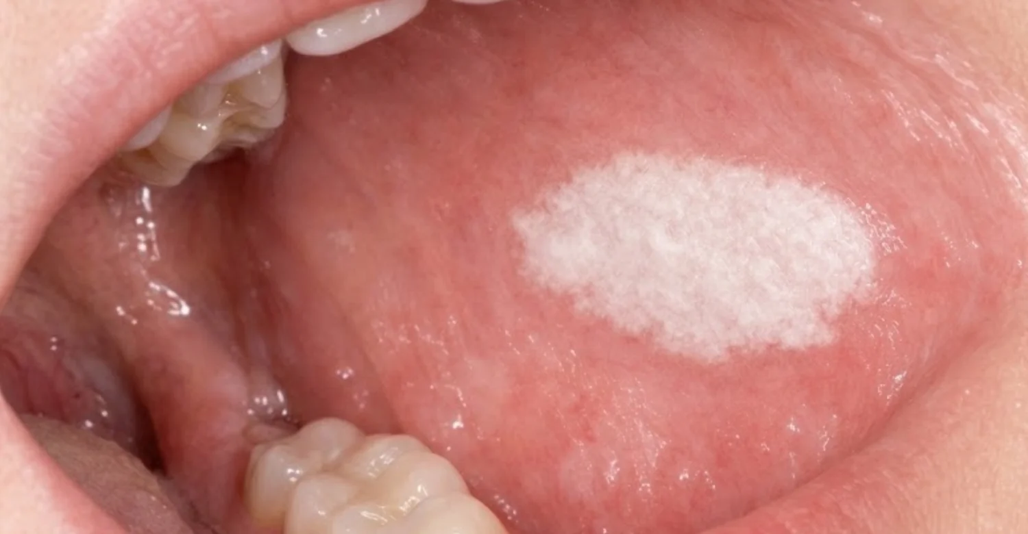
Most patches look flat, white, or grayish. Some are thin and slightly see-through, while others are thick and leathery. The surface may be smooth, wrinkled, or cracked like dried mud. Edges can be sharp or blend gradually into normal pink tissue. Some patches show red spots, small lumps, or a rough, wart-like texture. These mixed patterns deserve closer attention. Size varies from a few millimeters to several centimeters. A key feature is that gentle wiping with gauze does not remove the patch.
Where Do Leukoplakia White Patches in the Mouth Usually Appear?
White patches can appear almost anywhere inside the mouth. Common sites include the inner cheeks, gums, tongue edges, floor of the mouth, and palate. The lower lip and the area behind the last molars are also frequent sites. In many studies, the buccal mucosa, or inner cheek, is the most common location. The site often reflects habits. Smokeless tobacco users frequently develop patches where they hold the product. Patches on the floor of the mouth and tongue sides carry higher concern. Dentists record the exact site carefully at every visit.
Is Oral Leukoplakia Painful or Usually Painless?
Most patches are completely painless. This is exactly why many people miss them for months or years. Many lesions are first noticed during routine dental check-ups rather than by the patient. Pain is not a reliable warning signal in this condition. A painless patch can still contain abnormal cells. When pain, burning, or tenderness does appear, it may suggest ulceration, infection, or more advanced change. Any new discomfort in an existing patch should be checked promptly. Waiting for pain before booking an assessment is not a safe strategy.
Can Oral Leukoplakia Go Away on Its Own?
Some patches do fade, especially after the cause is removed. Stopping smoking is the clearest example. One classic Danish study found that about 56% of patches disappeared within a year of quitting. Removing a sharp tooth edge or loose denture can also help friction-related lesions. However, many patches persist despite lifestyle changes. Others shrink but never fully clear. Regression does not guarantee the tissue is normal underneath. For this reason, dentists still recommend follow-up even when a patch seems to improve.
Is Every White Patch in the Mouth Leukoplakia?
No. Many white patches have simple, harmless explanations. Oral thrush, cheek biting, burns from hot food, and lichen planus can all look white. Leukoplakia is diagnosed by exclusion. Dentists first rule out conditions with a known cause. If a white patch remains unexplained after about two to four weeks, it is treated as leukoplakia. A biopsy then confirms the tissue changes. This careful process prevents both unnecessary worry and missed early cancers.
Ready to transform your smile?
Talk to our team and get a personal FREE treatment plan.
What Are the Early Signs and Symptoms of Oral Leukoplakia?
Early signs are usually subtle and painless. A small white area may appear and slowly become more noticeable. Patients may feel a rough or slightly raised spot with the tongue. Many people notice nothing at all. Research suggests a large share of lesions are found incidentally during dental visits. Knowing the signs helps patients seek care earlier. Early assessment matters because treatment is simpler when changes are found sooner.
Early Signs of Leukoplakia Patients Should Recognize
Watch for a white or gray patch that does not rub away. Notice any area that feels rough, thickened, or different from nearby tissue. A patch that persists beyond two weeks deserves a dental appointment. Look for slow changes in color, size, or texture. Red specks mixed with white areas are especially important. A patch that returns after healing is also a meaningful sign. Taking a dated phone photo can help you track changes accurately. Bring these photos to your dentist.
Oral Leukoplakia Symptoms on the Tongue, Gums, and Inner Cheeks
On the tongue, patches often appear along the sides or underneath. These sites are considered higher-risk areas in many studies. On the gums, a patch may look like a thick white band around teeth. It can be mistaken for gum irritation or plaque. On the inner cheeks, patches often appear near the biting line or opposite rough teeth. Cheek lesions are frequently linked to tobacco or friction. Each location changes how a dentist interprets the lesion. Location also influences biopsy decisions and follow-up intervals.
Can Leukoplakia Cause Burning, Irritation, or Discomfort?
Most patches cause no discomfort. Some patients report mild burning when eating spicy, acidic, or salty foods. Others describe a sense of roughness or tightness. Discomfort may come from a secondary fungal infection on the patch. It may also reflect small cracks or ulcers on the surface. Persistent burning, stinging, or soreness needs professional assessment. These symptoms do not confirm cancer. They do signal that the lesion should be examined without delay.
How Does Leukoplakia Change as It Progresses?
A patch may stay stable for years without obvious change. In other cases, it slowly thickens or spreads. Some homogeneous white patches develop red areas, nodules, or a speckled surface. This shift toward a nonhomogeneous pattern can signal higher risk. Proliferative forms may spread to several sites over time. Studies show transformation can occur many years after first diagnosis. That is why long-term monitoring is important even for stable lesions. Change, rather than appearance alone, often prompts a repeat biopsy.
Which Changes in a White Mouth Patch Require Prompt Evaluation?
Seek prompt assessment if a patch develops red areas. Do the same if the surface becomes lumpy, ulcerated, or wart-like. Rapid growth over weeks is another warning sign. Bleeding with light touch also needs evaluation. A patch that becomes hard or fixed to deeper tissue is concerning. New pain, numbness, or difficulty swallowing should never be ignored. Swollen neck lymph nodes alongside a mouth patch need urgent attention. These signs do not always mean cancer, but they need timely examination.
What Causes Oral Leukoplakia?
Leukoplakia develops when the lining of the mouth thickens in response to damage. The tissue produces extra keratin, a tough protective protein. This creates the visible white color. Tobacco is the strongest known cause worldwide. Alcohol, areca nuts, and chronic friction also play roles. In some patients, no clear cause is found. These idiopathic cases need special attention because some studies link them to higher risk.
Oral Leukoplakia Causes Linked to Tobacco Use
Tobacco is the leading cause of white patches in the mouth. Chemicals in smoke and smokeless products damage the cells lining the mouth. Over time, this damage can alter cell growth and DNA. Studies consistently show oral leukoplakia is several times more common in smokers. Some research suggests smokers face around six times higher prevalence than nonsmokers. Risk rises with the number of years smoked and daily amount. Smokeless tobacco often causes patches exactly where it is placed. Quitting is the single most effective preventive step.
Does Alcohol Increase the Risk of Leukoplakia?
Alcohol is an independent risk factor for many oral lesions. Its link with white patches is weaker than tobacco but still important. Alcohol may make the mouth lining more permeable to carcinogens. It also breaks down into acetaldehyde, a substance classified as carcinogenic. The combination of drinking and smoking is particularly harmful. Studies show combined use multiplies oral cancer risk rather than simply adding to it. Reducing alcohol supports healing and lowers long-term risk.
Can Chronic Irritation From Teeth or Dentures Contribute to White Patches?
Repeated rubbing from sharp teeth, broken fillings, or poorly fitting dentures can cause white areas. These are often frictional keratosis rather than true leukoplakia. The difference matters for diagnosis. Frictional patches usually improve once the irritation is removed. Dentists may smooth sharp edges or adjust dentures, then review the area. If the patch fades within two to four weeks, friction was likely the cause. If it persists, further assessment or biopsy may be needed.
Can Leukoplakia Develop Without an Obvious Cause?
Yes. Some patients have no tobacco, alcohol, or friction history. These cases are called idiopathic leukoplakia. Several studies suggest idiopathic lesions may carry higher malignant potential than tobacco-related ones. This finding is especially reported in nonsmoking women with tongue lesions. The reasons are not fully understood. Genetic changes within the tissue may play a role. Never assume a patch is harmless simply because you do not smoke.
Is Leukoplakia Caused by an Infection?
Leukoplakia itself is not considered an infection. It is not contagious and cannot spread through kissing or sharing utensils. However, fungal infection with Candida can sit on top of white patches. This may worsen appearance and sometimes the cell changes. Researchers have also studied human papillomavirus, or HPV, in mouth lesions. Current evidence does not show HPV as a major cause of typical leukoplakia. Hairy leukoplakia is different and is linked to Epstein-Barr virus.
What Are the Main Oral Leukoplakia Risk Factors?
Risk factors increase the chance of developing a white patch or its progression. Tobacco and areca nuts are the strongest. Alcohol, older age, and certain clinical features also matter. Risk is cumulative, so multiple factors compound each other. Knowing your risk profile helps your dentist plan follow-up. It also helps you decide which habits to change first.
Smoking, Smokeless Tobacco, and Other Tobacco Products
Cigarettes, cigars, pipes, and waterpipes all expose the mouth to harmful chemicals. Smokeless forms such as chewing tobacco, snuff, and gutka are equally concerning. These products sit directly against tissue for long periods. Reverse smoking, practiced in parts of India, is strongly linked to palate lesions. Waterpipe smoking is common in the Middle East and Turkey. Many users wrongly believe water filtering makes it safe. Any tobacco product raises risk compared with no tobacco use.
Betel Quid, Areca Nut, and Paan Use
Areca nuts are chewed by an estimated 600 million people worldwide. It is often wrapped in betel leaf with lime, called betel quid or paan. The International Agency for Research on Cancer classifies areca nuts as a human carcinogen. Users frequently develop leukoplakia and oral submucous fibrosis. Adding tobacco to the quid further increases risk. These habits are especially common in South and Southeast Asia. Migrant communities in the UK and US may continue them.
Alcohol Consumption and Combined Lifestyle Risks
Heavy drinking raises the risk of oral cancer independently. When combined with smoking, risk rises dramatically. Some studies estimate heavy smokers who drink heavily face many times higher oral cancer risk. A poor diet low in fruits and vegetables may add to this risk. Poor oral hygiene and untreated dental disease can contribute too. Lifestyle factors tend to cluster in the same person. Addressing several at once gives the greatest benefit.
Age, Oral Health History, and Other Relevant Clinical Factors
Leukoplakia is most often diagnosed after age 40. Many studies report peak diagnosis between 50 and 70 years. Men were historically affected more often, though the gap is narrowing. A previous oral cancer raises the risk of new lesions. Immunosuppression after organ transplant may increase risk. Certain rare conditions, such as dyskeratosis congenita, also predispose patients. Your dentist will ask about all of these factors.
Can Leukoplakia Affect People Who Have Never Smoked?
Absolutely. A meaningful share of patients have never used tobacco. These lesions are often idiopathic. Research suggests nonsmokers with white patches may face higher transformation rates than smokers. One explanation is that smokers' lesions may be reactive and reversible. Nonsmokers' lesions may reflect intrinsic genetic changes. Never-smokers should not delay assessment because they feel low risk. A persistent patch always deserves professional review.
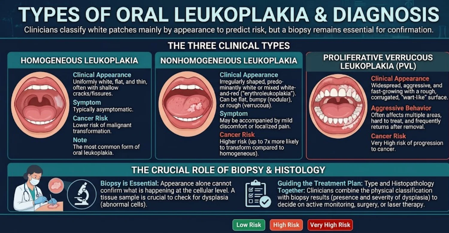
Homogeneous Oral Leukoplakia: Appearance and Characteristics
Homogeneous patches are uniformly white and flat. The surface is smooth, wrinkled, or finely cracked. Color and texture remain consistent across the whole lesion. This is the most common type in most studies. It generally carries lower malignant risk than other types. Lower risk is not zero risk. Homogeneous lesions still need assessment, documentation, and regular follow-up.
Nonhomogeneous Oral Leukoplakia: Red, Nodular, or Irregular Patches
Nonhomogeneous lesions show mixed features. They include speckled patches with red and white areas, called erythroleukoplakia. Nodular lesions show small raised lumps. Verrucous lesions have a rough, wart-like surface. Studies consistently find nonhomogeneous forms carry several times higher transformation risk. Red areas are particularly concerning because they often show dysplasia. These lesions usually need biopsy and often removal.
Proliferative Verrucous Leukoplakia: How It Differs From Other Forms
Proliferative verrucous leukoplakia, or PVL, is a rare but aggressive form. It often begins as a flat white patch and spreads slowly. Over years, it becomes multifocal and wart-like. It is more common in older women and often occurs without tobacco use. A 2021 meta-analysis estimated malignant transformation in about half of PVL cases. Recurrence after treatment is very common. Patients need lifelong specialist monitoring.
What Is the Difference Between Homogeneous and Nonhomogeneous Leukoplakia?
The main difference is uniformity. Homogeneous lesions look the same throughout. Nonhomogeneous lesions combine white, red, nodular, or rough areas. Risk differs significantly between them. Studies report nonhomogeneous lesions transform to cancer far more often. Dysplasia is also found more frequently in these mixed lesions. Management differs too, with nonhomogeneous patches usually treated more actively.
Feature | Homogeneous | Nonhomogeneous |
Color | Uniform white | White with red areas |
Surface | Flat, smooth, or finely cracked | Nodular, speckled, or verrucous |
Frequency | Most common | Less common |
Relative risk | Lower | Several times higher |
Typical approach | Biopsy and monitoring | Biopsy, often removal |
Which Types of Leukoplakia May Require Closer Monitoring?
Nonhomogeneous lesions need closer monitoring. PVL needs the most frequent and long-term follow-up. Patches larger than 200 square millimeters may also carry higher risk. Lesions on the tongue sides or floor of the mouth require attention. Patches in nonsmokers deserve careful review. Any lesion with moderate or severe dysplasia needs close specialist care. Follow-up may be every three to six months.
Oral Leukoplakia
Other Causes of White Patches in the Mouth
Many conditions produce white areas in the mouth. Some are infections, some are immune-related, and some are simple irritations. Distinguishing them is the first step in diagnosis. Each condition has different causes and different treatments. Dentists use history, examination, and sometimes biopsy to tell them apart. Self-diagnosis from online images is unreliable.
Oral Leukoplakia vs. Oral Thrush: Key Differences
Oral thrush is a fungal infection caused mainly by Candida albicans. Its white, creamy patches usually wipe off, leaving red tissue beneath. Leukoplakia does not wipe off. Thrush often follows antibiotics, steroid inhalers, diabetes, or dentures. It commonly responds to antifungal treatment within one to two weeks. Leukoplakia does not resolve with antifungals alone. If a patch persists after antifungal treatment, further assessment is needed.
Hairy Leukoplakia vs. Oral Leukoplakia
Hairy leukoplakia is caused by Epstein-Barr virus. It typically appears on the sides of the tongue. The surface has vertical ridges that look hairy or corrugated. It is mostly seen in people with weakened immune systems, such as HIV. Unlike typical leukoplakia, it is not considered potentially malignant. Its presence may prompt testing for immune problems. Treatment focuses on the underlying condition.
Oral Leukoplakia vs. Oral Lichen Planus
Oral lichen planus is a chronic immune condition. It often appears as lacy white lines called Wickham striae. Lesions are usually on both cheeks symmetrically. Some forms are red, eroded, and painful. Leukoplakia is usually a single solid patch without a lacy pattern. Lichen planus also carries a small malignant risk, estimated around 1% in many reviews. A biopsy helps separate these conditions when appearance overlaps.
Leukoplakia vs. Frictional Keratosis From Cheek Biting
Frictional keratosis results from repeated rubbing or biting. It typically appears along the biting line of the cheeks or tongue edges. The surface is often shaggy, rough, or frayed. It improves once the habit or irritation stops. Leukoplakia, by contrast, persists after the irritant is removed. Dentists may suggest a night guard or smoothing of teeth. They then review the area after a few weeks.
Leukoplakia vs. Oral Candidiasis and Other White Oral Lesions
Chronic hyperplastic candidiasis can mimic leukoplakia closely. It often appears at the corners of the mouth inside the cheeks. It may not wipe off like ordinary thrush. Treatment with antifungals may reduce or clear it. Other white lesions include leukoedema, white sponge nevus, and chemical burns. Leukoedema is a harmless milky film that fades when stretched. Accurate diagnosis may require several visits and a biopsy.
Can You Tell the Difference Between Oral Leukoplakia and Oral Cancer by Appearance Alone?
No. Appearance alone cannot reliably exclude cancer. Early cancers can look exactly like a simple white patch. Some studies report unexpected cancer in a small percentage of biopsied white lesions. Experienced clinicians still rely on biopsy for certainty. Red areas, ulcers, and firmness raise suspicion but are not definitive. Only histopathology under a microscope can confirm the diagnosis.
Is Oral Leukoplakia Precancerous?
Oral leukoplakia is considered potentially malignant rather than strictly precancerous. Most lesions never become cancer. A meta-analysis by Iocca and colleagues estimated an overall transformation rate of about 9.5%. Another 2021 review reported a similar figure near 9.8%. Annual risk is usually estimated at around 1% to 2%. Risk varies widely by lesion type, site, and dysplasia grade. This variability explains why individual assessment is essential.
What Is the Relationship Between Leukoplakia and Oral Cancer Risk?
Patients with leukoplakia have higher oral cancer risk than the general population. Some oral cancers arise from previously visible white patches. Others develop in normal-looking tissue nearby. The patch may act as a marker of wider tissue damage, called field cancerization. Globally, lip and oral cavity cancers caused about 389,000 new cases in 2022. Early detection improves survival significantly. Five-year survival exceeds 80% for localized cancers in many datasets.
Can Oral Leukoplakia Turn Into Oral Squamous Cell Carcinoma?
Yes, in a minority of cases. Oral squamous cell carcinoma is the most common oral cancer. It accounts for over 90% of malignant mouth tumors. Transformation usually takes years and passes through dysplasia stages. Not all cancers follow a predictable path. Some lesions without dysplasia still transform. Regular follow-up helps detect changes before they become advanced.
How Does Oral Epithelial Dysplasia Affect Cancer Risk?
Dysplasia describes abnormal cells within the lining of the mouth. It is graded as mild, moderate, or severe. Risk generally rises with dysplasia grade. A meta-analysis by Mehanna and colleagues reported transformation in about 10% of mild or moderate cases. Severe dysplasia showed transformation in around 24% of cases. Dysplasia grading is subjective and varies between pathologists. Some centers use a binary low-risk or high-risk system instead.
Does the Location of Leukoplakia Affect Its Risk?
Yes. Location is an important risk factor. Lesions on the lateral and ventral tongue carry higher risk. The floor of the mouth is another high-risk site. These areas have thinner tissue and may absorb carcinogens more easily. Lesions on the gums or palate generally carry lower risk. Still, any site can transform. Dentists consider location when deciding on biopsy and removal.
Which Clinical Features May Suggest a Higher-Risk Lesion?
Several features raise concern. These include red areas, nodules, ulceration, and verrucous surfaces. Large size, especially over 200 square millimeters, is associated with higher risk. Tongue and floor-of-mouth sites carry more concern. Long duration and nonsmoking status may also matter. Female sex has been linked to higher risk in some studies. The presence of dysplasia on biopsy remains the strongest predictor.
Higher-risk feature | Why it matters |
Red or speckled areas | Often linked with dysplasia |
Nodular or verrucous surface | Associated with higher transformation |
Tongue or floor of mouth | Thinner, more vulnerable tissue |
Size over 200 mm² | Linked with higher risk in studies |
Moderate or severe dysplasia | Strongest histological predictor |
No tobacco history | May reflect intrinsic genetic change |
Can Leukoplakia Return After Treatment?
Yes. Recurrence is common after removal. Studies report recurrence in roughly 10% to 35% of treated lesions. Rates vary by technique, lesion type, and continued tobacco use. PVL recurs far more often than other forms. New patches can also appear at different sites. Removal does not eliminate the underlying risk in the surrounding tissue. Long-term follow-up remains essential after treatment.
How Is Oral Leukoplakia Diagnosed?
Diagnosis combines medical history, clinical examination, and usually a biopsy. Dentists first exclude conditions with known causes. They often remove possible irritants and recheck the area after two to four weeks. If the patch persists, a tissue sample is usually taken. A pathologist examines it for keratosis, dysplasia, or cancer. Results guide the treatment plan and follow-up interval.
What Happens During a Leukoplakia Examination?
The dentist begins by asking about symptoms, duration, and habits. Questions cover tobacco, alcohol, areca nut, and medical history. The whole mouth is then examined under good light. The patch is measured, photographed, and gently wiped to check adherence. The dentist feels the area for firmness. The neck is checked for enlarged lymph nodes. The visit usually takes 20 to 40 minutes.
How Dentists Assess White Patches and Exclude Other Conditions
Dentists look at color, texture, borders, and location. They wipe the patch to rule out thrush. They check for sharp teeth or denture irritation nearby. A short antifungal course may be prescribed if candidiasis is suspected. Irritants may be removed, followed by review in two to four weeks. Adjunct tools such as special lights or dyes are sometimes used. These tools support decisions but do not replace biopsy.
Oral Leukoplakia Diagnosis and Biopsy: When Is Tissue Sampling Needed?
A biopsy is recommended when a white patch persists without a clear cause. It is especially important for nonhomogeneous, large, or high-risk site lesions. Any patch with ulceration, red areas, or firmness needs sampling. Biopsy is also advised for lesions in nonsmokers. Changes in a previously stable patch may require repeat biopsy. Most guidelines favor sampling rather than prolonged observation.
Incisional vs. Excisional Biopsy for Leukoplakia
An incisional biopsy removes a small part of the lesion. It is used for large patches or areas that look most suspicious. Multiple samples may be taken from different zones. An excisional biopsy removes the whole lesion with a small margin. It suits smaller patches that can be removed easily. Excision provides both diagnosis and treatment in one step. The choice depends on size, site, and clinical suspicion.
What Does a Leukoplakia Biopsy Test For?
The pathologist examines tissue under a microscope. They assess the surface keratin layer and the shape of cells. They look for dysplasia and grade its severity. They check whether abnormal cells have invaded deeper tissue. Fungal organisms may also be identified. The report helps decide between monitoring, removal, or cancer treatment.
Hyperkeratosis and Other Tissue Changes
Hyperkeratosis means the surface keratin layer is thicker than normal. It explains the white color of most patches. It may appear alone without abnormal cells. Acanthosis, or thickening of the cell layer, is often present too. These findings are generally considered lower risk. They still require follow-up because some lesions change over time.
Mild, Moderate, and Severe Oral Epithelial Dysplasia
Mild dysplasia affects the lower third of the lining. Moderate dysplasia extends into the middle third. Severe dysplasia involves more than two-thirds of the thickness. When the full thickness is affected, it may be called carcinoma in situ. Studies show risk increases with each grade. Severe dysplasia usually leads to removal and close follow-up.
Findings That May Indicate Oral Cancer
Cancer is diagnosed when abnormal cells break through the basement membrane. This boundary separates surface tissue from deeper layers. Invasion into connective tissue confirms squamous cell carcinoma. The report may describe tumor depth and differentiation. These details guide staging and treatment. Patients are then referred to a head and neck cancer team.
Can Leukoplakia Be Diagnosed Without a Biopsy?
A clinical diagnosis can be made without a biopsy. However, this only describes appearance. It cannot reveal dysplasia or early cancer. Brush cytology and light-based tools are sometimes used. These adjuncts have limited accuracy compared with surgical biopsy. Guidelines generally recommend biopsy for persistent unexplained patches. Biopsy remains the gold standard.
How Long Do Leukoplakia Biopsy Results Take?
Results typically take five to ten working days. Some laboratories report within three days. Complex cases or second opinions may take two to three weeks. Urgent cases can sometimes be prioritized. Your clinician will usually arrange an appointment to explain the findings. Ask how and when you will receive results before leaving.
Why Follow-Up Examinations Matter After Diagnosis
Leukoplakia can change silently over time. Follow-up visits allow early detection of new red areas or growth. Photographs help compare the patch across visits. Repeat biopsies may be needed if changes appear. Follow-up also checks for new lesions elsewhere in the mouth. Intervals range from three months to one year, depending on risk.
What Are the Oral Leukoplakia Treatment Options?
Oral leukoplakia treatment depends on biopsy findings, lesion type, site, and patient factors. Options include monitoring, surgical removal, laser treatment, and risk factor control. No treatment has been proven to fully prevent cancer in all cases. Systematic reviews show limited evidence that any single approach eliminates future transformation. For this reason, treatment is always combined with follow-up. Decisions should be shared between patient and clinician.
When Is Monitoring Appropriate Instead of Immediate Treatment?
Monitoring may suit small homogeneous lesions without dysplasia. It is also used for patches in lower-risk sites. Patients must stop tobacco and attend regular reviews. Photos and measurements track any change. Monitoring may also suit patients unfit for surgery. If the patch changes, biopsy or removal is reconsidered. Monitoring is active care, not neglect.
Surgical Removal of Leukoplakia
Scalpel excision removes the patch with a margin of healthy tissue. It is usually done under local anesthesia. The removed tissue is sent for full pathology. This confirms the diagnosis and checks margins. Small lesions heal within one to two weeks. Larger excisions may need stitches or tissue grafts. Surgery is often preferred for dysplastic lesions.
Laser Treatment for Oral Leukoplakia
CO₂ lasers are commonly used to remove or vaporize white patches. Laser excision allows tissue to be sent for pathology. Laser vaporization destroys tissue without providing a sample. For that reason, biopsy should always come first. Lasers typically cause less bleeding and minimal scarring. Studies report recurrence rates broadly similar to scalpel surgery. Healing usually takes two to four weeks.
When Is an Excisional Biopsy Also a Treatment?
When a small lesion is removed completely, the biopsy becomes treatment. The pathologist checks whether the margins are clear. If margins are clear and no severe dysplasia exists, monitoring may follow. If margins contain abnormal cells, further removal may be advised. This approach is efficient for small, accessible patches. It avoids separate procedures.
Can Medication Treat Leukoplakia?
Several medications have been studied. These include topical retinoids, beta-carotene, and lycopene. Some produced temporary improvement in lesion size. However, Cochrane reviews found no proven effect on preventing cancer. Patches often return once medication stops. Side effects can be significant with some agents. Medication is not currently a standard first-line cure.
Can Leukoplakia Be Treated Without Surgery?
Some lesions improve with nonsurgical measures. Quitting tobacco can lead to significant regression. Removing friction sources can resolve reactive patches. Antifungal treatment helps when Candida is involved. Photodynamic therapy is being studied as an alternative. Nonsurgical options suit selected lower-risk lesions. Monitoring remains essential with any nonsurgical approach.
How Does Treatment Change When Dysplasia Is Present?
Dysplasia usually shifts management toward removal. Mild dysplasia may be removed or closely monitored, depending on site and size. Moderate and severe dysplasia are generally removed when feasible. Follow-up becomes more frequent, often every three months initially. Specialists may involve oral medicine or maxillofacial surgery teams. Risk factor control becomes even more important.
What Happens if Leukoplakia Is Left Untreated?
Many untreated lesions remain stable for years. Some regress, especially after quitting tobacco. A minority progress to dysplasia or cancer. The problem is that no one can predict which lesions will progress. Untreated and unmonitored patches may reach advanced cancer before diagnosis. Advanced oral cancer has much lower survival rates. Monitoring at minimum is strongly recommended.
Why Can Leukoplakia Return After Removal?
Removing a patch does not change the surrounding tissue. Nearby cells may already carry genetic damage. This concept is called field cancerization. Continued smoking or drinking increases recurrence risk. Incomplete removal can leave abnormal cells behind. PVL is particularly prone to returning. Recurrence is not treatment failure but a reason for ongoing follow-up.
Leukoplakia Treatment Side Effects and Recovery
Most patients recover well from biopsy or removal. Discomfort is usually mild and short-lived. Recovery time depends on lesion size, site, and technique. Small biopsies heal within seven to fourteen days. Larger excisions may take three to four weeks. Following aftercare instructions reduces complications.
Pain, Swelling, and Bleeding After Biopsy or Removal
Mild pain usually peaks in the first two to three days. Swelling is common and fades within a week. Minor oozing may occur during the first day. Pressing gauze for 15 to 20 minutes usually controls it. Cold compresses on the cheek can reduce swelling. Contact your clinician if bleeding is heavy or persistent.
Eating, Speaking, and Oral Hygiene During Recovery
Choose soft, cool foods for several days. Avoid spicy, crunchy, acidic, or very hot foods. Tongue procedures may temporarily affect speech. Keep brushing gently while avoiding the wound site. Rinse as directed, often starting the day after surgery. Avoid smoking and alcohol throughout healing. Both delay recovery and increase infection risk.
Possible Scarring, Wound Complications, and Recurrence
Most wounds heal with minimal scarring. Larger excisions may leave tight or firm tissue. Infection is uncommon but possible. Signs include increasing pain, swelling, pus, or fever. Temporary numbness can occur near nerves. Recurrence may appear months or years later. Regular follow-up detects it early.
Oral Leukoplakia Relief: What Can Help With Discomfort?
Most patches are painless, but some cause irritation. Relief measures can improve comfort while you await assessment. They cannot treat the underlying condition. Gentle care, diet changes, and avoiding irritants usually help. Persistent discomfort should always prompt professional review. Comfort measures should never delay diagnosis.
How to Relieve Mouth Irritation While Waiting for an Appointment
Avoid tobacco and alcohol completely. Choose mild, soft foods at moderate temperatures. Use a soft toothbrush and fluoride toothpaste without harsh flavoring. Some patients find sodium lauryl sulfate-free toothpaste gentler. Stay hydrated to keep the mouth moist. Avoid touching or rubbing the patch with your tongue. Book an appointment rather than waiting for symptoms to settle.
Can Saltwater Rinses Help With Oral Discomfort?
Warm saltwater rinses can soothe irritated tissue. Mix half a teaspoon of salt in a glass of warm water. Swish gently for 30 seconds, then spit. Repeat two to three times daily. Saltwater helps keep the area clean after biopsy. It does not treat leukoplakia itself. Avoid alcohol-based mouthwashes, which may sting.
Which Foods and Drinks May Irritate an Oral Lesion?
Spicy foods often cause burning. Citrus fruits, tomatoes, and vinegar can sting due to acidity. Crunchy foods such as chips may scrape the patch. Very hot drinks can aggravate sensitive tissue. Alcohol and carbonated drinks may also irritate. Keep a short food diary if certain items cause discomfort. Share it with your dentist.
Are Over-the-Counter Pain Relievers Appropriate After Oral Biopsy?
Simple pain relievers are usually adequate after biopsy. Paracetamol or ibuprofen are commonly recommended. Always follow your clinician's advice and dosage instructions. Some patients cannot take ibuprofen due to stomach, kidney, or bleeding issues. Aspirin may increase bleeding and is often avoided. Ask your clinician before taking any medication.
Why Pain Relief Does Not Replace Diagnosis or Treatment
Pain relief only masks symptoms. It does not change the cells within the patch. A comfortable patch can still contain dysplasia. Relying on painkillers can delay important assessment. Early diagnosis offers the best outcomes. Use relief measures alongside, not instead of, professional care.
Which Home Remedies Should Patients Avoid?
Avoid scraping, burning, or cutting the patch at home. Do not apply undiluted essential oils, hydrogen peroxide, or aspirin directly. Avoid harsh herbal pastes or caustic powders. These can burn tissue and disguise warning signs. Online "cures" lack evidence and may cause harm. Turmeric or aloe products have limited research support. Discuss any remedy with your dentist first.
What Is the Average Cost of Oral Leukoplakia Diagnosis and Treatment in the US, UK, and Turkey?
Oral leukoplakia costs vary widely by country, clinic, and complexity. The main components are consultation, biopsy, pathology, removal, and follow-up. Insurance and public healthcare can reduce costs significantly. Prices below are approximate 2026 estimates for guidance only. Final costs depend on clinical findings. Always request an itemized written quote.
Average Leukoplakia Examination and Biopsy Cost in the United States
US costs are among the highest globally. Fees vary by state, provider type, and insurance coverage. Many dental and medical insurance plans cover biopsy when medically necessary. Uninsured patients may pay several hundred to several thousand dollars. Hospital-based procedures usually cost more than office-based ones. Always confirm coverage before treatment.
Dentist or Specialist Consultation, Biopsy, and Pathology Fees
A general dental exam typically costs $75 to $200. A specialist consultation may cost $150 to $400. Office biopsy fees usually range from $250 to $800. Pathology fees add roughly $150 to $500. Specialist stains or second opinions cost extra. Total diagnostic costs commonly reach $500 to $1,500 without insurance.
Additional Costs for Surgical Removal and Follow-Up
Surgical excision commonly costs $800 to $3,000. Laser removal may range from $1,000 to $4,000. Hospital or sedation fees can increase totals further. Follow-up visits typically cost $100 to $250 each. Repeat biopsies add similar costs to the first. Long-term monitoring adds ongoing expense.
US service | Approximate cost |
Specialist consultation | $150 – $400 |
Biopsy procedure | $250 – $800 |
Pathology report | $150 – $500 |
Surgical or laser removal | $800 – $4,000 |
Follow-up visit | $100 – $250 |
Average Leukoplakia Examination and Biopsy Cost in the United Kingdom
UK patients can access NHS or private care. NHS care is heavily subsidized but may involve waiting times. Private care offers faster access at higher cost. Many patients start with an NHS dentist and receive a hospital referral. Suspected cancer referrals follow faster pathways. Private insurance may cover specialist care.
NHS Assessment and Referral vs. Private Dental or Specialist Care
An NHS dental examination costs $27.40 in England under 2025 Band 1 charges. Hospital referral, biopsy, and pathology are usually free on the NHS. Suspected cancer referrals target diagnosis within 28 days. Routine referrals may wait several weeks or months. Private care avoids waiting but carries full fees. Charges differ across Scotland, Wales, and Northern Ireland.
Private Biopsy, Histopathology, and Surgical Treatment Costs
Private specialist consultations usually cost $150 to $350. A private biopsy often costs $300 to $900. Histopathology may add $100 to $350. Surgical or laser removal typically ranges from $800 to $3,000. Follow-up appointments cost around $100 to $250. London clinics often charge more.
UK private service | Approximate cost |
Specialist consultation | $150 – $350 |
Biopsy procedure | $300 – $900 |
Histopathology | $100 – $350 |
Surgical or laser removal | $800 – $3,000 |
Follow-up visit | $100 – $250 |
Average Leukoplakia Examination and Biopsy Cost in Turkey
Turkey offers lower private costs than the US and UK. Clinics in Istanbul and Antalya treat many international patients. Savings of 50% to 70% are commonly reported. Quality varies, so credentials matter. Patients should confirm the specialist's qualifications and laboratory standards. Clear follow-up planning is essential for travelers.
Private Specialist Consultation, Biopsy, and Pathology Costs
Private consultations usually cost $50 to $150. A biopsy procedure often costs $150 to $450. Pathology fees typically range from $50 to $200. Some clinics offer package pricing for diagnosis. Results may take five to ten working days. Plan your stay accordingly.
Surgical Removal, Follow-Up, and International Patient Expenses
Surgical or laser removal typically costs $300 to $1,500. Follow-up visits are often $40 to $100. International patients should budget for flights, hotels, and transfers. Healing time may require a stay of one to two weeks. Remote follow-up with photos can help after returning home. Local follow-up arrangements are still important.
Turkey service | Approximate cost |
Specialist consultation | $50 – $150 |
Biopsy procedure | $150 – $450 |
Pathology report | $50 – $200 |
Surgical or laser removal | $300 – $1,500 |
Follow-up visit | $40 – $100 |
What Factors Affect the Total Cost of Leukoplakia Treatment?
Lesion size and number strongly affect cost. Location matters because some sites require specialist surgery. Biopsy type and laboratory testing add variable fees. Anesthesia choice, from local to sedation, changes the total. Specialist experience and clinic location also influence pricing. Recurrence may require repeat procedures. Long-term monitoring adds ongoing expense.
Does the Cost Include Biopsy, Laboratory Testing, and Follow-Up?
Not always. Many clinics quote the procedure fee separately from pathology. Follow-up visits may be charged individually. Medications and post-operative supplies may cost extra. Ask whether the quote includes consultation, biopsy, histopathology, removal, and reviews. Request everything in writing. Hidden costs often appear in laboratory fees.
How Can Patients Compare Leukoplakia Treatment Quotes Across Countries?
Compare itemized quotes rather than headline prices. Check what each quote includes and excludes. Confirm specialist qualifications and laboratory accreditation. Ask how results will be communicated. Consider travel, accommodation, and time off work. Factor in follow-up needs after returning home. The cheapest option is not always the best value.
Note : All images used are for editorial and illustrative purposes only and may not originate from the original news provider or associated company.
How to Prevent Oral Leukoplakia From Worsening
Prevention focuses on reducing exposure to known risk factors. Lifestyle changes can stabilize or shrink many lesions. Regular dental care detects changes early. Prevention cannot guarantee a patch will never change. It does meaningfully lower risk. Combining several measures gives the strongest benefit.
Why Quitting Tobacco Is an Important Preventive Step
Quitting tobacco is the most effective preventive measure. Studies show many patches shrink or disappear after stopping. Quitting also lowers oral cancer risk over time. Risk continues to decline with each tobacco-free year. Support programs, nicotine replacement, and medications improve success rates. Ask your dentist or doctor about cessation support. Combined counseling and medication often works best.
Reducing Alcohol Consumption and Avoiding Betel Quid
Lowering alcohol intake reduces oral cancer risk. Avoiding binge drinking is particularly important. Stopping areca nut and betel quid use is essential. These products cause direct tissue damage. Community programs can help users quit. Combining tobacco and alcohol reduction gives the largest benefit.
Maintaining Oral Hygiene and Addressing Irritating Dental Appliances
Brush twice daily with fluoride toothpaste. Clean between teeth daily. Replace broken fillings and smooth sharp tooth edges. Have dentures checked for fit regularly. Remove dentures overnight to rest the tissues. Good hygiene reduces fungal infection risk. Regular cleanings allow early lesion detection.
Can Lifestyle Changes Make Leukoplakia Disappear?
Lifestyle changes can make some patches disappear. Tobacco-related lesions respond best. Research shows regression in a substantial share of patients after quitting. Idiopathic lesions respond less predictably. Disappearance does not end the need for follow-up. Abnormal cells may remain beneath normal-looking tissue.
How Often Should Patients Have Follow-Up Examinations?
Frequency depends on risk. Low-risk lesions without dysplasia may need review every six to twelve months. Lesions with mild dysplasia often need review every three to six months. Moderate or severe dysplasia usually requires review every three months. PVL needs lifelong specialist monitoring. Your clinician will tailor the schedule.
Can Leukoplakia Be Prevented Completely?
Complete prevention is not possible. Some lesions develop without known risk factors. However, avoiding tobacco, areca nut, and heavy alcohol greatly reduces risk. Good oral hygiene and regular checkups help too. Early detection prevents small problems becoming serious ones. Prevention and monitoring work best together.
When Should You See a Dentist or Doctor for Oral Leukoplakia?
Any unexplained white patch lasting over two weeks needs assessment. Red patches, ulcers, and lumps need prompt evaluation. Do not wait for pain to develop. Early evaluation leads to simpler treatment and better outcomes. Your dentist is usually the best first contact. Referral to a specialist follows if needed.
When a Persistent White Patch in the Mouth Needs Assessment
Book an appointment if a patch lasts longer than two weeks. Do the same if it returns after healing. Assessment is important even when the patch is painless. Smokers and heavy drinkers should be especially vigilant. Nonsmokers should not dismiss patches either. Early review provides peace of mind or early treatment.
Red and White Patches, Ulcers, or Lumps That Need Prompt Evaluation
Mixed red and white patches carry higher risk. Ulcers that do not heal within three weeks need urgent assessment. Lumps or thickening in the mouth also need prompt review. UK guidelines recommend urgent referral for unexplained ulcers persisting over three weeks. These signs need evaluation within days, not months.
Warning Signs Such as Bleeding, Firmness, or Rapid Changes
Bleeding with minimal contact is a warning sign. Firmness or hardness beneath a patch is concerning. Rapid growth over weeks needs urgent evaluation. Loose teeth without gum disease may signal deeper problems. A patch becoming fixed to underlying tissue is serious. Seek care promptly if any of these appear.
Persistent Pain, Difficulty Swallowing, or Unexplained Numbness
Ongoing pain in the mouth or ear needs assessment. Difficulty swallowing or chewing is a red flag. Numbness of the tongue, lip, or chin needs urgent review. Voice changes and persistent sore throat also need evaluation. A lump in the neck lasting over three weeks requires prompt referral.
Which Specialist Should Evaluate Suspected Leukoplakia?
Your general dentist is usually the first step. They may refer you to an oral medicine specialist. Oral and maxillofacial surgeons perform biopsies and removals. Oral pathologists interpret tissue samples. Ear, nose, and throat specialists may be involved for some sites. Head and neck cancer teams manage confirmed cancers.
Why Waiting for Pain to Develop Is Not a Safe Approach
Most patches and many early cancers are painless. Pain often appears only when disease is advanced. Waiting for symptoms can delay diagnosis by months. Delayed diagnosis is linked with worse outcomes. Early oral cancers have far higher survival than late-stage disease. Act on persistence, not pain.
Ready to transform your smile?
Talk to our team and get a personal FREE treatment plan.
What We Notice Clinically
In clinical practice, white patches are often found incidentally. Patients usually come for cleaning, cosmetic work, or a toothache. Many are surprised to learn they have a lesion. Some have noticed it for years without concern. These observations shape how we communicate findings. They also explain why we examine soft tissues at every visit.
Why a Painless White Patch Should Not Be Automatically Dismissed
Patients often believe that painless means harmless. In our experience, this is a common reason for delayed care. Many dysplastic lesions cause no symptoms. A painless patch can still need biopsy. We encourage patients to judge by persistence rather than comfort. A quick assessment can prevent years of uncertainty.
Common Patient Misconceptions About Leukoplakia
Many patients assume a white patch is a thrush. Others believe only smokers develop concerning lesions. Some think mouthwash or scraping will remove it. Others fear every patch means cancer. Both underestimating and overestimating risk can cause harm. Clear information helps patients make balanced decisions.
Why Clinical Appearance Alone Cannot Reliably Exclude Dysplasia
Two patches can look identical yet show very different histology. Homogeneous patches sometimes contain dysplasia. Nonhomogeneous patches sometimes show only keratosis. Studies show visual assessment alone misses some abnormal lesions. This is why biopsy remains central. We do not rely on appearance alone.
How Tobacco Exposure, Lesion Location, and Biopsy Findings Influence Assessment
We consider several factors together. Tobacco history affects cause and expected response to quitting. Location influences risk and biopsy urgency. Biopsy findings determine treatment intensity and follow-up frequency. Size, texture, and duration also contribute. Combining these factors creates a personalized risk profile.
Clinical Note
Clinical experience reinforces the importance of careful soft tissue examination. Many oral lesions are discovered before patients notice any symptoms. A structured approach helps avoid both missed lesions and unnecessary procedures.
Dr. Rifat Alsaman, on the Importance of Assessing Persistent Oral White Patches
Dr. Rifat Alsaman emphasizes that persistent white patches should never be dismissed. He advises patients to seek assessment for any patch lasting over two weeks. He notes that early evaluation offers patients the widest range of options. A structured examination, photographs, and timely referral are central to this approach. Clear communication helps patients understand their risk without unnecessary alarm.
Why Confirming the Diagnosis Should Come Before Cosmetic or Routine Dental Treatment
Cosmetic treatment should not proceed while an unexplained lesion remains. Veneers, crowns, or implants may alter tissue appearance and complicate monitoring. Confirming the diagnosis first protects patient safety. It also ensures treatment planning considers the patient's full oral health. Once the lesion is managed, cosmetic care can continue safely.








Tips for Patients
Patients play an active role in managing leukoplakia. Simple habits support accurate diagnosis and safe monitoring. Preparation makes appointments more productive. Good questions improve understanding of results. These practical tips help you stay engaged in your care.
What to Record Before a Leukoplakia Appointment
Note when you first noticed the patch. Record any changes in size, color, or texture. List tobacco, alcohol, and areca nut use, including amounts. Bring a list of current medications. Note any symptoms such as burning or pain. Bring dated photographs if available.
How to Monitor Changes in a White Patch Without Delaying Care
Take clear photos in good light every two to four weeks. Use the same angle and lighting each time. Note any new red areas, lumps, or ulcers. Do not use monitoring as a reason to delay assessment. Self-monitoring complements, but never replaces, professional review.
What to Ask About Biopsy Results and Treatment Recommendations
Ask what the biopsy showed and whether dysplasia was found. Ask about the grade if dysplasia is present. Ask whether margins were clear after excision. Ask what treatment options exist and their risks. Ask how often follow-up is needed. Request a written copy of your pathology report.
Medical Tips for Safe Oral Care Before and After a Biopsy
Tell your clinician about blood thinners and medical conditions. Eat a light meal before the procedure unless told otherwise. Avoid smoking and alcohol before and after biopsy. Follow aftercare instructions carefully. Use prescribed rinses as directed. Contact the clinic if bleeding, swelling, or fever develops.
Habits to Avoid, Including Scraping Patches or Using Unproven Remedies
Never scrape, pick, or burn a white patch. Avoid applying caustic substances or undiluted oils. Avoid unproven supplements promising cures. Do not stop follow-up because a patch looks better. Avoid tobacco in every form, including waterpipes. These habits protect both tissue and diagnosis.
How to Prepare for Follow-Up Visits and Long-Term Monitoring
Keep a calendar reminder for each review. Bring updated photos and symptom notes. Report any lifestyle changes, such as quitting smoking. Keep copies of all pathology reports. Ask whether intervals should change based on findings. Consistent follow-up provides the best long-term protection.
Oral Leukoplakia Assessment and Personalized Care at Vitrin Clinic
At Vitrin Clinic, soft tissue assessment is part of every comprehensive dental examination. Persistent white patches are documented, photographed, and evaluated carefully. Patients receive clear explanations of findings and next steps. Where needed, referral to appropriate specialists is coordinated. The goal is safe, evidence-based, personalized care.
Assessing Persistent White Patches Before Recommending Dental Treatment
Before cosmetic or restorative treatment, the clinic examines the entire mouth. Any persistent patch is assessed before treatment begins. This prevents masking lesions under new restorations. It also ensures safe treatment timing. Patient health always comes before cosmetic goals.
Personalized Evaluation and Referral for Leukoplakia Diagnosis
Each patient receives an individualized evaluation. History, habits, and lesion features are reviewed together. Possible irritants are identified and addressed. When appropriate, patients are referred for specialist evaluation. Referrals are explained clearly, including why they are needed.
Understanding When Biopsy or Specialist Assessment May Be Appropriate
Not every white patch needs a biopsy immediately. Some patches are reviewed after removing irritants. Persistent, nonhomogeneous, or high-risk lesions usually need specialist assessment. The clinic explains the reasoning behind each recommendation. Patients are encouraged to ask questions.
Coordinating Care Based on Clinical Findings and Histopathology
Care is planned around clinical findings and pathology results. Dental treatment is timed to support healing and monitoring. Communication between specialists keeps care consistent. Patients receive clear updates at each stage.
Distinguishing Oral Mucosal Concerns From Conditions Requiring Other Specialists
Some white patches relate to systemic or immune conditions. Others need oral surgery, oral medicine, or ENT expertise. The clinic helps identify which specialist is appropriate. This avoids delays and unnecessary appointments. Patients are guided toward the right care pathway.
How Vitrin Clinic Supports Your Treatment Journey
Vitrin Clinic supports patients from first consultation to follow-up. Each stage focuses on clarity, safety, and personalized care. International and local patients receive organized coordination. Costs and timelines are explained clearly. Patients are never rushed into decisions.
Initial Consultation and Review of Oral Health Concerns
The first visit includes a full oral examination. Concerns, symptoms, and history are discussed in detail. Soft tissues are inspected and photographed. The dentist explains initial findings clearly. Next steps are agreed together.
Individualized Assessment and Diagnostic Planning
Assessment considers lesion features and patient risk factors. Diagnostic plans may include irritant removal and review. Specialist evaluation or biopsy may be recommended where appropriate. Each plan reflects the patient's individual situation.
Coordinating Specialist Appointments and Relevant Investigations
The clinic helps arrange specialist appointments when needed. Relevant records and photos are shared with consent. This reduces repeated tests and delays. Patients receive guidance on preparation for each appointment.
Explaining Findings, Treatment Recommendations, and Expected Costs
Findings are explained in plain language. Treatment options are discussed with benefits and risks. Expected costs are provided in writing where possible. Patients have time to ask questions before deciding.
Follow-Up Coordination and Guidance for Ongoing Oral Health Care
Follow-up schedules are planned according to risk. Reminders help patients keep appointments. International patients receive guidance on remote follow-up. Ongoing dental care is coordinated around monitoring needs.
Ready to transform your smile?
Talk to our team and get a personal FREE treatment plan.
Key Takeaways
Leukoplakia is common, often painless, and occasionally serious. Understanding the basics helps patients act early. These key points summarize what matters most.
What Patients Should Know About Leukoplakia Symptoms and Causes
Oral leukoplakia appears as a white patch that does not wipe off. Most patches are painless. Tobacco, areca nut, and alcohol are major causes. Some lesions develop without obvious causes. Global prevalence is estimated at about 4%.
Why Persistent White Patches Need Professional Assessment
Appearance cannot reliably exclude dysplasia or cancer. Many concerning lesions cause no symptoms. Assessment is recommended for patches lasting over two weeks. Early evaluation offers the best outcomes.
How Biopsy Findings Guide Treatment and Cancer-Risk Evaluation
Biopsy reveals keratosis, dysplasia, or cancer. Dysplasia grade strongly influences risk. Findings determine monitoring, removal, or referral. Overall transformation rates near 9.5% highlight the need for careful evaluation.
Why Risk Reduction and Long-Term Follow-Up Matter
Quitting tobacco can shrink or clear many patches. Recurrence occurs in roughly 10% to 35% of treated lesions. Follow-up detects changes early. Lifelong monitoring may be needed for higher-risk forms.
When to Seek Professional Care Without Delay
Seek urgent care for red areas, ulcers, lumps, or bleeding. Do the same for numbness, swallowing problems, or neck lumps. Do not wait for pain. Prompt assessment saves time and protects health.
At Vitrin Clinic
At Vitrin Clinic, we believe healthy soft tissues come before any smile design. Every comprehensive examination includes careful assessment of the gums, cheeks, tongue, and palate. If we find a persistent white patch, we explain what it may mean. We coordinate specialist referral and diagnostic steps where needed. OurTreatment plans and costs are explained clearly in writing. If you have noticed a white patch in your mouth, book a consultation with our team. Early assessment brings clarity, safety, and peace of mind.
Reference
FAQs
Dr. Rifat Alsaman has more than 5 years of clinical experience in dentistry and currently serves as the Head of the Medical Team at Vitrin Clinic. He is dedicated to providing exceptional patient care, overseeing treatment planning, and ensuring the highest clinical standards across the team. His expertise, attention to detail, and commitment to continuous professional development have helped countless patients achieve healthier, more confident smiles.


