General Dentistry
October 15, 2026
Oral Mucosal Melanoma: What Are the Symptoms, Prognosis, and Survival Rates?
 Dr. Rifat Alsaman
Oral mucosal melanoma is a rare, potentially aggressive cancer that develops from pigment-producing cells in the lining of the mouth. A persistent dark patch, unusual pigmentation, or a changing lump can understandably cause worry. Most oral pigmentation is harmless, but suspicious changes still need professional assessment. This guide explains the symptoms, possible causes, diagnosis, treatment, prognosis, and when to seek specialist care. Early recognition and timely evaluation may widen treatment options and support better planning.
.jpg)
.jpg)
.jpg)
.jpg)
.jpg)
.jpg)
.jpg)
.jpg)
Ready to transform your smile?
Talk to our team and get a FREE treatment plan.
Oral Mucosal Melanoma: What Is It and Where Does It Develop?
Oral mucosal melanoma is a malignant tumor that arises from melanocytes, the cells that produce pigment. Melanocytes sit in the deepest layer of the mouth's lining. When they grow uncontrollably, they can form a cancer that invades nearby tissue. Most cases appear on the hard palate and upper gums. The disease is uncommon, yet it is clinically significant because it is often found late. It also differs from melanoma that starts elsewhere, usually the skin, and later spreads to the mouth.
Which Areas of the Mouth Can Be Affected?
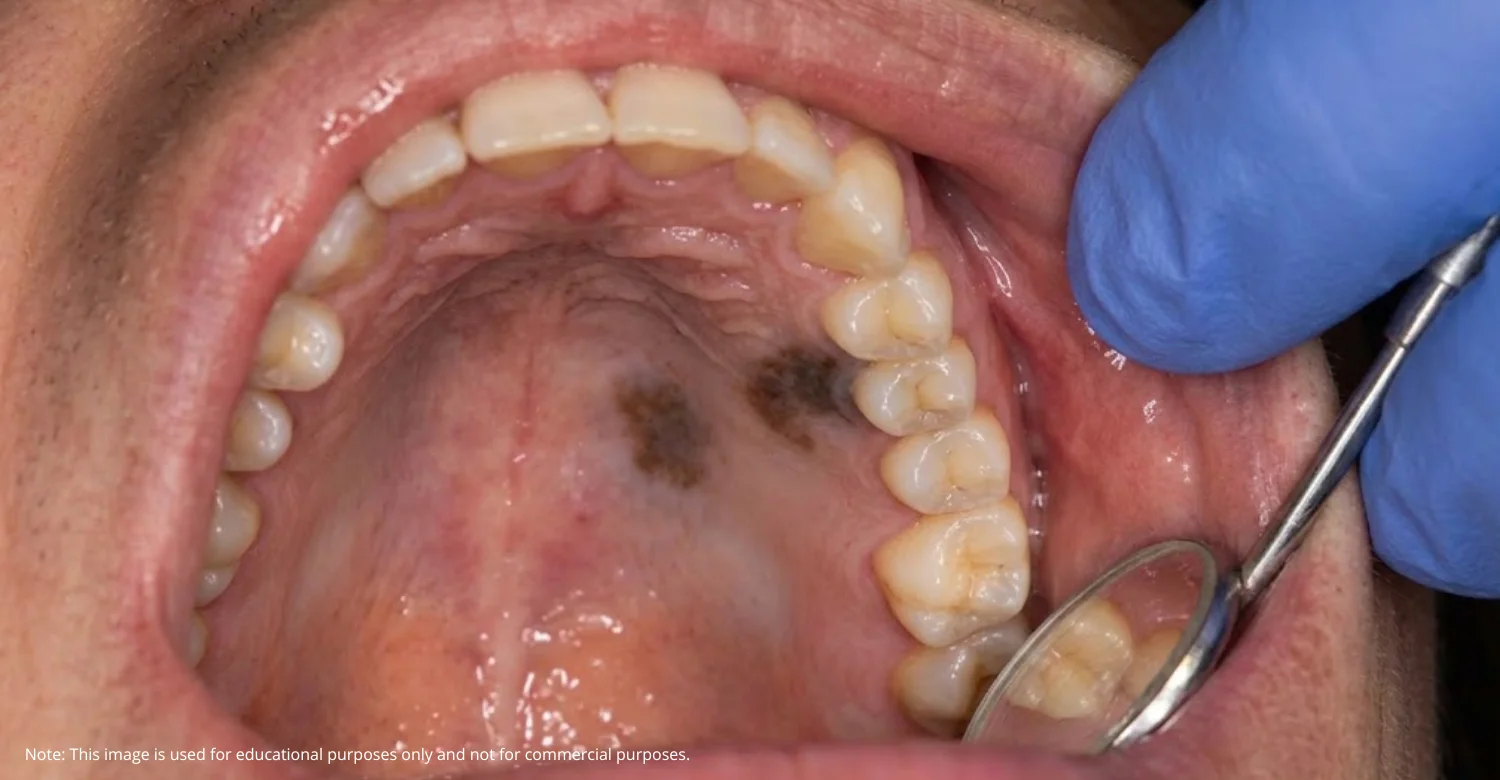
The hard palate, or roof of the mouth, is the most frequently reported site. The upper gums are the second most common location. Lesions may also develop on the lower gums, inner cheeks, lips, tongue, or floor of the mouth, although these sites are less common. Appearance often varies with location. A palatal lesion may look like a flat, spreading stain, while a gum lesion may appear raised, lumpy, or close to the teeth.
Who Can Develop Oral Mucosal Melanoma?
This cancer mainly affects adults, and it is usually diagnosed in later life, often after the age of 50. It is rare in children. Many patients have no recognized risk factors at all. Men and women can both be affected. Because of this, a person's age, general health, or medical history cannot confirm or rule out the condition. Only a proper clinical assessment, followed by a biopsy when indicated, can provide a reliable answer.
Oral Mucosal Melanoma Symptoms: What Changes Should You Look For?
Oral mucosal melanoma symptoms are often subtle at first. Early lesions are usually painless and may be discovered by chance during a dental examination. Oral melanoma early symptoms tend to be visual rather than physical. Common signs of melanoma in the mouth include a new dark patch, uneven coloring, or a slowly spreading stain. Later, a lesion may become raised, ulcerated, or prone to bleeding. Any persistent, unexplained change deserves professional attention, even when it causes no discomfort.
What Does an Early Oral Melanoma Look Like?
An early lesion often appears as a new brown, black, gray, or blue-black patch. It may be asymmetrical, with irregular borders and several shades within the same area. The patch may slowly enlarge or change in appearance over weeks or months. However, some melanomas contain little or no pigment. These amelanotic lesions may look pink, red, or similar to normal tissue. This makes them harder to recognize and is one reason assessment matters.
Can Oral Melanoma Cause Pain, Bleeding, or Swelling?
Pain is frequently absent during the early stages. As the lesion progresses, it may ulcerate, bleed without an obvious cause, or form a raised lump. Swelling can also develop around the affected area. Advanced lesions may interfere with chewing, speaking, or wearing dentures. Teeth near a gum lesion may loosen. These symptoms are not specific to melanoma. Infections, injuries, and other conditions can cause similar problems, so proper assessment is essential.
Are Oral Melanoma Symptoms Always Easy to Notice?
No. Lesions on the roof of the mouth or upper gums are difficult to see without a mirror and good light. Many people rarely look at these areas. The absence of pain also removes an important warning signal. As a result, a lesion may grow unnoticed for some time. Pay attention to persistent changes, even when eating and speaking feel completely normal. A routine dental visit may be when such changes are first noticed.
Note: All images used are for editorial and illustrative purposes only and may not originate from the original news provider or associated company.
Oral Mucosal Melanoma Causes: What Is Known About Its Development?
Oral mucosal melanoma causes remain poorly understood. Researchers know the disease begins when melanocytes acquire genetic changes. These changes allow the cells to multiply without normal control and invade surrounding tissue. However, the triggers behind these changes are not clearly identified. There is no evidence that poor oral hygiene, routine dental procedures, or a specific food directly causes this cancer. Research continues, but most cases currently have no single, identifiable cause.
What Are the Recognized or Suspected Risk Factors?
Evidence about oral mucosal melanoma risk factors is limited. Older age is the clearest association. Studies show that mucosal melanomas often carry different genetic changes from skin melanoma, including KIT and NRAS mutations, while BRAF mutations are less common. Established skin melanoma risks do not necessarily apply here. Ultraviolet exposure is not considered the usual cause, since the inside of the mouth receives little sunlight. Tobacco use and chronic irritation have been studied, but no clear link is proven.
Can Dental Irritation or Oral Pigmentation Cause Melanoma?
There is no strong evidence that dental irritation causes melanoma. Many dark spots have harmless explanations. Benign pigmentation, bruising, and amalgam tattoos from old silver fillings are common examples. A dark spot near a restoration may therefore have several possible causes. Irritation should not be automatically blamed for a suspicious lesion, though. Some melanomas are preceded by pigmentation present for months or years. Pigmentation that is new, changing, unexplained, or persistent should be examined.
What Other Symptoms Can Occur Alongside Oral Mucosal Melanoma?
Visible pigmentation is often the first finding, but other changes can appear as the lesion develops. These depend on the tumor's location, size, and stage. A lesion on the gums may affect nearby teeth. A palatal lesion may cause swelling or surface breakdown. Spreading to lymph nodes may produce a neck lump. Even so, having several symptoms does not establish a cancer diagnosis. Many benign conditions produce similar findings and need careful clinical assessment.
Which Oral Changes Should You Take Seriously?
Some changes deserve prompt professional attention:
A persistent ulcer or an area that repeatedly bleeds.
A growing lump or thickened area of tissue.
Unexplained swelling or discomfort.
A new pigmented lesion that changes in shape, color, or size.
Loose teeth without an obvious dental cause.
A lump in the neck, which may need assessment for lymph node involvement.
These signs do not confirm cancer. They do mean that waiting and watching is no longer appropriate.
Can Oral Melanoma Develop Without Pain?
Yes. Many oral melanomas are painless for a long time. Visible changes, such as darkening, spreading, or new color variation, often appear well before any discomfort. Pain may only develop once the lesion ulcerates or involves deeper structures. This is why pain is not a reliable way to judge whether a lesion is benign or malignant. A painless lesion that persists or changes still requires investigation by a qualified professional.
Oral Mucosal Melanoma vs Oral Pigmentation: How Are They Different?
When comparing oral mucosal melanoma vs oral pigmentation, remember that "pigmentation" describes a visible finding, not a diagnosis. Dark areas in the mouth are common and usually harmless. Benign pigmentation is often stable, symmetrical, and evenly colored. It may have a clear explanation, such as genetics or medication. Melanoma, by contrast, is rare and tends to change over time. Features like irregular borders, mixed colors, or growth make further investigation more important.
What Can Cause Benign Dark Spots Inside the Mouth?
Several harmless conditions can darken oral tissue:
Physiological pigmentation, common in people with naturally darker skin.
Medication-related pigmentation, linked to drugs such as certain antimalarials or minocycline.
Smoking-associated pigmentation.
Melanotic macules, which are small, flat, benign brown spots.
Amalgam tattoos, caused by particles from previous silver fillings.
Systemic conditions, such as some hormonal disorders.
A professional can often identify these causes, though uncertain lesions may still need a biopsy.
Which Changes Make a Pigmented Lesion More Concerning?
Certain features raise concern:
Recent onset without an obvious explanation.
Irregular borders or uneven coloration.
Progressive enlargement or changes in appearance.
Ulceration, bleeding, or a raised component.
These findings increase the need for investigation. However, visual appearance alone cannot reliably distinguish melanoma from benign lesions. Some early melanomas look innocent, and some benign spots look alarming. A biopsy is the only dependable way to settle the question.
.jpg)
Ready to transform your smile?
Talk to our team and get a FREE treatment plan.
Oral Mucosal Melanoma Diagnosis: Which Tests May Be Needed?
Oral mucosal melanoma diagnosis starts with a careful clinical examination and medical history. If a lesion looks suspicious, the next step is usually a biopsy. A pathologist then examines the tissue under a microscope to confirm or exclude melanoma. Special laboratory stains help identify melanocytic cells. If melanoma is confirmed, further tests assess whether it has spread. This staging process guides treatment decisions. Each step builds on the previous one.
What Happens During an Oral Examination?
The examiner reviews the lesion's location, size, color, borders, and surface texture. They ask when it appeared and whether it has changed. Your medications, medical history, previous dental work, and symptoms are also discussed, as these may explain the pigmentation. Nearby oral tissues are checked for additional lesions. When clinically indicated, the neck is examined for enlarged lymph nodes. Photographs may be taken to document the lesion's current appearance.
Why Is a Biopsy Important?
A biopsy is the only reliable way to confirm melanoma. An incisional biopsy removes a small tissue sample, while an excisional biopsy removes the whole lesion. The choice depends on the lesion's size, location, and clinical assessment. A pathologist then examines the tissue for malignant cells, often using special stains. Photographs or visual examination alone cannot confirm melanoma. Without a biopsy, any diagnosis remains an educated guess rather than a certainty.
How Do Doctors Determine Whether Cancer Has Spread?
Staging describes how far a cancer extends. It considers the tumor's size and depth, lymph node involvement, and spread to distant organs. Imaging may include CT, MRI, or PET-CT scans, depending on the findings. Neck lymph nodes may be checked by examination or ultrasound. Notably, current staging systems classify all head and neck mucosal melanomas as at least moderately advanced, reflecting their aggressive nature. Staging guides treatment planning and helps estimate prognosis.
Oral Mucosal Melanoma Treatment: What Are the Available Options?
Oral mucosal melanoma treatment depends on the confirmed diagnosis, tumor size, location, extent of spread, and the patient's overall health. Options may include surgery, radiotherapy, immunotherapy, or targeted therapy. Often, a combination is used. Because the disease is rare, care should be coordinated by a specialist cancer team. This team usually includes head and neck surgeons, oncologists, pathologists, and radiologists experienced in mucosal melanoma. Treatment plans are highly individual.
Can Oral Mucosal Melanoma Be Treated Without Surgery?
Surgery is generally the main treatment for localized disease when complete removal is feasible. Medication, home remedies, or simple observation should not replace a recommended definitive treatment plan. Delaying surgery may allow the tumor to grow or spread. Nonsurgical approaches, such as radiotherapy or systemic therapy, may be considered in selected situations. Examples include tumors that cannot be safely removed or patients unable to undergo surgery. These decisions require specialist assessment.
When Is Surgery Recommended?
Surgery is usually recommended when the tumor can be completely removed with clear margins. This means removing the tumor along with a border of healthy tissue. The extent of surgery depends on the lesion's size and anatomical location. Palatal or gum tumors may require removal of underlying bone. Reconstruction may then help restore chewing, speech, or appearance. Nearby lymph nodes may also need evaluation, with further surgery depending on individual findings.
Can Immunotherapy or Radiotherapy Be Used?
Yes, in selected cases. Radiotherapy may be used after surgery to improve local control, or when surgery is not possible. Immunotherapy helps the immune system recognize and attack cancer cells. Checkpoint inhibitors are commonly used for advanced, recurrent, or unresectable melanoma, although responses in mucosal melanoma are generally lower than in skin melanoma. Targeted drugs may suit tumors with specific mutations, such as KIT. Selection depends on specialist assessment and tumor characteristics.
What Can You Expect During Recovery?
Recovery varies with the extent of surgery and any additional treatment. Temporary changes in eating, speaking, oral comfort, or appearance are possible. Some patients need a soft diet or nutritional support for a period. Others may benefit from speech therapy or a dental prosthesis after palatal surgery. Careful oral hygiene supports healing. Regular follow-up appointments are essential. No specific recovery period or cosmetic outcome can be promised, as every case differs.
Oral Mucosal Melanoma Prognosis: What Affects the Outlook?
Oral mucosal melanoma prognosis varies significantly between individuals. In general, the outlook is more guarded than for skin melanoma. Several factors influence it. These include tumor size and depth, its exact location, and whether surgeons achieved clear margins. Lymph node involvement and distant spread have a major impact. Overall health and response to treatment also matter. A specialist team can explain how these factors apply to an individual case.
What Is the Oral Mucosal Melanoma Survival Rate?
The oral mucosal melanoma survival rate is usually expressed as five-year survival. This measures the percentage of patients alive five years after diagnosis. It cannot predict an individual's outcome. Published figures vary widely between studies and patient groups, and many report results considerably lower than for skin melanoma. These statistics are not interchangeable with figures for all oral cancers or skin melanoma. A single percentage should never be treated as a personal prediction.
Can Oral Mucosal Melanoma Return After Treatment?
Yes. Recurrence is a recognized risk, even after successful surgery. The cancer may return at the original site, in nearby lymph nodes, or in distant organs such as the lungs. For this reason, long-term surveillance is usually recommended. Follow-up may include regular oral and head-and-neck examinations. Imaging or other investigations may be scheduled according to individual risk. Reporting new symptoms promptly between appointments is also important.
When Should You Seek Medical Attention for a Suspicious Oral Lesion?
Home monitoring stops being appropriate once a lesion is new, unexplained, changing, or persists beyond about two weeks. Prompt assessment is particularly important for pigmented areas that grow or change color. Rapidly growing lesions, unexplained bleeding, persistent ulcers, and neck lumps warrant particular attention. Do not wait for pain to develop before booking an appointment. Many serious lesions remain painless for a long time. Early assessment provides clarity and, when needed, earlier treatment.
What Should You Do If You Notice a New Dark Patch?
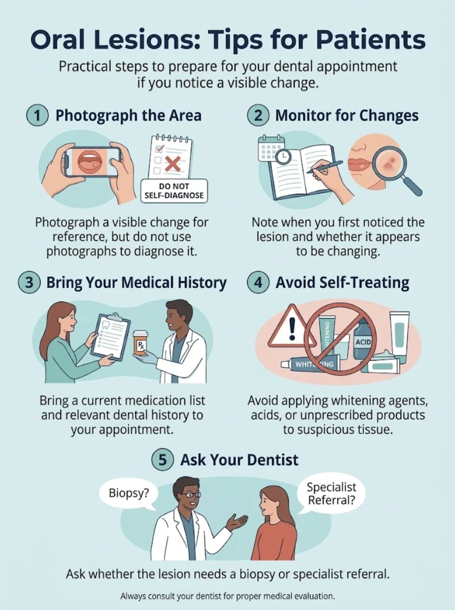
Arrange an assessment with an appropriately qualified dental or medical professional. Record when you first noticed the patch, if you know. Note any changes in size, color, or texture. Bring a list of your current medications and details of relevant medical and dental history. Do not attempt to remove, scrape, burn, or chemically treat the lesion at home. Such actions can cause injury and may complicate later diagnosis.
Patient Reviews
Ready to transform your smile?
Talk to our team and get a FREE treatment plan.
What We Notice Clinically
Dr. Rifat Alsaman, Head of the Medical Team and cosmetic dentist at Vitrin Clinic, leads a clinical approach built on comprehensive oral assessment before treatment planning. Within this approach, unexplained oral changes are assessed before elective cosmetic procedures proceed. A suspicious lesion is not treated as a cosmetic concern. Instead, it calls for appropriate diagnostic referral. Cosmetic treatment never replaces cancer investigation. Patient safety and accurate diagnosis take priority over the timing of aesthetic work.
Why Should Oral Pigmentation Be Assessed Before Cosmetic Dental Treatment?
Documenting an unexplained lesion may be more important than proceeding immediately with elective treatment. Assessment helps determine whether further investigation, such as a biopsy, is needed. It also creates a baseline record for comparison. Restorations placed over or near an unassessed lesion could make monitoring harder. Veneers, crowns, whitening, and other cosmetic procedures cannot diagnose or treat melanoma. Clarifying the nature of a lesion first allows safer, better-informed treatment planning.
Clinical Note
This article provides general educational information and cannot establish an individual diagnosis. Oral mucosal melanoma is rare, and many pigmented oral lesions are benign. Clinical examination, and biopsy when indicated, are necessary to distinguish harmless lesions from malignancy. Treatment decisions and prognosis require individualized specialist assessment. A suspicious lesion should never be ignored or treated with home remedies. If you notice an unexplained change in your mouth, please seek professional evaluation promptly.
How Can You Reduce Oral Health Risks and Monitor Changes?
No established strategy can reliably prevent oral mucosal melanoma. Its causes and risk factors remain incompletely understood. Good oral hygiene supports overall oral health, but it does not prevent this cancer. The most practical approach focuses on awareness and early detection. Routine oral examinations allow professionals to spot unusual changes. Prompt evaluation of new or changing lesions helps ensure that any problem is investigated early rather than late.
Which Habits Can Help You Notice Changes Earlier?
Become familiar with the normal appearance of your mouth. Use a mirror and good lighting, including for the roof of your mouth and gums. Look for new or changing patches, lumps, ulcers, or unexplained bleeding. Attend routine dental appointments, where your whole mouth can be examined. If you notice something unusual, report it. Avoid repeatedly checking the same spot for weeks without seeking professional advice.
Takeaway: Why Early Assessment of Oral Mucosal Melanoma Matters
This cancer is rare. It may be painless at first and can resemble harmless oral pigmentation. These features make it easy to overlook. Unexplained or changing lesions should therefore receive prompt professional assessment, with biopsy when indicated. Early evaluation brings clarity, whether the result is reassuring or not. Your next step is simple: seek an appropriate dental or medical evaluation. Avoid home treatment, and do not wait for symptoms to worsen.
Vitrin Clinic: The Importance of Oral Assessment Before Cosmetic Treatment
Vitrin Clinic in Istanbul, Türkiye, assesses oral and periodontal health before restorative or cosmetic treatment. Dental examinations may identify changes that require further investigation, although a routine dental assessment cannot independently confirm melanoma. Clinical examination and dental imaging are used when indicated, but imaging does not replace biopsy for a suspicious pigmented lesion. Under Dr. Rifat Alsaman, Head of the Medical Team and cosmetic dentist, treatment is individually planned, with referral when findings fall outside routine dental care.






Ready to transform your smile?
Talk to our team and get a FREE treatment plan.
References:
FAQs
Dr. Rifat Alsaman has more than 5 years of clinical experience in dentistry and currently serves as the Head of the Medical Team at Vitrin Clinic. He is dedicated to providing exceptional patient care, overseeing treatment planning, and ensuring the highest clinical standards across the team. His expertise, attention to detail, and commitment to continuous professional development have helped countless patients achieve healthier, more confident smiles.

.webp&w=3840&q=75)


