Dental Implant
September 29, 2026
Alveolar Ridge Preservation After Tooth Extraction: What You Need to Know ?
 Dr. Rifat Alsaman
Alveolar ridge preservation after tooth extraction helps limit the changes that naturally follow tooth removal. The procedure aims to maintain useful ridge dimensions for future treatment. Bone remodeling starts soon after extraction and continues throughout healing.
The goal is not to stop natural healing or eliminate all bone remodeling. Instead, treatment aims to reduce unnecessary dimensional loss. Your dentist considers socket anatomy, bone walls, gum condition, and the planned restoration. Vitrin Clinic uses individualized assessment before recommending any preservation procedure. This article explains indications, techniques, recovery, risks, timing, and planning costs.
What Is Alveolar Ridge Preservation After Tooth Extraction?
Alveolar ridge preservation after tooth extraction is a technique that maintains the shape of the extraction site. A clinician may place graft material inside the empty socket. The objective is to reduce dimensional changes after the tooth is removed. This approach is especially relevant when future implant placement is planned. Studies generally show less horizontal and vertical reduction with preservation than with natural healing. Evidence does not support one universal material for every socket.
What happens to the jawbone after a tooth is removed?
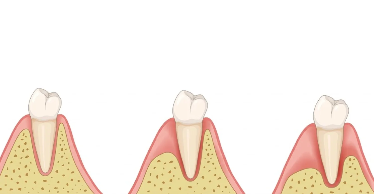
The bone surrounding a tooth develops partly in response to the tooth's presence. After removal, that functional stimulus changes quickly. A blood clot first fills the empty socket. Granulation tissue then develops and gradually changes into connective tissue and immature bone. Remodeling continues as the new bone matures over the following months. The ridge may become narrower and shorter during this process. The facial or buccal portion often shows notable dimensional changes.
How does socket preservation help limit alveolar bone loss?
Socket preservation places a scaffold or graft material within the healing socket. The material supports space maintenance while natural healing proceeds. A systematic review found measurable advantages for ridge width and several height measurements. The reported benefit was approximately 1.89 millimeters for buccolingual width. Midbuccal height showed an average difference of about 2.07 millimeters. Alveolar ridge preservation after tooth extraction can therefore reduce dimensional loss without completely stopping remodeling.
Is alveolar ridge preservation the same as a bone graft?
The terms overlap, but they are not always identical. Socket preservation describes the purpose and timing of treatment. During socket preservation, a graft may be placed immediately after extraction. Other protocols may use collagen materials, membranes, or platelet concentrates. A contained extraction socket differs from a ridge that has already collapsed. Your dentist selects the approach based on the defect and the future restoration.
Socket preservation vs. ridge augmentation
Socket preservation is usually performed immediately after extraction. Its purpose is to limit anticipated dimensional changes. Ridge augmentation usually addresses bone that has already been lost. A dentist may preserve a socket today and reassess the ridge later. A patient who arrives months later may need a more extensive augmentation procedure. Clinical examination and imaging determine which approach fits each situation.
Socket preservation vs. immediate dental implant placement
Immediate implant placement inserts an implant during the extraction appointment. Socket preservation usually focuses on maintaining the site for later treatment. These approaches can sometimes be combined, depending on the socket. Immediate placement requires adequate implant stability and appropriate tissue anatomy. The correct sequence depends on infection, remaining bone walls, stability, and esthetic demands. A preserved socket does not automatically mean delayed implantation is mandatory.
Why Does Alveolar Bone Loss Happen After Tooth Extraction?
Bone loss follows tooth removal because supporting tissues no longer receive the same biological stimulus. The socket therefore remodels toward a new anatomical balance. Evidence indicates that substantial dimensional changes can occur during early healing. The EFP reports that the first six months are particularly important. Average horizontal changes in one reviewed body of evidence reached about 3.79 millimeters. This is why alveolar ridge preservation after tooth extraction is assessed case by case.
How does the extraction socket naturally heal?
Healing begins with bleeding and clot formation immediately after extraction. The clot provides a temporary framework for cellular repair. Granulation tissue gradually replaces the early clot. New bone begins forming within the healing socket. The final architecture is not identical to the original tooth-supporting anatomy. Instead, the body creates a remodeled ridge adapted to the missing tooth. Infection, smoking, surgical trauma, and general health can all influence the course.
Why does the alveolar ridge shrink after tooth removal?
The tooth and its supporting tissues help maintain the surrounding alveolar architecture. Once the tooth is removed, this structure is no longer maintained in the same way. Research consistently describes horizontal and vertical reduction after extraction. Alveolar ridge preservation after tooth extraction aims to reduce these changes rather than eliminate them. New bone can form while the overall ridge still becomes smaller. This distinction matters when implants or visible tooth replacement are planned.
Which parts of the ridge are most affected?
The buccal or facial portion of the ridge can show substantial dimensional reduction. Thin facial bone can remodel noticeably after extraction. The amount differs according to socket anatomy and baseline bone thickness. Clinicians therefore assess both the facial and lingual walls. They also examine ridge width, ridge height, soft tissues, and neighboring structures. A three-dimensional assessment may be useful when implant placement is planned.
How quickly does bone remodeling begin after extraction?
Bone remodeling starts immediately after the extraction. Dimensional changes are particularly active during the early months. The first six months often show the most significant ridge reduction. This timing explains why alveolar ridge preservation after tooth extraction is planned before or during removal. The original socket may no longer provide the same contained environment. Tooth type, bone thickness, infection, and periodontal status all matter. Follow-up imaging can help determine the current anatomy.
Check out the comparison of Immediate Dental Implants vs Delayed Implants
When Is Alveolar Ridge Preservation Needed?
Preservation is considered when future treatment could benefit from maintaining the extraction site. The decision is not automatic for every extraction. Some sites may lose important width or height without intervention. Systematic reviews support dimensional benefits, but they do not establish one universal indication. Alveolar ridge preservation after tooth extraction is therefore a targeted choice, not a routine add-on.
When is socket preservation recommended for a future dental implant?
It may be recommended when maintaining ridge dimensions could simplify later implant placement. This can be especially relevant when the facial bone is thin. The clinician considers the implant position before deciding. A prosthetically driven plan shows whether existing bone can support the intended restoration. The extraction and preservation strategy can then be coordinated with later implant treatment. The socket then becomes the first stage of a broader treatment plan.
Does every tooth extraction require bone preservation?
No. Bone preservation is not required after every tooth extraction. The decision depends on the prosthetic plan, socket anatomy, remaining bone, and tissue conditions. A dentist may recommend natural healing when preservation offers limited practical benefit. Evidence supports alveolar ridge preservation after tooth extraction as a way to reduce dimensional changes. It does not show that every patient needs the procedure.
Which extraction sites may have a higher need for ridge preservation?
Sites with thin facial bones can be more vulnerable to contour changes. Anterior upper sites therefore receive particular attention. Periodontal disease can create pre-existing bone defects. Damaged socket walls reduce the intact anatomy available for natural healing. The need for preservation is therefore site-specific. A clinical examination helps identify whether maintaining the current anatomy has meaningful future value.
Front teeth and visible ridge changes
Front teeth often carry high esthetic demands. The facial bone can be relatively thin in this region. After extraction, remodeling may change the gum profile and ridge contour. Alveolar ridge preservation after tooth extraction may support later restorative and implant planning here. However, it does not guarantee an unchanged gum line. Digital planning can help coordinate implant position with prosthetic contours.
Extraction sites with thin or damaged bone walls
A thin or missing socket wall can increase the complexity of future treatment. The clinician first determines whether the defect can heal within the existing anatomy. Bone grafting may help fill the socket and support space maintenance. Preservation can be considered early instead of waiting for greater ridge collapse. A three-dimensional assessment can reveal wall deficiencies that visual inspection cannot fully show.
Teeth affected by periodontal disease or infection
Periodontal disease may leave an extraction site with reduced supporting bone. The baseline anatomy can therefore differ significantly from an intact socket. A 2024 systematic review found measurable preservation benefits in periodontally compromised molar sockets. The certainty of that evidence was low to very low. Active infection does not automatically exclude regenerative treatment. Treatment timing should reflect the biological condition of the socket.
When might socket preservation not be necessary?
Preservation may offer limited value when no future tooth replacement is planned. It may also be unnecessary when existing anatomy already meets restorative requirements. Other patients have enough remaining bone for straightforward future treatment. A clinician may recommend natural healing when the expected benefit is small. The decision should balance anticipated benefits against treatment burden. Evidence presents alveolar ridge preservation after tooth extraction as an option, not a mandatory step.
What factors influence the decision?
Several factors can change the treatment recommendation. These include the intended restoration, socket anatomy, soft tissues, periodontal health, and implant position. The dentist also considers the facial wall, lingual wall, and neighboring teeth. Preservation is generally performed immediately after removal when indicated. Digital planning can connect today's extraction with future restorative needs.
Planned dental implant placement
A future implant is one of the most common reasons to consider preservation. The clinician works backward from the planned implant position. A preserved site may provide a better starting point for later planning. Some patients still require additional augmentation after healing. Imaging and clinical reassessment show whether the preserved site has reached the required condition.
Remaining bone volume and socket anatomy
The clinician assesses the socket's existing dimensions and wall integrity. The shape and depth of the socket influence how well a graft is contained. A contained socket generally behaves differently from a site with missing walls. Materials for alveolar ridge preservation after tooth extraction should match the defect. No graft material is universally superior for every clinical situation.
Gum and periodontal health
Healthy periodontal tissues support better treatment planning. Active periodontal disease can affect surrounding tissues and the future implant environment. The clinician may assess gum inflammation, periodontal pockets, plaque control, and existing bone defects. Disease control should therefore remain part of implant planning. A 2024 review found measurable benefits in such sites, although certainty remained limited.
Overall treatment plan and future prosthetic needs
The final restoration should influence the extraction strategy. A dentist considers tooth position, bite, esthetic expectations, and adjacent structures. Extraction, healing, imaging, and implant stages should be planned together. Vitrin Clinic approaches treatment planning through individualized assessment. The aim is to coordinate each stage rather than treat extraction as a standalone event.
Explore the best choice between No-Prep Veneers vs Traditional Veneers
Who May Benefit From Bone Grafting After Tooth Extraction?
Patients can benefit when their future treatment depends on maintaining ridge dimensions. Patients with thin facial bones may also have greater concern about contour changes. Preservation can be particularly useful when the site has visible esthetic importance. The evidence shows measurable dimensional improvements compared with spontaneous healing. However, alveolar ridge preservation after tooth extraction does not remove every future surgical need.
Patients planning dental implants later
Patients who plan implants months after extraction may discuss preservation before removal. A later implant requires adequate bone volume and a suitable three-dimensional position. Ridge preservation may reduce the amount of dimensional loss during that waiting period. Some patients still need additional grafting because substantial remodeling occurred. A treatment plan should therefore identify the intended implant position early.
Patients at risk of significant ridge shrinkage
Risk may increase when the facial bone is thin or already damaged. Anterior sites deserve particular attention because contour changes may be more visible. Large extraction sockets may require careful graft stabilization. For these patients, alveolar ridge preservation after tooth extraction is often part of the discussion. A systematic review found preservation can reduce dimensional changes in compromised sockets. The certainty of that evidence remained low to very low.
.jpg)
.jpg)
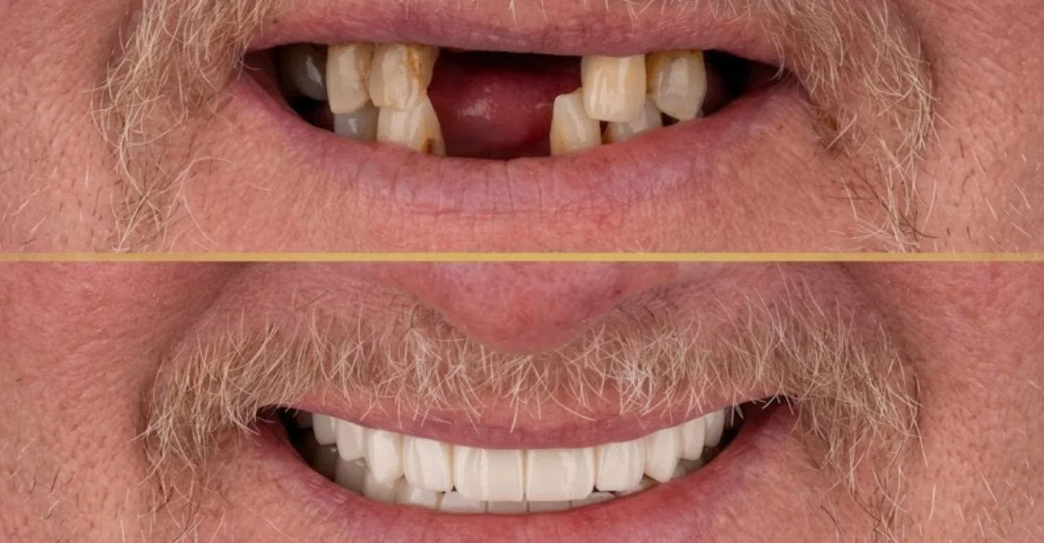
.jpg)
.jpg)
Patients with an extraction site where maintaining ridge contours is clinically important
Ridge contour can matter for esthetics, prosthetic support, and future implant positioning. A visible front-tooth site may therefore receive careful preservation planning. Preservation aims to maintain a useful foundation for future restoration. It does not freeze the ridge in its original shape. Digital photographs and scans can document the anatomy before extraction.
Can socket preservation reduce the need for additional bone augmentation later?
It can reduce the likelihood or extent of additional augmentation in some cases. However, it cannot guarantee that no further grafting will be required. A 2024 systematic review of periodontally compromised molar sites reported reduced augmentation needs. The final need depends on remaining bone, implant position, ridge anatomy, and healing. Patients should view alveolar ridge preservation after tooth extraction as risk reduction rather than a guarantee. The implant assessment after healing remains essential.
How Is Alveolar Ridge Preservation Performed?
The procedure usually begins once the tooth has been removed. The clinician inspects the socket and evaluates the remaining bone walls. Graft material can be placed into the site. A membrane or collagen-based sealing material may follow. The technique varies with socket anatomy and treatment objectives. Evidence supports alveolar ridge preservation after tooth extraction for reducing dimensional changes.
What happens during an extraction socket bone graft?
First, the tooth is carefully removed while protecting the remaining socket walls. Material is placed to occupy the available space without excessive compression. The graft is then covered or stabilized. A membrane, collagen plug, or other sealing approach may be selected. The procedure is normally performed under local anesthesia. Sedation can be considered for selected patients who feel anxious.
What materials can be used for socket preservation?
Several graft categories are used in clinical practice. These include autogenous bone, allografts, xenografts, and synthetic materials. Platelet concentrates may be incorporated into some protocols. Current evidence does not establish one material as universally superior. For alveolar ridge preservation after tooth extraction, selection depends on the defect and clinician experience. Patient factors and future treatment requirements also guide the choice.
Autogenous bone grafts
Autogenous grafts use bone obtained from the same patient. This approach can provide living bones and supporting biological components. However, harvesting creates an additional surgical site. Because a socket is often a contained defect, another graft source may be more practical. The decision depends on defect size and regenerative needs. Patients should ask where the graft originates.
Allografts
Allografts are derived from human donor tissue. They are processed and screened for clinical use before placement. Evidence indicates that allografts can help limit post-extraction dimensional changes. Their practical advantage includes avoiding a separate bone-harvesting procedure. Patients should receive clear information about the material and its intended use.
Xenografts
Xenografts come from animal-derived sources. Bovine and porcine materials are commonly used in regenerative dentistry. A recent systematic review associated xenograft use with favorable dimensional preservation. The authors also emphasized the need for larger and longer studies. Residual particles can remain while new bone develops around them. Patients should discuss the source and characteristics of any recommended material.
Alloplastic or synthetic materials
Alloplastic materials are manufactured rather than harvested from humans or animals. Synthetic options may avoid donor-tissue concerns for some patients. Their behavior depends on composition, porosity, particle structure, and resorption rate. Evidence supports several synthetic options for ridge management. The clinician selects the material based on socket anatomy and the planned treatment.
When is a membrane used?
A membrane may be used to stabilize the regenerative area. It can help keep the graft within the socket. Membranes can also support space maintenance and protect the healing site. Resorbable membranes break down over time, while non-resorbable types may need removal. Membrane selection depends on the defect, closure plan, and clinician preference. Patients should ask whether a membrane is planned and why.
Can platelet concentrates be used with socket preservation?
Platelet concentrates can be used in selected socket preservation protocols. These materials are prepared from the patient's own blood. They may provide a fibrin scaffold and concentrated biological mediators. Results vary, and protocols differ substantially between studies. Platelet concentrates should therefore be viewed as adjuncts rather than universal requirements. In alveolar ridge preservation after tooth extraction, the clinician decides whether they add value.
Is the graft placed immediately after tooth extraction?
Usually, graft placement occurs immediately after the tooth is removed. This timing lets the clinician address the socket before early healing progresses. The selected graft is then placed according to the treatment plan. Immediate grafting does not mean immediate implant placement. A socket can be grafted today and assessed for an implant later. The implant decision depends on subsequent healing and available bone.
How long does the procedure usually take?
Socket preservation is usually performed during the same appointment as extraction. More complex sites may need additional preparation or membrane management. Duration depends on extraction difficulty, socket anatomy, graft quantity, and closure requirements. Patients should focus on the planned clinical steps rather than a fixed number of minutes. A written treatment plan can explain the expected appointment sequence.
Understand the causes and treatment for Anodontia
How Is Socket Preservation Diagnosed and Planned?
Planning starts with examination rather than graft selection. The clinician reviews the tooth, surrounding tissues, and future restorative goals. Additional imaging can clarify bone thickness and anatomical relationships. For implant planning, three-dimensional information can be particularly valuable. The AAOMR recommends clinically justified cross-sectional imaging for implant site assessment. A plan for alveolar ridge preservation after tooth extraction should reflect both the socket and future restoration.
What does a dentist examine before recommending ridge preservation?
The dentist evaluates the tooth scheduled for extraction and its surrounding tissues. The examination can include gum health, bone support, infection, and neighboring teeth. The socket's shape and wall integrity are especially important. Facial bone thickness may influence the preservation strategy. Smoking, medications, and previous healing problems can affect surgical planning. This individualized approach shows whether preservation provides meaningful benefit.
When are dental X-rays or CBCT useful?
Two-dimensional radiographs show roots, surrounding anatomy, and existing bone levels. The AAOMR states that cross-sectional imaging should be clinically justified. CBCT provides three-dimensional information about implant sites with a focused field of view. It can help evaluate ridge width, vertical dimensions, and nearby anatomical structures. Not every extraction requires CBCT. Imaging should be selected according to the clinical question being asked.
How is the extraction site assessed for future implant placement?
The dentist considers where the future implant should be positioned. The available bone is then compared with that planned position. Assessment includes ridge dimensions and wall integrity. A prosthetically driven approach considers the future crown before finalizing implant position. Digital planning may help visualize the relationship between implant, bone, and restoration.
Ridge width and height
Ridge width describes the horizontal thickness of available bone. Ridge height describes the vertical dimension available for treatment. The clinician also considers the exact location of the planned implant. A ridge can appear adequate in one area and deficient in another. Alveolar ridge preservation after tooth extraction aims to reduce loss across these measurements. Research has reported measurable differences in both width and height.
Buccal and lingual bone walls
The buccal wall lies toward the cheek or lip. The lingual wall lies toward the tongue, or the palate in the upper jaw. The integrity and thickness of these walls influence healing and graft containment. The facial wall is especially important in esthetic implant cases. Clinicians therefore assess each wall rather than treating the socket as an empty hole.
Adjacent teeth and periodontal tissues
Neighboring teeth can influence both healing and future implant positioning. Their roots and periodontal structures must be respected. The dentist may evaluate gum thickness, recession, and tissue inflammation. This information shows whether additional soft-tissue management is needed. The implant site should be considered within its surrounding periodontal environment.
Anatomical structures relevant to implant planning
Important structures can include the mandibular canal, mental foramen, nasal cavity, and maxillary sinus. Their location can influence implant length, angulation, and surgical approach. The AAOMR emphasizes clinically justified cross-sectional imaging for implant site assessment. Good planning reduces reliance on assumptions about hidden anatomy.
Why is individualized assessment important?
No two extraction sites are identical. Teeth differ in root shape, bone thickness, infection status, and periodontal support. One patient may need an implant, while another may choose a bridge. Evidence reviews also show substantial variation among preservation studies. Individualized planning lets the clinician decide whether to preserve, augment, or allow natural healing. That flexibility makes alveolar ridge preservation after tooth extraction more purposeful and predictable.
What We Notice Clinically
Patients often focus on the visible empty socket. The underlying bone changes are less obvious because they occur beneath the gum. The socket may look closed while the ridge continues remodeling. Preservation is most useful when connected to a specific restorative objective. Research supports measurable dimensional benefits, but preservation does not eliminate biological remodeling.
Patients often think extraction alone preserves the shape of the jaw
The gum may close over the socket, creating the impression of full healing. However, bone dimensions can continue changing underneath. A healed gum surface does not guarantee preservation of the original ridge. Earlier planning shows whether alveolar ridge preservation after tooth extraction offers meaningful value. A dentist can explain the difference using clinical examination and imaging.
A bone graft does not completely stop natural ridge remodeling
Grafting reduces dimensional changes rather than freezing the ridge. The evidence shows measurable reductions in bone loss compared with extraction alone. One meta-analysis found about 1.89 millimeters less buccolingual reduction. Healing continues long after the initial grafting procedure. Patients should not think of preservation as permanent anatomical protection. Future assessment remains part of responsible treatment planning.
Socket preservation is different from rebuilding an already-collapsed ridge
Preservation is preventive in purpose. When a significant bone has already disappeared, the anatomy may require a different surgical strategy. A preserved socket starts with more of the original ridge available. This distinction can influence cost, healing time, and implant scheduling. Timely alveolar ridge preservation after tooth extraction may simplify later treatment in selected patients.
A planned implant can change the timing and preservation strategy
The future implant position can determine whether preservation is necessary. Immediate and delayed implant placement have different clinical requirements. The treatment sequence should therefore be designed around the final restoration. This is often called prosthetically driven planning. Early communication between surgical and restorative providers reduces avoidable changes later.
Clinical Note: Dr. Rifat Alsaman on Planning Bone Preservation Around Tooth Extraction
Dr. Rifat Alsaman, Head of the Medical Team at Vitrin Clinic and cosmetic dentist, emphasizes planning around the future restoration. The extraction site should be evaluated before removal whenever possible. The objective is not simply filling an empty socket. The objective is preparing the site for the patient's broader dental plan. In this approach, alveolar ridge preservation after tooth extraction serves a defined restorative goal. The final recommendation remains individualized for each patient.
How the extraction site is evaluated before deciding on preservation
The clinician examines the tooth, surrounding gum, and supporting bone. Preoperative radiographs may identify root anatomy or existing bone defects. CBCT can provide three-dimensional information when clinically justified. This helps determine whether ridge dimensions are important for that site. A preservation procedure should have a clear treatment objective. The recommendation should explain the problem it is expected to address.
Why the future restoration should be considered before extraction
The future crown or prosthesis influences the implant's ideal position. Implant position then influences the required bone envelope. A tooth may be removed today, but that surgery affects future restorative options. A prosthetically driven plan can identify preservation needs before extraction. This supports a more coordinated treatment sequence.
What Are the Benefits of Alveolar Ridge Preservation?
The main benefit is reduced dimensional change after extraction. This can help maintain a more useful ridge for future treatment. Research reports improvements in horizontal and vertical dimensions compared with natural healing. Some selected cases may also need less augmentation later. However, alveolar ridge preservation after tooth extraction does not guarantee ideal bone after healing. The procedure is best presented as a risk-reduction strategy rather than a guarantee.
Can ridge preservation reduce post-extraction bone loss?
Evidence indicates that preservation can reduce post-extraction ridge reduction. A major systematic review found average advantages in width and several height measurements. The EFP reports that extraction alone can produce substantial changes within six months. Its summary cites average horizontal reduction of around 3.79 millimeters. Individual outcomes vary significantly. Alveolar ridge preservation after tooth extraction is most useful when ridge dimensions have clear clinical value.
How can socket preservation support future dental implants?
Maintaining more ridge volume can provide a better starting point for implant planning. Some patients may avoid additional augmentation entirely. Others may need a smaller or more localized procedure. A systematic review found preservation methods effective when preparing sites for prosthetic treatment. Still, the implant position remains decisive. Bone must support the planned implant rather than simply fill the socket.
Can it help maintain the width and contour of the extraction ridge?
Yes, preservation can reduce loss of ridge width and height. This helps retain a more favorable contour. Thin facial walls, periodontal defects, and socket shape can change the result. The EFP summarizes evidence showing measurable reductions in horizontal and vertical bone changes. Esthetic areas may benefit from closer contour management. Clinical photographs and scans can help monitor changes over time.
Does ridge preservation guarantee that an additional bone graft will not be needed?
No. Additional augmentation can still be necessary after preservation. A 2024 meta-analysis found that preservation reduced some dimensional losses. The need for further grafting depends on final bone volume and the intended implant position. The preserved ridge is evaluated before committing to the next surgical stage. Realistic expectations make alveolar ridge preservation after tooth extraction easier to understand and accept.
Find out what means a Black Spot Inside Your Cheek
What Are the Possible Side Effects and Risks?
Socket preservation is a surgical procedure, so complications are possible. Common concerns include discomfort, swelling, bleeding, and infection. A recent systematic review of oral bone regeneration identified these among frequent complications. Wound dehiscence, or reopening of the incision, was also reported. Risk varies according to procedure complexity and patient factors. Smoking can increase postoperative healing problems in oral surgery. Understanding these risks is part of informed consent for alveolar ridge preservation after tooth extraction.
Is socket preservation painful?
The procedure is normally performed under local anesthesia. Patients should not feel sharp surgical pain during treatment. Some soreness is common after the anesthetic wears off. Its severity depends on extraction difficulty and surgical extent. Pain that steadily worsens instead of improving deserves professional assessment. Patients should follow the prescribed pain-relief plan. They should avoid taking medicines outside their dentist's instructions.
What swelling or discomfort can occur after treatment?
Mild swelling and tenderness can occur after extraction and graft placement. Cold compresses may be recommended during the initial period. Medication should follow the clinician's instructions. Swelling that becomes progressively worse can indicate a problem. Fever or increasing pain can also require assessment. Patients should not compare their recovery with another patient's timeline. Surgical complexity and individual healing can differ considerably.
What complications can occur after an extraction socket graft?
Potential complications include membrane exposure, graft exposure, wound opening, infection, bleeding, and delayed healing. Management depends on size, timing, tissue condition, and the presence of infection. Patients should contact the treating clinician when unexpected symptoms appear. Early review can stop a minor problem from becoming more significant.
Membrane exposure
Membrane exposure occurs when the barrier becomes visible through the soft tissues. Some exposures can be monitored carefully. Others require intervention, especially when infection is present. Non-resorbable membranes generally need more specific management when exposed. Patients should avoid touching an exposed membrane unless specifically instructed.
Infection or delayed healing
Infection can interfere with graft healing and soft-tissue closure. Signs include increasing pain, swelling, drainage, fever, or an unpleasant taste. Smoking, infection, surgical trauma, and local tissue conditions may contribute. A dentist should assess symptoms rather than relying on online comparisons. Prompt treatment often protects the graft and the final result.
Graft-material exposure or loss
Small amounts of graft material can sometimes become visible during healing. The significance depends on the material, site, and amount exposed. A loose particle does not automatically mean the entire graft failed. Patients should not replace displaced material themselves. The dentist can determine whether the site remains adequately protected.
Inadequate ridge preservation
Preservation may achieve less dimensional maintenance than expected. Socket anatomy, missing bone walls, infection, smoking, and biological variation can influence the outcome. The goal of alveolar ridge preservation after tooth extraction is dimensional reduction, not perfect duplication. A later scan may still show a need for augmentation. This outcome changes the plan but rarely closes the door on implants.
What symptoms are not expected during normal healing?
Increasing pain after initial improvement is concerning. Rapidly worsening swelling also deserves professional evaluation. Fever, persistent bleeding, foul drainage, or significant pus should not be ignored. Numbness or altered sensation requires assessment when it persists or worsens. Patients should contact their dentist rather than assuming every symptom is normal.
Relief and Recovery After Socket Preservation
Recovery usually includes soft-tissue healing followed by ongoing bone remodeling. The visible gum can improve before the underlying bone has fully matured. A common implant-planning interval is several months. Patients should protect the surgical site during early healing. Smoking should be avoided because it can impair oral surgical recovery. Follow-up appointments help confirm healing before the next treatment stage.
What can help relieve normal discomfort after the procedure?
Follow the medication instructions provided by the dental team. Pain relievers are commonly used when clinically appropriate. Rest can also reduce discomfort during the initial recovery period. Patients should not place aspirin directly on the gum. They should also avoid unapproved remedies around the surgical site. The treating clinician's instructions should always take priority over generic online advice.
How can patients protect the extraction site while it heals?
Avoid pulling, touching, or probing the grafted area. Maintain oral hygiene around the site without disturbing the surgical materials. Brush nearby teeth carefully according to the postoperative instructions. Soft foods may reduce mechanical irritation during the early period. Chewing on the opposite side can also help. Follow-up visits should be kept even when symptoms seem normal.
What should patients avoid after a bone graft?
Patients should avoid activities that can disturb the surgical site. Tobacco exposure is particularly important because smoking can increase healing risks. Each restriction exists to protect the clot, graft, and soft tissues. The dental team will explain how long each precaution should last.
Smoking and tobacco exposure
Smoking can compromise oral wound healing. Evidence links smoking with increased postoperative complications. Patients should follow the treating clinician's smoking-cessation recommendations. The exact restriction period depends on the procedure and individual risk. Nicotine products and vaping should be discussed with the dentist as well.
Disturbing the surgical site
Do not pick at sutures or attempt to reposition graft materials. Mechanical disturbance can disrupt healing tissues. The tongue naturally explores new areas, but conscious touching should be avoided. Patients should also avoid pulling the lip or cheek to look at the site. Contact the clinic if something feels loose or unexpectedly exposed.
Strenuous activity during the initial recovery period
Heavy physical activity can increase discomfort or bleeding in some patients. The dentist should provide a personalized activity restriction. The appropriate duration depends on extraction and graft complexity. Light walking is usually easier to tolerate than intense exercise. Patients should gradually resume normal activity as advised.
Food and oral-hygiene habits that may interfere with healing
Hard, sharp, or sticky foods can irritate healing tissue. Very hot foods may also be uncomfortable immediately after surgery. Oral hygiene should remain a priority without traumatizing the surgical area. Use the prescribed cleaning method and follow instructions about brushing near the site. Soft, nutritious meals and good hydration support comfortable recovery.
When should pain or swelling be assessed by a dentist?
Pain should generally become more manageable over time. Swelling should also trend toward improvement rather than continued progression. Severe pain that does not respond to prescribed medication also requires review. The clinician may inspect the wound, membrane, graft, and surrounding tissues. Early intervention is often easier than waiting for the next routine appointment.
Note : All images used are for editorial and illustrative purposes only and may not originate from the original news provider or associated company.
Learn about causes and prevention for Throat Ulcers
How Long Does Socket Preservation Take to Heal?
Healing occurs in stages rather than on one specific date. A commonly cited interval for implant planning is about three to six months. Some sites need longer because of anatomy or additional grafting. Imaging and clinical examination determine readiness for the next stage. For alveolar ridge preservation after tooth extraction, readiness is judged by findings rather than dates alone.
What is the typical socket preservation healing time?
Many clinicians reassess the site after several months. Around three to six months is frequently used for implant planning. Healing varies with graft material, defect size, infection, and patient factors. Complex defects may require additional time. The dentist should confirm readiness instead of relying only on a calendar date.
When does soft tissue healing occur?
Soft-tissue closure usually begins soon after extraction. Visible tissue healing can occur within the early weeks. Even when the gum looks healthy, the deeper bone may remain immature. This is why implant timing should not rely only on surface appearance. Follow-up allows the clinician to evaluate both soft tissue and underlying bone.
When does new bone formation occur?
New bone formation begins during the healing process. Graft particles can remain while new bone forms around them. Bone healing may therefore continue after the gum has closed. Clinical studies commonly reassess sockets after several months before implant placement. Imaging at that stage shows how the new bone has developed.
How long might a dentist wait before placing a dental implant?
A common clinical interval is approximately three to six months. The decision depends on bone maturity, ridge dimensions, soft-tissue health, and stability requirements. A follow-up scan can show whether the anatomy supports the planned implant. Timing following alveolar ridge preservation after tooth extraction is set by these findings.
Why healing time varies between patients
Patients heal differently because biological and local factors differ. Infection, periodontal disease, smoking, surgical trauma, and defect size can affect recovery. The selected graft and membrane can also change the healing environment. Medical history and medications may influence the clinician's precautions. Conditions such as poorly controlled diabetes can slow healing. For this reason, online timelines should be treated as general planning information.
Why graft material and site anatomy can affect timing
Different materials remodel at different rates. Socket anatomy also determines how well the graft is contained. A simple contained socket may follow a relatively straightforward healing course. A defect with missing walls can require additional regenerative treatment. The clinician therefore evaluates the actual site rather than using one fixed timeline.
Does the graft remain permanently in the jaw?
Not necessarily. Some graft materials are gradually resorbed and replaced by new bone. The clinical objective is a useful healed ridge, not complete disappearance of every particle. Residual particles are generally compatible with later implant placement. The final result depends on material choice, patient healing, and treatment conditions.
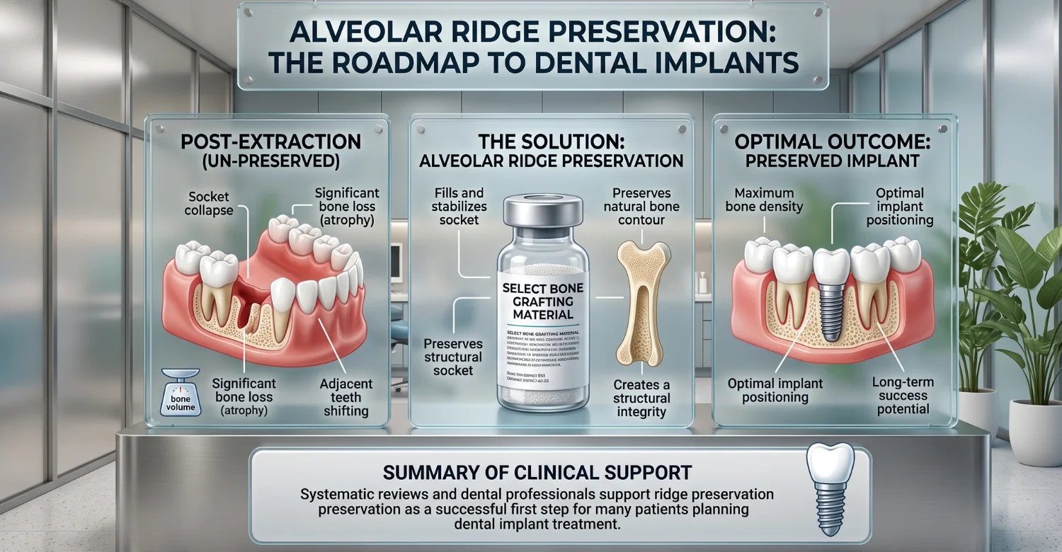
How does ridge preservation support future dental implants?
It may maintain more width and height than spontaneous healing. It can also support better implant positioning in selected cases. However, implant planning depends on three-dimensional anatomy. The preserved ridge must match the planned implant's location and dimensions. Volume alone is not enough if the bone sits in the wrong place.
When can an implant be placed after socket preservation?
Many clinical protocols reassess the site around three to six months later. Some sites need additional healing or augmentation first. The clinician evaluates bone quality, ridge dimensions, soft tissues, and restorative requirements. Imaging and clinical findings determine the actual timing. Patients traveling for treatment can plan visits around this reassessment window.
What happens if there is still insufficient bone?
Insufficient bone does not necessarily prevent implant treatment. Further grafting may be considered before or during implant placement. The exact technique depends on the missing dimension and its location. The dentist will explain the options and their effect on the overall timeline.
Additional bone augmentation
A localized augmentation procedure can rebuild areas that remain deficient. The graft may be placed before or together with implant treatment. The approach depends on the size and shape of the defect. Small deficiencies are often managed during the implant appointment itself.
Ridge augmentation
Ridge augmentation focuses on rebuilding an already deficient ridge. This differs from alveolar ridge preservation after tooth extraction, which protects a fresh socket. It can require a larger regenerative procedure and longer healing time. A staged approach is common when both width and height are missing.
Alternative implant planning when appropriate
Sometimes the implant position or size can be adjusted to fit the available anatomy. Digital planning can help explore suitable options. Shorter or narrower implants may be considered in selected situations. Such alternatives are case-dependent and must remain prosthetically acceptable.
Is ridge preservation always necessary before a dental implant?
No. Some implants can be placed without prior socket preservation. The decision depends on the socket, remaining bone, implant timing, and treatment objectives. Immediate implant placement may sometimes provide an alternative sequence. Delayed implant placement may also be selected. The key issue is not whether preservation is universally required. The key issue is whether it improves the clinical pathway for that particular site.
Alveolar Ridge Preservation Cost: USA vs UK vs Turkey
Costs vary substantially between clinics and treatment plans. One U.S. oral-surgery practice lists socket preservation at about 800–900. UK examples commonly fall around $500–$850. Turkish sources show prices from about $350, or roughly $350–$600. These figures are planning estimates, not fixed quotes. The final cost depends on the individual treatment plan.
Average Cost Comparison
Country | Typical Planning Range Per Socket | Approximate Midpoint | Currency | Main Cost Factors |
USA | 800–1,200 | $1,000 | USD | Graft, membrane, imaging, sedation, complexity |
UK | $500–$750 | $625 | GBP | Graft, membrane, extraction, imaging, follow-up |
Turkey | 350–700 | $525 | USD | Graft material, membrane, imaging, surgical complexity |
Midpoints are simple planning estimates based on the stated ranges. They are not national statistical averages.
How much does socket preservation cost in the USA?
U.S. pricing varies by region and clinical complexity. One Indianapolis oral-surgery practice publishes fees around 800–900. Other practices report approximately 300–800. Some cases may reach around $1,200 when anatomy or materials add complexity. Imaging, sedation, and membrane use can also affect the final invoice. Patients should request an itemized estimate before treatment.
How much does socket preservation cost in the UK?
Current private dental examples vary. Published fees include approximately $500, $750, and $850. The final price may depend on whether the quote includes graft, membrane, extraction, imaging, or follow-up. Patients should compare complete treatment plans rather than one procedure name. Regional differences can also be significant within the UK.
How much does socket preservation cost in Turkey?
Published Turkish medical-travel sources show substantial variation. One source reports prices from about $350. Local pricing also varies with graft material and membrane use. The final Vitrin Clinic price should follow examination and a clear procedure plan. International patients should also clarify what their treatment package includes.
What is included in the price of a bone graft after extraction?
An itemized quote may include several components. Patients should confirm each item before treatment begins.
Cost Component | What It May Include |
Examination and imaging | Consultation, dental X-rays, or CBCT when clinically appropriate |
Graft material | Selected graft type and required quantity |
Membrane or regenerative materials | Barrier membrane, collagen plug, or platelet concentrate |
Surgical procedure | Graft placement, suturing, and surgical care |
Follow-up | Healing review and possible suture removal |
What can make the final cost higher or lower?
Pricing depends on the actual clinical requirements.
Type and quantity of graft material
More material generally increases material and surgical costs. Different graft categories also carry different prices.
Number and location of extraction sites
Multiple sockets can increase total treatment costs. Difficult anatomical locations may require more surgical time.
Need for CBCT or additional procedures
CBCT, sedation, complex extraction, or additional regeneration can increase the final cost.
Whether implant placement is planned at the same or a later stage
Immediate implant treatment involves different surgical and restorative fees. Delayed treatment creates a separate later stage with its own costs.
What are realistic 2026 planning ranges?
The ranges below are broad planning estimates. They should not be treated as guaranteed clinic prices. Each figure refers to one socket and excludes unrelated treatment.
USA: approximately 800–1,200 per socket
The approximate midpoint is $1,000 per socket. Simpler sites may cost less, while complex cases can exceed this range.
UK: approximately $500–$750 per socket
The approximate midpoint is $625 per socket. Published examples can reach around $850 when treatment requirements differ.
Turkey: approximately 350–700 per socket
The approximate midpoint is $525 per socket. Published prices vary according to materials, anatomy, and included services.
Why these figures are estimates rather than fixed treatment prices
Final pricing depends on examination findings, graft volume, materials, imaging, extraction complexity, and additional procedures. A personalized quote should follow clinical assessment.
How Can You Preserve Bone After Tooth Extraction?
Preservation begins before extraction. Careful planning can reduce unnecessary trauma and identify future treatment requirements. When future implants are possible, preservation should be discussed before the tooth is removed. Patients can also support healing by following postoperative instructions. Smoking cessation, good oral hygiene, and appropriate nutrition support the recovery environment. Together, these steps help alveolar ridge preservation after tooth extraction achieve its intended purpose.
Can careful extraction techniques help preserve the socket?
Yes. Minimally traumatic extraction can help protect the remaining bone walls. The objective is to remove the tooth while minimizing damage to surrounding tissues. Root separation or other techniques may be used when appropriate. A careful extraction does not prevent all remodeling. It simply helps preserve the best possible starting anatomy.
When should socket preservation be discussed?
Ideally, it should be discussed before extraction. The discussion should include implant plans, expected healing, materials, cost, and alternatives. International patients should also discuss timing because travel schedules affect follow-up planning. Early discussion of alveolar ridge preservation after tooth extraction avoids rushed decisions on the day.
What can patients do to support healthy healing?
Follow the postoperative instructions closely. Take prescribed medicines according to the dental team's directions. Avoid smoking and unnecessary manipulation of the site. Maintain oral hygiene using the recommended technique. Attend all scheduled reviews. Healing is affected by many factors, but patient behavior influences the surgical environment.
Can good periodontal health affect future implant planning?
Yes. Periodontal health is relevant to the long-term implant environment. A patient may need periodontal treatment before implant placement. This can include professional cleaning, periodontal therapy, or improved home care. The preservation decision should consider the entire mouth rather than one socket alone. Healthy gums also support the long-term stability of implant restorations.
When Should You Contact a Dentist After Socket Preservation?
Most patients receive clear postoperative instructions before leaving the clinic. Increasing pain, worsening swelling, fever, persistent bleeding, or visible graft exposure can require assessment. A recent systematic review identified infection, wound dehiscence, and graft or membrane exposure as important complications. Early contact lets the clinician decide whether a change is normal or needs treatment.
Warning signs that need professional assessment
Symptoms that worsen rather than improve should be reviewed. A reversal of steady improvement is often the earliest sign of a problem.
Increasing rather than improving pain
Pain that becomes stronger after initial improvement may indicate a complication. Contact the treating dentist for assessment. Do not simply increase medication without professional advice.
Persistent or worsening swelling
Swelling that keeps increasing may indicate infection or another complication. Its timing and severity both matter. Swelling that spreads toward the eye or neck needs prompt attention.
Fever or other signs of infection
Fever, drainage, bad taste, and increasing swelling can occur with infection. Professional assessment is recommended. Antibiotics should only be taken as prescribed.
Persistent bleeding
Bleeding that does not settle with the clinic's recommended pressure technique requires dental review. Slight oozing on the first day is usually expected.
Graft or membrane exposure
Visible graft or membrane material should be reported. The dentist determines whether observation or treatment is required. A photograph can help the clinic assess the situation remotely.
When is urgent medical or dental care appropriate?
Urgent care is appropriate when symptoms become severe or progress rapidly. Examples include uncontrolled bleeding, significant facial swelling, breathing difficulty, or severe systemic symptoms. Patients should follow local emergency guidance for serious symptoms. Routine postoperative questions can usually go to the treating dental team. When in doubt, it is safer to seek advice early.
Tips for Patients
Patients can make treatment planning easier by asking specific questions before extraction. The key issue is understanding why preservation is being recommended. Questions about materials, healing time, costs, follow-up, and implant timing support informed decisions. Patients should also provide complete medical and medication information.
Ask whether preserving the extraction socket fits your future treatment plan
Ask whether you are likely to need an implant, bridge, denture, or another restoration. Then ask whether maintaining ridge dimensions matters for that restoration. A clear explanation helps you understand the purpose of treatment. It also helps you weigh the benefit against cost and healing time.
Ask which graft material and membrane, if any, are being recommended
Ask about the source, composition, and reason for choosing the proposed material. Ask whether a membrane is needed and whether it is resorbable. You should understand what will be placed and why. Mention any preferences regarding animal-derived or donor materials.
Ask how long healing is expected to take before implant assessment
Ask for an estimated timeline rather than a single fixed promise. The clinician can explain when the first reassessment is expected. Healing time varies with anatomy and grafting requirements. Knowing the window helps you plan work, travel, and other commitments.
Follow the dentist's instructions for oral hygiene, diet, medication, and activity
Postoperative instructions are individualized for a reason. Following them helps protect the surgical site. Do not substitute online advice for the treating dentist's instructions. Ask for written instructions if verbal guidance feels hard to remember.
Tell your dentist about smoking, medications, medical conditions, and previous healing problems
Complete information supports safer surgical planning. The clinician may change medication instructions, timing, or monitoring based on your history. Blood thinners, diabetes, and bone-related medications are especially important to mention. Never withhold relevant information because you expect it to complicate treatment.
At Vitrin Clinic: Assessment for Dental Implant Treatment After Extraction
Vitrin Clinic evaluates extraction sites within the context of future Dental Implant Treatment. The team considers the tooth, gums, oral tissues, remaining bone, and intended restoration. Assessment can include digital X-rays, CBCT, and three-dimensional scanning when appropriate. Any decision about alveolar ridge preservation after tooth extraction is based on actual anatomy. It is never made on the basis of extraction alone.
How the Vitrin Clinic team evaluates the extraction site
The team reviews the extraction area and the surrounding oral tissues. Existing bone conditions are considered before future implant planning. This approach shows whether preservation, additional grafting, or natural healing is most appropriate. Findings are explained to the patient before any decision is made.
Digital X-rays and CBCT when clinically appropriate
Digital radiographs provide useful information about roots and surrounding structures. CBCT adds three-dimensional information when implant planning requires cross-sectional imaging. Its use should always be clinically justified. Vitrin Clinic incorporates imaging findings into individualized digital planning.
Assessing bone volume and the potential implant site
The team evaluates ridge width, ridge height, and socket-wall conditions. The planned implant position is considered alongside the available bone. This shows whether the site is likely to suit implant placement after healing.
Developing an individualized Dental Implant Treatment plan
The treatment plan connects extraction, preservation, healing, implant placement, and restoration. Each stage is considered in relation to the final result. The plan may change after healing if the anatomy develops differently than expected.
Deciding whether socket preservation, additional grafting, or another approach is appropriate
Preservation is not selected automatically. The choice depends on socket anatomy, bone volume, soft tissues, periodontal health, and restorative needs. Some patients need additional grafting. Others heal well without further regeneration.
Internal-link opportunity: Dental Implant Treatment page
This article can link to the Dental Implant Treatment page. Suggested anchor text is "Dental Implant Treatment." The link should guide readers from preservation planning toward the broader implant journey.
How Vitrin Clinic Supports Your Treatment Journey
Vitrin Clinic can coordinate treatment stages for patients considering implants after extraction. Scheduling, imaging, treatment sequencing, and follow-up all need coordination. The objective is a clear pathway from assessment to final restoration.
Initial consultation and individualized treatment assessment
The process begins with a review of the patient's dental condition and treatment goals. Clinical assessment determines whether further records or imaging are needed. Patients can share existing X-rays or scans to support early planning.
Digital diagnostics and treatment planning
Digital records help the team assess anatomy and prepare a treatment sequence. CBCT is used when clinically appropriate for implant planning. Digital planning also helps patients visualize the proposed treatment.
Coordinating extraction, preservation, healing, and future implant stages
These stages should be connected rather than planned separately. Healing time can be built into the implant schedule. This reduces surprises and unnecessary gaps between appointments.
Treatment coordination for international patients
International patients need clear appointment sequencing and travel coordination. A coordinated schedule reduces unnecessary delays between clinical stages. The team can help align visits with healing and reassessment windows.
Follow-up and reassessment before the next treatment stage
The preserved site is reassessed before implant placement. Clinical findings and imaging confirm whether the planned next step remains appropriate. Adjustments are made when healing differs from expectations.
Ready to transform your smile?
Talk to our team and get a personal FREE treatment plan.
Key Takeaways
Preservation is designed to reduce the dimensional changes that naturally follow tooth removal. Evidence supports benefits for ridge width and several height measurements.
Alveolar ridge preservation can reduce but cannot completely eliminate post-extraction ridge changes
Bone remodeling continues after treatment. Preservation reduces dimensional loss rather than stopping biology. Some change should always be expected.
The decision depends on the extraction site and the patient's future dental treatment plan
Socket anatomy and the future restoration are central to decision-making. A procedure that helps one patient may add little for another.
Socket preservation can be particularly relevant when future dental implant treatment is being considered
Maintaining useful bone dimensions can support later implant planning. It may also reduce the extent of future grafting.
Bone graft materials and membranes should be selected according to the individual clinical situation
Current evidence does not establish one universally superior combination. The defect and treatment goals guide the choice.
Pain, swelling, bleeding, or other symptoms that worsen rather than improve should be professionally assessed
Progressive symptoms should not be dismissed as normal healing. Early review is usually simpler and safer.
The most appropriate timing for implant placement depends on clinical and radiographic healing
Three to six months is a common planning interval, but individual timing varies. Examination and imaging confirm readiness.
Medical tips for protecting the extraction site and supporting recovery
Avoid smoking, protect the surgical area, and follow postoperative instructions. Attend every scheduled review appointment.
Relief options and safe postoperative care should follow individualized dental instructions
Medication and activity advice should come from the treating dental team. Generic online advice may not suit your case.
Possible side effects should be explained before treatment, including infection, membrane exposure, graft exposure, and delayed healing
These complications are documented across oral regenerative procedures. Knowing them in advance supports informed consent.
At Vitrin Clinic
Vitrin Clinic approaches tooth extraction with future dental implant needs in mind. The extraction site is assessed before deciding whether preservation could support later implant placement. Clinical examination evaluates the teeth, gums, oral tissues, and remaining bone. Digital X-rays and CBCT may be used when clinically appropriate for implant planning. Digital planning helps assess the potential implant position and available bone volume. The team considers whether socket preservation could support the planned implant site. Additional bone grafting may be considered when the remaining bone is insufficient. Each case receives an individualized Dental Implant Treatment plan based on clinical findings. International patients can also receive coordinated scheduling for extraction, healing, implant placement, and restoration. Patients can begin with a personalized assessment of their potential implant treatment pathway.
Reference
FAQs
Dr. Rifat Alsaman has more than 5 years of clinical experience in dentistry and currently serves as the Head of the Medical Team at Vitrin Clinic. He is dedicated to providing exceptional patient care, overseeing treatment planning, and ensuring the highest clinical standards across the team. His expertise, attention to detail, and commitment to continuous professional development have helped countless patients achieve healthier, more confident smiles.


