General Dentistry
September 1, 2026
Tooth Cusps: Anatomy, Types, Problems, and Dental Treatment
 Dr. Rifat Alsaman
Tooth cusps are small, defined elevations on the biting surface of a tooth. They allow efficient chewing by helping break down food into smaller pieces before swallowing. Cusps also play a central role in occlusion, guiding how upper and lower teeth meet and function together. The number and shape of tooth cusps vary depending on tooth type, with molars carrying more cusps than incisors.
Tooth Cusp Anatomy
Tooth cusp anatomy begins with a hard enamel layer covering each raised point, protecting the softer dentine beneath. Dentine surrounds the pulp, which houses nerves and blood vessels. Cusp height, shape, and position directly affect how efficiently a tooth manages chewing forces. Well-formed tooth cusps distribute pressure evenly across the arch.
Tooth Cusp Diagram: Understanding the Main Features
A tooth cusp diagram typically highlights the crown, individual cusps, grooves, fissures, enamel, dentine, and pulp chamber. Patients reviewing a tooth cusp diagram should note how grooves separate each cusp. Fissures around these grooves can also trap plaque. Dentists use this same visual framework during a clinical examination, assessing cusp height, wear patterns, and groove depth. How to understand Cbct Cad Subperiosteal Implant Design
What Is a Cuspid Tooth?
What is a cuspid tooth? A cuspid tooth is another common term for a canine, positioned between the incisors and premolars in each dental arch. The canine cuspid tooth plays a major role in biting and tearing food. It also helps guide jaw movement during chewing and supports facial aesthetics by shaping the smile line. Unlike posterior teeth, a cuspid tooth typically has one dominant point. This makes its structure distinct from molars and premolars.
Cuspid Tooth vs Other Teeth
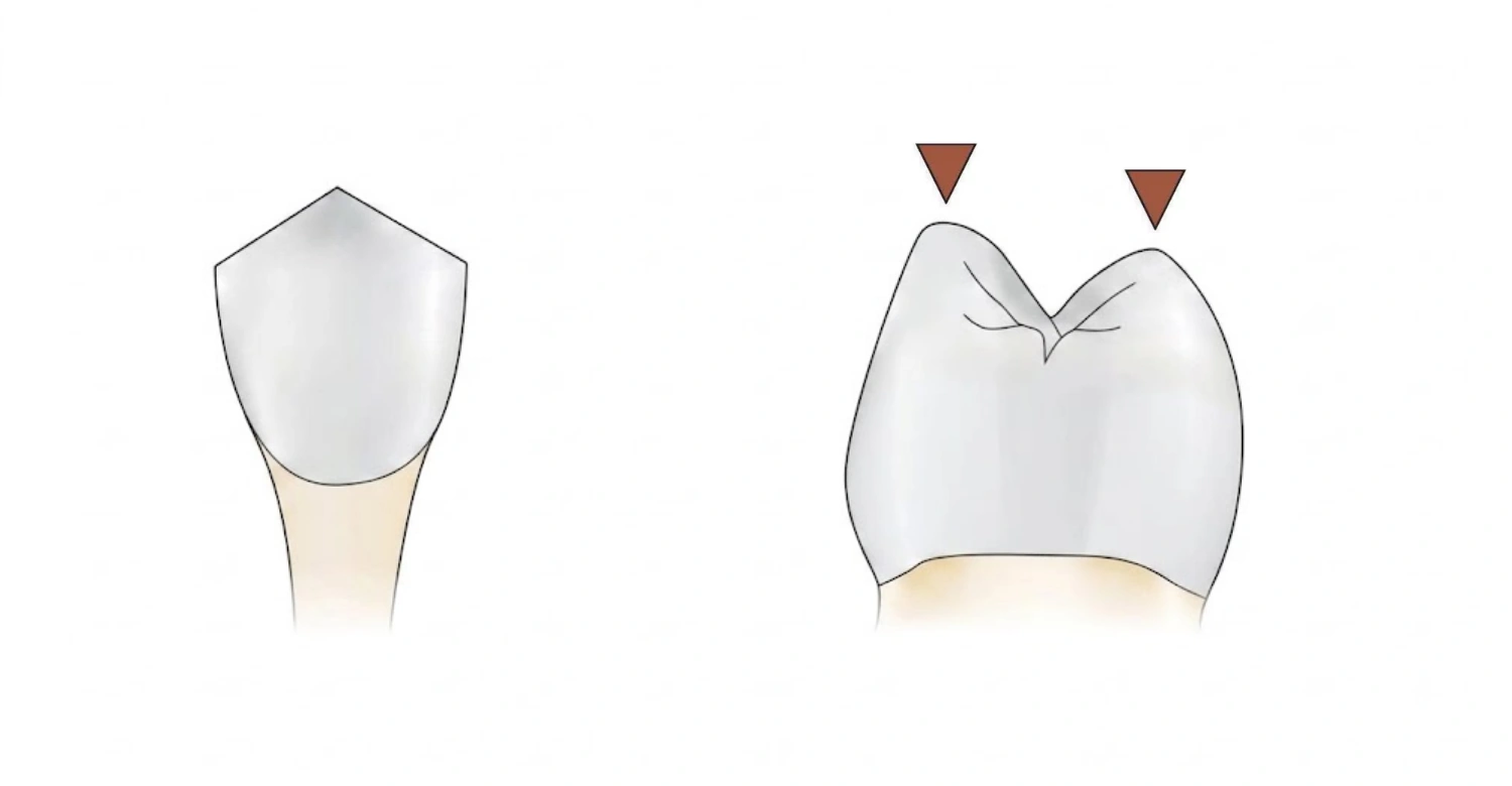
A cuspid tooth differs from premolar and molar tooth cusps in both shape and purpose. Canine teeth are built for tearing, so they feature one long, pointed cusp rather than several shorter ones. Premolars usually carry two cusps for crushing food, while molars have four or more for grinding. This is why a canine cuspid tooth rarely develops the same wear patterns seen around multi-cusp posterior teeth. Overview of Subperiosteal Implants Vs All On 4
Common Types and Variations of Tooth Cusps
Cusp of Carabelli Tooth
Cusp of Carabelli Tooth is an anatomical variation most often found on upper first molars. It typically appears on the palatal or lingual surface of the tooth. A cusp of a Carabelli tooth can range from a barely noticeable groove to a distinctly raised extra point. Its presence is considered a normal developmental variation. It does not, by itself, indicate disease, decay, or any dental problem requiring treatment.
Talon Cusp Tooth
A talon cusp tooth is an extra, claw-shaped projection that usually develops on the back surface of an anterior tooth. Because a talon cusp tooth often extends toward the biting edge, it can create plaque retention and increase caries risk. It may also interfere with occlusion or irritate soft tissue. In some cases, it affects the pulp.
Extra Cusp on Tooth
An extra cusp on tooth may simply reflect normal developmental variation in tooth cusps. In other cases, it may signal something that needs closer review. It's important to differentiate a longstanding developmental feature from a newly appeared swelling. A new swelling could point to underlying pathology rather than anatomy.
Tips for Patients
Do not attempt to file or reshape a cusp at home.
Brush carefully around grooves and unusual tooth anatomy.
Use fluoride toothpaste regularly.
Maintain routine dental examinations.
Report pain, sensitivity, fracture, or changes in tooth shape.
Avoid using excessive force when brushing.
Follow personalised recommendations if a dentist identifies a high-risk groove or cusp.
Ask whether a developmental variation requires monitoring or treatment.
Keep existing restorations and treated teeth under regular review. Essential information on Subperiosteal Implants Turkey Price
Common Problems Associated With Tooth Cusps
Cusp Fracture
Heavily loaded or weakened tooth cusps can fracture under normal biting forces. This is especially true where a tooth already has large fillings or cracks. Symptoms often include pain when biting down or sharp sensitivity to hot and cold temperatures. A fractured cusp exposes deeper tooth structure to bacteria and further damage.
Cusp Wear and Flattening
Cusp wear and flattening happen gradually through normal ageing, tooth grinding, acid erosion, or abnormal bite forces. Mild flattening is common and often harmless. Wear becomes clinically significant when it causes sensitivity, changes the bite, exposes dentine, or accelerates further breakdown of tooth cusps.
Tooth Decay Around Cusps and Grooves
Deep grooves surrounding tooth cusps are natural plaque traps, making them common sites for early decay. Anatomy alone doesn't determine risk. Oral hygiene habits, diet, and saliva quality all interact with groove depth to influence caries development.
Cusp-Related Bite Problems
An unusually prominent cusp can interfere with how teeth meet, creating premature contact points during biting or chewing. This uneven contact may cause localized discomfort, muscle strain, or accelerated wear on opposing teeth. Because the bite functions as a complete system, dentists assess the full arch rather than isolating one cusp. Don't miss out on Subperiosteal Implant Failure
How Are Tooth Cusp Problems Diagnosed?
Clinical Examination
Diagnosis starts with a visual inspection of tooth shape and surface, checking each cusp of the tooth for irregularities. Dentists assess occlusion to see how the bite comes together and evaluate for cracks, caries, wear, and sensitivity. When indicated, pulp vitality testing determines whether the nerve remains healthy.
Dental Imaging
Dental X-rays become useful when a dentist needs to see beneath the visible surface of tooth cusps. Imaging helps evaluate the relationship between an abnormal cusp and the pulp. It can reveal depth, proximity, and any early decay not visible clinically. Not every cusp variation requires imaging. It's typically reserved for cases involving pain, suspected fracture, or structural concerns a visual exam can't confirm.
Digital Dental Assessment
Digital scans and photographic documentation offer another layer of assessment for unusual tooth cusps. These tools capture precise detail on tooth shape, occlusal contacts, and wear patterns over time. Digital technology supports more accurate treatment planning by allowing comparisons between visits, helping identify subtle changes early.
Treatment Options for Tooth Cusp Problems
Monitoring and Preventive Care
Many anatomical variations in tooth cusps require no active treatment at all. Regular examinations allow a dentist to track any changes over time. Enhanced oral hygiene around deep grooves also reduces decay risk. Monitoring is often the most conservative and appropriate approach for stable, symptom-free variations.
Selective Cusp Reduction
Controlled reduction of an overly prominent cusp may be considered in select cases, particularly where it interferes with bite function. This approach prioritizes conservative removal of minimal tooth structure. Because reducing a large or deep cusp can bring treatment close to the pulp, dentists carefully evaluate pulp proximity beforehand.
Dental Sealants
Dental sealants can help protect susceptible grooves surrounding tooth cusps in selected cases, particularly deep fissures prone to plaque retention. Sealants serve a preventive rather than restorative purpose, forming a thin protective barrier over vulnerable surfaces. They're most useful before decay develops, sealing off hard-to-clean grooves.
Dental Bonding and Restorative Treatment
Composite bonding can restore a damaged or altered cusp when structural loss is moderate rather than severe. This approach repairs minor fractures and reshapes uneven tooth cusps while preserving as much natural structure as possible. Bonding offers a relatively quick, conservative solution for cosmetic and minor functional concerns.
Crowns and Other Restorations
Significant damage to tooth cusps may require stronger restorative protection than bonding alone can provide. Crowns and similar restorations reinforce weakened teeth, distributing biting forces more evenly across the remaining structure.
Root Canal Treatment
Root canal treatment becomes necessary when cusp-related damage leads to irreversible pulpal disease, such as infection or persistent nerve inflammation. An unusual cusp on its own does not mean root canal treatment is required. Full report on Subperiosteal Vs Bone Graft Implant
Benefits and Expected Results of Tooth Cusp Treatment
Functional Benefits
Treating tooth cusp problems can improve chewing efficiency by restoring even contact across the bite. Better occlusal balance reduces strain on individual teeth when clinically indicated. Protecting weakened tooth structure also lowers the risk of further fracture or breakdown.
Oral Health Benefits
Addressing problematic grooves around tooth cusps makes daily cleaning noticeably easier, reducing areas where plaque tends to accumulate. In appropriately treated cases, this lowers overall plaque-retention risk. Areas vulnerable to recurrent decay can be better managed once anatomy or restorations are optimized.
Aesthetic Benefits
Conservative reshaping of tooth cusps can create more harmonious tooth proportions where cusp shape visibly affects appearance. Aesthetic improvements are approached carefully, without compromising function or biological health. The goal isn't a purely cosmetic outcome but one that balances appearance with long-term durability.
Before and After Expectations
Results after tooth cusp treatment depend heavily on the original anatomy and the specific treatment selected. Some cases only require ongoing monitoring, while others need restorative intervention to protect function. Outcomes vary between patients, so realistic expectations matter more than assuming identical results for every case.
Clinical Note
Not every prominent, unusual, or additional cusp is a dental problem. The decision to treat should rest on clinical findings, symptoms, occlusion, caries risk, tooth structure, and pulp proximity. Dr. Rifat Alsaman's perspective prioritizes conservative treatment whenever clinically appropriate. Removing unnecessary tooth structure can create avoidable sensitivity or pulpal complications. Scientific facts about Chemotherapy Affect Teeth
When Should You See a Dentist About Tooth Cusps?
See a dentist if a cusp suddenly changes shape, a piece of the tooth breaks, or biting causes pain. Hot or cold sensitivity is also worth checking. So is food repeatedly trapping around a cusp, or persistent irritation to the tongue or cheek.
What We Notice Clinically
Dr. Rifat Alsaman, Head of the Medical Team at Vitrin Clinic and a cosmetic dentist, evaluates unusual cusp anatomy. This is done through clinical findings rather than appearance alone. Tooth structure, occlusion, aesthetics, symptoms, and long-term function all factor into assessing prominent or unusual tooth cusps.
When an Unusual Cusp Requires Attention
Attention is warranted when there's pain or sensitivity, recurrent decay around grooves, or difficulty cleaning the area. Premature contact with opposing teeth, tooth fracture, or excessive wear are additional warning signs. Soft-tissue irritation and any sign of pulpal involvement also call for prompt review.
Tooth Cusp Treatment at Vitrin Clinic
At Vitrin Clinic, assessment of tooth cusps combines clinical examination with modern diagnostic and digital dentistry approaches. Individualized treatment planning matters for unusual tooth anatomy, since no two cases present identically. Digital photography, intraoral scanning, digital treatment planning, and appropriate imaging support accurate diagnosis where clinically indicated. Conservative treatment and preservation of healthy tooth structure remain guiding priorities throughout. If you're noticing unusual cusp anatomy, sensitivity, fracture, or aesthetic concerns, a professional evaluation at Vitrin Clinic can clarify what, if anything, needs attention. Did you know about Sparkling Water And Teeth
References:
FAQs
Dr. Rifat Alsaman has more than 5 years of clinical experience in dentistry and currently serves as the Head of the Medical Team at Vitrin Clinic. He is dedicated to providing exceptional patient care, overseeing treatment planning, and ensuring the highest clinical standards across the team. His expertise, attention to detail, and commitment to continuous professional development have helped countless patients achieve healthier, more confident smiles.

.webp&w=3840&q=75)


