
Understanding tooth pulp health is vital for maintaining a functional smile. At Vitrin Clinic, we specialize in identifying why the living tissue inside your tooth fails. Necrosis occurs when the dental pulp dies. This is the innermost layer, and it contains nerves and blood vessels. Trauma or infection usually causes it. This process can be silent or excruciating. Treating it promptly involves removing the dead tissue. It also means managing the infection to save the natural structure. Early intervention at Vitrin Clinic helps you avoid systemic complications and bone loss.
Understanding Tooth Necrosis: The Basics
Tooth necrosis marks the final stage of dental pulp inflammation. When people ask, "pulp tooth meaning," they are referring to the life source of the tooth. When this source is cut off, the tooth becomes necrotic. At Vitrin Clinic, we focus on the pulp dentistry aspect of care. We make sure patients understand that a dead tooth isn't just a cosmetic issue. It is a biological failure within the dental pulp cavity. It requires professional medical attention to prevent the spread of bacteria. Accidental trauma or deep decay can lead to painful tooth pulp exposure.
What Is Tooth Necrosis?
To define dental pulp necrosis, one must look at the cessation of blood flow. Essentially, it is the death of the dental pulp within the pulp cavity of the tooth. Once the tissue dies, it can no longer mount an immune response against bacteria. This leads to a cavity pulp environment where infection thrives. At Vitrin Clinic, we explain that the hard enamel remains intact. But the internal tooth pulp anatomy has ceased to function. This means immediate clinical intervention is necessary.
How Does Dead Tooth Pulp Develop?
The development of dead tooth pulp usually follows a predictable path of degradation. It begins with reversible pulpitis, where the dental pulp is irritated but can heal. If the irritation persists, it becomes irreversible. Eventually, the blood supply in the pulp canal is severed. Without oxygen and nutrients, the tissue undergoes necrosis. Vitrin Clinic experts note that this transition can happen fast, following a high-impact injury. Or it can happen slowly, through deep, untreated tooth decay. Inflammation of this tissue leads directly to the debate of reversible vs irreversible pulpitis.
Common Causes of Pulp Decay
The primary driver of pulp decay is bacterial invasion. When a standard cavity pulp reaches the inner layers, bacteria feast on the organic material. Other causes include physical trauma. Trauma can sever the blood vessels at the root tip. Repeated dental procedures on the same tooth are another cause. Once the dental pulp chamber is breached, the risk of necrosis spikes. Vitrin Clinic notes that even minor cracks can let pathogens reach the sensitive pulpal anatomy.
Recognizing the Warning Signs of Tooth Necrosis
Identifying signs of dental pulp exposure or death early can save your tooth. Many patients wonder, "is pulp part of a tooth that gives warnings?" Yes, it is. However, a necrotic tooth can sometimes be deceptive. Dead nerves don't send pain signals. Vitrin Clinic advises patients to watch for subtle shifts in how their teeth feel and look. Recognizing these shifts allows for more conservative treatments. This happens before the infection spreads to the surrounding jawbone. When this vital tissue dies, patients begin exhibiting tooth necrosis symptoms.
Early Symptoms to Watch For
Early warnings often involve extreme sensitivity to temperature. If you feel a lingering ache after drinking cold water, your dental pulp may be struggling. You might also notice a localized bad taste or smell. This can indicate a cavity pulp infection. At Vitrin Clinic, we tell patients that "silent" symptoms matter too. A slight dullness in the tooth's luster is one example. These are the first indicators that the tooth pulp chamber is under significant biological stress.
Pain Patterns and Sensitivity Changes
Pain associated with the pulp cavity of teeth often changes as necrosis progresses. Initially, there is sharp, acute pain as the nerves die. Once the dental pulp is fully necrotic, the pain might disappear entirely. This is a dangerous sign. Later, a different, throbbing pain may emerge as pressure builds within the pulp tooth. Vitrin Clinic specialists explain why this happens. Gas and pus from the infection get trapped inside the rigid pulpal chamber.
Visual Indicators: Discoloration and Swelling
A dead tooth often undergoes a color change, turning grayish, yellow, or dark brown. This happens as the decomposing dental pulp leeches into the porous dentin. Swelling near the gum line is another sign. So is the appearance of a "pimple," or abscess. Either one signals that the infection has exited the pulp canal and entered the soft tissues. At Vitrin Clinic, we use these visual markers to quickly assess the severity of the pulp cavity damage. If the nerve is only slightly exposed, a dental pulp cap can often save it.
When to Consult a Dentist
You should see a pulp dentist the moment you notice spontaneous pain or significant discoloration. Waiting until an abscess forms makes treatment more complex. At Vitrin Clinic, we recommend a check-up after any facial trauma. This applies even if the tooth isn't broken. Damage to the radicular pulp can occur internally without visible cracks. Early consultation helps preserve the anatomy of dental pulp and contain the infection.
Diagnostic Approaches in Modern Dentistry
Modern pulp dentistry relies on a blend of technology and clinical skill. At Vitrin Clinic, we don't just guess. We use precise metrics to determine the health of your dental pulp. Understanding whether a tooth is alive or dead requires looking inside the tooth pulp chamber. Various diagnostic lenses make this possible. This phase is crucial. It dictates whether we can perform a simple filling, or whether we must proceed with more intensive pulp cavity treatments.
Clinical Examination Techniques
During a clinical exam at Vitrin Clinic, we perform percussion and palpation tests. By gently tapping the tooth, we can tell if inflammation has spread beyond the dental pulp to the periodontal ligament. We also check for mobility and gum recession. Either can expose the radicular pulp. These manual checks give us a baseline understanding of pulp health. They help us map out the necessary steps for your restorative plan.
Imaging and Radiographs for Pulp Health
Digital X-rays are essential for visualizing the dental pulp cavity. The dental pulp itself is soft tissue, so it doesn't show up clearly. But we can see the dark shadows at the root tip that indicate an infection. Radiographs allow us to inspect the pulp canal for blockages or narrowing. Vitrin Clinic uses high-resolution imaging for every case. This ensures every corner of the pulpal anatomy is accounted for before any procedure begins. The study and treatment of this inner tissue define what endo is in dentistry.
Pulp Vitality Tests Explained
To answer "do teeth have pulp that is still alive?", we use vitality tests. This involves thermal testing, using cold or heat, and electric pulp testing. A healthy pulp tooth will respond to the stimulus. A necrotic one will feel nothing. At Vitrin Clinic, these tests are performed with precision to ensure accuracy. Checking the pulpal horns and nerve response is the definitive way to confirm if the tooth pulp has truly died.
Treatment Options for Necrotic Teeth
Once necrosis is confirmed within the dental pulp chamber, action must be taken. You cannot leave dead tissue inside a living body. Vitrin Clinic offers several pathways, depending on the extent of the decay. This includes saving the tooth through advanced pulp dentistry. It also includes performing a clean extraction. Our goal is always to restore your oral health. We prioritize the most conservative methods possible. This keeps the anatomy of dental pulp functional for as long as possible.
Root Canal Therapy: Step-by-Step
Root canal therapy is the gold standard for treating a dead pulp tooth. First, we access the pulp chamber and remove the infected tissue. Next, the pulp canal is cleaned and disinfected to kill all bacteria. Finally, the empty dental pulp cavity is filled with a biocompatible material called gutta-percha. Vitrin Clinic ensures this process is painless. It seals the tooth and prevents future bacterial entry into the sensitive pulpal anatomy.
Pulp Capping and Pulp Preservation
If the dental pulp is exposed but still healthy, we may use pulp capping. This involves placing a medicated material over the exposed area. It encourages the tooth to heal itself. This is a vital part of pulp dentistry aimed at avoiding a full root canal. At Vitrin Clinic, we use bio-inductive materials that stimulate the dental pulp. This creates a new layer of dentin, protecting the pulpal horns and maintaining tooth vitality.
Tooth Extraction: When It Becomes Necessary
Sometimes the damage to the pulp cavity is too severe. Or the tooth structure is too weak to support a restoration. In these cases, extraction is the safest option. It prevents the infection from spreading to the jaw. Vitrin Clinic surgeons perform extractions with the utmost care. After removing the tooth, we discuss replacement options like implants. This fills the gap left behind and maintains your facial structure and chewing function.
Innovative Materials and Techniques in Pulp Dentistry
Vitrin Clinic stays at the forefront of dental science. We use bioceramic sealers and rotary endodontics. These materials bond better with the dental pulp cavity walls. This provides a superior seal against bacteria. Our microscopic technology lets us see deep into the pulp canal. No hidden branches of the radicular pulp are missed. This high-tech approach increases the success rate of treatments, even for the most complex tooth pulp anatomy cases.
Benefits of Prompt Treatment
Treating pulp tooth issues immediately offers benefits far beyond pain relief. By addressing the dental pulp the moment symptoms appear, you save time, money, and your natural smile. Vitrin Clinic emphasizes that "waiting and seeing" is never a good strategy with pulp decay. Prompt action stops the biological clock on tooth loss. Our patients find that early intervention leads to faster recovery times. It also brings much more predictable long-term results for their overall oral health.
Preventing Infection and Further Decay
A necrotic pulp tooth is a breeding ground for bacteria. If left alone, this infection can lead to a painful abscess. It can even cause systemic health issues like sepsis. By cleaning the pulp chamber and sealing the pulp canal, we eliminate the source of the infection. Vitrin Clinic's proactive treatments stop the decay from migrating to adjacent teeth. They also prevent it from eroding the surrounding bone, keeping your entire mouth safe.
Maintaining Natural Tooth Structure
Nothing functions quite as well as your original tooth. Treating the dental pulp through root canal therapy, rather than extracting it, maintains natural stimulation of your jawbone. This prevents the bone thinning that often follows tooth loss. At Vitrin Clinic, we are dedicated to preserving the tooth pulp anatomy framework. Keeping your natural tooth helps maintain your bite alignment. It also prevents other teeth from shifting into the empty space.
Expected Outcomes: Before and After
Patients often come to Vitrin Clinic in significant distress due to pulp decay. After treatment, the "after" is characterized by a total absence of pain. The tooth becomes fully functional again. Visually, a crown can restore the appearance of a tooth darkened by pulp necrosis. The long-term outcome is a healthy mouth where the pulp cavity is no longer a liability. Most patients report a massive improvement in their quality of life and confidence.
Dental Care Tips to Protect Your Tooth Pulp
Prevention is the best form of pulp dentistry. Protecting the dental pulp involves more than just brushing. It requires a comprehensive approach to oral hygiene and lifestyle choices. At Vitrin Clinic, we believe in empowering patients with the knowledge to keep their pulp tooth healthy. The tooth pulp anatomy is buried deep. So you must protect the outer layers of the tooth. This keeps the inner sanctum sterile and vital throughout your life.
Daily Oral Hygiene Practices
To protect the dental pulp, you must keep the enamel intact. Brushing twice a day with fluoride toothpaste and flossing daily prevents the cavity pulp interactions that lead to necrosis. Focus on the gum line, where bacteria can enter and affect the radicular pulp. Vitrin Clinic recommends a soft-bristled brush. This avoids wearing down the enamel, the primary shield for the dental pulp chamber and the nerves within.
Diet and Lifestyle Considerations
Sugar is the enemy of the dental pulp. High sugar intake fuels the acid-producing bacteria that cause pulp decay. Lifestyle habits like smoking also matter. Smoking reduces blood flow to the gums, which can hurt the health of the pulpal anatomy. Vitrin Clinic advises a diet rich in calcium and vitamins to strengthen the teeth. Wearing a mouthguard during sports is also crucial. It prevents trauma that could sever the pulp canal's blood supply.
Preventive Check-Ups and Early Intervention
Regular visits to Vitrin Clinic allow us to spot tiny cracks or early decay before they reach the tooth pulp. We use diagnostic tools to monitor the pulp cavity of teeth that were previously injured. Catching an issue while it is still in the enamel or dentin is much easier. Treating a full-blown dental pulp infection is far harder. Preventive care is the most cost-effective way to avoid the pain of necrosis.
Patient Education: Understanding Your Pulp Health
Educated patients make better decisions for their smiles. Knowing the pulp definition dental terms helps you understand what happens during your appointment. At Vitrin Clinic, we take the time to explain the anatomy of dental pulp. This way, you aren't just a passive participant in your care. When you understand the function of the pulp chamber and the risks of exposure, you are more likely to prioritize the habits that keep your teeth alive.
What Is Pulp and Why Is It Important?
When asking, "what is pulp of teeth?", think of it as the tooth's heart. It consists of nerves, blood vessels, and connective tissue. Its primary job is to create dentin during tooth development. It also provides sensory feedback. Without the dental pulp, a tooth becomes brittle. Vitrin Clinic emphasizes that the pulp tooth lets you sense temperature and pressure. It acts as an early warning system for the rest of your body.
Pulp Anatomy: Chambers, Canals, and Horns
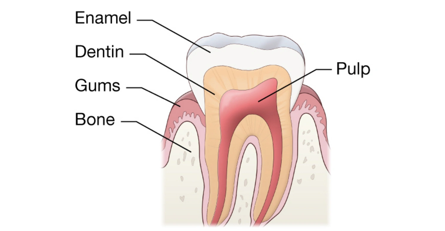
The tooth pulp anatomy is complex. The pulp chamber sits in the crown. The pulp canal extends down into the roots. The pulpal horns are extensions of the chamber that reach toward the chewing surface. Understanding these structures is vital, because bacteria can enter through any of them. Vitrin Clinic specialists use this anatomical knowledge to precisely target treatments. Every part of the pulpal anatomy is cleaned and protected during restorative procedures.
Signs of Dental Pulp Exposure and Risk Factors
Signs of dental pulp exposure include seeing a small red or pink spot inside a deep cavity or a broken tooth. This is a dental emergency. Risk factors include poor hygiene and a high-sugar diet. "Bruxism," or teeth grinding, is another risk factor. It can wear down the tooth until the pulp chamber is vulnerable. Vitrin Clinic warns that once the dental pulp is exposed to the oral environment, infection and necrosis are only a matter of time.
How Knowledge Can Improve Your Dental Decisions
Knowing the pulp definition teeth experts use helps you weigh the pros and cons of treatments. For instance, if you know the radicular pulp is infected, you'll understand why a simple filling won't suffice. You'll also see why a root canal is necessary. At Vitrin Clinic, we find that patients who understand pulp dentistry are less anxious. They are also more satisfied with their results. Knowledge transforms a scary procedure into a logical step toward long-term health and comfort.
Insights Into Vitrin Clinic's Expertise
Vitrin Clinic is a leader in pulp dentistry. We combine years of clinical experience with the latest technological advancements. We understand that the dental pulp is a delicate tissue. It requires a gentle touch and extreme precision. Our team is trained to handle everything from routine pulp cavity fillings to complex necrotic restorations. We pride ourselves on saving teeth that others might give up on. Our deep understanding of tooth pulp anatomy makes this possible.
Advanced Pulp Diagnostics and Treatments
At Vitrin Clinic, we use 3D CBCT imaging to get a 360-degree view of your pulp canal. This lets us see anatomical variations that traditional 2D X-rays miss. Whether we are treating a pulp tooth with an unusual canal shape, or performing a retrograde filling, our advanced tools ensure success. Our commitment to the best technology means your dental pulp treatment will be as efficient and effective as modern science allows.
Technology-Driven Solutions for Long-Lasting Results
We use laser-assisted disinfection and ultrasonic instruments to clean the dental pulp cavity. These tools remove bacteria from the microscopic nooks of the pulpal anatomy far better than traditional methods. Vitrin Clinic's focus on technology ensures that once a pulp tooth is treated, it stays healthy for decades. We don't just fix the problem. We provide a durable solution that respects the biological integrity of your entire mouth.
Patient-Centered Approach and International Care
Vitrin Clinic welcomes patients from around the world. We offer a seamless experience that prioritizes comfort. We understand that issues with the dental pulp can be stressful, especially when traveling. Our staff is multilingual and dedicated to providing a supportive environment. From the moment you walk in to discuss your signs of dental pulp exposure, you are treated with the highest level of care. We combine world-class pulp dentistry with a warm, patient-first philosophy.
Taking the Next Step and Protect Your Smile
Your journey to a healthy, pain-free smile begins with a single step. If you suspect your tooth pulp is in trouble, do not wait for the pain to become unbearable. Vitrin Clinic is here to help you navigate your options and restore your health. We believe everyone deserves access to top-tier pulp dentistry. By taking action today, you can save your natural teeth. You'll also avoid the complications of untreated necrosis and infection.
At Vitrin Clinic, we are currently offering a free consultation to help you assess your dental pulp health. During this session, we will examine your pulp cavity and provide a personalized treatment plan. Whether you need a simple check-up or complex pulp dentistry, our experts will guide you. Don't let pulp decay dictate your life. Contact Vitrin Clinic today to schedule your free consultation and take the first step toward protecting your smile forever.
What Is Tooth Pulp Made Of?
To fully answer "what is pulp of teeth?", it helps to look at its histological structure. The dental pulp is a soft connective tissue organized into distinct layers. The outermost layer is the odontoblastic layer, made up of specialized cells that produce dentin. Beneath it sits the cell-free zone, followed by the cell-rich zone. Finally comes the pulp core, which houses the larger blood vessels and nerve trunks. At Vitrin Clinic, understanding this layered anatomy lets our team predict how deep decay must travel before it threatens the dental pulp chamber. This helps us plan pulp dentistry treatment accordingly. This same histological detail also explains why dental pulp calcification and pulp stone formation develop in the first place. Calcified tissue tends to build up in exactly these layered zones over time.
Specific Pathological Conditions Affecting Dental Pulp
Beyond simple decay, the dental pulp can be affected by a range of specific pathological conditions. These conditions don't always present with the classic pain patterns associated with pulp necrosis. That's why Vitrin Clinic relies on a combination of clinical and radiographic findings to catch them early.
Dental Pulp Calcification and Pulp Stones
Dental pulp calcification is a common finding, where mineralized tissue forms inside the pulp chamber or canal. This can appear as diffuse calcification along the pulp walls. Or it can appear as discrete pulp stone formation, small, hardened nodules that develop within the pulp tissue itself. Pulp stones are often asymptomatic and are discovered incidentally on X-rays. But in some cases, they can narrow the pulp canal and complicate future root canal therapy. Vitrin Clinic factors dental pulp calcification into treatment planning whenever it's visible on imaging. It can change how a canal is located and cleaned. Cases involving pulp stone formation often need extra time under the microscope to bypass the obstruction safely.
Pulpotomy vs. Pulpectomy: Reversible Conservative Steps
When comparing pulpotomy vs pulpectomy, the key difference lies in how much of the dental pulp is removed. A pulpotomy removes only the inflamed pulp tissue in the crown portion of the tooth. It leaves the healthy pulp in the root canals intact. This is a more conservative step, often used when the pulp is still vital and inflammation is limited. A pulpectomy, by contrast, removes the entire pulp from both the crown and the root canals. It's typically the step taken once irreversible pulpitis or necrosis is confirmed. Vitrin Clinic favors the pulpotomy approach whenever pulp vitality tests suggest the tissue still has healing potential. This preserves as much natural structure as possible. The decision between pulpotomy vs pulpectomy ultimately comes down to how much of the dental pulp is still alive and capable of healing.
The Regeneration Potential of Vital Tissue
A fully necrotic pulp cannot heal itself. But vital dental pulp has a notable capacity for repair. This regenerative potential is a major reason Vitrin Clinic prioritizes conservative treatments, like pulp capping and pulpotomy, whenever the tissue is still alive.
Dental Pulp Stem Cells (DPSCs) and Homeostasis
Dental pulp stem cells, often abbreviated DPSCs, are undifferentiated cells found within the pulp tissue. They play a central role in reparative dentine homeostasis. When the pulp is mildly irritated, these cells can differentiate into odontoblast-like cells. They lay down new reparative dentin to wall off the irritation, maintaining homeostasis even under mild biological stress. This process is a key area of interest in dental tissue engineering. Researchers are exploring how dental pulp stem cells might one day regenerate damaged pulp tissue, rather than replace it with filling materials. Vitrin Clinic follows developments in dental tissue engineering closely, as it represents the future direction of conservative pulp dentistry.
Can Tooth Pulp Grow Back After Necrosis?
A common question patients ask is, can tooth pulp grow back after it has died? Unfortunately, once the dental pulp is fully necrotic, it cannot regenerate. Necrotic tissue has no blood supply and no living cells left to rebuild it. This is different from a mildly inflamed or partially exposed pulp. With prompt care, that kind of pulp may still repair itself. Vitrin Clinic emphasizes this distinction, because it's exactly why early diagnosis matters so much. The window for regeneration closes once necrosis sets in. Treatment then shifts from preservation to root canal therapy or extraction. So when patients ask whether tooth pulp can grow back once symptoms have gone silent, the honest answer is no. Silence is usually a sign the damage is already done.
Can a Tooth Function and Live Without Vital Pulp?
Many patients also ask, can a tooth live without pulp? The answer is yes, functionally. After root canal therapy removes the necrotic dental pulp and the canal is sealed, the tooth no longer has living internal tissue. But it can still be used normally for biting and chewing. What it loses is its ability to sense temperature and pressure. It also becomes slightly more brittle over time, since it no longer receives nutrients from within. This is why Vitrin Clinic typically recommends a crown following root canal therapy. It protects the now non-vital tooth structure from fracture during everyday use. In short, can a tooth live without pulp and still serve its owner for decades? With the right restoration, yes.
Neurological Processing of Dental Pulp Sensation
The dental pulp is one of the most densely innervated tissues in the body. It is supplied primarily by branches of the trigeminal nerve. These nerve fibers include fast-conducting A-delta fibers. They're responsible for the sharp, immediate pain felt with cold stimuli or a cracked tooth. Slower C-fibers produce the dull, lingering ache associated with more advanced inflammation. Pulpal nerves share pathways with other facial structures. Because of this, pain from the dental pulp can sometimes be felt as referred pain in a neighboring tooth, the jaw, or even the ear. At Vitrin Clinic, understanding this neurological overlap helps our team correctly localize which tooth is actually the source of a patient's discomfort before starting treatment. For patients who want a deeper explanation of this pain pathway, a resource like the cleveland clinic dental pulp note offers a useful general overview of how pulpal nerves are structured.
What are the 4 zones of pulp?
The dental pulp is histologically organized into four distinct zones, moving from the outer wall of the pulp chamber inward:
Odontoblastic zone – the outermost layer, lined with odontoblasts, the cells responsible for producing dentin throughout the tooth's life.
Cell-free zone (zone of Weil) – a thin layer just beneath the odontoblasts with relatively few cells, containing capillary and nerve networks.
Cell-rich zone – denser in fibroblasts and undifferentiated cells, including the reserve cells that can become odontoblasts if repair is needed.
Pulp core (central zone) – the innermost region, containing the larger blood vessels, nerve trunks, and connective tissue that supply the entire pulp.
At Vitrin Clinic, this layered structure explains why early pulp exposure doesn't always mean the whole pulp is compromised. Sometimes only the outer zone is affected. That's exactly the scenario a pulp capping procedure is designed to treat. This same four-zone breakdown is also referenced in general dental education sources, including the cleveland clinic dental pulp note, which describes the same layered anatomy from a patient-facing angle.
How Vitrin Clinic Manages Dental Pulp Health
Vitrin Clinic brings together diagnostic precision and conservative treatment planning for every stage of dental pulp health, from early dental pulp calcification to full necrosis. Rather than defaulting to the most invasive option, Vitrin Clinic assesses pulp vitality thoroughly first. We then choose the least aggressive treatment that still protects the patient's long-term oral health.
What We Notice Clinically
According to Dr. Rifat Alsaman, Head of the Medical Team at Vitrin Clinic and a cosmetic dentist, one common pattern stands out in clinic. Patients often delay a visit until pain has already disappeared, mistakenly assuming the problem resolved on its own. Dr. Rifat Alsaman notes that this silent phase is often when the dental pulp has actually progressed to necrosis. Dead nerve tissue can no longer transmit pain signals. Dr. Rifat Alsaman explains that this is exactly why Vitrin Clinic encourages patients to treat any history of sharp pain followed by sudden relief as a warning sign worth investigating, not a reason to relax. Dr. Rifat Alsaman also points out that questions like can tooth pulp grow back, and can a tooth live without pulp, come up constantly in consultations. Giving patients a clear, honest answer early on helps them make faster, better-informed treatment decisions.
Clinical Note
Pulp inflammation often progresses silently. Early pulpitis can cause mild sensitivity that fades on its own, giving a false sense that the problem resolved. In reality, the nerve tissue may already be transitioning toward irreversible damage. Vitality testing and X-rays are what actually confirm whether the dental pulp is still alive. A tooth can look and feel normal on the outside while the pulp inside is already necrotic.
Tips for the Patients
If a tooth becomes suddenly sensitive to cold or sweets, and the sensitivity lingers for more than a few seconds, don't wait for it to turn into constant pain before seeing a dentist. That lingering response is often the pulp's way of signaling early, treatable inflammation. Avoid biting down hard on a tooth that already feels "different." Added pressure on an irritated pulp can push reversible inflammation into an irreversible state faster.
References:
At the end, the tooth pulp is the living core of every tooth. It plays a vital role in its growth, nourishment, and long-term function. Supporting this, the referenced study highlights that regenerative endodontic procedures for immature permanent teeth with necrotic pulp can promote continued root development. This improves the chances of preserving natural teeth, and it underscores the importance of early diagnosis and appropriate pulp therapy.
https://pubmed.ncbi.nlm.nih.gov/25470507/
https://pmc.ncbi.nlm.nih.gov/articles/PMC2837315/
https://pmc.ncbi.nlm.nih.gov/articles/PMC12652750/
https://www.jendodon.com/article/S0099-2399(14)00792-4/abstract
FAQs

Dr. Rifat Alsaman has more than 5 years of clinical experience in dentistry and currently serves as the Head of the Medical Team at Vitrin Clinic. He is dedicated to providing exceptional patient care, overseeing treatment planning, and ensuring the highest clinical standards across the team. His expertise, attention to detail, and commitment to continuous professional development have helped countless patients achieve healthier, more confident smiles.


